R E S E A R C H
Open Access
Lipopolysaccharide binding protein,
interleukin-10, interleukin-6 and C-reactive
protein blood levels in acute ischemic stroke
patients with post-stroke infection
Hans Worthmann
1*, Anita B Tryc
1, Meike Dirks
1, Ramona Schuppner
1, Korbinian Brand
2, Frank Klawonn
3,4,
Ralf Lichtinghagen
2†and Karin Weissenborn
1,5†Abstract
Background:Ischemic stroke patients are prone to infection by stroke-induced immunodepression. We hypothesized that levels of lipopolysaccharide binding protein (LBP), interleukin-10 (IL-10), IL-6 and C-reactive protein (CRP) are early predictors for the development of stroke-associated infection.
Methods:Fifty-six patients with ischemic stroke (n = 51) and transient ischemic attack (TIA) (n = 5) who presented within 6 hours after symptom onset and who were free of detectable infection on admission were included in the study. Of these, 20 developed early infections during the first week. Blood samples were taken at 6, 12, and 24 hours and at 3 and 7 days after stroke onset. Levels of LBP, Il-10, IL-6 and CRP, as well as S100B, were measured as markers of inflammation and brain damage by commercially available immunometric tests.
Results:In the univariate analysis, levels of LBP, IL-10, IL-6 and CRP significantly differed between patients who developed an infection and those who did not. In the binary logistic regression analysis, which was adjusted for National Institutes of Health Stroke Scale (NIHSS) on admission, stroke subtype and S100B peak levels, as indicator of the extent of brain damage, IL-10 at 6 hours, CRP at 6 hours and NIHSS on admission were identified as independent predictors of infection (IL-10:P= 0.009; CRP:P= 0.018; NIHSS:P= 0.041). The area under the curve (AUC) of the receiver operating characteristic (ROC) curves in relation to the dichotomized status of the infection (infection versus no infection) was 0.74 (95% confidence interval: 0.59 to 0.88) for CRP at 6 hours, 0.76 (0.61 to 0.9) for IL-10 at 6 hours, 0.83 (0.71 to 0.94) for NIHSS on admission and 0.94 (0.88 to 1) for the combination of CRP, IL-10 and NIHSS. In a subanalysis, 16 patients with early infections were matched with 16 patients without infection according to S100B peak levels. Here, the temporal pattern of LBP, IL-10, IL-6 and CRP significantly differed between the patient groups. Conclusions:Our data show that blood levels of inflammation markers may be used as early predictors of stroke-associated infection. We propose prospective studies to investigate if the calculated cut-offs of CRP, IL-10 and NIHSS might help to identify patients who should receive early preventive antibiotic treatment.
Keywords:lipopolysaccharide binding protein (LBP), interleukin-10, Interleukin-6, C-reactive protein, stroke, infection, inflammation
* Correspondence:worthmann.hans@mh-hannover.de †Equal contributors
1
Department of Neurology, Hannover Medical School, Carl-Neuberg-Str. 1, 30623 Hannover, Germany
Full list of author information is available at the end of the article
Background
After acute ischemic stroke, urinary tract infections or pneumonia often complicate the clinical course and worsen the outcome [1]. Infections occurring within the first week after stroke onset are regarded as stroke-associated. They are correlated with stroke severity, since, on the one hand, a severe neurological deficit may facili-tate infection and on the other hand severe infections may trigger neurological worsening.
Experimental and clinical studies have shown that a pronounced anti-inflammatory response may cause a state of stroke-induced immunodeficiency [2,3] via mechanisms that include the hypothalamic pituitary ad-renal axis, the sympathetic nervous system and the vagus nerve (for a review see [4,5]). Thereby, stroke patients might be at a higher risk of infections. Clinical studies found increased levels of the anti-inflammatory cytokine Interleukin-10 (IL-10) in patients with post-stroke infec-tion [6,7]. But also pro-inflammatory mediators such as C-reactive protein (CRP) and Interleukin-6 (IL-6) are increased in ischemic stroke patients who develop an infection [6,8].
Lipopolysaccharide-binding protein (LBP) is essential for the response to bacterial lipopolysaccharides. As a type I acute phase response protein, it is also produced in acute inflammation for a review see [9]. Recently, LBP levels have been shown to be increased in stroke patients with infection [10].
Considering the worse outcome in patients with post-stroke infection, the prevention of infections by early application of antibiotics has repeatedly been discussed. So far, respective studies have shown controversial re-sults. According to a meta-analysis that included five clinical studies, antibiotics reduced neither the fre-quency of dependency nor death [11]. However, early identification of patients at risk for infection by mo-lecular markers might improve selection criteria for preventive antibiotic treatment and, thereby, treatment effects. We hypothesized that early levels of the inflam-matory and anti-inflaminflam-matory markers LBP, IL-10, IL-6 and CRP are biomarker candidates for the prediction of post-stroke infections in acute ischemic stroke patients.
Methods
Study population
Between August 2007 and February 2009, 56 patients with acute ischemic stroke (n = 51) or transient ischemic attack (TIA) (n = 5), who were admitted to the stroke unit of the Department of Neurology at Hannover Medical School, Germany within 6 hours after symptom onset, were enrolled. The patient cohort derived from a cohort of a former study [12]. Ischemic stroke was defined as an acute-onset focal neurological deficit combined with neu-roimaging evidence of cerebral infarction by cranial
computed tomography (CCT) or magnetic resonance im-aging (MRI). TIA was defined as a transient episode of neurological dysfunction caused by focal brain ischemia without acute infarction. Exclusion criteria were history of malignant tumour, hemorrhagic stroke, any detectable infection prior to stroke onset or any immunosuppressive treatment for example, dexametasone. Post-stroke infec-tion was defined as any infecinfec-tion occurring within the first week after the event. Patients were examined for signs of infection (that is, productive cough, tachypnea, dysuria, flank pain or fever) as part of the daily ward round by the treating physician. At the beginning of clical symptoms, a diagnostic workup was performed in-cluding clinical, laboratory and radiological examination (for example, chest radiograph or abdominal ultrasound imaging). The criteria used for diagnosis and anti-infective therapy were according to the local clinical practice. In the study population, post-stroke infection was identified (median interval from stroke onset to the diagnosis of infection was 3 days) in 20 patients.
On admission, clinical and demographic data of the pa-tients, including age, sex, stroke etiology classified accord-ing to Trial of Org 10172 in Acute Stroke Treatment (TOAST) criteria [13], white blood cell count (WBC), cre-atinine, estimated glomerular filtration rate (evaluated by CKD-EPI equation), vascular risk factors (arterial hyper-tension, diabetes mellitus, hyperlipidemia, and smoking status) and intravenous treatment with recombinant tissue-type plasminogen activator (rt-PA) were recorded. Initial stroke severity was evaluated by the National Institutes of Health Stroke Scale (NIHSS) at admission. Clinical outcome was assessed by NIHSS at 90 days.
The study was approved by the appropriate ethics gov-erning board (ethics committee Hannover Medical School). Patients or relatives gave written informed consent.
Blood sampling and marker quantification
Serum, EDTA- and heparin-plasma samples were drawn from patients at 6, 12 and 24 hours and 3 and 7 days after onset of symptoms. The samples were immediately cen-trifuged at 1,600 × g for 15 minutes (Thermo Scientific Heraeus Multifuge 3SR plus Centrifuge). The super-natant was stored at -80°C until analysis.
system for CRP (six point calibration with c.f.a.s. calibra-tor, Roche Diagnostics, Mannheim Germany) and the cobas e411 analyzer for S100B (two point calibration with S100 CalSet, Roche Diagnostics). LBP was measured in EDTA-plasma and IL-10 in heparin-plasma using the Immulite 2000 for LBP (two point calibration with calibra-tors LLBL0116 and LLBH0116, Siemens Healthcare Diag-nostics) and the Immulite 1000 for IL-10 (two point calibration with calibrators LXPL0114 and LXPH0114, Siemens Healthcare Diagnostics).
The interassay precision was 6.5% for IL-6, 5.5% for Il-10, 5.0% for S100B, 4.0% for CRP, and 5.6% for LBP. The intra-assay precision was 4.5% for IL-6, 3.1% for IL-10, 2.1% for S100B, 4.6% for CRP and 3.2% for LBP.
Statistics
Data were analyzed using SPSS software package ver-sion 11.5, SigmaPlot 11.0 and R verver-sion 3.1.1 with the package pROC [14]. Baseline characteristics are shown as percentages or median with interquartile range. The differences of demographics, clinical characteristics and blood marker levels between stroke patients with and
without infection were detected by Mann-Whitney U
-test (continuous variables) or Chi-Square--test (propor-tions). The binary logistic regression analysis included initial levels of IL-10, IL-6 and CRP adjusted for NIHSS on admission, stroke subtype, and peak levels of S100B, using the method of backward stepwise. Receiver Oper-ating Characteristic (ROC) curves and the correspond-ing area under the curve (AUC) were computed for CRP at 6 h, IL-10 at 6 h and NIHSS on admission and also for the combination of the three markers. Random forests (RF) were used to combine the three single bio-markers. In order to avoid overfitting, scores for the ROC curves of the combination of the biomarkers were calculated on the basis of jackknife or leave-one-out method, so that the random forest did not use the cor-responding patient for training. Other classifiers like lin-ear discriminant analysis (LDA) and support vector machines (SVM) yielded similar results. In a subanaly-sis, for comparison of time courses of marker levels, 16 patients with infection and 16 without infection were matched according to S100B peak levels (differences in levels <20%, respectively). Between-group comparisons were analyzed by the Mann-Whitney test, and within-group comparisons between initial (6 h) and follow-up time points were analyzed by Friedman test and Wilcoxon test. For within group comparisons, after Bonferroni correction for multiple testing, a probability value ≤0.01 was considered statistically significant. The relation between marker levels was assessed by Spear-man rank correlation analysis. After Bonferroni correc-tion for multiple testing, a probability value≤0.003 was considered statistically significant.
Results
Demographics
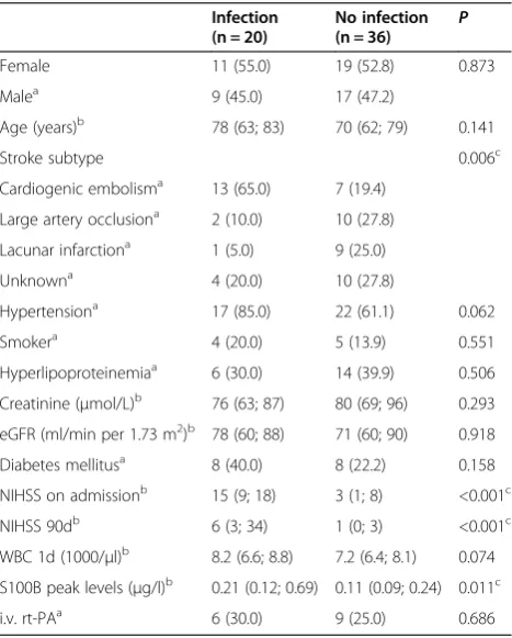
The study population consisted of 56 patients. Stroke-associated infection was diagnosed in 20 of these tients. Demographic and clinical characteristics for pa-tients with and without infection are shown in Table 1. Here, patients differed significantly for stroke subtype, clinical severity (NIHSS on admission), clinical outcome (NIHSS at day 90), and the extent of brain damage (S100B peak levels). The infections were pneumonia (n = 9), urinary tract infection (n = 10), and cholangitis (n = 1).
Levels of LBP, IL-10, IL-6 and CRP differed in patients with and without infection
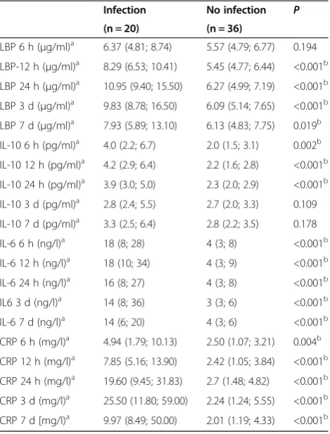
In the univariate analysis, levels of LBP, IL-10, IL-6 and CRP were compared between patients with and without infection (Table 2). LBP differed significantly between 12 hours and 7 days (12 h, 24 h, 3 d:P <0.001; 7 d:P= 0.019), while IL-10 levels differed significantly during the first day (6 h: P= 0.002; 12 h, 24 h: P< 0.001). IL-6 and CRP levels were significantly different at each time point (IL-6 6 h, 12 h, 24 h, 3 d, 7 d: P< 0.001; CRP 6 h: P= 0.004; 12 h, 24 h, 3 d, 7 d:P< 0.001).
Table 1 Clinical characteristics of patients with and without infection
Infection (n = 20)
No infection (n = 36) P
Female 11 (55.0) 19 (52.8) 0.873
Malea 9 (45.0) 17 (47.2)
Age (years)b 78 (63; 83) 70 (62; 79) 0.141
Stroke subtype 0.006c
Cardiogenic embolisma 13 (65.0) 7 (19.4) Large artery occlusiona 2 (10.0) 10 (27.8) Lacunar infarctiona 1 (5.0) 9 (25.0)
Unknowna 4 (20.0) 10 (27.8)
Hypertensiona 17 (85.0) 22 (61.1) 0.062
Smokera 4 (20.0) 5 (13.9) 0.551
Hyperlipoproteinemiaa 6 (30.0) 14 (39.9) 0.506 Creatinine (μmol/L)b 76 (63; 87) 80 (69; 96) 0.293 eGFR (ml/min per 1.73 m2)b 78 (60; 88) 71 (60; 90) 0.918 Diabetes mellitusa 8 (40.0) 8 (22.2) 0.158 NIHSS on admissionb 15 (9; 18) 3 (1; 8) <0.001c NIHSS 90db 6 (3; 34) 1 (0; 3) <0.001c WBC 1d (1000/μl)b 8.2 (6.6; 8.8) 7.2 (6.4; 8.1) 0.074 S100B peak levels (μg/l)b 0.21 (0.12; 0.69) 0.11 (0.09; 0.24) 0.011c
i.v. rt-PAa 6 (30.0) 9 (25.0) 0.686
eGFR, estimated glomerular filtration rate; NIHSS, National Institutes of Health Stroke Scale; i.v. rt-PA, intravenous recombinant tissue-type plasminogen acti-vator; WBC, white blood cell count.Data are presented as numbers (percent-ages)a
or median (interquartile range)b
Independent association of initial levels of IL-10 and CRP with infection
For determination if initial levels (at 6 h) of IL-10, IL-6 and CRP are independently associated with infection in ischemic stroke patients, we performed a binary logistic regression analysis that was adjusted for NIHSS on ad-mission, stroke etiology, and peak levels of S100B. The analysis revealed that IL-10 at 6 h, CRP at 6 h and NIHSS on admission are independently associated with
infection (IL-10: P= 0.009; CRP: P= 0.018; NIHSS:
P= 0.041) (Table 3).
Prediction of infection by initial CRP, IL-10 and National Institutes of Health Stroke Scale
After identification of CRP at 6 h and IL-10 at 6 h and NIHSS on admission as independent determinants of infec-tion, ROC analyses were performed (Figure 1. A-D). The area under the curve (AUC) for CRP in relation to dichoto-mized status of infection (infection versus no infection) was 0.74 (95% confidence interval: 0.59 to 0.88), 0.76 (95% con-fidence interval: 0.61 to 0.9) for IL-10 and 0.83 (95% confi-dence interval: 0.71 to 0.94) for NIHSS (Figure 1. A-C). For the combination of CRP and NIHSS the AUC was 0.86 (95% confidence interval: 0.77 to 0.96), 0.87 (95% confi-dence interval: 0.76 to 0.98) for IL-10 and NIHSS and 0.94 (95% confidence interval: 0.88 to 1) for CRP, IL-10 and NIHSS, when the method of RF was used (Figure 1. D). For a specificity of 97% the ROC curve-derived sensitivity was 60% for the combination of CRP, IL-10 and NIHSS. Results using the method of LDA or SVM for the combination were comparable.
Comparison of time courses of LBP, IL-10, IL-6 and CRP in patients with and without infection
For comparison of time courses of LBP, IL-10, IL-6 and CRP 16 patients with infection and 16 without infection were matched for S100B peak levels (differences in levels <20%, respectively) because the extent of the unspecific systemic inflammatory response after ischemic stroke de-pends on the size of brain damage. Of note, in 4 out of 20 patients with infection, no fitting matches could be identi-fied according to S100B levels. Clinical characteristics did not differ between both patient groups (P >0.05). Fig-ure 2A-D demonstrates the time courses. The temporal pattern differed significantly for each marker between the patient groups; levels in patients with infection were sig-nificantly elevated (LBP 12 h: P= 0.001, 24 h: P <0.001, 3 d:P= 0.007; IL-10 6 h:P= 0.006, 12 h:P <0.001, 24 h:
P= 0.004; IL-6 6 h:P<0.001, 12 h:P<0.001, 24 h:P= 0.004, 3 d: P= 0.003, 7 d: P= 0.008; CRP 6 h: P= 0.007, 12 h:
P<0.001, 24 h:P<0.001, 3 d:P<0.001, 7 d:P= 0.002). For within-group comparisons of marker levels be-tween initial (6 h) and follow-up time points, a signifi-cant increase was detected in patients with infection for LBP (6 h versus 12 h: P= 0.001, 6 h versus 24 h: P=
0.001, 6 h versus 3 d: P= 0.01) and CRP (6 h versus
12 h: P= 0.002, 6 h versus 24 h: P= 0.001, 6 h versus 3 d:P= 0.002). In patients without infection, marker levels significantly increased for LBP (6 h versus 24 h: P= 0.005, 6 h versus 3 d: P= 0.005, 6 h versus 7 d: P= 0.007) and CRP (6 h versus 24 h:P= 0.001).
Correlation between LBP, IL-10, IL-6 and CRP in patients with and without infection
The correlation analysis revealed a significant correlation between LBP and CRP at 6 h (P<0.001), 12 h (P= 0.003)
Table 2 LBP, Il-10, Il-6 and CRP in patients with and without infection
Infection No infection P (n = 20) (n = 36)
LBP 6 h (μg/ml)a 6.37 (4.81; 8.74) 5.57 (4.79; 6.77) 0.194 LBP-12 h (μg/ml)a 8.29 (6.53; 10.41) 5.45 (4.77; 6.44) <0.001b LBP 24 h (μg/ml)a 10.95 (9.40; 15.50) 6.27 (4.99; 7.19) <0.001b LBP 3 d (μg/ml)a 9.83 (8.78; 16.50) 6.09 (5.14; 7.65) <0.001b LBP 7 d (μg/ml)a 7.93 (5.89; 13.10) 6.13 (4.83; 7.75) 0.019b IL-10 6 h (pg/ml)a 4.0 (2.2; 6.7) 2.0 (1.5; 3.1) 0.002b IL-10 12 h (pg/ml)a 4.2 (2.9; 6.4) 2.2 (1.6; 2.8) <0.001b IL-10 24 h (pg/ml)a 3.9 (3.0; 5.0) 2.3 (2.0; 2.9) <0.001b IL-10 3 d (pg/ml)a 2.8 (2.4; 5.5) 2.7 (2.0; 3.3) 0.109 IL-10 7 d (pg/ml)a 3.3 (2.5; 6.4) 2.8 (2.2; 3.5) 0.178 IL-6 6 h (ng/l)a 18 (8; 28) 4 (3; 8) <0.001b IL-6 12 h (ng/l)a 18 (10; 34) 4 (3; 9) <0.001b IL-6 24 h (ng/l)a 16 (8; 27) 4 (3; 8) <0.001b IL6 3 d (ng/l)a 14 (8; 36) 3 (3; 6) <0.001b IL-6 7 d (ng/l)a 14 (6; 20) 4 (3; 6) <0.001b CRP 6 h (mg/l)a 4.94 (1.79; 10.13) 2.50 (1.07; 3.21) 0.004b CRP 12 h (mg/l)a 7.85 (5.16; 13.90) 2.42 (1.05; 3.84) <0.001b CRP 24 h (mg/l)a 19.60 (9.45; 31.83) 2.7 (1.48; 4.82) <0.001b CRP 3 d (mg/l)a 25.50 (11.80; 59.00) 2.24 (1.24; 5.55) <0.001b CRP 7 d [mg/l)a 9.97 (8.49; 50.00) 2.01 (1.19; 4.33) <0.001b Data are presented as median (interquartile range)a
.P<0.05 was considered statistically significantb
.
Table 3 Independent early determinants of infection after acute ischemic stroke
Parameters Β SE Pvalue
IL-10a 3.328 0.461 0.009
CRPb 2.136 0.320 0.018
NIHSSc 1.192 0.086 0.041
Binary logistic regression including IL-6, IL-10a and CRPb
at 6 hours, stroke severity (National Institutes of Health Stroke Scale (NIHSS) on admission)c
and 3 d (P<0.001), IL-10 and IL-6 at 7 d (P<0.001), and IL-6 and CRP at 7 d (P= 0.001) in patients with infec-tion (Table 4). In patients without infecinfec-tion LBP versus IL-6 at 3 d (P= 0.003) and IL-6 versus CRP at 3 d (P= 0.003) were significantly correlated (Table 4).
Discussion
The main findings of the present study are that, in acute stroke patients, levels of LBP, IL-10, IL-6 and CRP show a different time course in patients with and without post-stroke infection and that IL-10 and CRP at 6 h and NIHSS on admission are independent pre-dictors of stroke-associated infections.
After the acute event of stroke, concentrations of the pro-inflammatory cytokine IL-6 and the acute phase protein CRP in brain tissue and peripheral blood are in-creased since they are rapidly released by activated cells
[12,15]. Also, in the acute stage after stroke, a strong anti-inflammatory reaction results in a suppression of the immune system [5]. As a mechanism, immunode-pression is hypothesized to favor the development of infections, for example, from microaspiration to pneu-monia or from asymptomatic bacteriuria to urinary tract infection. IL-10 is a major player of the cellular and mo-lecular suppression of inflammation [16]. Our data show that levels of the pro- and anti-inflammatory markers IL-6, CRP and IL-10 differ as early as at 6 h after stroke onset between patients with and without post-stroke in-fection. In patients with infection, levels remained ele-vated until 24 h in the case of IL-10 and until day 7 in the case of IL-6 and CRP, respectively.
In contrast to this, early levels of LBP at 6 h after stroke were not significantly associated with occurrence of infec-tion. At later time points, LBP levels were significantly
Figure 2A-D Temporal profile of LBP, IL-10, IL-6 and CRP in patients with and without infection.Comparison of time courses between patients with (n = 16) and patients without (n = 16) infection matched for S100B peak levels for the extent of brain damage. Data are presented as median with interquartile range. Intergroup comparisons between patients with and without infection: *P≤0.05; **P≤0.01; ***P≤0.001. Within-group comparisons of marker levels between initial (6 h) and follow-up time points: Patients with infection: significant differences were detected for LBP (6 h versus 12 h:P= 0.001, 6 h versus 24 h:P= 0.001, 6 h versus 3 d:P= 0.01) and CRP (6 h versus 12 h:P= 0.002, 6 h versus 24 h:P= 0.001, 6 h versus 3 d:P= 0.002). Patients without infection: significant differences were detected for LBP (6 h versus 24 h:P= 0.005, 6 h versus 3 d:P= 0.005, 6 h versus 7 d:P= 0.007) and CRP (6 h versus 24 h:P= 0.001).
Table 4 Correlation between LBP, IL-10, IL-6 and CRP in patients with and without infection
6 h 12 h 24 h 3 d 7 d
R P R P R P r P r P
Infection (n = 16)
LBP versus IL-10 −0.405 0.120 −0.314 0.237 −0.284 0.286 0.100 0.712 0.041 0.879
LBP versus IL-6 −0.018 0.948 0.376 0.151 0.292 0.273 0.322 0.224 0.281 0.292
LBP versus CRP 0.879 <0.001a 0.697 0.003a 0.450 0.080 0.820 <0.001a 0.571 0.021 IL-10 versus IL-6 −0.014 0.959 0.221 0.411 0.309 0.244 0.399 0.126 0.785 <0.001a IL-10 versus CRP −0.393 0.132 −0.247 0.356 −0.084 0.757 0.424 0.102 0.361 0.170
IL-6 versus CRP 0.078 0.774 0.302 0.255 0.336 0.204 0.427 0.099 0.725 0.001a
No infection (n = 16)
LBP versus IL-10 0.194 0.471 0.195 0.469 0.030 0.913 −0.233 0.384 0.183 0.498
LBP versus IL-6 0.028 0.929 0.426 0.100 0.252 0.346 0.687 0.003a 0.297 0.264
LBP versus CRP 0.478 0.061 0.453 0.078 0.178 0.509 0.444 0.085 0.615 0.011
IL-10 versus IL-6 −0.279 0.296 −0.037 0.893 −0.379 0.148 −0.326 0.218 0.270 0.313
IL-10 versus CRP 0.100 0.712 0.010 0.970 0.187 0.489 0.055 0.841 0.072 0.791
IL-6 versus CRP 0.280 0.293 0.402 0.122 0.407 0.117 0.689 0.003a 0.308 0.247
elevated until day 7 in patients with infection. In addition, we showed that LBP - as an acute-phase protein – not only increases in patients with infection but also increases in patients without infection during the first day in re-sponse to the infarction. Further investigations need to show whether the increase of LBP in circulating blood directly results from the site of infarction or represents a systemic reaction from peripheral blood cells.
Over the past years, several clinical trials have investigated preventive anti-infective treatment not only for lowering the rate of infection but also for ameliorating the clinical out-come. Mechanisms that may explain why the rate of infec-tion could directly influence neurological outcome are the potentially detrimental effects of hyperthermia, hypotension or hypoxia on neurons [17-19]. So far, only the Mannheim Infection in Stroke Study (MISS) has reached improvement for rate of infection and clinical outcome using the prophy-lactic mezlocillin plus sulbactam in severe stroke patients, who presented as bedridden within 24 h of stroke onset [20]. In contrast to these results, other clinical trials investi-gating treatment with anti-infective drugs have failed to im-prove outcome [21] or did not lower the rate of infection [22]. Acute ischemic stroke patients had significantly better outcome at 30 d when treated with minocycline within 24 h compared to placebo [23]. The favorable outcome after ad-ministration of minocycline has been recently confirmed in another cohort of stroke patients [24]. But it has been dis-cussed that minocycline acts independently from its anti-biotic effects via neuroprotection.
So far, it remains unclear as to whether an assessment of molecular inflammation markers or clinical variables might be useful for decision-making in preventive anti-infective treatment. Recently, the predictive ability of both, IL-6 and CRP within 3 days after stroke for post-stroke infection has been reported [25,26]. However, the time points used for the determination of inflammation markers might be too late to indicate the development of infection. In two of the clinical studies investigating preventive anti-infective treatment, the association of in-flammation markers and post-stroke infections was ana-lyzed. In the Preventive Antibacterial Therapy in Acute Ischemic Stroke (PANTHERIS) trial Klehmet et al. [27] showed that increased levels of IL-10 were predictive for post-stroke infections independently of preventive antibacterial treatment with moxifloxacin. Therefore, the authors concluded that IL-10 levels might indicate patients who would not respond to this treatment. Also, increased IL-10 levels within 24 h were associated with infection in patients from the Early Systemic Prophylaxis of Infection after Stroke (ESPIAS) trial, a randomized clinical trial using preventive antibacterial treatment with levofloxacin [6].
Since timing is decisive in acute stroke treatment, early administration of antibiotics in a selected cohort of
stroke patients might be the key to ameliorate outcome. Therefore, in contrast to former studies, we determined very early levels of inflammation markers IL-6, IL-10, CRP and LBP (at 6 h) to analyze the association with post-stroke infection. Of note, the multivariate analysis revealed an independent association of IL-10 and CRP with infections, suggesting a strong effect of the inflam-matory and anti-inflaminflam-matory reaction on the develop-ment of infections.
In a subanalysis of patients matched for S100B levels, we showed that differences in inflammation markers in patients with and without infection were independent from the extent of brain lesion. As previously shown, de-velopment of infection is associated with stroke severity [28,29]. The association of stroke severity and infection can be explained since clinical deficits result in condi-tions such as dysphagia or shallow respiration due to
disturbance of consciousness. But severe stroke does not obligatorily mean large infarction, as reflected by S100B peak levels, since a small infarction in the brainstem or internal capsule is related to a severe deficit, whereas a major infarction in the temporal and occipital lobe may be associated with only a mild deficit. In the current study, we found evidence for an additional independent effect of the individual inflammatory response of any pa-tient on the development of early infection. According to our data, we propose prospective studies to investi-gate if the combination of markers CRP, IL-10 and NIHSS might be useful as inclusion criteria for very early initiation of preventive anti-infective treatment.
Limitations
than the ones used in the current study could reproduce the findings.
Conclusions
The current study shows that the temporal pattern of circulating levels of LBP, IL-10, IL-6 and CRP differs between acute ischemic stroke patients with and without post-stroke infection. IL-10 and CRP at 6 h, as well as NIHSS on admission, were identified as independent pre-dictors of stroke-associated infection. Prospective studies are warranted to confirm these surrogate markers as in-dependent early predictors for infection in acute ischemic stroke patients. This might help to identify patients who should receive early preventive antibiotic treatment.
Abbreviations
AUC:area under the curve; CCT: cranial computed tomography; CRP: C-reactive protein; ESPIAS: early systemic prophylaxis of infection after stroke; IL-10: interleukin-10; IL-6: interleukin-6; LBP: lipopolysaccharide binding protein; MRI: magnetic resonance imaging; MISS: Mannheim Infection in Stroke Study; NIHSS: National Institutes of Health Stroke Scale; PANTHERIS: Preventive Antibacterial Therapy in Acute Ischemic Stroke; TIA: transient ischemic attack; WBC: white blood cell count.
Competing interests
RL received a research grant from Siemens Healthcare diagnostics. The other authors declare that they have no competing interests.
Authors’contributions
HW contributed to the conception and design of the study; the data acquisition, analysis, and interpretation; and the drafting and revision of the manuscript. KW contributed to the conception and design of the study, the data analysis and interpretation, and the revision of the manuscript. RL contributed to the conception and design of the study, the data acquisition and interpretation, and the revision of the manuscript. FK contributed to the data analysis and interpretation and to the revision of the manuscript. ABT, MD, RS and KB contributed to the data acquisition, analysis and
interpretation and to the revision of the manuscript. All authors read and approved the final manuscript.
Acknowledgments
The authors thank Frank Dsiosa, Klaus Burfeind and Bernadette Lüns for excellent technical assistance.
Source of Funding
This work was supported by research grants from Siemens Healthcare diagnostics, Germany and Boehringer-Ingelheim, Germany.
Author details
1Department of Neurology, Hannover Medical School, Carl-Neuberg-Str. 1,
30623 Hannover, Germany.2Department of Clinical Chemistry, Hannover
Medical School, Carl-Neuberg-Str. 1, 30623 Hannover, Germany.3Department
of Computer Science, Ostfalia University of Applied Sciences, Am Exer 2, 38302 Wolfenbuettel, Germany.4Biostatistics, Helmholtz Centre for Infection
Research, Inhoffenstr. 7, 38124 Braunschweig, Germany.5Center for Systems
Neuroscience (ZSN), Buenteweg 2, 30559 Hannover, Germany.
Received: 22 September 2014 Accepted: 21 December 2014
References
1. Aslanyan S, Weir CJ, Diener HC, Kaste M, Lees KR. GAIN International Steering Committee and Investigators. Pneumonia and urinary tract infection after acute ischaemic stroke: a tertiary analysis of the GAIN International trial. Eur J Neurol. 2004;11:49–53.
2. Prass K, Meisel C, Höflich C, Braun J, Halle E, Wolf T, et al. Stroke-induced immunodeficiency promotes spontaneous bacterial infections and is
mediated by sympathetic activation reversal by poststroke T helper cell type 1-like immunostimulation. J Exp Med. 2003;198:725–36.
3. Chamorro A, Urra X, Planas AM. Infection after acute ischemic stroke: a manifestation of brain-induced immunodepression. Stroke. 2007;38:1097–103. 4. Worthmann H, Tryc AB, Deb M, Goldbecker A, Ma YT, Tountopoulou A, et al.
Linking infection and inflammation in acute ischemic stroke. Ann N Y Acad Sci. 2010;1207:116–22. Review.
5. Chamorro Á, Meisel A, Planas AM, Urra X, van de Beek D, Veltkamp R. The immunology of acute stroke. Nat Rev Neurol. 2012;8:401–10.
doi: 10.1038/nrneurol.2012.98.
6. Chamorro A, Amaro S, Vargas M, Obach V, Cervera A, Torres F, et al. Interleukin 10, monocytes and increased risk of early infection in ischaemic stroke. J Neurol Neurosurg Psychiatry. 2006;2006:1279–81.
7. Urra X, Cervera A, Obach V, Climent N, Planas AM, Chamorro A. Monocytes are major players in the prognosis and risk of infection after acute stroke. Stroke. 2009;40:1262–8.
8. Emsley HC, Smith CJ, Gavin CM, Georgiou RF, Vail A, Barberan EM, et al. An early and sustained peripheral inflammatory response in acute ischaemic stroke: relationships with infection and atherosclerosis. J Neuroimmunol. 2003;139:93–101.
9. Villar J, Maca-Meyer N, Pérez-Méndez L, Flores C. Bench-to-bedside review: understanding genetic predisposition to sepsis. Crit Care. 2004;8:180–9. 10. Wartenberg KE, Stoll A, Funk A, Meyer A, Schmidt JM, Berrouschot J.
Infection after acute ischemic stroke: risk factors, biomarkers, and outcome. Stroke Res Treat. 2011;2011:830614.
11. Westendorp WF, Vermeij JD, Vermeij F, Den Hertog HM, Dippel DW, van de Beek D, et al. Antibiotic therapy for preventing infections in patients with acute stroke. Cochrane Database Syst Rev. 2012;1:CD008530.
12. Worthmann H, Tryc AB, Goldbecker A, Ma YT, Tountopoulou A, Hahn A, et al. The temporal profile of inflammatory markers and mediators in blood after acute ischemic stroke differs depending on stroke outcome. Cerebrovasc Dis. 2010;30:85–92.
13. Adams Jr HP, Bendixen BH, Kappelle LJ, Biller J, Love BB, Gordon DL, et al. Classification of subtype of acute ischemic stroke. Definitions for use in a multicenter clinical trial. TOAST. Trial of Org 10172 in Acute Stroke Treatment. Stroke. 1993;24:35–41.
14. Robin X, Turck N, Hainard A, Tiberti AN, Lisacek F, Sanchez J-C, et al. pROC: an open-source package for R and S+ to analyze and compare ROC curves. BMC Bioinformatics. 2011;12:77.
15. Lambertsen KL, Biber K, Finsen B. Inflammatory cytokines in experimental and human stroke. J Cereb Blood Flow Metab. 2012;32:1677–98. 16. O’Farrell AM, Liu Y, Moore KW, Mui AL. IL-10 inhibits macrophage activation
and proliferation by distinct signaling mechanisms: evidence for Stat3-dependent and -inStat3-dependent pathways. EMBO J. 1998;17:1006–18. 17. Kammersgaard LP, Jørgensen HS, Rungby JA, Reith J, Nakayama H, Weber
UJ, et al. Admission body temperature predicts long-term mortality after acute stroke: the Copenhagen Stroke Study. Stroke. 2002;33:1759–62. 18. Leonardi-Bee J, Bath PM, Phillips SJ, Sandercock PA. IST Collaborative Group.
Blood pressure and clinical outcomes in the International Stroke Trial. Stroke. 2002;33:1315–20.
19. Rocco A, Pasquini M, Cecconi E, Sirimarco G, Ricciardi MC, Vicenzini E, et al. Monitoring after the acute stage of stroke: a prospective study. Stroke. 2007;38:1225–8.
20. Schwarz S, Al-Shajlawi F, Sick C, Meairs S, Hennerici MG. Effects of prophylactic antibiotic therapy with mezlocillin plus sulbactam on the incidence and height of fever after severe acute ischemic stroke: the Mannheim infection in stroke study (MISS). Stroke. 2008;39:1220–7. 21. Harms H, Prass K, Meisel C, Klehmet J, Rogge W, Drenckhahn C, et al.
Preventive antibacterial therapy in acute ischemic stroke: a randomized controlled trial. PLoS One. 2008;3:e2158.
22. Chamorro A, Horcajada JP, Obach V, Vargas M, Revilla M, Torres F, et al. The Early Systemic Prophylaxis of Infection After Stroke study: a randomized clinical trial. Stroke. 2005;36:1495–500.
23. Lampl Y, Boaz M, Gilad R, Lorberboym M, Dabby R, Rapoport A, et al. Minocycline treatment in acute stroke: an open-label, evaluator-blinded study. Neurology. 2007;69:1404–10.
24. Padma Srivastava MV, Bhasin A, Bhatia R, Garg A, Gaikwad S, Prasad K, et al. Efficacy of minocycline in acute ischemic stroke: a single-blinded, placebo-controlled trial. Neurol India. 2012;60:23–8.
mortality in the elderly after an ischemic stroke. Exp Gerontol. 2013;48:960–5.
26. Fluri F, Morgenthaler NG, Mueller B, Christ-Crain M, Katan M. Copeptin, procalcitonin and routine inflammatory markers-predictors of infection after stroke. PLoS One. 2012;7:e48309.
27. Klehmet J, Harms H, Richter M, Prass K, Volk HD, Dirnagl U, et al. Stroke-induced immunodepression and post-stroke infections: lessons from the preventive antibacterial therapy in stroke trial. Neuroscience.
2009;158:1184–93.
28. Walter U, Knoblich R, Steinhagen V, Donat M, Benecke R, Kloth A. Predictors of pneumonia in acute stroke patients admitted to a neurological intensive care unit. J Neurol. 2007;254:1323–9.
29. Sellars C, Bowie L, Bagg J, Sweeney MP, Miller H, Tilston J, et al. Risk factors for chest infection in acute stroke: a prospective cohort study. Stroke. 2007;38:2284–91.
Submit your next manuscript to BioMed Central and take full advantage of:
• Convenient online submission
• Thorough peer review
• No space constraints or color figure charges
• Immediate publication on acceptance
• Inclusion in PubMed, CAS, Scopus and Google Scholar
• Research which is freely available for redistribution