Open Access
Methodology
Protocol: a highly sensitive RT-PCR method for detection and
quantification of microRNAs
Erika Varkonyi-Gasic*, Rongmei Wu, Marion Wood, Eric F Walton and
Roger P Hellens
Address: HortResearch, Mt Albert Research Centre, Private Bag 92169, Auckland, New Zealand
Email: Erika Varkonyi-Gasic* - [email protected]; Rongmei Wu - [email protected];
Marion Wood - [email protected]; Eric F Walton - [email protected]; Roger P Hellens - [email protected] * Corresponding author
Abstract
MicroRNAs (miRNAs) are a class of small non-coding RNAs with a critical role in development and environmental responses. Efficient and reliable detection of miRNAs is an essential step towards understanding their roles in specific cells and tissues. However, gel-based assays currently used to detect miRNAs are very limited in terms of throughput, sensitivity and specificity. Here we provide protocols for detection and quantification of miRNAs by RT-PCR. We describe an end-point and real-time looped RT-PCR procedure and demonstrate detection of miRNAs from as little as 20 pg of plant tissue total RNA and from total RNA isolated from as little as 0.1 μl of phloem sap. In addition, we have developed an alternative real-time PCR assay that can further improve specificity when detecting low abundant miRNAs. Using this assay, we have demonstrated that miRNAs are differentially expressed in the phloem sap and the surrounding vascular tissue. This method enables fast, sensitive and specific miRNA expression profiling and is suitable for facilitation of high-throughput detection and quantification of miRNA expression.
Introduction
MicroRNAs (miRNAs) are families of short non-coding transcripts, arising from larger precursors with a character-istic hairpin secondary structure [reviewed in [1]]. Together with short interfering RNAs (siRNAs), miRNAs belong to a class of 19- to 25-nucleotide (nt) small RNAs that are essential for genome stability, development and differentiation, disease, cellular communication, signal-ing, and adaptive responses to biotic and abiotic stress [1-4]. A large proportion of miRNAs are highly conserved among distantly related species, from worms to mammals in the animal kingdom [1], and mosses to high flowering eudicots in plants [5,6].
Currently, over 4000 miRNA sequences from vertebrates, flies, worms, plants and viruses are annotated in the Sanger Centre miRBase Database [version 9.0, October 2006; [7]]. In animals, miRNAs appear predominantly to inhibit translation by targeting partially complementary sequences located within the 3' untranslated region (UTR) of mRNA [reviewed in [8]]. The majority of animal miR-NAs appear to be operating at several levels, regulating multiple targets implicated in various molecular functions and biological processes [1]. In plants, miRNAs repress gene expression by acting either on near-perfect comple-mentary sequences in mRNA coding region to guide cleav-age and translational repression [9-12], or in at least one example, on DNA to guide chromatin remodelling [13].
Published: 12 October 2007
Plant Methods 2007, 3:12 doi:10.1186/1746-4811-3-12
Received: 18 July 2007 Accepted: 12 October 2007
This article is available from: http://www.plantmethods.com/content/3/1/12
© 2007 Varkonyi-Gasic et al; licensee BioMed Central Ltd.
The majority of plant miRNA targets are developmentally important transcription factors [14,15] and stress-regu-lated genes [16,17]. Thus, ectopic expression of miRNAs [9,10,13,18-20] or misexpression of miRNA-resistant tar-get mRNAs can induce strong developmental phenotypes [reviewed in [21]]. It has been proposed that plant miR-NAs act mainly by clearing of the mRNA of the target reg-ulatory genes during the cell-fate changes [15,22,23]. There is also evidence for quantitative action of plant miR-NAs in quenching the target gene activity rather than elim-inating it completely [24,25]. Furthermore, several miRNAs were detected in the phloem sap, suggesting a long-distance signaling role [26], in contrast to several miRNAs with demonstrated cell-autonomous expression and effects [27,28].
This complexity in miRNA modes of action demonstrates that reliable detection and quantification of miRNA expression in specific tissues is essential for better under-standing of miRNA-mediated gene regulation. Although miRNA represent a relatively abundant class of transcripts, their expression levels vary greatly among different cells and tissues. Conventional technologies such as cloning, northern hybridization and microarray analysis are widely used but may not be sensitive enough to detect less abundant miRNAs. Furthermore, intensive small RNA sequencing revealed a very complex small RNA popula-tion in plants. Unlike mammals, which have relatively simple small RNA populations comprising mainly miR-NAs and no siRmiR-NAs [29], plants have a hugely complex small RNA fraction. It is comprised of both miRNAs and endogenous siRNAs derived from repetitive sequences, intergenic regions and genes [14,30]. This complexity renders miRNAs highly under-represented in the small RNA fraction and further affects detection methods such as cloning and microarray hybridization.
Poor sensitivity and low throughput of conventional tech-nologies can be overcome by using a sensitive reverse tran-scription-polymerase chain reaction (RT-PCR) detection method. However, because of their small size, detection of miRNAs by PCR is technically demanding. A number of specific quantitative RT-PCR (qRT-PCR) techniques were developed and optimised for miRNA detection, including real-time methods based upon reverse transcription (RT) reaction with a stem-loop primer followed by a TaqMan PCR analysis [31,32]. The stem-loop reverse transcription primers provide better specificity and sensitivity than lin-ear primers because of base stacking and spatial constraint of the stem-loop structure [31]. Detection sensitivity is further increased by a pulsed RT reaction [32]. However, these methods were optimized for detection of mamma-lian miRNAs and require individual miRNA-specific fluo-rescent probes, making them very costly to many laboratories and not amenable for high-throughput
anal-ysis of a large number of miRNAs. Alternatively, qRT-PCR miRNA detection kits and primer sets are commercially available, but are very costly and thus not suitable for high-throughput miRNA analysis. Indeed, the available plant miRNA expression data are mainly results of gel-based assays.
Here we describe and provide protocols for an end-point and real-time looped RT-PCR procedure. We demonstrate detection of miRNAs from as little as 20 pg of plant tissue total RNA and from total RNA isolated from as little as 0.1
μl of phloem sap.
The expression of a miRNA was detected using a two-step process. First, the stem-loop RT primer designed accord-ing to Chen et al. [31] was hybridized to the miRNA mol-ecule, and then reverse transcribed in a pulsed RT reaction. Next, the RT product was amplified using a miRNA-specific forward primer and the universal reverse primer (Figure 1A). The amplification product was visual-ized on an agarose gel by ethidium bromide staining. The amplification was also performed in real-time, using the SYBR Green I assay (Figure 1B). In addition, we have developed a Universal ProbeLibrary (UPL; Roche Diag-nostics) RT-PCR method for miRNA detection and quan-tification that provides increased specificity when analysing expression of low abundant miRNAs (Figure 1C).
Materials
Plant material and phloem sap collection
Arabidopsis thaliana Columbia, Cucurbita maxima 'Crown' (pumpkin) and Cucumis sativus 'Telegraf' (cucumber) plants were grown in a greenhouse under natural daylight conditions. Phloem sap was collected from well-watered plants, as previously described [26].
miRNA sequences, primers and probes
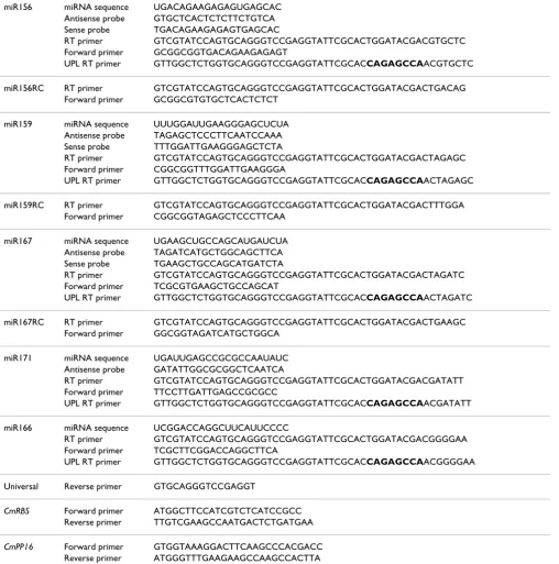
Plant miRNA genes were selected from the Sanger Insti-tute miRBase Sequence Database [33]. UPL probe #21 was obtained from universal probe library database (Roche Diagnostics). Stem-loop RT primers were designed according to Chen et al. [31]. Sequence data are presented in Table 1.
Protocols
Equipment
Standard laboratory equipment including a thermal cycler is required for pulsed reverse transcription and end-point PCRs. A real-time thermal cycler is required for SYBR Green I and UPL probe assays. All our reverse transcrip-tion reactranscrip-tions and end-point PCR analyses were per-formed on Mastercycler (Eppendorf, Hamburg, Germany). All real-time PCR analyses were performed on LightCycler 1.5 (Roche Diagnostics, Mannheim, Ger-many).
RNA template
RNA should be handled according to standard laboratory practices to avoid RNase contamination. Avoid RNA puri-fication methods that use RNA-binding glass-fiber filters that do not quantitatively recover small RNA species. In our hands, both non-denatured RNA and RNA denatured by incubation at 65°C for 5 minutes produced similar results.
NOTE: Total RNA used in our experiments was isolated using the TRIzol reagent (Invitrogen, Carlsbad, CA). High molecular weight (HMW)RNA was purified with RNAqueous kit (Ambion, Austin, TX). Low molecular weight (LMW)RNA present in the flow-through was precipitated as described previ-ously [26]. The concentration of RNA was determined using
the NanoDrop ND-1000 Spectrophotometer (NanoDrop Tech-nologies, Wilmington, DE).
For RNA gel blot analyses, HMW RNA was separated on a for-maldehyde-containing 1% agarose gel and transferred over-night to a Hybond-N+ membrane (GE Healthcare, formerly Amersham Biosciences, Buckinghamshire, UK). LMW RNA was separated on a 7 M urea/15% polyacrylamide gel and transferred overnight to a Hybond-XL membrane (GE Health-care). The membranes were UV cross-linked and prehybridized at 40–45°C for 1 h in hybridization buffer (0.5 M Na2HPO4, 1 mM EDTA, 1% BSA, and 7% SDS). DNA oligo probes were end labeled by the forward reaction using 10 units of T4 poly-nucleotide kinase (Invitrogen) with the supplied buffer, to which was added 300 nM [γ-32P]ATP (3000 Ci/mmol) for 10 min at 37°C. Unincorporated 32P-label was removed using ProbeQuant G-50 microcolumns (GE Healthcare). Probes were denatured at 94°C for 5 min, added to the hybridisation buffer and hybridisation was allowed to proceed at 40°C over-night. The membrane was then washed twice, 15 min each in 2× SSC, 0.1% SDS at 50°C. Hybridization signal was detected using a Typhoon scanner (GE Healthcare).
Stem-loop pulsed reverse transcription protocol
1. Prepare an RT master mix by scaling the volumes listed below to the desired number of RT reactions. If testing many RNA samples for one miRNA, prepare a 'no RNA'
Schematic showing stem-loop RT-PCR miRNA assays
Figure 1
Table 1: miRNA, primer and probe sequences
miR156 miRNA sequence UGACAGAAGAGAGUGAGCAC Antisense probe GTGCTCACTCTCTTCTGTCA Sense probe TGACAGAAGAGAGTGAGCAC
RT primer GTCGTATCCAGTGCAGGGTCCGAGGTATTCGCACTGGATACGACGTGCTC Forward primer GCGGCGGTGACAGAAGAGAGT
UPL RT primer GTTGGCTCTGGTGCAGGGTCCGAGGTATTCGCACCAGAGCCAACGTGCTC
miR156RC RT primer GTCGTATCCAGTGCAGGGTCCGAGGTATTCGCACTGGATACGACTGACAG Forward primer GCGGCGTGTGCTCACTCTCT
miR159 miRNA sequence UUUGGAUUGAAGGGAGCUCUA Antisense probe TAGAGCTCCCTTCAATCCAAA Sense probe TTTGGATTGAAGGGAGCTCTA
RT primer GTCGTATCCAGTGCAGGGTCCGAGGTATTCGCACTGGATACGACTAGAGC Forward primer CGGCGGTTTGGATTGAAGGGA
UPL RT primer GTTGGCTCTGGTGCAGGGTCCGAGGTATTCGCACCAGAGCCAACTAGAGC
miR159RC RT primer GTCGTATCCAGTGCAGGGTCCGAGGTATTCGCACTGGATACGACTTTGGA Forward primer CGGCGGTAGAGCTCCCTTCAA
miR167 miRNA sequence UGAAGCUGCCAGCAUGAUCUA Antisense probe TAGATCATGCTGGCAGCTTCA Sense probe TGAAGCTGCCAGCATGATCTA
RT primer GTCGTATCCAGTGCAGGGTCCGAGGTATTCGCACTGGATACGACTAGATC Forward primer TCGCGTGAAGCTGCCAGCAT
UPL RT primer GTTGGCTCTGGTGCAGGGTCCGAGGTATTCGCACCAGAGCCAACTAGATC
miR167RC RT primer GTCGTATCCAGTGCAGGGTCCGAGGTATTCGCACTGGATACGACTGAAGC Forward primer GGCGGTAGATCATGCTGGCA
miR171 miRNA sequence UGAUUGAGCCGCGCCAAUAUC Antisense probe GATATTGGCGCGGCTCAATCA
RT primer GTCGTATCCAGTGCAGGGTCCGAGGTATTCGCACTGGATACGACGATATT Forward primer TTCCTTGATTGAGCCGCGCC
UPL RT primer GTTGGCTCTGGTGCAGGGTCCGAGGTATTCGCACCAGAGCCAACGATATT
miR166 miRNA sequence UCGGACCAGGCUUCAUUCCCC
RT primer GTCGTATCCAGTGCAGGGTCCGAGGTATTCGCACTGGATACGACGGGGAA Forward primer TCGCTTCGGACCAGGCTTCA
UPL RT primer GTTGGCTCTGGTGCAGGGTCCGAGGTATTCGCACCAGAGCCAACGGGGAA
Universal Reverse primer GTGCAGGGTCCGAGGT
CmRBS Forward primer ATGGCTTCCATCGTCTCATCCGCC Reverse primer TTGTCGAAGCCAATGACTCTGATGAA
CmPP16 Forward primer GTGGTAAAGGACTTCAAGCCCACGACC Reverse primer ATGGGTTTGAAGAAGCCAAGCCACTTA
UPL probe #21 reverse complement sequence is highlighted in bold.
master mix; if testing for many different miRNAs in one sample, prepare a 'no RT primer' master mix. Include 10% overage to cover pipetting errors. Also prepare the minus RT controls by omitting reverse transcriptase from the reactions and water controls by adding nuclease-free water in place of RNA. Keep on ice and work in the cold room if handling large number of samples.
• For a 'no RNA' master mix, add the following components to a nuclease-free microcentrifuge tube:
0.5 μl 10 mM dNTP mix
1 μl of appropriate stem-loop RT primer (1
μM)
• For a 'no RT primer' master mix, add the follow-ing components to a nuclease-free microcentrifuge tube:
0.5 μl 10 mM dNTP mix
11.15 μl nuclease-free water
1 μl of appropriate RNA template
• Heat mixture to 65°C for 5 minutes and incubate on ice for 2 minutes.
• Centrifuge briefly to bring solution to the bottom of the tube and add:
4 μl 5× First-Strand buffer
2 μl 0.1 M DTT
0.1 μl RNaseOUT (40 units/μl)
0.25 μl SuperScript III RT (200 units/μl)
• Mix gently and centrifuge to bring solution to the bottom of the tube.
2. Assemble the RT reaction.
• Aliquot the appropriate amount of the RT master mix (19 μl).
• Add 1 μl RNA template to 'no RNA' master mix or
• Add 1 μl of appropriate stem-loop RT primer (1
μM) to 'no RT primer' master mix
• Mix gently and centrifuge to bring solution to the bottom of the tube.
3. Perform pulsed RT:
• Load thermal cycler and incubate for 30 min at 16°C, followed by pulsed RT of 60 cycles at 30°C for 30 s, 42°C for 30 s and 50°C for 1 s.
• Incubate at 85°C for 5 min to inactivate the reverse transcriptase.
NOTE: RT reaction volume can be scaled down to 10 μl.
End-point PCR protocol
1. Prepare a PCR master mix by scaling the volumes listed below to the desired number of amplification reactions. Include 10% overage to cover pipetting errors. Also prepare water controls by adding nuclease-free water in place of the RT product.
• Add the following components to a nuclease-free microcentrifuge tube:
15.4 μl nuclease-free water
2 μl 10× PCR buffer
0.4 μl 10 mM dNTP mix
0.4 μl forward primer (10 μM)
0.4 μl reverse primer (10 μM)
0.4 μl Advantage 2 Polymerase mix
• Mix gently and centrifuge to bring solution to the bottom of the tube.
2. Aliquot 19 μl of the PCR master mix into PCR tubes and add 1 μl RT product.
3. Place reactions in a preheated (94°C) thermal cycler heat block and incubate at 94°C for 2 min, followed by 20–40 cycles of 94°C for 15 s and 60°C for 1 min.
4. Analyse reaction products by electrophoresis on a 4% agarose gel in 1× TAE.
NOTE: We tested several commercial thermostable DNA polymerases. In our hands, the most consistent results were obtained using Advantage 2 PCR Polymerase Mix (Clontech, Mountain View, CA).
miRNA SYBR Green I assay protocol
1. Prepare 5× LightCycler FastStart SYBR Green I mas-ter mix (Roche Diagnostics) according to manufac-turer's instructions.
2. Prepare a PCR master mix by scaling the volumes listed below to the desired number of amplification reactions. Include 10% overage to cover pipetting errors.
• Add the following components to a nuclease-free microcentrifuge tube:
4 μl SYBR Green I master mix
1 μl forward primer (10 μM)
1 μl reverse primer (10 μM)
• Mix gently and centrifuge to bring solution to the bottom of the tube.
• Store in cooling block or on ice.
3. Perform real-time PCR:
• Place required number of LightCycler capillaries in precooled centrifuge adapters.
• Pipet 18 μl master mix into each LightCycler cap-illary.
• Add 2 μl RT product.
• Seal each capillary with a stopper.
• Place capillaries into the LightCycler carousel and spin in the carousel centrifuge.
• Incubate samples at 95°C for 5 min, followed by 35–45 cycles of 95°C for 5 s, 60°C for 10 s, and 72°C for 1 s.
• For melting curve analysis, denature samples at 95°C, then cool to 65°C at 20°C per second. Col-lect fluorescence signals at 530 nm wavelength continuously from 65°C to 95°C at 0.2°C per sec-ond.
4. Analyse results using the LightCycler software.
• Prepare standard curves for each primer set by using dilution series of the experimental sample expected to have the highest expression. Use at least 3 points (or one point per log of concentra-tion whichever is greater).
• Alternatively, standard curves can be prepared from dilution series of an appropriate RNA oligo-nucleotide, e.g. when detecting artificial miRNAs or siRNAs.
• Perform a Relative Quantification-Monocolor Analysis.
NOTE: PCR reaction volume can be scaled down to 10 μl. Per-form PCR in at least three replicates.
miRNA UPL probe assay protocol
1. Prepare 5× LightCycler TaqMan master mix (Roche Diagnostics) according to manufacturer's instructions.
2. Prepare a PCR master mix by scaling the volumes listed below to the desired number of amplification reactions. Include 10% overage to cover pipetting errors.
• Add the following components to a nuclease-free microcentrifuge tube:
11.8 μl nuclease-free water
4 μl TaqMan master mix
1 μl forward primer (10 μM)
1 μl reverse primer (10 μM)
0.2 μl UPL probe #21 (10 μM)
• Mix gently and centrifuge to bring solution to the bottom of the tube.
• Store in cooling block or on ice.
3. Perform real-time PCR as described for miRNA SYBR Green I assay (omit melting curve analysis).
4. Analyse results using the LightCycler software as described above.
Comments
The sensitivity of stem-loop RT-PCR assays
The specificity of stem-loop RT-PCR assays
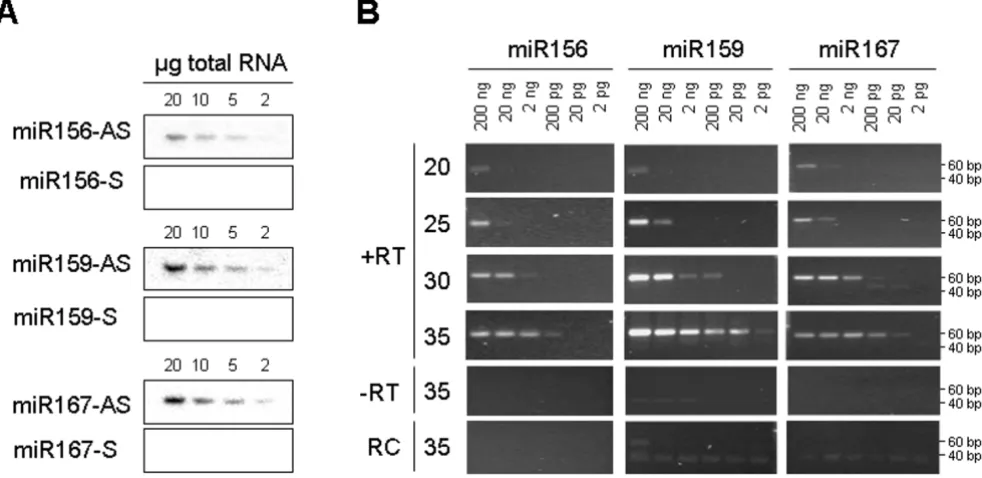
PCR amplification methods can lack specificity for mature miRNAs. Mature miRNAs are processed from large capped and polyadenylated transcripts (primary or pri-miRNA) that first give rise to short-lived hairpin intermediates (pre-miRNA) and finally to mature single stranded miR-NAs. In some instances, total RNA contains large amounts of pri-miRNAs, as established for pri-miR156 in pumpkin and cucumber tissues (Figure 3A). RNA gel blot analyses clearly show higher expression of pri-miR156 in the shoot tip and stem than in leaf tissue, and higher expression of mature miRNA in leaf than in shoot tip and stem. To investigate the ability of stem-loop RT-PCR assays to dif-ferentiate between mature miRNAs and their pri-miRNAs, the reactions were performed using pumpkin tissue total RNA and the high molecular weight (HMW) and low molecular weight (LMW) RNA purified from total RNA. Similar amounts of amplification product from total and LMW RNA were obtained after 25 and 30 cycles (Figure 3B). The slightly more efficient amplification of total RNA than LMW RNA was the result of RNA losses during puri-fication, as only 60–75% RNA was recovered. Some amplification visible in the HMW RNA fraction is more likely to be the result of contamination of the HMW RNA with LMW RNA, rather than amplification of the primary miRNA, as there is less amplification in the shoot tip HMW RNA than in leaf HMW RNA. Similar results were
obtained with cucumber tissue RNA (data not shown). These results suggest that the stem-loop RT-PCR assay is highly specific for mature miRNAs.
Detection of miRNAs from small amounts of tissue and phloem sap RNA
We further analysed the expression of several miRNAs in pumpkin tissues. RNA gel blot analysis suggested higher expression in leaf than in the shoot apex or stem tissue for miR156; highest expression in the shoot apex, followed by leaf and low expression in the stem tissue for miR159; miR167 and miR171 showed similar levels of expression across all analysed tissues (Figure 4A). Comparable results were obtained using 28 cycles of stem-loop RT-PCR (Fig-ure 4B).
To test the ability of the stem-loop RT-PCR assay to detect miRNA sequences in very small amounts of rare biologi-cal samples, pumpkin and cucumber phloem sap RNA were used. The RNA concentration in pumpkin and cucumber phloem sap is in the range of 300–400 ng/ml [26]. Phloem sap is enriched for some miRNAs and siR-NAs. miR156, miR159 and miR167 were cloned from the sap RNA and were detectable in RNA derived from 1 ml of the sap by RNA gel blot analysis [26]. The stem-loop RT-PCR identified miR156, miR159 and miR167 in RNA purified from as little as 0.1 μl phloem sap (Figure 4C).
The sensitivity of the stem-loop RT-PCR assay
Figure 2
Detection of miRNAs in tissue and phloem sap
Figure 4
Detection of miRNAs in tissue and phloem sap. A. Gel blot analyses of miRNA expression in pumpkin shoot apex (SA), leaf (L) and stem (S). An ethidium bromide-stained prominent band of tRNA was used as the loading control (LC). B. Stem-loop end-point RT-PCR analyses of miRNA expression. miRNAs were amplified using 28 cycles of PCR. Pumpkin RUBISCO (CmRBS) mRNA was amplified using 30 cycles of standard PCR. C. Stem-loop end-point RT-PCR analyses of miRNA expres-sion in pumpkin phloem sap. The number of PCR cycles is indicated on the top. miR156, miR159 and miR167, but not miR171 were detected.
The specificity of the stem-loop RT-PCR assay
Figure 3
miR171, previously shown not to be phloem-mobile [26,27], could not be detected in the phloem sap RNA by this assay.
Real-time detection of miRNAs using the SYBR Green I assay and UPL probe assay
An aliquot of cDNA previously used to establish the sen-sitivity of end-point PCRs was amplified in real-time, using 35 cycles of the SYBR Green I assay (Figure 5). We were again able to detect miR156, miR159 and miR167 from as little as 20 pg total RNA without significant ampli-fication in minus-RT control and the water control. How-ever, a larger number of amplification cycles often resulted in non-specific amplification in the 40–80 bp range that was difficult to distinguish from the desired amplification product. These non-specific products were found in the minus-RT and water controls and were often indistinguishable from the specific PCR products by melt-ing-curve analysis.
Dual labeled hydrolysis probes such as TaqMan (Applied Biosystems) and more recently UPL (Roche Diagnostics) are routinely used to increase specificity of real-time quan-titative PCR (qPCR) assays. TaqMan probes have been used successfully to detect mammalian miRNAs [31,32]. However, a unique TaqMan probe is required for each miRNA sequence, which may be impractical when screen-ing large numbers of miRNAs. To investigate the efficiency of a single, universal hybridization probe, we developed a miRNA UPL probe assay that utilises a short hydrolysis probe of 8 nucleotides, of which one is a locked nucleic acid (LNA) to increase binding specificity and melting temperature. The stem-loop oligonucleotides were rede-signed to include a UPL probe #21 (Roche Diagnostics) reverse complement sequence in the stem region between the miRNA-specific sequence and the universal reverse oligonucleotide sequence (Figure 1C). Pulsed stem-loop RT reactions were performed on an RNA dilution series, followed by UPL qPCR.
We compared miR166 amplification curves obtained using SYBR Green I assay and the UPL probe assay. At 40 cycles of SYBR Green I assay amplification we could detect non-specific products in the minus-RT and water controls (Figure 6A). Melting-curve analysis could not distinguish between the specific and non-specific PCR products, but cloning and sequencing of amplicons derived from minus-RT control revealed that they were concatenated primer sequences (data not shown).
At 45 cycles of UPL probe assay amplification, amplifica-tion curves correlated with the concentraamplifica-tion of the RNA (Figure 6B). Neither of the negative controls (minus-RT and water) gave a detectable signal, though non-specific amplification bands from minus-RT control and water
miRNA SYBR Green I assay
Figure 5
could be seen on the agarose gels (Figure 6C). Cloning and sequencing confirmed that plus-RT reaction products were the expected amplicons. Sequencing of the minus-RT control products revealed that they were concatenated primer sequences.
NOTE: The concept of stem-loop RT-PCRs has been used in multiplex assays. We have not tested multiplexing with the UPL probe assay.
Quantification of miRNA expression in vascular tissue and phloem sap
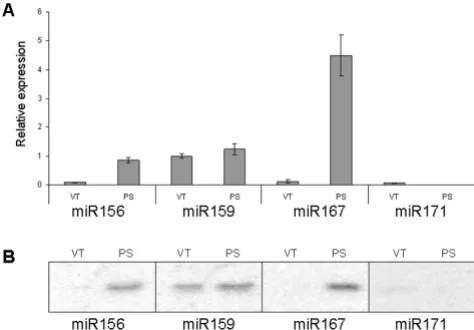
Finally, we used the miRNA UPL assay to quantify miRNA expression levels in pumpkin vascular tissue and the phloem sap. miRNA expression data were normalized to pumpkin phloem RNA binding protein, CmPP16, as this mRNA species was earlier shown to be expressed both in the vascular tissue and in the phloem sap [34].
This assay established that the phloem sap is enriched for miR156 and miR167; phloem sap showed a 10-fold increase in miR156 expression and a 20-fold increase in miR167 expression compared with the surrounding vas-culature. The abundance of miRNA159 was similar in the phloem sap and in the surrounding vascular tissue. Only relatively low levels of miR171 expression were detected in the vascular tissue, but very little or no expression was detectable in the phloem sap (greater than 10-fold below the levels detected in the vasculature; Figure 7A). The results were comparable to those obtained by RNA gel blot analyses (Figure 7B) and spatial analysis using in situ
hybridization [35] that revealed high expression of several miRNA, including miR159, but not miR167, in the
vascu-Expression of miRNAs in vascular tissue and phloem sap
Figure 7
Expression of miRNAs in vascular tissue and phloem sap. A. Expression of miRNAs in pumpkin vascular tissue (VT) and phloem sap (PS) detected by miRNA UPL assay. Samples containing 10 ng total RNA isolated from pumpkin vascular bundles and phloem sap each were subjected to stem-loop RT reactions and subsequent UPL qPCR. The PCR was performed in three replicates and miRNA expression was normalized against CmPP16 and expressed as a ratio with vascular tissue miR159 expression, which was set arbitrarily at 1. B. Expression of miRNAs detected by gel blot analyses.
miRNA UPL probe assay
Figure 6
lar bundles of Arabidopsis thaliana and Nicotiana benthami-ana.
Conclusion
The stem-loop pulsed RT-PCR assays described here are rapid, sensitive, specific and convenient for screening a large number of miRNAs quickly and inexpensively. They require considerably less tissue and time compared with standard gel-based methods. We have demonstrated their reliability, sensitivity and specificity using plant tissue RNA and phloem sap RNA. These assays enable miRNA expression profiling from as little as 20 pg RNA and as lit-tle as 0.1 μl phloem sap.
It is envisaged that this approach will have application in detection and quantification of miRNAs across kingdoms, as well as for other small RNA sequences such as artificial miRNAs and short interfering RNAs (siRNAs).
Competing interests
The author(s) declare that they have no competing inter-ests.
Authors' contributions
EV-G conceived of the project, designed the experiments, conducted the gel-blot, end-point and real-time PCR anal-yses and prepared the manuscript. RW carried out the real-time PCR, cloning and sequencing and prepared the man-uscript. MW conceived of the UPL probe assay. EFW acquired the funding and contributed to and edited the manuscript. All authors read and approved the final man-uscript. RPH has given final approval of the version to be published.
Acknowledgements
We thank Toshi Foster, Charles Ampomah-Dwamena, Anne Gunson and Sue Muggleston for critical reading of the manuscript.
References
1. Bartel DP: MicroRNAs: genomics, biogenesis, mechanism, and function. Cell 2004, 116(2):281-297.
2. Bartel B, Bartel DP: MicroRNAs: at the root of plant develop-ment? Plant Physiol 2003, 132(2):709-717.
3. Mallory AC, Vaucheret H: Functions of microRNAs and related small RNAs in plants. Nat Genet 2006, 38(Suppl):S31-36. 4. Zhang B, Wang Q, Pan X: MicroRNAs and their regulatory roles
in animals and plants. J Cell Physiol 2007, 210(2):279-289. 5. Axtell MJ, Snyder JA, Bartel DP: Common functions for diverse
small RNAs of land plants. Plant Cell 2007, 19(6):1750-1769. 6. Zhang B, Pan X, Cannon CH, Cobb GP, Anderson TA:
Conserva-tion and divergence of plant microRNA genes. Plant J 2006,
46(2):243-259.
7. Griffiths-Jones S, Grocock RJ, van Dongen S, Bateman A, Enright AJ:
miRBase: microRNA sequences, targets and gene nomencla-ture. Nucleic Acids Res 2006:D140-144.
8. Jackson RJ, Standart N: How do microRNAs regulate gene expression? Sci STKE 2007, 2007(367):re1.
9. Aukerman MJ, Sakai H: Regulation of flowering time and floral organ identity by a MicroRNA and its APETALA2-like target genes. Plant Cell 2003, 15(11):2730-2741.
10. Chen X: A microRNA as a translational repressor of APETALA2 in Arabidopsis flower development. Science 2004,
303(5666):2022-2025.
11. Llave C, Xie Z, Kasschau KD, Carrington JC: Cleavage of Scare-crow-like mRNA targets directed by a class of Arabidopsis miRNA. Science 2002, 297(5589):2053-2056.
12. Palatnik JF, Allen E, Wu X, Schommer C, Schwab R, Carrington JC, Weigel D: Control of leaf morphogenesis by microRNAs. Nature 2003, 425(6955):257-263.
13. Bao N, Lye KW, Barton MK: MicroRNA binding sites in Arabi-dopsis class III HD-ZIP mRNAs are required for methylation of the template chromosome. Dev Cell 2004, 7(5):653-662. 14. Llave C, Kasschau KD, Rector MA, Carrington JC: Endogenous and
silencing-associated small RNAs in plants. Plant Cell 2002,
14(7):1605-1619.
15. Rhoades MW, Reinhart BJ, Lim LP, Burge CB, Bartel B, Bartel DP:
Prediction of plant microRNA targets. Cell 2002,
110(4):513-520.
16. Sunkar R, Zhu JK: Novel and stress-regulated microRNAs and other small RNAs from Arabidopsis. Plant Cell 2004,
16(8):2001-2019.
17. Sunkar R, Kapoor A, Zhu JK: Posttranscriptional induction of two Cu/Zn superoxide dismutase genes in Arabidopsis is mediated by downregulation of miR398 and important for oxidative stress tolerance. Plant Cell 2006, 18(8):2051-2065. 18. Achard P, Herr A, Baulcombe DC, Harberd NP: Modulation of flo-ral development by a gibberellin-regulated microRNA. Development 2004, 131(14):3357-3365.
19. Li H, Xu L, Wang H, Yuan Z, Cao X, Yang Z, Zhang D, Xu Y, Huang H: The Putative RNA-dependent RNA polymerase RDR6 acts synergistically with ASYMMETRIC LEAVES1 and 2 to repress BREVIPEDICELLUS and MicroRNA165/166 in Ara-bidopsis leaf development. Plant Cell 2005, 17(8):2157-2171. 20. Schwab R, Palatnik JF, Riester M, Schommer C, Schmid M, Weigel D:
Specific effects of microRNAs on the plant transcriptome. Dev Cell 2005, 8(4):517-527.
21. Chen X: MicroRNA biogenesis and function in plants. FEBS Lett
2005, 579(26):5923-5931.
22. Juarez MT, Kui JS, Thomas J, Heller BA, Timmermans MC: micro-RNA-mediated repression of rolled leaf1 specifies maize leaf polarity. Nature 2004, 428(6978):84-88.
23. Kidner CA, Martienssen RA: Spatially restricted microRNA directs leaf polarity through ARGONAUTE1. Nature 2004,
428(6978):81-84.
24. Tang G, Reinhart BJ, Bartel DP, Zamore PD: A biochemical frame-work for RNA silencing in plants. Genes Dev 2003, 17(1):49-63. 25. Mallory AC, Reinhart BJ, Jones-Rhoades MW, Tang G, Zamore PD, Barton MK, Bartel DP: MicroRNA control of PHABULOSA in leaf development: importance of pairing to the microRNA 5' region. Embo J 2004, 23(16):3356-3364.
26. Yoo BC, Kragler F, Varkonyi-Gasic E, Haywood V, Archer-Evans S, Lee YM, Lough TJ, Lucas WJ: A systemic small RNA signaling system in plants. Plant Cell 2004, 16(8):1979-2000.
27. Parizotto EA, Dunoyer P, Rahm N, Himber C, Voinnet O: In vivo investigation of the transcription, processing, endonucleo-lytic activity, and functional relevance of the spatial distribu-tion of a plant miRNA. Genes Dev 2004, 18(18):2237-2242. 28. Alvarez JP, Pekker I, Goldshmidt A, Blum E, Amsellem Z, Eshed Y:
Endogenous and synthetic microRNAs stimulate simultane-ous, efficient, and localized regulation of multiple targets in diverse species. Plant Cell 2006, 18(5):1134-1151.
29. Mineno J, Okamoto S, Ando T, Sato M, Chono H, Izu H, Takayama M, Asada K, Mirochnitchenko O, Inouye M, Kato I: The expression profile of microRNAs in mouse embryos. Nucleic Acids Res
2006, 34(6):1765-1771.
30. Lu C, Tej SS, Luo S, Haudenschild CD, Meyers BC, Green PJ: Eluci-dation of the small RNA component of the transcriptome. Science 2005, 309(5740):1567-1569.
31. Chen C, Ridzon DA, Broomer AJ, Zhou Z, Lee DH, Nguyen JT, Bar-bisin M, Xu NL, Mahuvakar VR, Andersen MR, Lao KQ, Livak KJ, Gue-gler KJ: Real-time quantification of microRNAs by stem-loop RT-PCR. Nucleic Acids Res 2005, 33(20):e179.
Publish with BioMed Central and every scientist can read your work free of charge "BioMed Central will be the most significant development for disseminating the results of biomedical researc h in our lifetime."
Sir Paul Nurse, Cancer Research UK
Your research papers will be:
available free of charge to the entire biomedical community
peer reviewed and published immediately upon acceptance
cited in PubMed and archived on PubMed Central
yours — you keep the copyright
Submit your manuscript here:
http://www.biomedcentral.com/info/publishing_adv.asp
BioMedcentral 33. miRBase::Sequences [http://microrna.sanger.ac.uk/sequences/
index.shtml]
34. Xoconostle-Cazares B, Xiang Y, Ruiz-Medrano R, Wang HL, Monzer J, Yoo BC, McFarland KC, Franceschi VR, Lucas WJ: Plant paralog to viral movement protein that potentiates transport of mRNA into the phloem. Science 1999, 283(5398):94-98. 35. Valoczi A, Varallyay E, Kauppinen S, Burgyan J, Havelda Z: