R E S E A R C H
Open Access
Ameliorative effects of polyunsaturated fatty
acids against palmitic acid-induced insulin
resistance in L6 skeletal muscle cells
Keisuke Sawada
1, Kyuichi Kawabata
1,4, Takatoshi Yamashita
2, Kengo Kawasaki
3, Norio Yamamoto
3and
Hitoshi Ashida
1*Abstract
Background:Fatty acid-induced insulin resistance and impaired glucose uptake activity in muscle cells are fundamental events in the development of type 2 diabetes and hyperglycemia. There is an increasing demand for compounds including drugs and functional foods that can prevent myocellular insulin resistance.
Methods:In this study, we established a high-throughput assay to screen for compounds that can improve myocellular insulin resistance, which was based on a previously reported non-radioisotope 2-deoxyglucose (2DG) uptake assay. Insulin-resistant muscle cells were prepared by treating rat L6 skeletal muscle cells with 750μM palmitic acid for 14 h. Using the established assay, the impacts of several fatty acids on myocellular insulin resistance were determined.
Results:In normal L6 cells, treatment with saturated palmitic or stearic acid alone decreased 2DG uptake, whereas unsaturated fatty acids did not. Moreover, co-treatment with oleic acid canceled the palmitic acid-induced
decrease in 2DG uptake activity. Using the developed assay with palmitic acid-induced insulin-resistant L6 cells, we determined the effects of other unsaturated fatty acids. We found that arachidonic, eicosapentaenoic and
docosahexaenoic acids improved palmitic acid-decreased 2DG uptake at lower concentrations than the other unsaturated fatty acids, including oleic acid, as 10μM arachidonic acid showed similar effects to 750μM oleic acid. Conclusions:We have found that polyunsaturated fatty acids, in particular arachidonic and eicosapentaenoic acids prevent palmitic acid-induced myocellular insulin resistance.
Keywords:Insulin resistance, Glucose uptake, L6 skeletal muscle cells, Palmitic acid, Arachidonic acid, Eicosapentae-noic acid
Background
Insulin resistance is an impaired response to insulin in specific organs or cells such as liver, fat and muscle, and is strongly associated with the development of obesity and type 2 diabetes [1]. Elevated plasma free fatty acid levels is an important factor, because it causes insulin resistance in skeletal muscle, the major site for blood glucose disposal [2]. Thus, many studies have been reported on the relationship between fatty acids and insulin resistance, and revealed that saturated fatty
acids, particularly palmitic acid, induce insulin resistance in myotubes [3], whereas unsaturated fatty acids do not [4,5]. In skeletal muscle, insulin resistance is mediated by the intramyocellular accumulation of the metabolites of saturated palmitic acid, namely diacylglycerol (DAG) and ceramide. DAG downregulates insulin-sensitive glu-cose transporter type 4 (GLUT4) and insulin receptor (IR) by activating the inflammatory transcription factor nuclear factor (NF)-B [6]. Ceramide inhibits protein kinase B (PKB/Akt) activity which plays an important role in insulin signaling [7,8]. The levels of these meta-bolites progressively increase as insulin resistance wor-sens [9-11], and further decrease glucose uptake activity in myotubes.
* Correspondence: ashida@kobe-u.ac.jp 1
Department of Agrobioscience, Graduate School of Agricultural Science, Kobe University, Kobe, Hyogo 657-8501, Japan
Full list of author information is available at the end of the article
There is an increasing demand for drugs and func-tional foods that are capable of regulating blood glucose levels. Several unsaturated fatty acids, including palmito-leic and opalmito-leic acids, were reported to ameliorate palmitic acid-induced insulin resistance in myotubes [4,12,13]. Collet al.(2008) reported that oleic acid inhibited intra-myocellular DAG accumulation by enhancing b-oxida-tion of palmitoyl CoA and upregulating diacylglycerol acyltransferase 2, an enzyme that synthesizes triacylgly-cerol from DAG, which ultimately inhibited palmitic acid-induced downregulation of IR [12]. Chen et al. have reported that berberine, an isoquinoline alkaloid, improves palmitic acid-induced insulin resistance in L6 myotubes by inhibiting peroxisome proliferator-activated receptor (PPAR)-g [14]. Other than these compounds, little is known about inhibitors of palmitic acid-induced insulin resistance in muscle cells.
In these earlier studies, insulin resistance was evalu-ated based on downregulation of IR and GLUT4 expres-sion or decreased radioisotope-labeled glucose uptake [15-22]. We recently reported an enzymatic 2DG uptake assay, which had greater processing capacity compared with these conventional tools [23,24]. This enzymatic assay enables us to measure 2DG uptake into myotubes cultured in a 96-well microplate by measuring the fluor-escence of resorufin, which is derived from resazurin, and is appropriate to screen for compounds capable of regulating 2DG uptake, including polyphenols [25,26]. In this study, we report our development of a high-throughput procedure to screen for compounds that can prevent palmitic acid-induced myocellular insulin resis-tance using this enzymatic 2DG uptake assay, and assessment of the anti-insulin-resistant effects of unsatu-rated fatty acids.
Results
Determination of optimum treatment time and concentration of palmitic acid
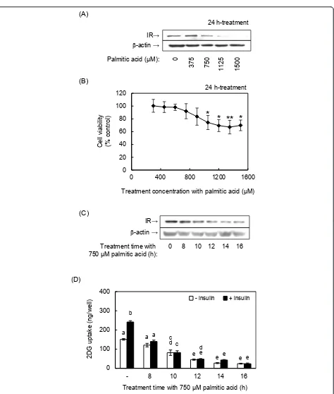
To induce insulin resistance in muscle cells, we treated differentiated L6 skeletal muscle cells with palmitic acid according to a method reported by Chaves et al. [7]. Treatment-time and concentration of palmitic acid were defined by evaluating the decreases in IR expression, cellviability and 2DG uptake activity. Treating cells with 750μM palmitic acid for 24 h significantly decreased IR expression (Figure 1A). After we confirmed that palmitic acid did not show significant cytotoxicity by 900μM, the treatment concentration was fixed at 750 μM according to the previous reports [4,6] (Figure 1B). This concentration of palmitic acid also decreased IR expres-sion after 14 and 16 h of treatment (Figure 1B), and decreased basal and insulin-induced 2DG uptake activity in a time-dependent manner (Figure 1C). The decrease in 2DG uptake activity was well correlated with the
decrease in IR expression. Based on these results, we prepared insulin-resistant cells in the following experi-ments by treating differentiated L6 cells with 750 μM palmitic acid for 14 h.
Effect of fatty acids on myocellular glucose uptake activity
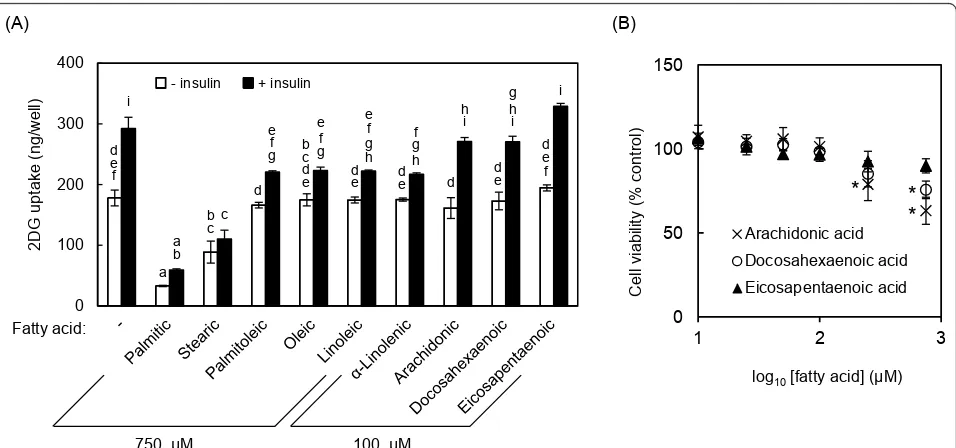
We used the enzymatic 2DG uptake assay to evaluate the effects of several saturated and unsaturated fatty acids commonly contained in food on the development of insulin resistance. Palmtic acid (16:0) significantly decreased glucose uptake activity to 19% and 33% in the absence and presence of insulin, respectively (Figure 2A). Stearic acid (18:0) also significantly decreased 2DG uptake, although its effect was weaker than that of pal-mitic acid.
On the other hand, the unsaturated forms of these fatty acids, such as palmitoleic (16:1), oleic (18:1), lino-leic (18:2) anda-linolenic acid (18:3), only affected 2DG uptake activity in the presence of insulin. Other polyun-saturated fatty acids, including arachidonic (20:4), doco-sahexaenoic (22:6) and eicosapentaenoic acids (20:5), were tested at a lower concentration (100μM) because arachidonic and docosahexaenoic acids were cytotoxic at 750 μM (Figure 2B). These three polyunsaturated fatty acids did not affect 2DG uptake activity (Figure 2A). Palmitic acid showed the strongest effect in terms of decreasing 2DG uptake activity. These results confirm that palmitic acid is an appropriate inducer of myocellu-lar insulin resistance.
Effects of unsaturated fatty acids on palmitic acid-induced insulin resistance
To confirm the effects of oleic acid on palmitic acid-induced insulin resistance, L6 myotubes were simulta-neously treated with palmitic acid and oleic acid. In this assay, oleic acid canceled palmitic acid-induced decreases in IR and GLUT4 expression (Figure 3A), as expected. Furthermore, oleic acid dose-dependently inhibited the palmitic acid-induced decrease in 2DG uptake activity (Figure 3B). These results indicate that the enzymatic 2DG uptake assay using palmitic acid-treated cells is useful to screen for compounds that reg-ulate insulin resistance in skeletal muscle cells.
Palmitic acid (ȝM): 0
375 750 1125 1500 (A)
0 8 10 12 14 16
Treatment time with
ȝ0SDOPLWLFDFLG(h):
(B)
E-actinĺ
IRĺ
E-actinĺ
IRĺ
2DG
upt
ake (ng/
w
ell)
- 8 10 12 14 16
a
a a b
c
d c d
e e
e e e e
(D)
24 h-treatment
7UHDWPHQWWLPHZLWKȝ0palmitic acid (h)
0 100 200 300 400
+ insulin - insulin
(C) Cell viability (% control)
0 20 40 60 80 100 120
0 400 800 1200 1600
Treatment concentration with SDOPLWLFDFLGȝ0
24 h-treatment
* *
**
*
much more effective. For example, 100μM docosahex-aenoic acid and eicosapentdocosahex-aenoic acid elicited similar effects to 750 μM oleic acid (Figure 4A). Moreover, ara-chidonic acid was more effective than the other polyun-saturated fatty acids at 10 and 50μM (Figure 4B), with significant effects at 10 μM. It was confirmed that eico-sapentaenoic acid was also effective at the lower concen-trations. As shown in Figure 4C, significant recovery
effect of arachidonic acid on 2DG uptake was observed at 10μM and reached maximum levels at 50μM and 75 μM in the presence and absence of insulin, respectively.
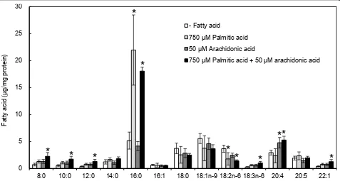
We further investigated whether treated fatty acids would incorporated into the cells by measuring fatty acid contents in the cells treated with 750μM palmitic acid or 50μM arachidonic acid (Figure 5). It was con-firmed that intracellular contents of the treated fatty 0
100 200 300 400
2DG
upt
ake (ng/
w
ell)
e f g
a b
b c
a
d e e
f g d
e f i
b c d e
e f g h d e
f g h d
e d
h i
d e f i g h i
c d
Cell viability
(%
control)
log10>IDWW\DFLG@ȝ0
(A) (B)
+ insulin - insulin
0 50 100 150
1 2 3
Arachidonic acid Docosahexaenoic acid Eicosapentaenoic acid
Fatty acid:
ȝ0 ȝ0
*
*
*
Figure 2Fatty acid-induced decreases in 2DG uptake activity and viability of L6 cells.(A)Differentiated L6 cells were incubated with the indicated fatty acid (750μM palmitic, stearic, palmitoleic, oleic, linoleic ora-linolenic acids; or 100μM arachidonic, docosahexaenoic or eicosapentaenoic acid) for 14 h. 2DG uptake activity was measured in the absence or presence of 100 nM insulin.(B)Cell viability was determined by crystal violet staining after treatment with the indicated concentrations of arachidonic, docosahexaenoic and eicosapentaenoic acids for 24 h. Different letters indicate significant changes (p< 0.05) for panel (A). * indicates significant difference vs control atp< 0.05 for panel (B)
0 100 200 300 400
GLUT4ĺ
E-actinĺ
IRĺ
ȝ0Palmitic acid:
ȝ0Oleic acid: --
-+ +
+
(A)
14 h-treatment
ȝ0Palmitic acid:
2OHLFDFLGȝ0
-᧩
-+ +
100
(B)
+ 300
+ 500
+ 750 a
a b
c d c d f a b c b
c d f
2DG
upt
ake (ng/
w
ell)
+ insulin - insulin
a a
c d f
f f
!" # $ % &' !" # $ % &' ( ) *) ! + , # ! " # $ % &' '# ./ 2 ,# !+ !" # $ % &' 3 3 3 3 3 3 3 3 3 3 3 3 % 3 % 3 % 3 % %
acids were significantly higher than those of non-treated cells; i.e., content of palmitic acid increased 3-fold while that of arachdonic acid 2-fold. Co-treatment with palmi-tic and arachidonic acids also increased both fatty acids in the cells.
Discussion
The in vitro evaluation of compounds that can affect insulin resistance in myotubes has long been performed by measuring the changes in uptake of radiolabeled glu-cose analogs and/or in the expression and phosphoryla-tion of components of the insulin signaling pathways [15-22]. However, these conventional approaches are unsuitable for high-throughput screening, because radi-olabeled analogs are costly and require specialized equipments, and both approaches have low processing capacity. Our research group previously reported a high-throughput enzymatic 2DG uptake assay system [23,24]. In the present study, we developed a high-throughput assay based on this previously reported assay system to screen for compounds that can ameliorate insulin resis-tance in L6 myotubes. Using this system, we found that arachidonic, eicosapentaenoic, and docosahexaenoic acids were more effective than oleic acid [12,13] in terms of ameliorating insulin resistance.
In this study, we first confirmed that saturated palmi-tic and stearic acids decreased glucose uptake by L6 myotubes, whereas their unsaturated forms (i.e., oleic,
palmitoleic, linoleic and a-linolenic acids) did not. These results are consistent with previous reports [4,5]. Indeed, it was reported that myocellular insulin resis-tance is mediated by the accumulation of the saturated fatty acid metabolites DAG and ceramide. DAG acti-vates novel PKCs that downregulate GLUT4 and IR expression by activating NF-B [6]. Ceramide inhibits PKB/Akt activity, which plays an important role in insu-lin signainsu-ling [7,8]. Palmitic acid is a stronger inducer of the accumulation of these metabolites and the transcrip-tional activity of NF-B compared as stearic acid [3], which is correlated with the greater decrease in glucose uptake activity in palmitic acid-treated cells. These facts and the obtained results indicate that our assay with palmitic acid-induced insulin resistant L6 myotubes is suitable to screen for compounds that can induce or prevent insulin resistance.
Using palmitic acid as an inducer of insulin resistance, we tested the anti-insulin resistant effects of several fatty acids. All of the unsaturated fatty acids tested in this study, namely palmitoleic, oleic, linoleic, linolenic, ara-chidonic, docosahexaenoic and eicosapentaenoic acids, ameliorated palmitic acid-induced decrease in 2DG uptake (Figure 4). Unsaturated fatty acids, in particular polyunsaturated fatty acids, are known to be oxidized easily. Therefore, to investigate whether the recovery of 2DG uptake was dependent on the exogenously added fatty acids per se or their oxidized derivatives, we
*
*
*
*
*
*
*
*
*
*
*
0 5 10 15 20 25 30
8:0 10:0 12:0 14:0 16:0 16:1 18:0 18:1n-9 18:2n-6 18:3n-6 20:4 20:5 22:1
- Fatty acid
ȝM Palmitic acid
ȝM Arachidonic acid
ȝ03DOPLWLFDFLGȝ0DUDFKLGRQLFDFLG
)DWW\
D
FLG
ȝJPJSURWHLQ
Figure 5Effect of exogenously added palmitic and oleic acids on cellular fatty acid contents. Differentiated L6 myotubes were treated with 750μM palmitic acid and/or 50μM arachidonic acid for 14 h. Intracellular fatty acid contents were measured by using a gas
conducted an experiment with an antioxidant, butylated hydroxytoluene (BHT). In the presence of 0.2 mM BHT in the medium, palmitic acid-induced decrease and its amelioration by arachidonic acid were confirmed(data not shown). However, 2DG uptake was decreased by treatment with BHT alone, indicating that BHT itself affected cellular functions under our experimental con-ditions. Meanwhile, Figure 5 shows that treated palmitic and arachidonic acid were increased in the cells. These results suggest that the exogenously added arachidonic acid and other unsaturated fatty acids are incorporated into the cells, and contribute to their anti-insulin resis-tant effects.
Of the tested unsaturated fatty acids, oleic acid has been a focus of many researches. The effects of oleic acid were reported to be dependent on its ligand activity for PPARawhich plays essential roles in regulation of lipid metabolism [27]. PPARaactivation improves intra-myocellular DAG accumulation by accelerating b-oxida-tion and upregulating DAG acyltransferase 2, an enzyme that synthesizes triacylglycerol from DAG [12]. The effects of other fatty acids would depend, at least in part, on PPARa because of its high affinity for unsatu-rated fatty acids [27,28]. Our results indicate that the effects of arachidonic, eicosapentaenoic and docosahex-aenoic acids at much lower concentrations compared with oleic acid (Figure 4A and 4C). This result strongly suggests that bioactive metabolites of these polyunsatu-rated fatty acids are involved in underlying mechanism. Indeed, prostaglandin E2, a metabolite of n-6 polyunsa-turated fatty acids, ameliorated the palmitic acid-induced inflammation, and recovered PKB/Akt phos-phorylation level and glucose uptake in muscle cells [29]. Therefore, the results obtained for polyunsaturated fatty acids in our assay are mainly due to their PPARa ligand activity or anti-inflammatory activity. However, the mechanisms by which arachidonic, eicosapentaenoic and docosahexaenoic acids ameliorated insulin resis-tance, which seem to be independent of the mechanisms exploited by the other unsaturated fatty acids tested, remain unclear. Further studies are needed to clarify the underlying mechanisms.
Conclusions
We developed a high-throughput assay to screen for compounds that can improve palmitic acid-induced insulin resistance by modifying our previously estab-lished enzymatic 2DG uptake assay [23,24]. Using the developed assay, we determined the ameliorative effects of several polyunsaturated fatty acids on insulin resis-tance. Notably, arachidonic, eicosapentaenoic and doco-sahexaenoic acids showed were more potent than the other unsaturated fatty acids tested in this study.
Methods Materials
Minimum essential medium (MEM), 2-deoxyglucose (2DG), glucose-6-phosphate dehydrogenase (G6PDH), resazurin and stearic acid (Sigma, St. Louis, MO); dia-phorase andb-nicotinamide adenine dinucleotide phos-phate (b-NADP+; Oriental Yeast, Tokyo, Japan); arachidonic acid, docosahexaenoic acid and eicosapen-taenoic acid (Cayman Chemical,Ann Arbor, MI); fetal bovine serum (FBS; Biological Industries, Kibbutz Beit Haemek, Israel); palmitoleic acid and bovine serum albumin (BSA; Nakarai Tesque, Kyoto, Japan) and Lumi-Light Plus Western Blotting Substrate® (Roche Diagnostics, Mannheim, Germany) were purchased from commercial sources. Anti-GLUT4, anti-IR, anti-goat IgG and anti-rabbit IgG antibodies were purchased from Santa Cruz Biotechnology (Santa Cruz, CA). All other reagents were purchased from WAKO Pure Chemicals (Osaka, Japan), unless otherwise specified.
Cell culture
Rat skeletal muscle L6 cells (passages 27-38) were main-tained in MEM supplemented with 10% FBS, 100 U/mL penicillin, and 100 μg/mL streptomycin at 37°C in a humidified atmosphere with 5% CO2. To induce differ-entiation into myotubes, L6 cells were seeded on 96-well plates (4 × 10³ cells/0.2 mL), 60-mm dishes (1.2 × 10⁵ cells/4 mL), or 35-mm dishes (4 × 10⁴ cells/1 mL) in culture medium. After 2 days, the medium was replaced with MEM containing 2% FBS and antibiotics, which was changed every other day. L6 cells were used for experiments after 5 days of differentiation. Fatty acid-containing media were prepared by conjugating fatty acids to BSA, as previously described [7], but with some modifications. Briefly, palmitate was dissolved in ethanol and diluted in MEM containing 2% (w/v) BSA. The cells were incubated for 14 h in MEM containing 2% FBS and 2% BSA in either the presence or absence of fatty acids. The treated cells were used for western blotting analysis and 2DG uptake assays.
Western blotting
min at 4°C, the supernatant was collected and used as whole protein. Aliquots of whole protein were separated on 10% polyacrylamide gels and transferred to polyviny-lidene difluoride membranes (Biotrace, Pall Corporation, Port Washington, NY). After blocking with Blocking one® (Nakarai Tesque), the membranes were treated with appropriate specific primary antibodies for 1 h at room temperature, followed by the corresponding horse-radish peroxidase-conjugated secondary antibody for 1 h at room temperature. Specific immune complexes were detected using ECL plus kits (GE Healthcare Bio-Sciences Corporation, Tokyo, Japan).
Glucose uptake assay
Glucose uptake in L6 myotubes was measured using an enzymatic 2DG uptake assay as previously described [23,24], with some modifications. L6 myotubes on a 96-well plate were serum-starved in 0.2% BSA/MEM for 18 h and were treated with DMSO and insulin (final con-centration, 0.1 μM) in the same media. The cells were washed twice with KRH buffer containing 0.1% (w/v) BSA and incubated with 1 mM 2DG in 0.1% (w/v) BSA/ KRH buffer for 20 min at 37°C in 5% CO2. They were then washed twice with 0.1% (w/v) BSA/KRH buffer, lysed with 0.1 N sodium hydroxide, warmed at 60°C for 10 min, and dried at 85°C for 50 min. The dried cell lysate was solubilized with 0.1 N hydrochloric acid and 200 mM triethanolamine (TEA) (pH 8.1), and gently stirred using a microplate shaker. The lysate was mixed with an assay cocktail [50 mM TEA, pH 8.1, 50 mM KCl, 0.02% (w/v) BSA, 0.1 mMb-NADP+, 2 units dia-phorase, 150 units G6PDH, 2μM resazurin] on another 96-well plate and incubated at 37°C for 50 min. The fluorescence of resorufin was measured at 570 nm with excitation at 530 nm using a Wallac 1420 ARVOsx (Per-kin-Elmer, Boston, MA). The 2DG concentration in each well was calculated based on a standard curve gen-erated with a 2DG-6-phosphate solution.
Cytotoxicity
The cytotoxicity of fatty acids was determined by crystal violet staining [25]. Differentiated L6 cells on 96-well microplate were treated with ethanol or fatty acids (300, 450, 600, 750, 900, 1050, 1200, 1350 and 1500μM for Figure 1B; and 10, 25, 50, 100, 250 and 750 μM for Fig-ure 2B) in MEM containing 2% BSA for 24 h, and then stained with 2% ethanol containing 0.2% (w/v) crystal violet for 10 min. The wells were washed three times with tap water, and the stained cells were extracted with 50% ethanol containing 0.5% (w/v) sodium dodecyl sul-fate. The absorbance at 570 nm with a reference wave-length of 630 nm was measured using the Wallac 1420 ARVOsx.
Measurement of fatty acid contents
L6 myotubes on a 35-mm culture dish were incubated for 14 h in MEM containing 2% FBS and 2% BSA in either the presence or absence of palmitic and arachido-nic acids. The treated cells were washed with 2% BSA/ KRH for 3 times, and with phosphatydyl buffer serine twice, and collected with phosphate buffered saline (137 mM sodium chloride, 8.10 mM sodium phosphate diba-sic, 2.68 mM potassium chloride, 1.47 mM potassium phosphate monobasic). Intracellular fatty acid contents were measured by using a gas chromatography accord-ing to previous report with slight modification [30]. Briefly, lipids were extracted from the cells by chloro-form:methanol (2:1, v/v). hydolized and methylated with potassium hydroxide in methanol, and subjected to a GC2010 gas chromatograph (Shimadzu Co., Kyoto, Japan) equipped with DB23 column (Agilent J&W, Fol-som, CA).
Statistical analysis
Data are shown as means ± SD of results from three independent experiments. Statistical analyses were per-formed using the Tukey-Kramer test, except for the cytotoxicity tests (Dunnet test, for Figures 1B and 2B). Values ofp< 0.05 were considered significant.
Abbreviations
BSA: Bovine serum albumin; BHT: Butylated hydroxytoluene; DAG: Diacylglycerol; 2DG: 2-deoxyglucose; FBS: Fetal bovine serum; GLUT4: Glucose transporter type 4; G6PDH: Glucose-6-phosphate dehydrogenase; IR: Insulin receptor; KRH: Krebs-Ringer HEPES buffer;β-NADP+:β-nicotinamide adenine dinucleotide phosphate; NF-κB: Nuclear factor-κB; PKB/Akt: Protein kinase B; PPAR: Peroxisome proliferator-activated receptor.
Author details
1Department of Agrobioscience, Graduate School of Agricultural Science, Kobe University, Kobe, Hyogo 657-8501, Japan.2J-Oil Mills Inc., Yokohama, Kanagawa 230-0053, Japan.3Food Science Research Center, House Wellness Foods Corporation, Itami, Hyogo 664-0011, Japan.4Department of Bioscience, Fukui Prefectural University, Fukui 910-1195, Japan.
Authors’contributions
KS conceived the study, its design and coordination, performed all experiments and statistical analysis. KS and HA discussed results and made the manuscript. TK measured fatty acid contents. NY and HA participated in the design and coordination of the study and discussion of results. K Kawabata, TY, and K Kawasaki participated in the design and coordination of the study. All authors read and approved the final manuscript.
Competing interests
The authors declare that they have no competing interests.
Received: 11 January 2012 Accepted: 12 March 2012 Published: 12 March 2012
References
1. DeFronzo RA, Ferrannini E:Insulin resistance: A multifaceted syndrome responsible for NIDDM, obesity, hypertension, dyslipidemia, and atherosclerotic cardiovascular disease.Diabetes Care1991,14:173-194. 2. DeFronzo RA, Jacot E, Jequier E, Maeder E, Wahren J, Felber JP:The effect
calorimetry and hepatic and femoral venous catheterization.Diabetes 1981,30:1000-1007.
3. Hommelberg PPH, Plat J, Langen RC, Schols AM, Mensink RP:Fatty acid-induced NF-κB activation and insulin resistance in skeletal muscle are chain length dependent.Am J Physiol Endocrinol Metab2009,296: E114-E120.
4. Dimopoulos N, Watson M, Sakamoto K, Hundal HS:Differential effects of palmitate and palmitoleate on insulin action and glucose utilization in rat L6 skeletal muscle cells.Biochem J2006,399:473-481.
5. Lee JS, Pinnamaneni SK, Eo SJ, Cho IH, Pyo JH, Kim CK, Sinclair AJ, Febbraio MA, Watt MJ:Saturated, but not n-6 polyunsaturated, fatty acids induce insulin resistance: role of intramuscular accumulation of lipid metabolites.J Appl Physiol2006,100:1467-1474.
6. Jové M, Planavila A, Sánchez RM, Merlos M, Laguna JC, Vázquez-Carrera M: Palmitate induces tumor necrosis factor-αexpression in C2C12 skeletal muscle cells by a mechanism involving protein kinase C and nuclear factor-κB activation.Endocrinology2006,147:552-561.
7. Chavez J, Knotts TA, Wang LP, Li G, Dobrowsky RT, Florant GL, Summers SA: A role for ceramide, but not diacylglycerol, in the antagonism of insulin signal transduction by saturated fatty acids.J Biol Chem2003, 278:10297-10303.
8. Pickersgill L, Litherland GJ, Greenberg AS, Walker M, Yeaman SJ:Key role for ceramides in mediating insulin resistance in human muscle cells.J Biol Chem2007,282:12583-12589.
9. Haus JM, Kashyap SR, Kasumov T, Zhang R, Kelly KR, Defronzo RA, Kirwan JP:Plasma ceramides are elevated in obese subjects with type 2 diabetes and correlate with the severity of insulin resistance.Diabetes 2009,58:337-343.
10. Itani S, Ruderman NB, Schmieder F, Boden G:Lipid-induced insulin resistance in human muscle is associated with changes in diacylglycerol, protein kinase C, and IκB-α.Diabetes2002,51:2005-2011.
11. Moro C, Galgani JE, Luu L, Pasarica M, Mairal A, Bajpeyi S, Schmitz G, Langin D, Liebisch G:SR Smith: Influence of gender, obesity, and muscle lipase activity on intramyocellular lipids in sedentary individuals.J Clin Endocrinol Metab2009,94:3440-3447.
12. Coll T, Eyre E, Rodríguez-Calvo R, Palomer X, Sánchez RM, Merlos M, Laguna JC, Vázquez-Carrera M:Oleate reverses palmitate-induced insulin resistance and inflammation in skeletal muscle cells.J Biol Chem2008, 283:11107-11116.
13. Gao D, Griffiths HR, Bailey CJ:Oleate protects against palmitate-induced insulin resistance in L6 myotubes.Br J Nutr2009,102:1557-1563. 14. Chen Y, Li Y, Wang Y, Wen Y, Sun C:Berberine improves
free-fatty-acid-induced insulin resistance in L6 myotubes through inhibiting peroxisome proliferator-activated receptor ? and fatty acid transferase expressions.Metabolism2009,58:1694-1702.
15. Bhattacharya S, Dey D, Roy SS:Molecular mechanism of insulin resistance. J Biosci2007,32:405-413.
16. Zhang J, Wu W, Li D, Guo Y, Ding H:Overactivation of NF-?B impairs insulin sensitivity and mediates palmitate-induced insulin resistance in C2C12 skeletal muscle cells.Endocrine2010,37:157-166.
17. Kim JH, Lee JO, Lee SK, Jung JH, You GY, Park SH, Park M, Kim SD, Kim HS: Clozapine activates AMP-activated protein kinase (AMPK) in C2C12 myotube cells and stimulates glucose uptake.Life Sci2010,87:42-48. 18. Qu W, Zhao L, Peng X, Yang X, Ying C, Hao L, Sun X:Biphasic effects of
chronic ethanol exposure on insulin-stimulated glucose uptake in primary cultured rat skeletal muscle cells: role of the Akt pathway and GLUT4.Diabetes Metab Res Rev2011,27:47-53.
19. Wasada T, Yano T, Ohta M, Yui N, Iwamoto Y:ATP-Sensitive potassium channels modulate glucose transport in cultured human skeletal muscle cells.Endocr J2001,48:369-375.
20. Yin J, Gao Z, Liu D, Liu Z, Ye J:Berberine improves glucose metabolism through induction of glycolysis.Am J Physiol Endocrinol Metab2008,294: E148-156.
21. Powell DJ, Turban S, Gray A, Hajduch E, Hundal HS: Intracellular ceramide synthesis and protein kinase Cζactivation play an essential role in palmitate-induced insulin resistance in rat L6 skeletal muscle cells. Biochem J 2004,382:619-6129.
22. Koves TR, Ussher JR, Noland RC, Slentz D, Mosedale M, Ilkayeva O, Bain J, Stevens R, Dyck JRB, Newgard CB, Lopaschuk GD, Muoio DM:
Mitochondrial overload and incomplete fatty acid oxidation contribute to skeletal muscle insulin resistance.Cell Metab2008,7:45-56.
23. Yamamoto N, Sato T, Kawasaki K, Murosaki S, Yamamoto Y:A nonradioisotope, enzymatic assay for 2-deoxyglucose uptake in L6 skeletal muscle cells cultured in a 96-well microplate.Anal Biochem2006, 351:139-145.
24. Yamamoto N, Kawasaki K, Kawabata K, Ashida H:An enzymatic fluorimetric assay to quantitate 2-deoxyglucose and 2-deoxyglucose-6-phaophate for in vitro and in vivo use.Anal Biochem2010,404:238-240. 25. Kawabata K, Sawada K, Ikeda K, Fukuda I, Kawasaki K, Yamamoto N,
Ashida H:Prenylated chalcones 4-hydroxyderricin and xanthoangelol stimulate glucose uptake in skeletal muscle cells by inducing GLUT4 translocation.Mol Nutr Food Res2011,55:467-475.
26. Yamamoto N, Kawabata K, Sawada K, Ueda M, Fukuda I, Kawasaki K, Murakami A, Ashida H:Cardamonin stimulates glucose uptake through translocation of glucose transporter-4 in L6 myotubes.Phytother Res 2011,25:1218-1224.
27. Vega RB, Huss JM, Kelly DP:The coactivator PGC-1 cooperates with peroxisome proliferator-activated receptor in transcriptional control of nuclear genes encoding mitochondrial fatty acid oxidation enzymes.Mol Cell Biol2000,20:1868-1876.
28. Krey G, Braissant O, L’Horset F, Kalkhoven E, Perroud M, Parker MG, Wahli W: Fatty acids, eicosanoids, and hypolipidemic agents identified as ligands of peroxisome proliferator-activated receptors by coactivator-dependent receptor ligand assay.Mol Endocrinol1997,11:779-791.
29. Coll T, Palomer X, Blanco-Vaca F, Escolà-Gil JC, Sánchez RM, Laguna JC, Vázquez-Carrera M:Cyclooxygenase 2 inhibition exacerbates palmitate-induced inflammation and insulin resistance in skeletal muscle cells. Endocrinology2010,151:537-548.
30. Kiyohara R, Yamaguchi S, Rikimaru K, Takahashi H:Supplemental arachidonic acid-enriched oil improves the taste of thigh meat of Hinai-jidori chickens.Poult Sci2011,90:1817-1822.
doi:10.1186/1476-511X-11-36
Cite this article as:Sawadaet al.:Ameliorative effects of
polyunsaturated fatty acids against palmitic acid-induced insulin resistance in L6 skeletal muscle cells.Lipids in Health and Disease2012
11:36.
Submit your next manuscript to BioMed Central and take full advantage of:
• Convenient online submission
• Thorough peer review
• No space constraints or color figure charges
• Immediate publication on acceptance
• Inclusion in PubMed, CAS, Scopus and Google Scholar
• Research which is freely available for redistribution
![Figure 4 Effects of arachidonic acid and other fatty acids on palmitic acid-induced decreases in 2DG uptake activityacid)] for 14 h.uptake activity was measured in the absence or presence of 100 nM insulin](https://thumb-us.123doks.com/thumbv2/123dok_us/9171712.1915427/5.595.60.536.88.642/effects-arachidonic-palmitic-decreases-activityacid-activity-measured-presence.webp)