R E S E A R C H
Open Access
Engineering the cell surface display of cohesins
for assembly of cellulosome-inspired enzyme
complexes on
Lactococcus lactis
Andrew S Wieczorek, Vincent JJ Martin
*Abstract
Background:The assembly and spatial organization of enzymes in naturally occurring multi-protein complexes is of paramount importance for the efficient degradation of complex polymers and biosynthesis of valuable products. The degradation of cellulose into fermentable sugars byClostridium thermocellumis achieved by means of a multi-protein“cellulosome”complex. Assembled via dockerin-cohesin interactions, the cellulosome is associated with the cell surface during cellulose hydrolysis, forming ternary cellulose-enzyme-microbe complexes for enhanced activity and synergy. The assembly of recombinant cell surface displayed cellulosome-inspired complexes in surrogate microbes is highly desirable. The model organismLactococcus lactisis of particular interest as it has been metabolically engineered to produce a variety of commodity chemicals including lactic acid and bioactive compounds, and can efficiently secrete an array of recombinant proteins and enzymes of varying sizes.
Results:Fragments of the scaffoldin protein CipA were functionally displayed on the cell surface ofLactococcus lactis. Scaffolds were engineered to contain a single cohesin module, two cohesin modules, one cohesin and a cellulose-binding module, or only a cellulose-binding module. Cell toxicity from over-expression of the proteins was circumvented by use of thenisAinducible promoter, and incorporation of the C-terminal anchor motif of the streptococcal M6 protein resulted in the successful surface-display of the scaffolds. The facilitated detection of successfully secreted scaffolds was achieved by fusion with the export-specific reporter staphylococcal nuclease (NucA). Scaffolds retained their ability to associatein vivowith an engineered hybrid reporter enzyme,E.coli b-glucuronidase fused to the type 1 dockerin motif of the cellulosomal enzyme CelS. Surface-anchored complexes exhibited dual enzyme activities (nuclease andb-glucuronidase), and were displayed with efficiencies approaching 104complexes/cell.
Conclusions:We report the successful display of cellulosome-inspired recombinant complexes on the surface of Lactococcus lactis. Significant differences in display efficiency among constructs were observed and attributed to their structural characteristics including protein conformation and solubility, scaffold size, and the inclusion and exclusion of non-cohesin modules. The surface-display of functional scaffold proteins described here represents a key step in the development of recombinant microorganisms capable of carrying out a variety of metabolic processes including the direct conversion of cellulosic substrates into fuels and chemicals.
Background
Macromolecular enzyme complexes catalyze an array of biochemical and metabolic processes such as the degrada-tion of proteins [1,2] or recalcitrant polymers [3] as well as the high-yield synthesis of valuable metabolic products via substrate channeling [4]. From a biotechnological
perspective, mimicking such process by incorporating cat-alytic modules or enzymes of interest within synthetic complexes can significantly enhance the efficiency of such bioprocesses via substrate channeling [5] and increased enzyme synergy [3]. In a cellulosome, multiple enzymes assemble into a macromolecular complex by their associa-tion with a scaffold protein for the efficient degradaassocia-tion of cellulose [6]. In the case of the gram-positive thermophile Clostridium thermocellum, the cellulosome is anchored to * Correspondence: [email protected]
Department of Biology, Concordia University, Montréal, Québec, H4B 1R6, Canada
the surface of cells, resulting in one of the most efficient systems for bacterial cellulose hydrolysis [3,7].
Cellulosomal enzymes bear C-terminal type 1 dock-erin (dock1) modules, which target them to a central scaffold protein (CipA) via chemically and thermally stable non-covalent interactions with one of nine cohe-sin (coh) modules [8]. CipA also contains a type 3a cellulose-binding module (CBM3a), allowing the differ-ent cellulases to act in synergy on the crystalline sub-strate, as well as a type 2 dockerin module which
binds anchor proteins, ensuring the cellulosome’s
attachment to the cell [9,10]. Therefore, the architec-ture of the cellulosome establishes proximal and syner-gistic effects of enzymes within the complex when associated with the substrate [8,11,12]. These synergis-tic effects are further augmented by an extra level of synergy resulting from the cellulosome’s association with the surface of cells, yielding cellulose-enzyme-microbe (CEM) ternary complexes [6,7,13-18]. CEM ternary complexes benefit from the effects of microbe-enzyme synergy, ultimately limiting the escape of hydrolysis products and enzymes, increasing access to substrate hydrolysis products, minimizing the distance products must diffuse before cellular uptake occurs, concentrating enzymes at the substrate surface, pro-tecting hydrolytic enzymes from proteases and thermal degradation, as well as optimizing the chemical envir-onment at the substrate-microbe interface [6,7,13-16].
In this work, we describe the cell surface display of small cellulosome scaffold proteins inLactococcus lactis, a first and necessary step for the eventual engineering of extracellular protein complexes in this, and other bac-terial hosts.“Mini”scaffold proteins have been intracel-lularly expressed and purified from hosts such as Escherichia coli or Bacillus subtilisfor the purpose of assembling mini-cellulosomesin vitro [19-23]. The
pro-duction of mini-cellulosomes in vivo has also been
reported inClostridium acetobutylicum andB. subtilis, however, complex localization was limited to secretion into the culture supernatant [24,25]. More recently, the surface-display of mini-cellulosomes was described in Saccharomyces cerevisiae, in some cases enabling growth on cellulosic substrates [26-29]. However, there have been no reports on the recombinant assembly of cellulo-some-inspired complexes on the surface of bacterial cells. For this purpose, we choseL. lactis, a gram-posi-tive bacterium with established commercial value. L. lactisis of specific interest as it is generally regarded as safe (GRAS), has been used to produce valuable com-modity chemicals such as lactic acid [30] and bioactive compounds [31], and has been successfully engineered to secrete and/or display on its cell surface, a wide vari-ety of proteins ranging from 9.8 to 165 kDa [32]. The metabolic engineering tools available in conjunction
with the successful controlled expression and high-yield production of enzymes and proteins [32] make it an ideal candidate for the recombinant expression of cellu-losomal components. UsingL. lactisas a surrogate host, we successfully secreted fragments of CipA (CipAfrags)
to the cell surface and the scaffolds retained functional-ity. All scaffolds containing functional cohesins were capable of associating with an engineered test enzyme, E. coli b-glucuronidase (UidA) fused with a dockerin. We envision expanding on this work to eventually engi-neer larger scaffolds that will serve as the basis for assembling and immobilizing large extracellular enzyme complexes.
Results
Regulated expression of CipAfragsyields the surface-display of scaffold proteins
L. lactis HtrA NZ9000 cells were successfully trans-formed with either the pAW500 series or pAW300 ser-ies of vectors (Fig. 1A), resulting in strains expressing fragments of CipA (CipAfrags) alone, or as fusions with
the NucA export-specific reporter, and/or the cwaM6for
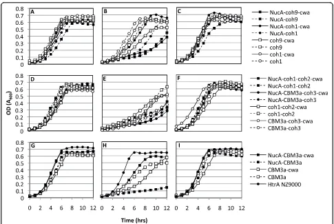
anchoring of the scaffold to the cell-surface (Fig. 1B). Growth curves of engineeredL. lactisstrains were used to determine if the expression and secretion of scaffold proteins resulted in growth inhibition. Results from the
growth experiments showed a correlation between
cipA-frag gene expression and growth inhibition (Fig. 2). The
constitutive over-production of recombinant proteins targeted to the cell surface in L. lactis may interfere with the integrity of the cell wall [33], whereas in C. thermocellum, the constitutive expression of CipA is modulated through catabolite repression [34]. In the absence of the inducer nisin, all cipAfrag-expressing
strains grew similarly to the control L. lactis HtrA
NZ9000 with a final cell density corresponding to an OD600 approaching 0.7 (Fig. 2A, D, G). This indicated
that little change in growth profile resulted from any leaky expression of the recombinant proteins. Nisin induction at inoculation resulted in cellular toxicity, as demonstrated by extended lag phases, lower growth rates and final cell yields (Fig. 2B, E, H). In all cases, when induction of protein expression was carried out after 4 hrs of growth (corresponding to an OD600 ≈0.3),
cultures did not display growth retardation and final cell densities were similar to those attained with no induc-tion (Fig. 2C, F, I). Expression of the various cipAfrags
from the constitutiveP59promoter consistently resulted
in plasmid rearrangements as observed by restriction digest analysis of the rescued plasmids from bothE. coli and L. lactis(data not shown). From these results, we hypothesized that unregulated high-level expression of the CipAfragproteins was toxic to the cells and using a
Figure 1pAW series ofcipAfragexpression vectors and strategy for complex assembly.(A)Vectors were designed for facilitated insertion
of fragments of the gene encoding the cellulosomal scaffold protein CipA, intoAscI-NotI restriction sites. Scaffolds can be optionally expressed with or without an N-terminal nuclease reporter and/or a C-terminal cell wall anchor motif. pAW304 is designed for expression, secretion, and cell wall-targeting of CipA fragments (CipAfrags) as fusions with the N-terminal NucA reporter. pAW305 is designed for the expression and
secretion of CipAfragsas a fusion with the N-terminal NucA reporter, but without the C-terminal anchor motif. pAW504 is designed for
expression, secretion, and cell wall-targeting of CipAfragswithout the N-terminal NucA reporter. pAW505 is designed for the expression and
secretion of CipAfragswith neither the N-terminal NucA reporter nor the C-terminal anchor motif.(B)Graphic depiction of the surface-display
strategy of engineered scaffolds and their association with theb-glucuronidase-dockerin fusion protein (UidA-dock1). All successfully displayed CipAfragsare portrayed as fusions with both NucA and a cell wall anchor, however were also expressed and tested without these two
rearrangements that abolished or reduced cipAfrag
expression. These results confirmed the necessity for regulating expression of the proteins, which was achieved using the PnisA promoter. With the exception
of cell wall anchored scaffold containing only a cellu-lose-binding module (CBM3a-cwa) (Fig. 2H), removal of the NucA lowered or eliminated toxicity to the cells, as observed by improved growth rates and yields.
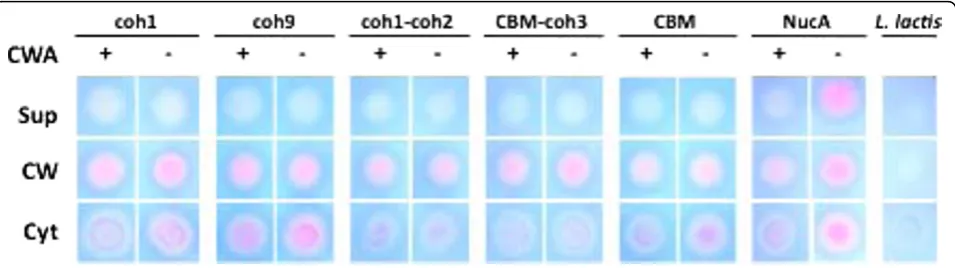
NucA-CipAfragproteins are localized to the cell wall of L. lactis
In order to quickly evaluate our success at recombinant
protein secretion in L. lactis, a nuclease enzyme was
fused to the CipA fragments to be displayed on the cell surface. L. lactis cells harboring the pAW300 series of vectors all displayed a NucA+ phenotype on plates over-laid with TBD agar, confirming that all variants of the NucA-CipAfragproteins were successfully secreted and
that the nuclease retained its function when expressed
as an N-terminal fusion to CipAfrags. To determine the
cellular localization of the expressed CipAfrag fusion
pro-teins, cell fractionations were performed, and cytoplas-mic, cell wall, and supernatant fractions were spotted on
TBD agar. Of the secreted NucA-CipAfrag proteins,
almost all detectable nuclease activity was found in the cell wall fractions corresponding to proteins released from lysozyme/lysostaphin treatments, suggesting suc-cessful cell wall targeting of the proteins (Fig. 3).
CipA-frag proteins were not detected in the supernatant,
suggesting that secreted proteins remained localized to the cell wall due to the activity of lactococcal sortase.
Unexpectedly, the NucA-CipAfrag fusions lacking the
cell wall anchor domain were also detected primarily in the cell wall fractions (Fig. 3) suggesting that fusion of
NucA with CipAfrags caused the scaffolds to remain
associated with the cell wall, even without covalent cross-linking by sortase. All of the cytoplasmic fractions were also found to contain varying levels of expressed
Figure 2Growth profiles ofL. lactisexpressing CipAfragsalone or as fusions with M6cwaand/or NucA. Panels A, D and G represent cultures not induced with nisin, panels B, E, H represent cultures induced with 10 ng/mL nisin at inoculation (t = 0 hrs), and panels C, F, I represent cultures induced with 10 ng/mL nisin in log phase corresponding to an OD600≈0.3 (t = 4 hrs). Constructs were grouped according to
scaffolds, a finding consistent with observations pre-viously made while exporting recombinant proteins in L. lactis[35-38]. We hypothesize that these cytoplasmic proteins were either in the process of being synthesized and exported by the cell via cytoplasmic chaperones, or had evaded the sec-pathway due to a lack of recognition of the signal sequence. In certain instances, the net charge of N-terminal residues downstream of the signal peptide can also contribute to the poor secretion effi-ciency of recombinant proteins [39]. As expected from previous studies [36,37] in the absence of a cell wall anchor domain, NucA was secreted into the supernatant but remained associated to the cell wall if the anchor domain was present (Fig. 3).
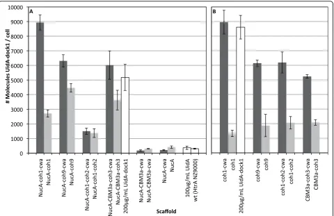
Cell surface displayed CipAfragscaffolds bind UidA-dock1 In vivobinding assays were performed to determine if a dockerin-containing enzyme could associate with cell surface displayed CipAfragscaffold proteins.L. lactiscells
expressing cell wall and supernatant-targeted scaffolds
were incubated with purifiedb-glucuronidase enzymes
fused to a dockerin module (UidA-dock1). After incuba-tion, washed cells were assayed forb-glucuronidase activ-ity, allowing a relative comparison of CipAfragdisplay
efficiencies between engineered constructs. All constructs containing cohesin modules as part of their scaffolds suc-cessfully bound UidA-dock1, while those lacking cohe-sins as well as the plasmid-freeL. lactisHtrA NZ9000 failed to do so (Fig. 4). Binding experiments using UidA lacking dock1 resulted in no successful“docking”onto L. lactisdisplaying NucA-CBM3a-coh3 (Fig. 4A) or any other recombinant scaffolds (data not shown). These results demonstrated that functional recombinant scaf-folds could be expressed on the surface ofL. lactisand that cell surface complex formation was dependent on the presence of both cohesin and dockerin modules. Among those strains secreting and displaying functional
scaffolds, significant variation in display efficiency was observed. Assuming a 1:1 enzyme-to-cohesin ratio, the approximate number of cohesins and/or scaffolds per cell was determined. The strains that displayed the greatest number of nuclease bearing scaffolds (~9 × 10³scaffolds/ cell) were those expressing the cohesin 1 module alone (coh1-cwa and NucA-coh1-cwa) (Fig. 4). Strains expres-sing coh9-cwa, NucA-coh9-cwa, coh1-coh2-cwa, CBM3a-coh3-cwa and NucA-CBM3a-coh3-cwa, were estimated to display between 5.0 × 10³ and 6.3 × 10³ scaffolds/cell. These results suggested that the size of the CipAfragis not necessarily the limiting factor influencing
scaffold display. This was further observed with the
rela-tively lower amount of enzymes binding to L. lactis
displaying NucA-coh1-coh2-cwa (1.5 × 10³UidAdock1/
cell). Essentially, NucA-coh1-coh2-cwa is of similar size to NucA-CBM3a-coh3-cwa (approx. 68 kDa), contains twice as many cohesins, yet host cells where able to bind one quarter the amount of UidA-dock1 molecules. The predicted molecular weights of the engineered scaffolds were used in order to estimate the net amount of recom-binant protein on the cell surface of strains producing scaffolds with a single cohesin. The culture producing the highest net yield of functional recombinant protein was the strain anchoring NucA-CBM3a-coh3-cwa on its surface. Cultures produced and displayed approximately 0.72 mg/mL of recombinant scaffolds, which remained cell-associated and fully functional.
The effect of the N-terminal nuclease reporter on secretion efficiency was also analyzed by comparing the binding capacity ofL. lactisharboring the pAW300 ser-ies (nuclease fusions) with cells harboring the pAW500 (nuclease deficient) series of vectors. Initially included as a reporter to facilitate detection of exported scaffolds, we hypothesized that the nuclease fusion might also increase secretion efficiency, as has been previously observed [35,38]. Removal of NucA had no detrimental
Figure 3Cellular localization of NucA-CipAfragscaffolds expressed byL. lactiswith or without M6cwa. NucA activity was detected by spotting cell fractions on TBD-agar and analyzing for pink color formation. Fractions analyzed are supernatant (sup), cell wall (cw), and cytoplasm (cyt). Constructs are represented by their respective CipAfragcomponents and were expressed as fusions with NucA with or without cell wall
effects on scaffold display for all constructs (Fig. 4B), as similar amounts of anchor-containing scaffolds were located to the cell surface. Furthermore, removal of NucA resulted in a fourfold increase in the amount of coh1-coh2-cwa successfully displayed when compared to its NucA-containing counterpart. The presence of NucA appeared to interfere with the secretion of supernatant-targeted scaffolds from the cell, given that the cwa-deficient variants of coh1, coh9, and CBM3a-coh3 remained associated with the cell to a much larger extent than their NucA-deficient counterparts (Fig. 4).
Discussion
Several recent studies have reported on the recombi-nant expression of mini cellulosome scaffold proteins inSaccharomyces cerevisiae [26-29]. In these examples, the potential application of the engineered strains for the direct conversion of cellulosic biomass to ethanol was the driving factor for choosing S. cerevisiae as a
host. However, many more platform strains have been or are now being developed that will produce ethanol, biofuels other than ethanol, and non-biofuel chemicals [5,14,40-47]. The economics of these processes would be greatly improved if these engineered microbes could use cellulosic substrates. With this goal in mind, the first logical step in establishing this system was the successful secretion and display of cohesin-bearing scaffold proteins. Previous studies have demonstrated that controlled gene expression inL. lactis can reduce toxicity and increase net protein yields [33,48,49]. In our study, the constitutive expression of the scaffold proteins consistently led to cellular toxicity, a problem that was solved by delaying the onset of gene expres-sion until the cells had reached mid log-phase. In cell division, higher concentrations of recombinant cell wall-targeted proteins are localized to the septum, the site of cell wall biosynthesis [33]. It is thus likely that over-expression of our scaffold proteins targeted to the
Figure 4In vivobinding of UidAdock1 on live intactL. lactiscells displaying CipAfrags. CipAfragswere expressed and anchored as fusions
extracytoplasmic space early in the growth phase impaired cell wall biosynthesis and ultimately resulted in cell death. Removal of NucA from the scaffolds decreased or eliminated cellular toxicity for all cohe-sin-containing constructs (Fig. 2), and we thus suspect that accumulation of NucA in the cytoplasm may also contribute to this observed lag in the onset of growth when induced at t = 0 hrs. In addition, as a larger pro-portion of scaffolds lacking a cell wall anchor remained trapped in the cell wall when fused with NucA, it is also likely that part of this observed reduction in toxi-city is due to a decrease in the amounts of recombi-nant proteins being trapped in the cell wall and ultimately disrupting its integrity.
Quantification of cell surface displayed proteins in lactic acid bacteria was previously reported using fluor-escence-activated cell sorting, flow cytometry, or whole-cell ELISA [50]. In our assay, functionality of the
displayed CipAfrag scaffold proteins could be tested
directly through binding with a dockerin-containing reporter enzyme, attesting that the number of cohesins detected was a direct quantification of those that retained biochemical function. Of the four expressed CipA fragments containing at least one cohesin (coh1, coh9, coh1-coh2, CBM3a-coh3), coh1 was displayed with the highest efficiency (~9 × 10³ scaffolds per cell). Due to its small size and decreased number of modules compared with coh1-coh2 and CBM3a-coh3, we attri-bute part of this increase in display to the decrease in size of the scaffold itself. However, coh1 was also displayed more efficiently than coh9, which is approxi-mately the same size and similar in primary amino acid sequence. One possible explanation may relate to the position of coh1 relative to coh9 on native CipA scaf-fold. Coh1 is located at the N-terminus of the 200 kDa scaffold CipA, adjacent to the processing site of the signal peptide by the sec-pathway machinery of C. thermocellum[7]. It is possible that the increase in secretion efficiency of coh1 when compared with coh9 may be in some part due to differences in amino acid content adjacent to the signal peptide, possibly increas-ing its accessibility to the chaperones involved in its transport to the extracytoplasmic space [51]. This, how-ever, does not account for the differences in display effi-ciency between NucA-coh1 and NucA-coh9, as in both cases, NucA is adjacent to the signal sequence. The amount of sequence identity among cohesins perhaps provides a better explanation for these observed differ-ences. Of the nine cohesin modules on CipA, cohesins 3 through 8 show between 96 to 100% sequence identity, whereas among the remaining cohesins, coh1 and coh9 show the least amount of sequence identity (69 and 75%, respectively) [52]. These differences in amino acid
content may translate into differences in folding and solubility of the recombinantly expressed modules.
L. lactis was engineered to display a scaffold contain-ing 2 cohesin modules (coh1-coh2). Based on a 1:1 binding ratio of the enzyme-cohesin and assuming equivalent expression and secretion, we expected this strain to bind twice the amount of UidA when com-pared to scaffolds of similar size but containing a single cohesin module (i.e. CBM3a-coh3). However, coh1-coh2 bound similar amounts of UidA as CBM3a-coh3 (Fig 4B). This reduction in UidA binding was not attributed to CipAfrag size differences, since both mature scaffolds
have a theoretical molecular weight of 68 kDa, suggest-ing that other factors affected secretion and display effi-ciency. In fact, protein size is not regarded as a major bottleneck for protein secretion in L. lactis, as the size of successfully secreted heterologous proteins ranges from 6.9 kDa to a staggering 165 kDa [32]. We hypothe-size that the substitution of a cohesin module by CBM3a may have enhanced secretion by increasing the rate of folding of the scaffold into its soluble form. A similar effect was recently reported with the fusion of the highly insoluble Clostridium cellulovoranscellulase CelL with the CBM of cellulase CelD, which resulted in dramatic increases in its solubility [53].
Comparisons between amounts of UidA binding to cells expressing CipAfrags with or without the cwaM6
domain revealed that the cell wall anchor motif signifi-cantly increased the amounts of functional scaffolds dis-played on the cell (Fig. 4). With NucA present, CipAfrags
lacking cwaM6 remained cell-associated to a larger
extent (Fig. 3) and bound UidA (Fig. 4), suggesting that NucA fusion proteins remained trapped in the cell wall for reasons other than covalent cross-linking by the sor-tase, but yet the cohesin modules were accessible to UidA. This phenomenon is well-documented in other studies of protein secretion in L. lactis, as in some cases the fusion of two generally well-secreted proteins results in changes in the folding of the hybrid protein, and defi-ciencies in their release from cells [37,54]. While the exact mechanism of this phenomenon is not clear, hydrophobic domains resulting from fusing two recom-binant proteins may promote cell wall association [37].
Conclusions
and scaffold in a subsequent development of the strain. We thus envision that further development of this cellu-losome-inspired system may contribute to the efficient bioconversion of substrates into industrially relevant fuels and commodity chemicals, and that tailor-designed synthetic macromolecular complexes could be engi-neered to contain large permutations and combinations of desired enzymes of interest.
Methods
Bacterial strains and plasmids
The bacterial strains and plasmids used in this study are listed in Table 1. E. coli strains were grown in
Luria-Bertani medium at 37°C with shaking (220 rpm).
Lacto-coccus lactisHtrA NZ9000 was grown in M17 medium [62] supplemented with 1% (w/v) glucose (GM17) at
30°C without agitation. C. thermocellum was grown in
ATCC1191 medium at 55°C with 0.2% (w/v) cellobiose as a carbon source. Where appropriate, antibiotics were added as follows: forE. coli, ampicillin (100μg/mL),
ery-thromycin (150 μg/mL), chloramphenicol (10 μg/mL)
and kanamycin (30 μg/mL); forL. lactis, erythromycin
(5μg/mL) and chloramphenicol (10 μg/mL). General
molecular biology techniques for E. coliwere performed as previously described [63]. Genomic DNA was isolated from C. thermocellumas previously described [64]. To
make competent cells,L. lactiswas grown in M17
med-ium [62] supplemented with 1% (w/v) glucose, 25% (w/v) sucrose and 2% (w/v) glycine and cells were trans-formed as previously described [65]. M17 media was supplied by Oxoid, LB media was supplied by Novagen, all antibiotics, r-nitrophenyl-b-D-glucuronide and nisin were provided by Sigma, and X-gal and IPTG were sup-plied by Fermentas.
Assembly of cassettes for scaffold protein expression and targeting
The E. coli-L. lactis shuttle vectors pVE5524 and pVE5523 were used as backbone plasmids for targeting fragments of the CipA scaffold protein to the cell surface or supernatant, respectively [36]. The various CipAfrags
were produced as fusions with the N-terminal signal pep-tide from the lactococcal Usp45 secreted protein (spUsp45) and for targeting to the cell wall, as a fusion
with the C-terminal anchor from the Streptococcus
pyogenes M6 protein (cwaM6) (Fig. 1). Expression
cassettes were designed to allow the optional fusion of CipAfragswith an N-terminal nuclease reporter (NucA)
used for detection of the fusion proteins in the extracel-lular milieu [35,38] (Fig. 1). The strong constitutive lacto-coccal promoterP59 [36] and thePnisAnisin-inducible
promoter from thenisAgene ofL. lactis[66] were tested for optimal expression of the recombinant scaffolds. Two ribosome-binding sites were also tested, that of theusp45
gene (rbsusp45) [36] and that of thenisAgene (rbsnisA)
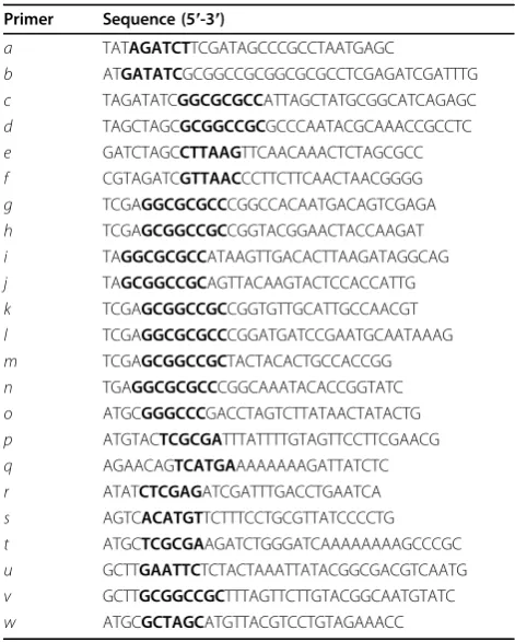
[66]. In order to facilitate the exchange of scaffold frag-ments in the expression cassette,AscI-NotI restriction sites were engineered just downstream ofnucA(Fig. 1). To achieve this, an 800-bp fragment containing thenucA
gene was PCR-amplified from pVE5524 using primersa
and b(Table 2), digested with SalI-EcoRV and ligated
into similarly digested pVE5524 and pVE5523, yielding
pAW004 and pAW005. To facilitate detection ofE. coli
clones that harborcipAfragments, alacZ-astuffer
frag-ment was PCR-amplified from pUC19 using primersc
andd, digested withAscI-NotI, and subsequently ligated into similarly cut pAW004 and pAW005, yielding
pAW004Z and pAW005Z, respectively. SinceL. lactis
HtrA NZ9000 is resistant to erythromycin, theery
mar-ker of the pAW vectors was replaced with thecatgene
from pSCNIII. Thecatgene was PCR-amplified using
primerseandf, digested withAflII andHpaI, and ligated into similarly digested pAW004Z and pAW005Z, yielding plasmids pAW004ZC and pAW005ZC, respectively. For inducible expression of the scaffolds, we replaced theP59 promoter withPnisAfrom pSIP502. ThePnisApromoter
was isolated using primers o and p, digested with
ApaI-NruI and ligated to similarly digested pAW004ZC and pAW005ZC, yielding pAW104 and pAW105, respectively.
Cloning ofcipAfragments fromC. thermocellum
Five uniquecipA fragments were PCR-amplified from
C. thermocellum genomic DNA using primer pairsg-h, i-j, g-k, l-m and n-m (Table 2), ligated into pGEM-T (Promega) and sequenced to verify the integrity of the gene sequence. The resulting pGEM plasmids were digested with AscI-NotI to release the cipAgene frag-ments and these were ligated into pAW004ZC and
pAW005ZC. The cipAfragments were chosen on the
basis of containing a single cohesin (coh1 or coh9), two cohesins of identical specificity (coh1-coh2), one cohesin and a cellulose-binding module (coh3-CBM3a) and only a cellulose-binding module (CBM3a) (Fig. 1). The result-ing spUsp45-nucA-cipAfrag-cwaM6 cassettes were under
control of theP59promoter and containedrbsusp45. The
same cipA fragments were cloned into pAW104 and
pAW105 for inducible expression of the scaffold proteins.
For the inducible expression of the fusion proteins under the control ofPnisAwith an intact
ribosome-bind-ing site from thenisA gene (rbsnisA),spUsp45-nucAwas
PCR-amplified from pAW004ZC using primersqandr,
creating aBspHI cut site at the 5′ end of the PCR
pro-duct. The PCR product was digested with BspHI and
XhoI and ligated to pSIP502 digested with NcoI-XhoI, effectively replacing the gusA gene withspUsp45-nucA,
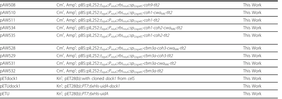
Table 1 Strains and plasmids used in this study
Strain Genotype/Decription Source
L. lactisHtrA NZ9000 MG1363 (nisRKgenes on the chromosome) [37]
E. coliTG1 supE thi-1Δ(lac-proAB)Δ(mcrB-hsdSM)5(rK- mK-) [F’traD36 proAB lacIqZΔM15] ATCC
E. coliDH5a fhuA2Δ(argF-lacZ)U169 phoA glnV44F80Δ(lacZ)M15 gyrA96 recA1 relA1 endA1 thi-1 hsdR17 Invitrogen
E. coliBL21 (DE3) F-ompT gal dcm lon hsdSB(rB
-mB
-)l(DE3 [lacI lacUV5-T7 gene 1 ind1 sam7 nin5]) Novagen
Plasmid
pVE5524 Eryr, Ampr; pBS::pIL252::t
trpA::P59::rbsusp45::spUsp45-nucA-cwaM6-t1t2 [36]
pVE5523 Eryr, Ampr; pBS::pIL252::t
trpA::P59::rbsusp45::spUsp45-nucA-t1t2 [36]
pSIP502 Eryr;P
nisA::rbsnisA::uidA [66]
pSCNIII Cmr J. Seegersa
pUC19 Ampr [69]
pET28(b) Kmr Novagen
pSIPsp-nuc Eryr;PnisA::rbsnisA::spUsp45-nucA This Work
pUC104 Ampr;t
trpA::PnisA::rbsusp45::spUsp45-nucA This Work
pUC104mod Ampr;t
trpA::P59::rbsusp45::spUsp45-nucA This Work
pUC304 Ampr;t
trpA::PnisA::rbsnisA::spUsp45-nucA This Work
pUC504 Ampr;ttrpA::PnisA::rbsnisA::spUsp45 This Work
pAW004 Eryr, Ampr; pBS::pIL252::ttrpA::P59::rbsusp45::spUsp45-nucA-MCS-cwaM6-t1t2 This Work
pAW005 Eryr, Ampr; pBS::pIL252::t
trpA::P59::rbsusp45::spUsp45-nucA-MCS-t1t2 This Work
pAW004Z Eryr, Ampr; pBS::pIL252::t
trpA::P59::rbsusp45::spUsp45-nucA-lacZa-cwaM6-tlt2 This Work
pAW005Z Eryr, Ampr; pBS::pIL252::ttrpA::P59::rbsusp45::spUsp45-nucA- lacZa-tlt2 This Work
pAW004ZC Cmr, Ampr; pBS::pIL252::ttrpA::P59::rbsusp45::spUsp45-nucA-lacZa-cwaM6-tlt2 This Work
pAW005ZC Cmr, Ampr; pBS::pIL252::ttrpA::P59::rbsusp45::spUsp45-nucA- lacZa-tlt2 This Work
pGEMc9 Ampr; pGEMT::with clonedcoh9fromcipA This Work
pGEMc1 Ampr; pGEMT::with clonedcoh1fromcipA This Work
pGEMc1-c2 Ampr; pGEMT::with clonedcoh1-coh2fromcipA This Work
pGEMcbm-c3 Ampr; pGEMT::with clonedcbm3a-coh3fromcipA This Work
pGEMcbm Ampr; pGEMT::with clonedcbm3afromcipA This Work
pAW104 Cmr, Ampr; pBS::pIL252::t
trpA::PnisA::rbsusp45::spUsp45-nucA-LacZa-cwaM6-tlt2 This Work
pAW105 Cmr, Ampr; pBS::pIL252::t
trpA::PnisA::rbsusp45::spUsp45-nucA-LacZa-tlt2 This Work
pAW301 Cmr, Ampr; pBS::pIL252::t
trpA::PnisA::rbsnisA::spUsp45-nucA-cwaM6-tlt2 This Work
pAW302 Cmr, Ampr; pBS::pIL252::ttrpA::PnisA::rbsnisA::spUsp45-nucA-tlt2 This Work
pAW304 Cmr, Ampr; pBS::pIL252::ttrpA::PnisA::rbsnisA::spUsp45-nucA-lacZa-cwaM6-tlt2 This Work
pAW305 Cmr, Ampr; pBS::pIL252::t
trpA::PnisA::rbsnisA::spUsp45-nucA-lacZa-tlt2 This Work
pAW307 Cmr, Ampr; pBS::pIL252::t
trpA::PnisA::rbsnisA::spUsp45-nucA-coh9-cwaM6-tlt2 This Work
pAW308 Cmr, Ampr; pBS::pIL252::t
trpA::PnisA::rbsnisA::spUsp45-nucA-coh9-tlt2 This Work
pAW310 Cmr, Ampr; pBS::pIL252::ttrpA::PnisA::rbsnisA::spUsp45-nucA-coh1-cwaM6-tlt2 This Work
pAW311 Cmr, Ampr; pBS::pIL252::ttrpA::PnisA::rbsnisA::spUsp45-nucA-coh1-tlt2 This Work
pAW334 Cmr, Ampr; pBS::pIL252::t
trpA::PnisA::rbsnisA::spUsp45-nucA-coh1-coh2-cwaM6-tlt2 This Work
pAW335 Cmr, Ampr; pBS::pIL252::t
trpA::PnisA::rbsnisA::spUsp45-nucA-coh1-coh2-tlt2 This Work
pAW328 Cmr, Ampr; pBS::pIL252::t
trpA::PnisA::rbsnisA::spUsp45-nucA-cbm3a-coh3-cwaM6-tlt2 This Work
pAW329 Cmr, Ampr; pBS::pIL252::ttrpA::PnisA::rbsnisA::spUsp45-nucA-cbm3a-coh3-tlt2 This Work
pAW331 Cmr, Ampr; pBS::pIL252::ttrpA::PnisA::rbsnisA::spUsp45-nucA-cbm3a-cwaM6-tlt2 This Work
pAW332 Cmr, Ampr; pBS::pIL252::t
trpA::PnisA::rbsnisA::spUsp45-nucA-cbm3a-tlt2 This Work
pAW504 Cmr, Ampr; pBS::pIL252::t
trpA::PnisA::rbsnisA::spUsp45-lacZa-cwaM6-tlt2 This Work
pAW505 Cmr, Ampr; pBS::pIL252::t
trpA::PnisA::rbsnisA::spUsp45-lacZa-tlt2 This Work
yielding pSIPSPNUC. For the insertion of an upstream transcriptional terminator and removal ofnucA, a 1500-bpSapI-XbaI fragment was temporarily removed from pAW104, and was ligated to similarly cut pUC19, yield-ing vector pUC104. To introduce theE. coli transcrip-tional terminator from the tryptophan synthase operon (ttrpA) upstream of PnisA and to introduce a BglII cut
site, a 200-bp fragment containingttrpA was
PCR-ampli-fied from pVE5524 using primerssandt, digested with AflIII-NruI and ligated to similarly-cut pUC104, yielding pUC104mod. Plasmid pSIPSPNUC was digested with BglII-XhoI and ligated to similarly-digested pUC104mod, yielding vector pUC304. This was the base vector har-boring the ttrpA-PnisA-rbsnisA-spUsp45-nucA cassette,
which was digested withApaI-AscI and ligated into the pAW100 series of vectors. Inserting this cassette into ApaI-EcoRV digested pAW110 and pAW111, yielding pAW301 and pAW302, respectively, created controls
lacking cipA fragments for expression of nucA alone.
For deletion of thenucA reporter and construction of
the pAW500 series, pUC304 was digested with
SalI-XhoI and self-ligated, yielding vector pUC504. ThettrpA
-PnisA-rbsnisA-spUsp45 cassette was released via digestion
withApaI-AscI, gel-purified, and ligated to similarly-cut pAW100 series vectors, yielding the pAW500 series of vectors. This cassette was also ligated into similarly cut pAW104 and pAW105 yielding base vectors containing thelacZ-astuffer fragment. The final expression vectors for this study included the pAW300 series of vectors for inducible expression and targeting of NucA-fused scaf-folds, and the pAW500 series of vectors for inducible expression and targeting of scaffolds lacking the N-terminal NucA reporter (Fig. 1).
Expression and localization of CipAfragsinL. lactis
L. lactis HtrANZ9000 was transformed with the
pAW300 and pAW500 series of vectors for the con-trolled expression of scaffolds. It contains chromosomal copies of the nisRand nisK genes necessary for nisin-inducible expression of cassettes under control of the nisApromoter, and is deficient in a major extracellular housekeeping protease, which has been shown pre-viously to be responsible for the proteolysis of exported recombinant proteins [37]. Growth curves were used to evaluate the potential of growth inhibition caused by the
over-expressed CipAfragproteins. Growth curves were
performed in 96 well plates and cells were induced with 10 ng nisin/mL at inoculation (t = 0 hrs), 4 hrs post-inoculation (t = 4 hrs) or were not induced. For the expression of CipAfrag proteins in L. lactisHtrA
NZ9000, overnight cultures were diluted 1/50 into fresh GM17 medium and were induced with 10 ng nisin/mL when an OD600 ≈0.3 was reached (4 hrs). After 20 hrs
growth, successful CipAfrag secretion was evaluated
using a nuclease assay consisting of spotting cells on TBD-agar and observing pink color formation [36]. For analysis of NucA-CipAfrag proteins in various cellular
locations, cell fractionation was performed as described previously [58], with the addition of lysostaphin (0.6 mg/mL) [67]. Aliquots of proteins were blotted on TBD-agar plates and formation of a pink color was ana-lyzed after a 1-hr incubation at 37°C.
Expression and purification of CipAfrag-binding
b-glucuronidase
The E. coli b-glucuronidase (UidA) was engineered to have a C-terminal dock1 module for binding onto Table 1 Strains and plasmids used in this study(Continued)
pAW508 Cmr, Ampr; pBS::pIL252::ttrpA::PnisA::rbsnisA::spUsp45-coh9-tlt2 This Work
pAW510 Cmr, Ampr; pBS::pIL252::t
trpA::PnisA::rbsnisA::spUsp45-coh1-cwaM6-tlt2 This Work
pAW511 Cmr, Ampr; pBS::pIL252::t
trpA::PnisA::rbsnisA::spUsp45-coh1-tlt2 This Work
pAW534 Cmr, Ampr; pBS::pIL252::t
trpA::PnisA::rbsnisA::spUsp45-coh1-coh2-cwaM6-tlt2 This Work
pAW535 Cmr, Ampr; pBS::pIL252::ttrpA::PnisA::rbsnisA::spUsp45-coh1-coh2-tlt2 This Work
pAW528 Cmr, Ampr; pBS::pIL252::t
trpA::PnisA::rbsnisA::spUsp45-cbm3a-coh3-cwaM6-tlt2 This Work
pAW529 Cmr, Ampr; pBS::pIL252::ttrpA::PnisA::rbsnisA::spUsp45-cbm3a-coh3-tlt2 This Work
pAW531 Cmr, Ampr; pBS::pIL252::ttrpA::PnisA::rbsnisA::spUsp45-cbm3a-cwaM6-tlt2 This Work
pAW532 Cmr, Ampr; pBS::pIL252::t
trpA::PnisA::rbsnisA::spUsp45-cbm3a-tlt2 This Work
pETdock1 Knr; pET28(b)::with cloneddock1fromcelS This Work
pETUdock1 Knr; pET28(b)::PT7::6xHis-uidA-dock1 This Work
pETU Knr; pET28(b)::PT7::6xHis-uidA This Work
a
Vector pSCNIII was a gift provided by Jos Seegers (unpublished data).
pAW100 series of vectors are nisin-inducible and contain an intactrbsusp45. pAW300 series vectors are nisin-inducible and contain an intactrbsnisA. pAW500 series vectors are pAW300 variants lacking an N-terminal NucA fusion.P59, constitutive lactococcal promoter;PT7, inducible T7 promoter;PnisA, induciblenisApromoter;
rbsusp45, Usp45 ribosome-binding site;rbsnisA,nisAribosome-binding site;spUsp45, signal sequence of Usp45;nucA, staphylococcal nuclease;cwaM6, anchor motif of
CipAfragscaffolds, as well as an N-terminal 6 × His-tag
for protein purification. The dock1 module of the C. thermocellum celS gene was amplified from C. thermocellum genomic DNA using primersu andv
(Table 2). PCR products were digested withEcoRI-NotI
and ligated to similarly-digested pET28(b), yielding
pET-dock1. TheuidAgene lacking a stop codon was
ampli-fied using primers w and x and pSIP502 as template.
The PCR product was digested with NheI-EcoRI and
ligated to similarly-cut pET28(b) and pETdock1, yielding His-tagged UidA proteins with and without a dock1 module (pETUdock1 and pETU). His-tagged proteins
were expressed in E. coli BL21(DE3). Cultures were
induced at an OD600of 0.5 with 1 mM IPTG and
incu-bated for an additional 5 hrs at 37°C. Cells were har-vested (1000 × g, 10 min, 4°C) and cell pellets were kept overnight at -80°C. Thawed cell pellets were suspended in 50 mM phosphate buffer, pH 7.5, containing 300 mM NaCl. Samples were subjected to sonication (15 sec pulse, 5 sec between pulses, 2 min total process time) and lysates were loaded on approximately 10 mL of Ni-NTA sepharose resin. The resin was washed with phos-phate buffer (50 mM, pH 6.0) containing 300 mM NaCl and 20 mM imidazole and eluted using the same buffer containing 250 mM imidazole. Fifty μL of each elution
fraction were added to 450 μL GUS buffer containing
50 mM sodium phosphate buffer (pH 7), 10 mMb
-mer-captoethanol, 1 mM ethylenediaminetetraacetic acid and 0.1% (v/v) Triton X-100. Samples were heated for 1 min, after whichp-nitrophenyl-b-D-glucuronide was added to a final concentration of 4 mg/mL [68]. The UidA-containing fractions were identified by the appearance of a yellow color. Proteins from the elution fractions showing UidA activity were visualized by SDS-PAGE on a 12% (w/v) gel to identify fractions contain-ing the highest purity of enzyme. The specific activities of UidA-dock1 and UidA were determined by colori-metric assays in a thermostated UV-Vis spectrophot-ometer (Cary 50 WinUv) at 405 nm, using a 1 cm (L) cuvette, and the known molar extinction coefficient of p-nitrophenol being 18 000 M-1cm-1. Quantification of the proteins was done using a Bradford protein assay kit (Pierce) and BSA as a standard. Specific activities were used to evaluate the amount of enzyme bound to cells in thein vivobinding assay described below.
Binding ofb-glucuronidase toL. lactis
L. lactis HtrA NZ9000 cells harboring the pAW300 or pAW500 series of vectors, as well as the plasmid-free strain were grown overnight in GM17 medium. Cultures were diluted 1/50 in 5 mL of fresh media and grown for an additional 4 hrs (OD600≈0.3) after which cells were
induced with 10 ng nisin/mL for scaffold expression. After 20 hrs of growth, cells from 1-mL of culture were harvested (4,300 × g, 5 min, 4°C) washed once in phos-phate buffer (50 mM, pH 6.0) containing 300 mM NaCl
and suspended in 100 μL of purified UidA-dock1 or
UidA at a concentration 100 μg/mL. To ensure that
saturation of all cohesin sites was achieved, binding
assay with 200 μg UidA-dock1/mL was tested for
L. lactisharboring pAW328. Binding was carried out at 4°C for 10 hrs. Cells were then washed 6 times to elimi-nate residual enzyme activity and suspended in 100μL of phosphate buffer (50 mM, pH 6.0) containing 300 mM NaCl for detection ofb-glucuronidase activity.
For quantification of bound UdiA-dock1, 50 μL of
washed cells were analyzed forb-glucuronidase activity. Reactions were stopped with 250μL of 1 M sodium car-bonate once a yellow color appeared, and the duration of each assay was recorded. The specific activities of the purified UidA-dock1 and UidA were used to determine
the amount of enzyme bound onto the L lactis cells.
Using the calculated molecular weight of UidA-dock1 and the known amount of cells present in each sample, the average number of enzyme units bound per cell was estimated. Assuming a 1:1 cohesin to dockerin ratio, the number of enzymes present per cell also is a representa-tion of the number of cohesins present on the cell surface. The calculated molecular weight of the scaffolds was used to estimate the net amount of recombinant Table 2 Primers used in this study
Primer Sequence (5′-3′)
a TATAGATCTTCGATAGCCCGCCTAATGAGC
b ATGATATCGCGGCCGCGGCGCGCCTCGAGATCGATTTG
c TAGATATCGGCGCGCCATTAGCTATGCGGCATCAGAGC
d TAGCTAGCGCGGCCGCGCCCAATACGCAAACCGCCTC
e GATCTAGCCTTAAGTTCAACAAACTCTAGCGCC
f CGTAGATCGTTAACCCTTCTTCAACTAACGGGG
g TCGAGGCGCGCCCGGCCACAATGACAGTCGAGA
h TCGAGCGGCCGCCGGTACGGAACTACCAAGAT
i TAGGCGCGCCATAAGTTGACACTTAAGATAGGCAG
j TAGCGGCCGCAGTTACAAGTACTCCACCATTG
k TCGAGCGGCCGCCGGTGTTGCATTGCCAACGT
l TCGAGGCGCGCCCGGATGATCCGAATGCAATAAAG
m TCGAGCGGCCGCTACTACACTGCCACCGG
n TGAGGCGCGCCCGGCAAATACACCGGTATC
o ATGCGGGCCCGACCTAGTCTTATAACTATACTG
p ATGTACTCGCGATTTATTTTGTAGTTCCTTCGAACG
q AGAACAGTCATGAAAAAAAAGATTATCTC
r ATATCTCGAGATCGATTTGACCTGAATCA
s AGTCACATGTTCTTTCCTGCGTTATCCCCTG
t ATGCTCGCGAAGATCTGGGATCAAAAAAAAGCCCGC
u GCTTGAATTCTCTACTAAATTATACGGCGACGTCAATG
v GCTTGCGGCCGCTTTAGTTCTTGTACGGCAATGTATC
w ATGCGCTAGCATGTTACGTCCTGTAGAAACC
protein anchored to cells in respective cultures. Experi-ments were repeated twice and true biological replicates (independent colonies and cultures) were performed in triplicate for all samples.
Acknowledgements
We are grateful to Dr. Alexandra Gruss and Dr. Isabelle Poquet for providing base expression vectors for LAB as well as strains ofL. lactis. The authors would like to acknowledge Dr. Andy Ekins for his help in reviewing the manuscript. This work was supported by research grants from the Natural Sciences and Engineering Research Council of Canada (NSERC) (grant numbers 312357-06 and 330781-06) the Canada Foundation for Innovation (grant number 202359) and a Canada Research Chair to V.J.J.M. A.S.W. is the recipient of graduate scholarships from NSERC and the Fonds Québécois de la Recherche sur la Nature et les Technologies.
Authors′contributions
VM defined the strategy described and supervised the project. AW designed and carried out all experiments. AW drafted the initial manuscript, VM helped draft the manuscript, and both AW and VM edited the manuscript. VM supervised the entire PhD project of AW. All authors read and approved the final manuscript.
Competing interests
The authors declare that they have no competing interests.
Received: 1 June 2010 Accepted: 14 September 2010 Published: 14 September 2010
References
1. Lowell GH, Ballou WR, Smith LF, Wirtz RA, Zollinger WD, Hockmeyer WT:
Proteosome-lipopeptide vaccines: enhancement of immunogenicity for malaria CS peptides.Science1988,240:800-802.
2. Lowell GH, Smith LF, Seid RC, Zollinger WD:Peptides bound to proteosomes via hydrophobic feet become highly immunogenic without adjuvants.J Exp Med1988,167:658-663.
3. Bayer EA, Belaich JP, Shoham Y, Lamed R:The cellulosomes: multienzyme machines for degradation of plant cell wall polysaccharides.Annu Rev Microbiol2004,58:521-554.
4. Conrado RJ, Varner JD, DeLisa MP:Engineering the spatial organization of metabolic enzymes: mimicking nature′s synergy.Curr Opin Biotechnol
2008,19:492-499.
5. Dueber JE, Wu GC, Malmirchegini GR, Moon TS, Petzold CJ, Ullal AV, Prather KL, Keasling JD:Synthetic protein scaffolds provide modular control over metabolic flux.Nat Biotechnol2009,27:753-759. 6. Lynd LR, Weimer PJ, van Zyl WH, Pretorius IS:Microbial cellulose
utilization: fundamentals and biotechnology.Microbiol Mol Biol Rev2002,
66:506-577, table of contents.
7. Schwarz WH:The cellulosome and cellulose degradation by anaerobic bacteria.Appl Microbiol Biotechnol2001,56:634-649.
8. Kruus K, Lua AC, Demain AL, Wu JH:The anchorage function of CipA (CelL), a scaffolding protein of theClostridium thermocellumcellulosome. Proc Natl Acad Sci USA1995,92:9254-9258.
9. Leibovitz E, Beguin P:A new type of cohesin domain that specifically binds the dockerin domain of theClostridium thermocellum cellulosome-integrating protein CipA.J Bacteriol1996,178:3077-3084.
10. Lemaire M, Ohayon H, Gounon P, Fujino T, Beguin P:OlpB, a new outer layer protein ofClostridium thermocellum, and binding of its S-layer-like domains to components of the cell envelope.J Bacteriol1995,
177:2451-2459.
11. Kosugi A, Amano Y, Murashima K, Doi RH:Hydrophilic domains of scaffolding protein CbpA promote glycosyl hydrolase activity and localization of cellulosomes to the cell surface ofClostridium cellulovorans.J Bacteriol2004,186:6351-6359.
12. Garcia-Campayo V, Beguin P:Synergism between the cellulosome-integrating protein CipA and endoglucanase CelD ofClostridium thermocellum.J Biotechnol1997,57:39-47.
13. Zverlov VV, Klupp M, Krauss J, Schwarz WH:Mutations in the scaffoldin gene, cipA, ofClostridium thermocellumwith impaired cellulosome
formation and cellulose hydrolysis: insertions of a new transposable element, IS1447, and implications for cellulase synergism on crystalline cellulose.J Bacteriol2008,190:4321-4327.
14. Lynd LR, van Zyl WH, McBride JE, Laser M:Consolidated bioprocessing of cellulosic biomass: an update.Curr Opin Biotechnol2005,16:577-583. 15. Lu Y, Zhang YH, Lynd LR:Enzyme-microbe synergy during cellulose hydrolysis byClostridium thermocellum.Proc Natl Acad Sci USA2006,
103:16165-16169.
16. Miron J, Ben-Ghedalia D, Morrison M:Invited review: adhesion
mechanisms of rumen cellulolytic bacteria.J Dairy Sci2001,84:1294-1309. 17. Bayer EA, Kenig R, Lamed R:Adherence ofClostridium thermocellumto
cellulose.J Bacteriol1983,156:818-827.
18. Ng TK, Weimer TK, Zeikus JG:Cellulolytic and physiological properties of
Clostridium thermocellum.Arch Microbiol1977,114:1-7.
19. Fierobe HP, Bayer EA, Tardif C, Czjzek M, Mechaly A, Belaich A, Lamed R, Shoham Y, Belaich JP:Degradation of cellulose substrates by cellulosome chimeras. Substrate targeting versus proximity of enzyme components.J Biol Chem2002,277:49621-49630.
20. Fierobe HP, Mechaly A, Tardif C, Belaich A, Lamed R, Shoham Y, Belaich JP, Bayer EA:Design and production of active cellulosome chimeras. Selective incorporation of dockerin-containing enzymes into defined functional complexes.J Biol Chem2001,276:21257-21261.
21. Fierobe HP, Mingardon F, Mechaly A, Belaich A, Rincon MT, Pages S, Lamed R, Tardif C, Belaich JP, Bayer EA:Action of designer cellulosomes on homogeneous versus complex substrates: controlled incorporation of three distinct enzymes into a defined trifunctional scaffoldin.J Biol Chem
2005,280:16325-16334.
22. Mingardon F, Chanal A, Tardif C, Bayer EA, Fierobe HP:Exploration of new geometries in cellulosome-like chimeras.Appl Environ Microbiol2007,
73:7138-7149.
23. Murashima K, Kosugi A, Doi RH:Synergistic effects on crystalline cellulose degradation between cellulosomal cellulases fromClostridium cellulovorans.J Bacteriol2002,184:5088-5095.
24. Perret S, Casalot L, Fierobe HP, Tardif C, Sabathe F, Belaich JP, Belaich A:
Production of heterologous and chimeric scaffoldins byClostridium acetobutylicumATCC 824.J Bacteriol2004,186:253-257.
25. Sabathe F, Soucaille P:Characterization of the CipA scaffolding protein andin vivoproduction of a minicellulosome inClostridium
acetobutylicum.J Bacteriol2003,185:1092-1096.
26. Ito J, Kosugi A, Tanaka T, Kuroda K, Shibasaki S, Ogino C, Ueda M, Fukuda H, Doi RH, Kondo A:Regulation of the display ratio of enzymes on the
Saccharomyces cerevisiaecell surface by the immunoglobulin G and cellulosomal enzyme binding domains.Appl Environ Microbiol2009,
75:4149-4154.
27. Tsai SL, Oh J, Singh S, Chen R, Chen W:Functional assembly of minicellulosomes on theSaccharomyces cerevisiaecell surface for cellulose hydrolysis and ethanol production.Appl Environ Microbiol2009,
75:6087-6093.
28. Lilly M, Fierobe HP, van Zyl WH, Volschenk H:Heterologous expression of a Clostridium minicellulosome inSaccharomyces cerevisiae.FEMS Yeast Res2009,9:1236-1249.
29. Wen F, Sun J, Zhao H:Yeast surface display of trifunctional
minicellulosomes for simultaneous saccharification and fermentation of cellulose to ethanol.Appl Environ Microbiol76:1251-1260.
30. Petrov K, Urshev Z, Petrova P:L+-lactic acid production from starch by a novel amylolyticLactococcus lactissubsp. lactis B84.Food Microbiol2008,
25:550-557.
31. Hernandez I, Molenaar D, Beekwilder J, Bouwmeester H, van Hylckama Vlieg JE:Expression of plant flavor genes inLactococcus lactis.Appl Environ Microbiol2007,73:1544-1552.
32. Le Loir Y, Azevedo V, Oliveira SC, Freitas DA, Miyoshi A, Bermudez-Humaran LG, Nouaille S, Ribeiro LA, Leclercq S, Gabriel JE,et al:Protein secretion inLactococcus lactis: an efficient way to increase the overall heterologous protein production.Microb Cell Fact2005,4:2.
33. Narita J, Okano K, Kitao T, Ishida S, Sewaki T, Sung MH, Fukuda H, Kondo A:
Display of alpha-amylase on the surface ofLactobacillus caseicells by use of the PgsA anchor protein, and production of lactic acid from starch.Appl Environ Microbiol2006,72:269-275.
34. Zhang YH, Lynd LR:Regulation of cellulase synthesis in batch and continuous cultures ofClostridium thermocellum.J Bacteriol2005,
35. Dieye Y, Hoekman AJ, Clier F, Juillard V, Boot HJ, Piard JC:Ability of
Lactococcus lactisto export viral capsid antigens: a crucial step for development of live vaccines.Appl Environ Microbiol2003,69:7281-7288. 36. Dieye Y, Usai S, Clier F, Gruss A, Piard JC:Design of a protein-targeting
system for lactic acid bacteria.J Bacteriol2001,183:4157-4166. 37. Miyoshi A, Poquet I, Azevedo V, Commissaire J, Bermudez-Humaran L,
Domakova E, Le Loir Y, Oliveira SC, Gruss A, Langella P:Controlled production of stable heterologous proteins inLactococcus lactis.Appl Environ Microbiol2002,68:3141-3146.
38. Ribeiro LA, Azevedo V, Le Loir Y, Oliveira SC, Dieye Y, Piard JC, Gruss A, Langella P:Production and targeting of theBrucella abortusantigen L7/L12 inLactococcus lactis: a first step towards food-grade live vaccines against brucellosis.Appl Environ Microbiol2002,68:910-916.
39. Langella P, Le Loir Y:Heterologous protein secretion inLactococcus lactis: a novel antigen delivery system.Braz J Med Biol Res1999,32:191-198. 40. Atsumi S, Hanai T, Liao JC:Non-fermentative pathways for synthesis of
branched-chain higher alcohols as biofuels.Nature2008,451:86-89. 41. Zhang M, Eddy C, Deanda K, Finkelstein M, Picataggio S:Metabolic
engineering of a pentose M\metabolism pathway in ethanologenic
Zymomonas mobilis.Science1995,267:240-243.
42. Wu CH, Mulchandani A, Chen W:Versatile microbial surface-display for environmental remediation and biofuels production.Trends Microbiol
2008,16:181-188.
43. Rittmann D, Lindner SN, Wendisch VF:Engineering of a glycerol utilization pathway for amino acid production byCorynebacterium glutamicum. Appl Environ Microbiol2008,74:6216-6222.
44. Lee SK, Chou H, Ham TS, Lee TS, Keasling JD:Metabolic engineering of microorganisms for biofuels production: from bugs to synthetic biology to fuels.Curr Opin Biotechnol2008,19:556-563.
45. Rogers PL, Jeon YJ, Lee KJ, Lawford HG:Zymomonas mobilisfor fuel ethanol and higher value products.Adv Biochem Eng Biotechnol2007,
108:263-288.
46. Steen EJ, Kang Y, Bokinsky G, Hu Z, Schirmer A, McClure A, Del Cardayre SB, Keasling JD:Microbial production of fatty-acid-derived fuels and chemicals from plant biomass.Nature463:559-562.
47. Shaw AJ, Podkaminer KK, Desai SG, Bardsley JS, Rogers SR, Thorne PG, Hogsett DA, Lynd LR:Metabolic engineering of a thermophilic bacterium to produce ethanol at high yield.Proc Natl Acad Sci USA2008,
105:13769-13774.
48. de Vos WM:Gene expression systems for lactic acid bacteria.Curr Opin Microbiol1999,2:289-295.
49. Bermudez-Humaran LG, Cortes-Perez NG, Le Loir Y, Alcocer-Gonzalez JM, Tamez-Guerra RS, de Oca-Luna RM, Langella P:An inducible surface presentation system improves cellular immunity against human papillomavirus type 16 E7 antigen in mice after nasal administration with recombinant lactococci.J Med Microbiol2004,53:427-433. 50. Leenhouts K, Buist G, Kok J:Anchoring of proteins to lactic acid bacteria.
Antonie Van Leeuwenhoek1999,76:367-376.
51. Gerngross UT, Romaniec MP, Kobayashi T, Huskisson NS, Demain AL:
Sequencing of aClostridium thermocellumgene (cipA) encoding the cellulosomal SL-protein reveals an unusual degree of internal homology. Mol Microbiol1993,8:325-334.
52. Lytle B, Myers C, Kruus K, Wu JH:Interactions of the CelS binding ligand with various receptor domains of theClostridium thermocellum
cellulosomal scaffolding protein, CipA.J Bacteriol1996,178:1200-1203. 53. Murashima K, Kosugi A, Doi RH:Solubilization of cellulosomal cellulases
by fusion with cellulose-binding domain of noncellulosomal cellulase engd fromClostridium cellulovorans.Proteins2003,50:620-628. 54. Bermudez-Humaran LG, Langella P, Miyoshi A, Gruss A, Guerra RT, Montes
de Oca-Luna R, Le Loir Y:Production of human papillomavirus type 16 E7 protein inLactococcus lactis.Appl Environ Microbiol2002,68:917-922. 55. Avall-Jaaskelainen S, Lindholm A, Palva A:Surface display of the
receptor-binding region of theLactobacillus brevisS-layer protein inLactococcus lactisprovides nonadhesive lactococci with the ability to adhere to intestinal epithelial cells.Appl Environ Microbiol2003,69:2230-2236. 56. Cortes-Perez NG, Azevedo V, Alcocer-Gonzalez JM, Rodriguez-Padilla C,
Tamez-Guerra RS, Corthier G, Gruss A, Langella P, Bermudez-Humaran LG:
Cell-surface display of E7 antigen from human papillomavirus type-16 in
Lactococcus lactisand inLactobacillus plantarumusing a new cell-wall anchor from lactobacilli.J Drug Target2005,13:89-98.
57. Lindholm A, Smeds A, Palva A:Receptor binding domain ofEscherichia coliF18 fimbrial adhesin FedF can be both efficiently secreted and surface displayed in a functional form inLactococcus lactis.Appl Environ Microbiol2004,70:2061-2071.
58. Piard JC, Hautefort I, Fischetti VA, Ehrlich SD, Fons M, Gruss A:Cell wall anchoring of theStreptococcus pyogenesM6 protein in various lactic acid bacteria.J Bacteriol1997,179:3068-3072.
59. Raha AR, Varma NR, Yusoff K, Ross E, Foo HL:Cell surface display system forLactococcus lactis: a novel development for oral vaccine.Appl Microbiol Biotechnol2005,68:75-81.
60. Ramasamy R, Yasawardena S, Zomer A, Venema G, Kok J, Leenhouts K:
Immunogenicity of a malaria parasite antigen displayed byLactococcus lactisin oral immunisations.Vaccine2006,24:3900-3908.
61. Yang Z, Liu Q, Wang Q, Zhang Y:Novel bacterial surface display systems based on outer membrane anchoring elements from the marine bacteriumVibrio anguillarum.Appl Environ Microbiol2008,74:4359-4365. 62. Terzaghi BE, Sandine WE:Improved medium for lactic streptococci and
their bacteriophages.Appl Microbiol1975,29:807-813.
63. Sambrook J, Russell DW:Molecular cloning: a laboratory manual.Cold Spring Harbor, N.Y.: Cold Spring Harbor Laboratory Press, 3 2001. 64. Wang WK, Wu JH:Structural features of theClostridium thermocellum
cellulase SS gene.Appl Biochem Biotechnol1993,39-40:149-158. 65. Holo H, Nes IF:High-frequency transformation, by electroporation, of
Lactococcus lactissubsp. cremoris grown with glycine in osmotically stabilized media.Appl Environ Microbiol1989,55:3119-3123. 66. Sorvig E, Gronqvist S, Naterstad K, Mathiesen G, Eijsink VG, Axelsson L:
Construction of vectors for inducible gene expression inLactobacillus sakeiandL plantarum.FEMS Microbiol Lett2003,229:119-126. 67. Steidler L, Viaene J, Fiers W, Remaut E:Functional display of a
heterologous protein on the surface ofLactococcus lactisby means of the cell wall anchor ofStaphylococcus aureusprotein A.Appl Environ Microbiol1998,64:342-345.
68. Axelsson L, Lindstad G, Naterstad K:Development of an inducible gene expression system forLactobacillus sakei.Lett Appl Microbiol2003,
37:115-120.
69. Yanisch-Perron C, Vieira J, Messing J:Improved M13 phage cloning vectors and host strains: nucleotide sequences of the M13mp18 and pUC19 vectors.Gene1985,33:103-119.
doi:10.1186/1475-2859-9-69
Cite this article as:Wieczorek and Martin:Engineering the cell surface display of cohesins for assembly of cellulosome-inspired enzyme complexes onLactococcus lactis.Microbial Cell Factories20109:69.
Submit your next manuscript to BioMed Central and take full advantage of:
• Convenient online submission
• Thorough peer review
• No space constraints or color figure charges
• Immediate publication on acceptance
• Inclusion in PubMed, CAS, Scopus and Google Scholar
• Research which is freely available for redistribution