Cytochrome C Oxidase Subunit-1(COX1) Gene in Tilapia
(Oreochromis Niloticus): its Cloning and Characterization
Iman M. K. Abumourad
Department of hydrobiology, National Research Center, Cairo, Egypt
Abstract
The current study aimed to identify cytochrome c oxidase subunit 1 (ONCOX1) in Tilapia (Oreochromis niloticus) immunized by formalin-killed Flavobacterium columnarae. Suppressive subtractive hybridization (SSH) was utilized to construct a cDNA library and a semi-quantitative RT-PCR analysis used to examine Oreochromis niloticus cy-tochrome c oxidase subunit 1 (COX1) gene expression. The complete sequence of the COX1 has been deposited in the GenBank Database and assigned the accession number (GE647880). COX1 cDNA is composed of 1139 bps with a 1107 bps open reading frame, the predicted gene product is 369 amino acid with molecular weight of 40.16 kDa. The amino acid sequence revealed high identity with subunit 1 from Oreochromis mossambicus. Compared to ß-actin, the semi-quantitative RT-PCR revealed that ONCOX1 expressed in tissues of stimulated fish as up-regulated gene suggesting that this member of COX genes is probably involved in the general immune response against the pathogenic bacteria.
Keywords Tilapia, Cytochrome C Oxidase , Gene Expression, Protein Characterization
1. Introduction
Cytochrome c is a small, highly conserved, heme- con-taining bifunctional protein; it has been studied extensively, not only for its role in electron transport, but also for its role in apoptosis, it releases from the mitochondria in response to specific apoptotic stimuli (Wang X., 2001). Cytochrome c oxidase (COX) is an oligomeric enzymatic complex located in the inner membrane of mitochondria which is consists of three large primarily catalyticsubunits encoded by the mi-tochondrial genome and 10 smallersubunits encoded by nuclear genes .The nuclear encoded subunitsare thought to modulate enzyme function. In vertebrates,COX is composed of 13-subunits, and a further 30 proteins arerequired for proper COX assembly (Ettickan et al, 2004). In eukaryotes this enzyme complex is located in the mitochondrial inner membrane. It is considered to be a major site of regulation of mitochondrialoxidative phosphorylation. This rate-limiting enzyme is also implicated in theproduction of reactive oxygen species (ROS) under oxidativestress conditions (Lee
et al, 2001, 2003 and Vijayasarthy et al., 2003). COX1 en-codes an important enzyme involved inthe oxidation phos-phorylation pathway and thus energy production. In an at-tempt to characterize differential gene expression during cDNA library construction from tilapia immunized by F. columnarae, we could recognize ONCOX1 as up-regulated
* Corresponding author:
imankam_2@yahoo.com (Iman M. K. Abumourad) Published online at http://journal.sapub.org/ijge
Copyright © 2011 Scientific & Academic Publishing. All Rights Reserved
gene. The specific hypothesis for this study is that gene characterization and detectable changesin gene expression directed by certain pathobiological conditionsmay identify genetic information that are important for understanding the host-pathogen relationship and host immune responses., thus in this paper we summarize the identification of a cyto-chrome c oxidase subunit 1 gene utilizing available protein structure prediction methods based on the predicted protein sequence. The sequence we have identified and its predicted corresponding structure is the first functional annotation for this family of proteins in O.niloticus.
2. Material and Methods
Tissue preparation and RNA extraction
Oreochromis niloticus 12 individuals were obtained from Wuhan, China. The fish were held in the laboratory, accli-matized in recirculating freshwater tank at room temperature under natural photoperiod for a week and fed with commer-cial pellets at a daily ration of 3% of their body weight. They were devided into two groups, the first were stimulated for four days each with intraperitonial injection of0.5 ml 107
formalin killed Flavobacterium columnarae, the other group were injected with PBS and used as a control. Two
(Invetrogen, USA). The mRNA was then isolated from total RNA using the poly Atract Isolation System (Promega). Concentration of mRNA was determined using a spectro-photometer.
SMART cDNA synthesis and Suppressive subtraction hybridization (SSH)
Complimentary DNA was synthesized andamplified using a CLONTECH cDNA Synthesis Kit by following the the manufacure's instruction. The mRNA obtained from tester and driver samples was subjected to SSH and selective PCR amplification using PCR-select cDNA subtraction kit to construct a cDNA library.
Cloning and screening for differential expression by dot blotting and sequence analysis
secondary PCRs products of stimulated and unstimulatd directly inserted into pGEM-T using a pGEM-T Easy Vector System ( Promega), which was then transformed into Es-cherichia coli and screened by the white colonies which were selected and amplified with nested PCR primer 1(5’-TCGAGCGGCCGCCCGGGCAGGT-3’) and nested PCR primer 2(5’- AGGGTGGTCGCGGCCGAGGT-3’). 3 µl of PCR product were denatured in 3 ml of 0.5 M NaOH. Two identical H-bond N+ nylon membranes were prepared
by loading 1 µl of denatured PCR product of each clone on the same location, after 5 min. neutralization in 0.5 M Tris-Hcl (pH7.5), the membranes were baked for 30 min at 120 °C to cross link the cDNAs.
Forward-subtracted cDNAs were digested with Rsa1 and labeled as probes with digoxigenin using a DIG High Prime system (Boehringer Mannheim) by following the manufac-ture’s instruction. Positive clones were sequenced using dedoxy chain sequencer (ABI Applied Biosystems Model 337).
Sequence and phylogenetic analysis
Sequences were compared with the sequences in the da-tabase using the BLASTX program at the web server of the National biotechnology information. Protein prediction was performed using software at the ExPASY Molecular Biology Sever(http:∕∕expasy.pku.edu.cn)and SAPS program (Statis-tical Analysis of Protein Sequences). Sequences were aligned, employing the distance matrix; a distance-joining tree was constructed and a Phylogenetic analysis was carried out for the deduced amino acid sequences of cytochrome c oxidase subunit 1(COX1) using Clustal W version 1.83(Thompson et al. 1994), Sequence database alignments and comparisons were done with the BLAST family of pro-grams (blastx,) against database specifications of non-redundant protein, SWISS-PROT which were available from the BLAST website at the National Center for Bio-technology Information webserver, (http://www.ncbi.nlm. nih. gov/ blast/) (Altschul et al. 1997). A signal peptide was predicted using Signal P 3.0 and analysis for transmembrane helices was applied using SOSUI dumbbellserver.
Semi - quantitative RT - PCR for tissue expression of COX1
Total RNA from different mixed tissues was treated with DNase, 2 µg RNA was reverse transcribed with M-MLV reverse transcriptase using hexanucleotides (Promega) to prim the reaction. The first strand cDNA was used as tem-plates for RT-PCR with a pair of COX1 specific primers designed, forward 5’- TTGGACACCCTGAAGT-3’ and reverse: 5’ GTGGCGGAAGTAAAGT3’.
The PCR cycling parameters were one cycle of 94℃ for 5 min, 35 cycles of 94℃ for 30 s, 48℃ for 30 s and 72℃ for 1 min, with a final extenssion step of 72℃ for 10 min. The RT-PCR products were analyzed by electrophoresis on 2% agarose gel with PCR products derived from beta actin of the infected and non infected tilapia as controls, documented with documentation system. Semi-quantitative assessment of mRNA levels were determined by quantifying the band's intensity of PCR products using GeneTools in the gene ex-pression relative to beta actin were tested between infected and uninfected fish using t-test.
3. Results
Isolation of differentially expressed clones
The comparison of pooled RNA samples from Oreo-chromis niloticus immunized by Flavobacterium columna-rae against unimmunized healthy Oreochromis niloticus
yielded multiple differentially expressed clones; some were suggested as up-regulated genes in the process of immuni-zation as revealed by dot blot analysis and RT-PCR. Characterization of cDNA sequence of ONCOX1
The complete sequence of the COX1 has been deposited in the GenBank Database (http://www.ncbi.nlm.nih.gov/ Genbank/) and was assigned the GenBank nucleotide ac-cession number (GE647880). Blast queries of the sequenced nucleotides of COX1 demonstrated that the deduced amino acid sequence contains 1139 nucleotide bps with 1107 bps open reading frame coding for a protein of 369 aa and mo-lecular weight 40.16 kda (Fig. 1).
Figure 1. Deduced amino acid sequences of ONCOX1 Protein structure prediction
anchor probability: 0.126 and the max cleavage site prob-ability: 0.017 between bps 44 and 45. Average of hydro-phobicity = 0.582114. The revealed isoelectric point (pI) of ONCOX1= 9.51.
Multiple methods of transmembrane prediction were used to enable a consensus confirmation of predicted transmem-brane helices. The TM region search for ONCOX1 by SO-SUI analysis program beta version suggests that ONCOX1 is a membrane protein with 8 transmembrane helices(TM). Transmembrane helices were found in the positions 29-53,72-95, 126-145, 152-171, 192-211, 218-237, 258-282 and 289-306 as showed in table 1.
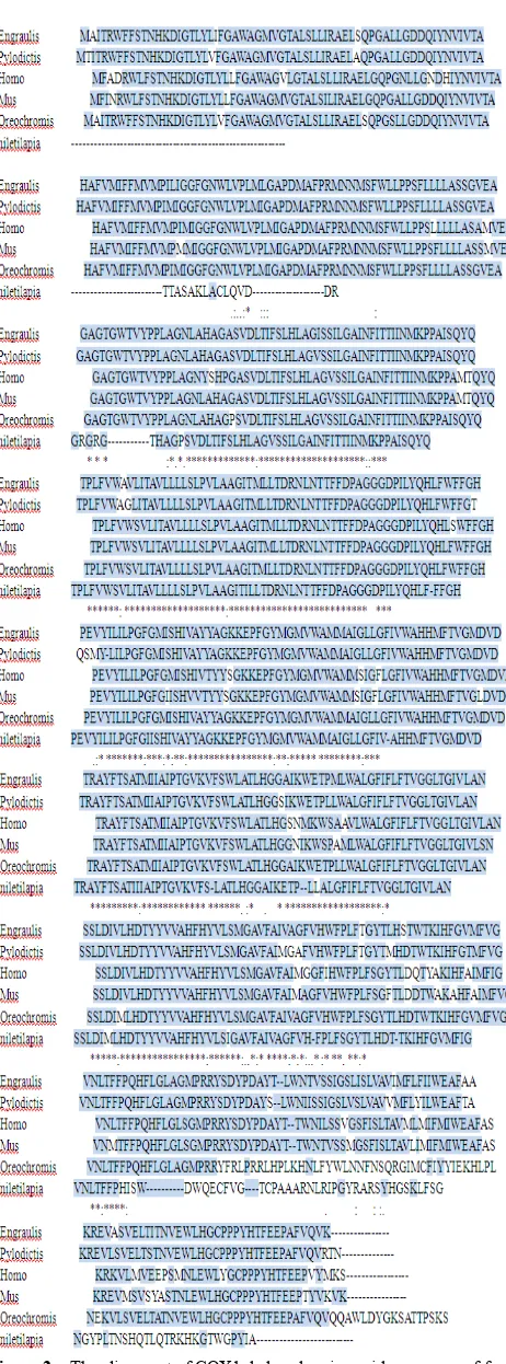
Alignment analysis and phylogenetic tree construction A Basic Genebee ClustalW 1.83 was performed on a va-riety of Cox1 genes from Engraulis japonicas; Pylodictis olivaris; Homo sapiens; Mus musculus; Oreochromis mos-sambicus and Oreochromis niloticus (accession nos. NP_203729; AAL91550; BAA07292; CAD54434; YP_ 272017 and GE647880 respectively). Fig.1. Oreochromis niloticus shares an overall identity of 89.2 - 99% with other known vertebrates. Using the Clustal W and the GeneBee programs, a phylogenetic tree was generated (Fig.2, 3). Expression of HSP9 in Oreochromis niloticus
ONCOX1 expression studied by RT-PCR in mixed tissues from infected and uninfected control fish was detected only in the infected tilapia (Fig.4).
4. Discussion
Suppression subtractive hybridization (SSH) has led to the enrichment of specific expression genes as the non-specific expression of beta actin was reduced to about 215 time lower
after subtraction, suggesting that non-specific expressed genes have been avoided and immunization- specific genes have been enriched efficiently in this study. The identifica-tion of new genes involved in the bacterial immunizaidentifica-tion provides the foundation for further research on the immu-noilogical interaction between host and the bacteria, In this study, cDNA library construction in case of O. niloticus
immunized by F. columnarae introduced some new immune genes, one of them was ONCOX1.
dynamic change in an environment, disease hazards and the host-pathogen relationship provided signals for identi-fying differentially expressed geneswhich may play an
im-portant role in the immune system responses. During con-struction of cDNA library from O. niloticus immunized by F. columnarae, we could identify and characterize cytochrome c oxidase subunit 1 gene as up-regulated gene.
ONCOX1 sequence was found to be homologous to other known cytochrome c oxidases with varying levels of se-quence identity and high similarity with Oreochromis mos-sambicus (accession no.YP_272017) and also to human (accession no.BAA07292) and several vertebrates COX1, it appears to have structural features similar to the largest subunit of the heme/copper-requiring cytochrome c.
The prediction of O-GalNAc (mucin-type) for glycosyla-tion sites in ONCOX1 proteins indicated the absence of any glycosylation sites. Multiple methods of transmembrane prediction were used to enable a consensus confirmation of predicted transmembrane helices. The TM region search for ONCOX1 by SOSUI analysis program beta version suggests that ONCOX1 is a membrane protein with 8 transmembrane helices(TM). Transmembrane helices were found in the positions 29-53,72-95, 126-145, 152-171, 192-211, 218-237, 258-282 and 289-306 as showed in table 1.The objective of this TM search step was to identify and map out regions of transmembrane helices to confirm our sequence database based hypothesis of the putative sequence being identified as a cytochrome c oxidase subunit I. Cythochrome c oxidases are known to have these transmembrane regions and the identification of these regions served as a confirmatory step in gauging the validity of the predicted structure (Mohamed
et al., 2003). The observation from the transmembrane pre-diction step is consistent with the subunit I of most other haem copper oxidases and acts as confirmatory data for correctness of the protein fold generated by the analysis program.
A final taxonomic system for the animal kingdom will probably include at least 10 million species partitioned among more than a million genera (Paul et al., 2003). Given such high diversity, there is a growing realization that it is critical to seek technological assistance for its initial de-scription and its subsequent recognition (Godfray 2002; Blaxter 2003). Recent investigations have suggested the feasibility of creating identification systems reliant on the analysis of sequence diversity in small segments of DNA (Tautz etal. 2003). Hebert et al. (2003) focused this discus-sion by proposing that a DNA barcoding system for animal life could be based upon sequence diversity in cytochrome c
Table 1. Analysis of COX1 transmembrane amino acid sequence (SOSUI dumbbell server)
No. N terminal transmembrane region C terminal type length
1 31 IFSLHLAGVSSILGAINFITTII 53 Secondary 23
2 73 LITAVLLLLSLPVLAAGITILLT 95 Primary 23
3 126 VYILILPGFGIISHIVAYYAG 146 Primary 21
4 153 YMGMVWAMMAIGLLGFIVAHHMF 175 Primary 23
5 188 TSATIIIAIPTGVKVFSLATLHG 210 Secondary 23
6 219 LALGFIFLFTVGGLTGIVLANSS 241 Primary 23
7 257 HYVLSIGAVFAIVAGFVHFPLFS 279 Primary 23
Figure 2. The alignment of COX1 deduced amino acid sequences of from
Engraulis japonicas; Pylodictis olivaris; Homo sapiens; Mus musculus; Oreochromis mossambicus and Oreochromis niloticus (accession nos. NP_203729; AAL91550; BAA07292; CAD54434; YP_272017 and GE647 880 respectively). Identical amino acids are indicated with an asterisk, similar amino acids are shaded. Dashes are gaps generated by alignment
Figure 3. phylogenetic distance tree showing the relationship among the COX1 from different vertebrates
Figure 4. Tissue specific expression revealed by RT-PCR in Oreochromis niloticus; 1=βactin 2=immunized fish, 3= unimmunized fish
Paul et al., 2003 in their study on COX1 divergences among closely related species indicated that an identification system for animal life based on the COI gene will be highly effective, although COXI divergences appear too low to regularly enable species diagnosis within the cnidarians, generic-level identifications in these organisms remain a prospect, as their study has established that species-level identifications are ordinarily achieved, COI analysis actually provides a taxonomic system that is chasing the last digit of animal diversity.
The mRNA expression of mixed tissues of F. columnare - immunized O. niloticus indicated that the COX1 enzyme coding gene was highly expressed in the immunized in con-trast to unimmunized fish, thus it is suggested to be included in the immune response against F. columnare.
Cytochrome
c
, which is a component of the mitochondrial electron transfer chain releases from the mitochondria in response to specific apoptotic stimuli (Wang X., 2001). It is initiates caspase activation when released from mitochondria during apoptosis (Liu et al.1996), Skulachev V.P.(1998) in his study on apoptosis has revealed that cytochrome c is a pro-apoptotic factor. It is released from its places on the outer surface of the inner mitochondrial membrane at early steps of apoptosis and, combining with some cytosolic pro-teins, activates conversion of the latent apoptosis-promoting protease pro-caspase-9 to its active form. Thus from this investigation, it is suggested that COX1 may be included in the immune response against bacterial infection, the higher level of expression may also imply its role as an apoptotic inducer.5. Conclusions
the immune response to bacterial infection, thus the present study provides some information for further research on the structure of COX1 gene as an immune gene in relation to bacterial infection.
REFERENCES
[1] Altschul, S.F.; Thomas, L.M.; Alejandro, A.S.; Jinghui Z.; Zheng Z.; Webb, M. and David, J.L.1997.Gapped BLAST and PSI-BLAST: a new generation of protein database search programs. Nucleic Acids Research,September, 25( 17): 3389-3402
[2] Blaxter, M. 2003. Counting angels with DNA. Nature 421:122–124
[3] Ettickan Boopathi , Nibedita Lenka , Subbuswamy K. Prabu , Ji-Kang Fang , Frank Wilkinson , Michael Atchison, Agata Giallongo, and Narayan G. Avadhani (2004). Regulation of Murine Cytochrome c Oxidase Vb Gene Expression during Myogenesis. J. Biol. Chem., Vol. 279, Issue 34:35242-35254 [4] Godfray, H. C. J. 2002. Challenges for taxonomy. Nature
417:17–19
[5] Hebert, P. D. N., Cywinska, A., Ball, S. L. & deWaard, J. R. 2003. Biological identifications through DNA barcodes. Proc. R. Soc. Lond. B 270: 313–322
[6] Lee, I., Bender, E., Arnold, S., and Kadenbach, B. 2001. Biol. Chem. 382: 1629–1636. Lee, I., Bender, E., and Kadenbach, B. 2003. Mol. Cell. Biochem. 234:63–70
[7] Liu X, Kim CN, Yang J, Jemmerson R, Wang X.1996. In-duction of apoptotic program in cell-free extracts: require-ment for dATP and cytochrome c. J. Cell.12; 86(1):147-57 [8] Mohamed .R; Raih, M.R.; Sailan A.T.; Zamrod Z.; Embi M.N.
2003. A predicted structure of the cytochrome c oxidase from Burkholderia Pseudomallei. J. Boitec. 6: 17-28
[9] Paul D. N. Hebert*, Sujeevan Ratnasingham and Jeremy R. deWaard.2003. Barcoding animal life:cytochrome c oxidase subunit 1 divergences among closely related species. Proc. R. Soc. Lond. B (Suppl.) 270:S96–S99
[10] Skulachev V.P. 1998. Bioenergetic aspects of apoptosis, necrosis and mitoptosis. J. APOPTOSIS 11 (4): 473-485 [11] Tautz, D., Arctander, P., Minelli, A., Thomas, R. H. & Vogler,
A. P. 2003. A plea for DNA taxonomy. Trends Ecol. Evol. 18:70–74
[12] Thompson, J.D.; Higgins, D.G. and Gibson, T.J. 1994. CLUSTAL W: improving the sensitivity of progressive mul-tiple sequence alignment through sequence weighting, posi-tions-specific gap penalties and weight matrix choice. J.Nucleic Acids Research. 22: 4673-4680
[13] Vijayasarthy, C., Damle, S., Prabu, S. K., Otto, C. M., and Avadhani, N. G. 2003.Eur. J. Biochem. 270: 871–879 [14] Wang, X. 2001.The expanding role of mitochondria in