R E S E A R C H
Open Access
Regulation of Toll-like receptor 4-mediated
immune responses through
Pasteurella multocida
toxin-induced G protein signalling
Dagmar Hildebrand, Aline Sähr, Sabine J Wölfle, Klaus Heeg
*and Katharina F Kubatzky
*Abstract
Background:Lipopolysaccharide (LPS)-triggered Toll-like receptor (TLR) 4-signalling belongs to the key innate defence mechanisms upon infection with Gram-negative bacteria and triggers the subsequent activation of adaptive immunity. There is an active crosstalk between TLR4-mediated and other signalling cascades to secure an effective immune response, but also to prevent excessive inflammation. Many pathogens induce signalling cascades via secreted factors that interfere with TLR signalling to modify and presumably escape the host response. In this context heterotrimeric G proteins and their coupled receptors have been recognized as major cellular targets. Toxigenic strains of Gram-negativePasteurella multocidaproduce a toxin (PMT) that constitutively activates the heterotrimeric G proteins Gαq, Gα13and Gαiindependently of G protein-coupled receptors through deamidation. PMT is known to induce signalling events involved in cell proliferation, cell survival and cytoskeleton rearrangement.
Results:Here we show that the activation of heterotrimeric G proteins through PMT suppresses LPS-stimulated IL-12p40 production and eventually impairs the T cell-activating ability of LPS-treated monocytes. This inhibition of TLR4-induced IL-12p40 expression is mediated by Gαi-triggered signalling as well as by Gβγ-dependent activation of PI3kinase and JNK.
Taken together we propose the following model: LPS stimulates TLR4-mediated activation of the NFĸB-pathway and thereby the production of TNF-α, IL-6 and IL-12p40. PMT inhibits the production of IL-12p40 by Gαi-mediated inhibition of adenylate cyclase and cAMP accumulation and by Gβγ-mediated activation of PI3kinase and JNK activation.
Conclusions:On the basis of the experiments with PMT this study gives an example of a pathogen-induced interaction between G protein-mediated and TLR4-triggered signalling and illustrates how a bacterial toxin is able to interfere with the host’s immune response.
Keywords:Monocytes, Toll-like receptor 4, Heterotrimeric G proteins,Pasteurella multocidatoxin, Interleukin-12, T lymphocytes, Immune evasion
Background
Monocytes are professional antigen presenting cells and carry out at least two important functions during infec-tion. First of all they represent a barrier against patho-gens through their antimicrobial activity and second they support the initiation of adaptive immune responses. The latter is exerted by the presentation of processed antigens on major histocompatibility complex (MHC)
molecules to T lymphocytes, the expression of various costimulatory proteins on the cell surface and the pro-duction of cytokines [1,2]. To direct these functions dur-ing an immune response, monocytes become activated through binding of conserved microbial structures to their respective pattern-recognition receptors (PRRs). Within the group of PRRs, Toll like receptors (TLRs) play a well-known role in the initiation of such immune responses. Up to now, 10 functional TLRs have been identified in humans [3]. Each TLR detects distinct PAMPs derived from viruses, bacteria, mycobacteria, fungi, and parasites [3]. Gram-negative bacteria are
* Correspondence:[email protected]; [email protected]
Department für Infektiologie, Medizinische Mikrobiologie und Hygiene, Im Neuenheimer, Feld 324, D-69120, Heidelberg, Germany
typically sensed through the cell wall constituent lipo-polysaccharide (LPS) that binds in complex with the LPS-binding Protein (LBP) to a receptor complex of TLR4, CD14 and an associated protein (MD-2) [4]. The TLR4-mediated signalling cascades then modulate the gene expression towards the production of a variety of pro-inflammatory cytokines such as Interleukin (IL)-6,
Tumour necrosis factor (TNF)-α and IL-12 [5]. In
addition, these signalling events enhance the costimula-tory function of monocytes [6]. However, this TLR4-mediated induction of inflammation and activation of adaptive immunity is actively targeted and modulated by a variety of pathogens, presumably to impair the immune response. At this, heterotrimeric G proteins play an im-portant role. Bacterial toxins that act on heterotrimeric G
proteins such as Cholera Toxin (CT) ofVibrio cholerae,
heat-labile enterotoxin (LT) of enteropathic Escherichia
coli or Pertussis Toxin (Ptx) of Bordetella pertussis can
therefore alter LPS-induced cytokine release. This is achieved by heterotrimeric G protein-mediated produc-tion of cAMP which eventually results in an altered cyto-kine release, most notably of IL-12, by human monocytic cells. The molecular mechanism of the effect is however only incompletely understood [7,8].
Another toxin that targets heterotrimeric G proteins is
produced by toxigenic strains of Gram-negative
Pasteur-ella multocidabacteria that can be isolated from chronic respiratory infections in animals and from humans after
cat or dog bites [9].Pasteurella multocidatoxin (PMT) is
known to activate Gαq, Gαiand Gα13 [10-13]
independ-ently of a G protein-coupled receptor through
deamida-tion resulting in the constitutively activadeamida-tion of the α
-subunit and release of theβγ-subunit [14]. This leads to
the downstream activation of signalling events such as
phospholipase Cβ activation, induction of the
mitogen-activated protein (MAP) kinase pathways, the RhoA/Rho kinase (ROCK) pathway and the Janus kinase (JAK)-sig-nal transducers of transcription (STAT) pathway [15-18].
Additionally, PMT activates Gαi, thus inhibiting adenylate
cyclase activity and cAMP accumulation [10].
In this study we investigated how the activation of het-erotrimeric G proteins through PMT influences the
TLR4-mediated activation of human blood-derived
monocytes (hBDMs) and their ability to induce T cell proliferation. Our data demonstrate that PMT modulates TLR4-mediated cytokine production. This effect was most pronounced for the release of IL-12p40, a cytokine important for T cell activation. The suppression of IL-12p40 release resulted in the inhibition of the T cell-activating ability of LPS-activated hBDMs. This block could be restored by adding IL-12-containing superna-tants of LPS-stimulated hBDMs to the mixed lympho-cyte reaction (MLR), showing that IL-12 was both, necessary and sufficient. The toxin mediates this
inhibition of TLR4-induced IL-12p40 expression by Gαi
-mediated inhibition of adenylate cyclase and subsequent
cAMP accumulation in combination with a Gβγ
-mediated activation of PI3kinase and the MAP kinase JNK. These findings give new insights into the modula-tion of an immune response by a bacterial toxin and we hypothesise that the decreased T cell activation results in increased survival of the pathogen.
Results
PMT modulates LPS-triggered cytokine release of human blood-derived monocytes
Antigen presenting cells regulate lymphocyte-mediated immune responses through presentation of antigens to lymphocytes via the major histocompatibility complex (MHC) on their surface, costimulatory or co-inhibitory signals that are delivered by ligands of the B7 family and via the release of cytokines [19].
Our study aimed to clarify whether the LPS-induced activation of hBDMs through TLR4 is modulated by the heterotrimeric G protein activator PMT and whether the toxin itself activates monocytes. We prestimulated
hBDMs with wildtype PMT (PMTwt) or the inactive
mu-tant PMTRBD, which contains the receptor binding
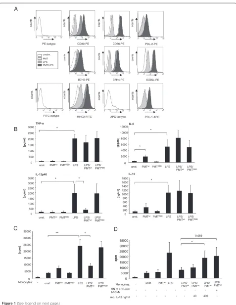
do-main (RBD) but lacks the C-terminal catalytic dodo-main (CD), for three hours, as the intracellularly-acting toxin needs time to enter the cell and to translocate to the cytoplasm [20]. LPS was then added in co-stimulated samples for overnight incubation. The first experiments addressed whether the expression of immune-relevant ligands on monocytes was changed in the presence of PMT compared to unstimulated or LPS-treated samples. The surface expression of MHC II and the B7 family members B7.1 (CD80), B7.2 (CD86), inducible co-stimulator ligand (ICOS-L), programmed death-1 ligand (PD-L1), programmed death-2 ligand (PD-L2), B7-H3 and B7-H4 were determined by FACS analysis. The
results demonstrate that PMTwt alone activates
mono-cytes only to a small extent as can be seen by the nearly unchanged expression patterns compared to the un-stimulated samples. Samples that had been treated with
LPS and PMTwtshowed an increase in the level of CD80
when compared to the LPS-treated control and a smaller increase was also detectable for B7H3, B7H4 and MHC2
(Figure 1A). Stimulation with the inactive PMTRBD did
not change the expression pattern of untreated and LPS-treated hBDMs at all (data not shown). We therefore
conclude that PMTwt by itself is unable to stimulate
monocyte activation and that it only marginally increases monocyte activation induced by LPS.
Next, we investigated, whether PMT modulates the in-flammatory response of monocytes by inducing or inhi-biting their ability to release cytokines. We stimulated
CD80-PE CD86-PE
MHC2-FITC PDL-1-APC
PDL-2-PE
B7H3-PE B7H4-PE ICOSL-PE PE-isotype
FITC-isotype APC-isotype
counts counts counts counts
counts counts counts
counts counts counts counts
unstim.
PMT LPS PMT/LPS
A
B
TNF-0 500 1000 1500 2000 2500 3000
unst. PMTwt PMTRBD LPS LPS/
PMTwt
LPS/ PMTRBD
[pg/ml]
IL-6
0 2000 4000 6000 8000 10000 12000
[pg/ml]
IL-12p40
0 500 1000 1500 2000 2500 3000 3500
[pg/ml]
unst. PMTwt PMTRBD LPS LPS/
PMTwt
LPS/ PMTRBD
unst. PMTwt PMTRBD LPS LPS/
PMTwt
LPS/ PMTRBD *
*
*
* *
Monocytes: 0 5000 10000 15000 20000 25000 30000 35000
unst. PMTwt PMTRBD LPS LPS/
PMTwt
LPS/ PMTRBD
[cpm]
* **
C
D
-
IL-10
0 200 400 600 800 1000 1200 1400 1600 1800
[pg/ml]
unst. PMTwt PMTRBD LPS LPS/
PMTwt
LPS/ PMTRBD *
Monocytes: unst. PMTwt LPS LPS/ PMTwt
LPS/ PMTwt
SN of LPS-stim hBDMs
- LPS/
PMTwt
LPS/ PMTwt
- - - - - - - - +
- - - - - - 40 400 - rec. IL-12 ng/ml
0 5000 10000 15000 20000 25000 30000 35000
cpm
*
0.059
combination with LPS, as described before, and
mea-sured the release of pro-inflammatory IL-6, TNF-α and
IL-12p40 and anti-inflammatory IL-10 by ELISA analyses (Figure 1B). As expected, LPS-stimulated monocytes
released elevated levels of all four cytokines. PMTwtonly
enhanced the secretion level of IL-6 in a statistically rele-vant manner. While the toxin had a minor influence on
LPS-triggered TNF-α secretion and IL-10 production, it
elevated the LPS-induced IL-6 level considerably, but al-most totally abrogated the IL-12p40 release of activated cells. Since the truncated toxin again did not affect hBDMs we assigned the observed modulation to the catalytic function of the toxin and continued the studies
with PMTwt.
PMT attenuates T cell-activating ability of LPS-activated monocytes
T cell activation by antigen presenting cells requires the interaction between MHC complexes and T cell recep-tors as well as the binding of costimulatory B7 molecules to the CD28 receptor on T cells [21]. Additionally, IL-12 exerts a strong synergistic effect with the B7/CD28 inter-action in inducing proliferation and cytokine production in T cells [22].
As PMTwt only slightly elevated TLR4-mediated
ex-pression of costimulatory ligands on hBDMS (Figure 1A) and almost totally prevented LPS-stimulated IL-12p40 production (Figure 1B), we asked whether the toxin would affect T cell proliferation induced by the presence of LPS-treated hBDMs. To this end we performed mixed
lymphocyte reactions with LPS- and/or PMTwt
-stimu-lated monocytes and allogeneic T cells. The quantification of T cell proliferation after three days of co-culture revealed that PMT-stimulated monocytes did not differ significantly from unstimulated cells in mediating T cell proliferation. However, the toxin strongly inhibited the ability of LPS-stimulated hBDMs to activate T cell prolif-eration (Figure 1C). This pronounced suppression of T cell activation was reversed by adding recombinant IL-12 or supernatants from LPS-treated cells known to have a high level of IL-12p40 (Figure 1B and D). We therefore
conclude that the suppressive influence of PMTwt on
monocyte-mediated T cell activation is due to a change in cytokine release, and in particular caused by the abro-gated IL-12p40 production.
PMT does not alter the cytokine release of LPS-activated hBDMs via the cyclooxygenase (COX)-2/Prostaglandin E2 (PGE2) pathway
To identify the mechanism behind the PMT-modulated cytokine secretion of LPS-activated hBDMs, we first examined the COX/PGE2 pathway. The lipid mediator PGE2 is a well-established immunomodulatory substance which is produced during the immune response of anti-gen presenting cells. PGE2 is known to be a potent in-ducer of IL-10 and a strong inhibitor of IL-12 production [23,24]. COX proteins play a key role in the synthesis of prostaglandins. Whereas COX-1 is constitu-tively expressed and induces a basal level of PGE2, COX-2 is specifically induced by inflammatory stimuli [COX-25]. To check whether PGE2 could be responsible for the
PMT-induced blockade of IL-12p40, we treated
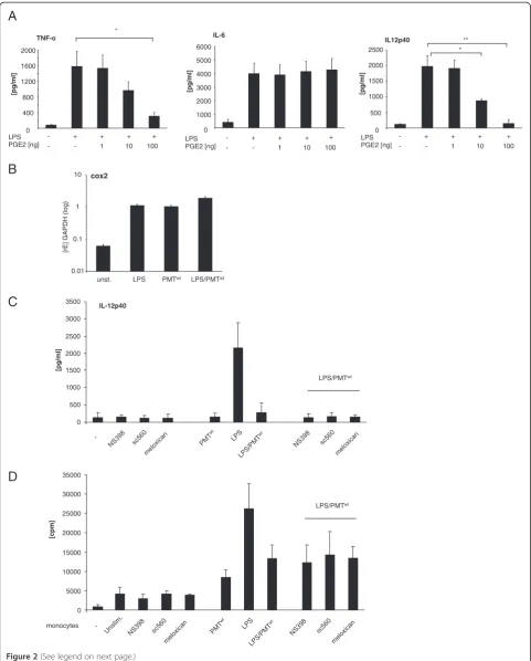
LPS-stimulated hBDMs with increasing concentrations of re-combinant PGE2 and quantified the release of cytokines. Whereas there was no significant influence detectable on
IL-6 production, LPS-induced TNF-α and IL-12p40
se-cretion were diminished by PGE2 (Figure 2A). We next determined the mRNA level of COX-2 in LPS and/or
PMTwt-treated hBDMs to investigate whether PGE2
could be released in this setting. The performed
quanti-tative real-time PCRs showed that LPS and PMTwt both
triggered COX-2 gene transcription but that there was no pronounced synergistic effect between PMT and LPS (Figure 2B). To investigate whether the minor change in COX-2 expression could explain the PMT-induced change in cytokine release, we used various COX-2 inhi-bitors. Neither SC560 nor Meloxicam or NS398 neutra-lised the blockade of IL-12p40 production (Figure 2C) or
changed the pattern of released IL-6 and TNF-α (data
not shown). Mixed lymphocyte reactions additionally revealed that the COX-2 inhibitors did not restore the T cell-activating ability of LPS-treated monocytes after
PMTwt-stimulation (Figure 2D). Based on these data we
exclude a prominent impact of the COX2/PGE2 pathway
(See figure on previous page.)
on the PMTwt-mediated effect on endotoxin-stimulated hBDMs.
PMT-induced modulation of LPS-triggered IL-12 release
involves the Gαi/cAMP pathway
In order to characterize the PMT-induced signalling pathway that modulates cytokine production of LPS-activated monocytes, we next focused on the
heterotri-meric G protein Gαi that is directly activated by PMT
[10]. While the exact mechanism is still under
investiga-tion, it is commonly accepted that Gαi- and Gαs
-mediated alterations in intracellular cAMP metabolism play a key role in modulating IL-12 production. At this,
GPCR ligands of adenylate cyclase-activator Gαs as well
as GPCR agonists of adenylate cyclase-inhibitor Gαiwere
shown to decrease the capacity of antigen presenting cells to produce IL-12 [8]. Whereas the connection of adenyl-ate cyclase-activation and IL-12 decrease may be more
generally accepted, investigations with the Gαi-activator
mastoparan, the Gαi-inhibitor Ptx as well as studies using
Gαi-deficient mice clearly revealed an inhibitory effect of
this G protein on LPS-induced cytokine release in differ-ent cells types, including human monocytes [26-28]. To
investigate the role of Gαion the PMT-mediated
manipu-lation of TLR4 signalling we pre-treated LPS and/or
PMTwt-stimulated monocytes with Ptx or mastoparan.
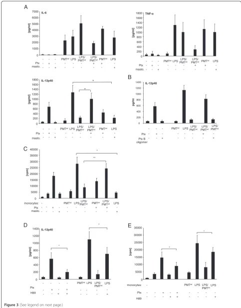
The performed cytokine ELISAs show that neither Ptx nor mastoparan significantly influenced IL-6 production of unstimulated or stimulated cells. The alteration of
TNF-α was more influenced but statistically not
signifi-cant. The impact of Gαi was most pronounced on
IL-12p40. Mastoparan mimicked the inhibition of IL-12p40 and Ptx could almost fully restore the LPS-induced pro-duction of IL-12p40 after toxin-treatment (Figure 3A). To confirm that the Ptx-mediated reconstitution of IL-12 production is due to the influence of G proteins, we included the B oligomer subunit of Ptx in our studies. The Ptx B oligomer is the receptor binding domain but can also initiate signal transduction pathways [29]. How-ever, it does not play a role in the activation of G-protein signalling that is initiated by the catalytic subunit. As it exerted no influence on the PMT-mediated suppression of IL-12p40 (Figure 3B), we assigned the Ptx effect to the
inhibition of Gαi.
Moreover, in mixed lymphocyte reactions using allor-eactive T cells it was revealed that Ptx reconstituted the
PMTwt-blocked activation of T cells proliferation through
LPS-stimulated monocytes, while mastoparan/LPS-trea-ted monocytes on the other hand were less activating than cells treated with LPS only (Figure 3B). These data
implicate that the PMTwt-activated Gαiprotein is
respon-sible for the abolishment of LPS-activated IL-12p40
re-lease, while its impact on IL-6 and TNF-α is only
marginal.
In order to further analyse the link between PMTwt
-activated Gαiand IL-12p40 suppression, we next
concen-trated on protein kinase A (PKA), as elevated cAMP concentrations can result in PKA activation [30]. This serine/threonine kinase mediates phosphorylation of substrates and subsequently induces the transcription of a variety of genes via the binding of transcription factors such as CRE-binding protein (CREB) to the DNA bind-ing motif cAMP-response element (CRE) present in the promoter region of cAMP-regulated genes [31,32]. To mimic the PMT-induced inhibition of PKA caused by an inhibition of adenylate cyclase and cAMP accumulation, we included the PKA-specific inhibitor H89 in our stud-ies. As a control, we verified that the inhibitor was able to block Ptx-induced IL-12p40 production (Figure 3C).
Compared to PMTwt, H89 did not totally abolish
LPS-induced IL-12p40 release but significantly diminished the LPS-induced release of the cytokine (Figure 3C) and as a consequence decreased the T cell-activating ability of hBDMs (Figure 3D). These data indicate a prominent role for PKA in the suppression of LPS-induced IL-12p40 production.
PMT-activated JNK contributes to the suppression of IL-12 production
Next we investigated whether other known regulators of IL-12p40 may contribute to the PMT-induced abolish-ment of LPS-mediated IL-12p40 release. Mitogen acti-vated protein kinases (MAP kinases) are described to modulate IL-12 production in myeloid cells. Whereas ERK (p42/44) and JNK were shown to inhibit IL-12p40 release [33,34], p38 is known as an inducer of IL-12p40 [35]. We therefore determined the activation status of
ERK, JNK and p38 after PMTwttreatment and the
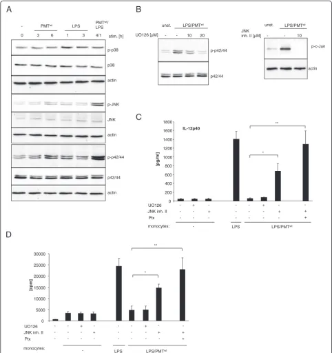
modu-lation of LPS-activated MAP kinases. The performed western blot analyses revealed that PMT, as expected, neither significantly activated p38 nor modulated the slight LPS-induced p38 phosphorylation. However, ERK
activation was enhanced after six hours of PMTwt
treat-ment and the LPS-mediated phosphorylation was se-verely enhanced by the toxin. In addition, the low JNK
activation detectable after six hours of PMTwt and one
hour of LPS-treatment was strongly enhanced by simul-taneous stimulation with both stimuli (Figure 4A). To verify whether this observed activation of ERK and JNK could account for the suppression of LPS-mediated IL-12p40 production, we used specific MAP kinase tors. Monocytes were pre-treated with the MEK1 inhibi-tor UO126 (upstream of ERK) or JNK inhibiinhibi-tor II and
stimulated with PMTwt and LPS. The blots in
Figure 4B show that 20 μM of UO 126 reduced LPS/
PMTwt-induced ERK phosphorylation to basal levels and
10μM of JNK inhibitor II totally blocked the activation
A
B
IL-6
0 1000 2000 3000 4000 5000 6000
[pg/ml]
LPS PGE2 [ng]
C
D
IL-12p40
0 500 1000 1500 2000 2500 3000 3500
[pg/ml]
0 5000 10000 15000 20000 25000 30000 35000
[cpm]
cox2
0.01 0.1
1 10
unst. LPS PMTwt LPS/PMTwt
[rE] GAPDH (log)
[pg/ml]
TNF-0 400 800 1200 1600 2000
*
LPS PGE2 [ng]
IL12p40
0 500 1000 1500 2000 2500
[pg/ml]
**
LPS PGE2 [ng]
*
- -
+ 1
+ 100 + 10 +
-
- -
+ 1
+ 100 + 10 +
-
- -
+ 1
+ 100 + 10 +
-
LPS/PMTwt
LPS/PMTwt
monocytes -
supernatants of these cells demonstrated that while the inhibition of ERK did not affect LPS-induced IL-12p40 release, the suppression of JNK restored LPS-induced cytokine production (Figure 4C). Simultaneous inhibition
of JNK and Gαi activation through Ptx treatment
abol-ished the PMT effect (Figure 4C) and restored the T
cell-activating ability of LPS-treated cells after PMTwt
-stimu-lation (Figure 4D). These data indicate that PMT inhibits
LPS-mediated IL-12p40 expression via Gαi-mediated
sig-nalling and the activation of JNK.
PMT-stimulated JNK-activation involves PI3-kinase
Our next experiments addressed the question how PMT activates JNK. Recent publications suggest PI3 kinase as
a mediator between Gαi-proteins and Ras/MAP kinase
activation [36,37]. Interestingly, PMT activates
PI3-kinase γ through dissociation of Gβγ subunits from the
Gαisubunit [38]. Here, we demonstrate by western blot
analysis that PMTwt induces the phosphorylation of the
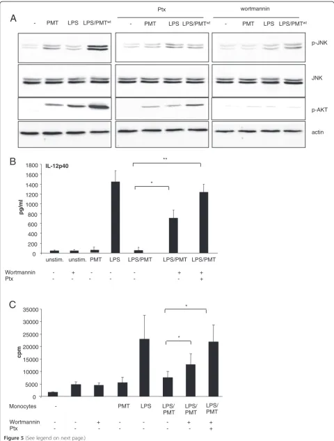
PI3-kinase target Akt in primary monocytes (Figure 5A). To check whether the activation of JNK depends on PI3-kinase activation we blocked PI3 PI3-kinase using wortman-nin and observed a reduction of JNK phosphorylation in
PMTwt- and most notably in LPS/PMTwt-stimulated cells
(Figure 5A, right section). Furthermore, the inhibition of
Gαi-activation by treatment with Ptx diminished the
sti-mulated Akt activation downstream of PI3 kinase and impaired the activation of JNK (Figure 5A, middle
sec-tion). This suggests that PI3 kinase mediates PMTwt
-sti-mulated JNK activation and that the βγsubunits of Gαi
and other PMT-activated Gαproteins induce the
activa-tion of the kinase. ELISA experiments confirmed that the inhibition of JNK activation through wortmannin could reduce the PMT-mediated IL-12p40 suppression.
Finally, the inhibition of both, JNK and Gαi-mediated
signalling through Ptx, totally restored the LPS-induced production of the cytokine (Figure 5B). Performed MLRs confirmed the impact of the restored IL-12-production on the T cell-proliferation (Figure 5C).
Taken together we propose the following model: LPS
stimulates TLR4-mediated activation of the NFĸ
B-pathway and modulates the production of TNF-α, IL-6
and IL-12p40. PMT inhibits the production of IL-12p40
through Gαi-mediated inhibition of adenylate cyclase
and cAMP accumulation as well as by PI3kinase/Akt
and JNK activation mediated by the dissociated Gβγ
sub-unit (Figure 6).
Discussion
Antigen presenting cells are crucial for the first-line host defence and act as mediators between innate and adap-tive immunity. One major pathway of these cells involves TLR-mediated signalling events, which can detect and react to extracellular as well as intracellular microbial components [39,40]. To be effective, immune responses need the crosstalk between TLR-mediated and other sig-nalling cascades that play a role in immune responses or other cellular pathways, respectively. These interactions can have synergistic effects but can also antagonise responses in order to prevent excessive inflammation. To
modify and presumably escape the host’s immune
re-sponse pathogens have developed various strategies to interfere with signalling cascades of the host [41,42]. Be-sides direct inhibition or modification of TLR signalling, induction of alternative regulative or antagonistic en-dogenous signalling cascades is another strategy in im-mune evasion. Here, we investigated how the activation of heterotrimeric G proteins through the bacterial toxin PMT modulates TLR4-mediated activation of human blood-derived monocytes and how this potentially affects cells of the adaptive immune system such as T cells.
Our data on the expression of MHC and costimulatory B7 family members show that PMT alone does not acti-vate monocytes by itself and only slightly enhances LPS-triggered cell activation. However, PMT is able to modu-late the TLR4-mediated cytokine production of these cells significantly leading to an almost complete block of IL-12p40 production. Bioactive IL-12 is a potent activa-tor of natural killer (NK) cell and T cells and, together with the B7/CD28 interaction between antigen present-ing cells and T cells, IL-12 has a strong synergistic effect in inducing T cell proliferation [22,43]. Our data show that due to the absence of IL-12, PMT suppresses T cell activation induced by LPS-activated hBDMs. This effect
(See figure on previous page.)
Figure 2Contribution of the COX-2/PGE2 pathway to the PMT-mediated modulation of LPS-activated monocytes.A. Monocytes were stimulated overnight with LPS (30 ng/ml) and treated in parallel with PGE2 in increasing concentrations. Supernatants were used to quantify the release of TNF-α, IL-6 and IL-12p40 by ELISA. Presented is the mean and standard deviation of three independent experiments. B. Cells were stimulated with LPS (30 ng/ml; 1 h), PMTwt(1μg/ml; 4 h) or PMTwt/LPS (4 h/1 h) or left unstimulated. RNA was extracted, cDNA was prepared and used for quantitative PCR analysis with specific primers for COX2. The results are presented as relative expression to the reference gene (GAPDH). Shown is one representative experiment with 10% standard error. C. Monocytes were pre-treated for 2 hours with the cox-inhibitors NS398 (3.8μM), sc560 (6.3μM) or meloxican (4.7μM). Then cells were stimulated with PMTwt(1
A
C
D
E
[cpm] monocytes: IL-12p40 0 200 400 600 800 1000 1200 1400 [pg/ml] * * 0 5000 10000 15000 20000 25000 30000 35000 [cpm] * * monocytes: masto. TNF-0 200 400 600 800 1000 1200 1400 1600 1800 [pg/ml] LPS/
PMTwt PMTwt LPS
PMTwt LPS LPS/
PMTwt Ptx - - - + - - + - - - - - - + - + - - + - - [pg/ml] IL-6 0 1000 2000 3000 4000 5000 6000 7000 LPS/
PMTwt PMTwt LPS
PMTwt LPS LPS/
PMTwt Ptx - - - + - - + - - - - - - + - + - - + - - masto. [pg/ml] * IL-12p40 0 200 400 600 800 1000 1200 1400 1600 1800 * masto. LPS/
PMTwt PMTwt LPS
PMTwt LPS LPS/
PMTwt Ptx - - - + - - + - - - - - - + - + - - + - - 0 ** * 5000 10000 15000 20000 25000 30000 35000 40000 masto. LPS/ PMTwt LPS
PMTwt LPS LPS/
PMTwt Ptx - - - + - - + - - - - - - + - + - - + - PMTwt - Ptx H89 PMTwt + - - - - + - - - - + + - + -
- Ptx
H89 + - - - - + - - - - + + - + - - PMTwt LPS LPS/ LPS
- - - -
PMTwt LPS LPS/
PMTwt LPS
- - - IL-12p40 0 200 400 600 800 1000 1200 1400 pg/m l Ptx B oligomer LPS/ PMTwt
PMTwt LPS LPS/
PMTwt Ptx - - - + - - + - - - - - - + - - - PMTLPS/ wt
- +
B
strictly depends on IL-12, as activity could be restored
by adding IL-12-containing supernatants of
LPS-stimulated hBDMs or recombinant IL-12 to the mixed lymphocyte reaction. Although IL-10 is a known sup-pressor of IL-12 activity [44], a dominant role of IL-10 could be excluded, since PMT only slightly enhanced the LPS-induced IL-10 production.
Bagley et al.. demonstrated that PMT also suppresses LPS-stimulated IL-12 release of human macrophage-derived dendritic cells and they speculate that this
block-ade is caused by Gαq-mediated Phospholipase C-β
activa-tion and subsequent intracellular calcium mobilizaactiva-tion, but they did not further investigate this [45]. For human monocytes we suggest that PMT prevents
TLR4-mediated IL-12p40 production through Gαi- and βγ
-mediated signalling cascades, respectively. PMT activates
Gαi and thereby inhibits adenylate cyclase activity and
cAMP accumulation, independently of Gαqand Gα12/13
[10]. Our data point to Gαisignalling as the Gαiagonist
mastoparan mimicked the suppression of LPS-induced IL-12p40 production. Recently it became clear that data obtained from experiments using mastoparan have to be interpreted with care as mastoparan can bind the toxic lipid A portion of the LPS molecule [46]. Nevertheless, as in our studies mastoparan only blocked LPS-stimulated IL-12p40 production and did not block TLR4-mediated
IL-6 and TNF-αexpression, we assign the result primarily
to Gαi-mediated signalling. Additionally, the Gαi
-inhibi-tor Pertussis toxin (Ptx) reversed the PMT-mediated blockage of IL-12-release and restored the T cell-activating ability of LPS-treated monocytes.
In the past it has been shown that both Gαiand Gαs,
although having opposite function and acting as negative regulators for each other, are able to downregulate IL-12 production [8,47]. This suggests that alterations in intra-cellular cAMP metabolism in general play a role in modulating the IL-12 production. The molecular mech-anism, however, is only poorly understood. Our data in-dicate that Protein kinase A (PKA) is involved in the PMT-mediated suppression of IL-12p40 production induced by LPS. In our experiments the PKA-specific in-hibitor H89 significantly diminished LPS-induced release
of IL-12p40 and decreased the T cell-activating ability of hBDMs. It thereby mimicked the PMT-induced effect on monocytes at least to some extent. H89 totally blocked Ptx-induced IL-12p40 production, demonstrating that
the Gαi-protein-inhibitor Ptx induces IL-12 via PKA.
Our experiments suggest that signalling mechanisms
directly dependent on Gαiaccount partially for the
PMT-triggered abrogation of LPS-induced IL-12p40 produc-tion, but they also indicate that other pathways are involved. Another signalling cascade connected to IL-12 production is the MAP kinase pathway, whereas ERK (p42/44) and JNK are known inhibitors of IL-12 produc-tion [35]. Our investigaproduc-tions demonstrate that simultan-eous stimulation of the cells with PMT and LPS induced a strong activation of JNK. Furthermore, inhibition of JNK restored LPS-induced IL-12 production after PMT-treatment. Although G proteins are known to activate the MAP kinase cascade, the exact mechanism is not well defined. However, a potential link to known PMT-mediated signalling pathways comes from a publication that showed the activation of this pathway was induced by phospholipase C and protein kinase C [48]. These
pro-teins are known to be activated by PMT through Gαq
ac-tivation [16]. Additionally, βγ subunits dissociated from
their Gαisubunit were also identified as initiators of the
Ras/MAP kinase pathway [37,49]. Our experiments point
to Gαiproteins as well as their respective dissociatedβγ
subunits as mediators of the PMT-stimulated JNK
phos-phorylation, as the Gαiinhibitor Ptx significantly
dimin-ished the PMT/LPS-induced activation of the kinase. It is
also possible thatβγ subunits of other PMT-activated G
proteins may additionally be involved, as the inhibition of
Gβγ release through Ptx did not completely suppress
JNK activation. The possibility thatβγsubunits of various
G proteins are involved has also been discussed previ-ously by Preuss et al.. [38].
Kranenburg et al. claim from their experiments in COS cells and fibroblasts that the missing link between
Gβγ and Ras/MAP kinase activation is PI3 kinase [37].
In addition, Lopez-Ilasaca et al. confirmed that overex-pressed PI3 kinase activates MAP kinases [36,37]. Recent
studies showed that PMT activates PI3 kinase γthrough
(See figure on previous page.)
Figure 3Gαi-mediated modulation of LPS-induced IL-12 production.A. Monocytes were stimulated overnight with Pertussis toxin (Ptx; 200 ng/ml) or left unstimulated. The next day PMTwt(1μg/ml) or mastoparan (20μM) was added. After three hours LPS was added for overnight incubation (30 ng/ml). The release of TNF-α, IL-6 and IL-12p40 were measured by ELISA. Shown is the mean of three independent experiments (mean ± SD; n = 3). B.As additional control cells were treated with Ptx B oligomer and ELISAs were performed with collected supernatant. (mean ± SD; n = 2) C. Monocytes were stimulated as described in A. After washing, co-cultures of CD14+hBDMs and CD3+alloreactive T cells were prepared. After 3 days proliferation of T cells was analysed by [3H]-thymidine incorporation and presented as counts per minute (cpm), mean ± SD; n = 3. D. Cells were stimulated as followed: H89 (10μM; 2 h pre-stimulation) and LPS (30 ng/ml) or Ptx (200 ng/ml) overnight, PMTwt(1
A B
C
UO126 [µM]
LPS/PMTwt
20
p-p42/44
p42/44
p-c-Jun
actin unst.
10 - -
JNK inh. II [µM]
LPS/PMTwt
unst.
10 - -
p-p42/44
p42/44
actin p-JNK
JNK
actin p-p38
p38
actin stim. [h]
0 3 6 1 3 4/1
- PMTwt PMT
wt/ LPS LPS
IL-12p40
0 200 400 600 800 1000 1200 1400 1600 1800
- LPS LPS/PMTwt
[pg/ml]
* **
D
0 5000 10000 15000 20000 25000 30000
[cpm]
* ** UO126 JNK inh. II
Ptx
- + + - - -
- - - +
+ - -
-
- -
- - - - -
+ -
+
UO126 JNK inh. II
Ptx
+ -
- + - - - - -
- +
- + -
- -
- - -
- -
- - -
+ -
+
monocytes:
LPS LPS/PMTwt
monocytes:
-
Figure 4Involvement of MAPkinases in the modulation of LPS-induced IL-12 release.A. Monocytes were stimulated with PMTwt(1μg/ml) and/or LPS (30 ng/ml) for the indicated time points or left unstimulated. The cells were lysed and extracted with RIPA buffer as described under Experimental procedures. Total amounts of p42/44, JNK and p38 and the phosphorylation levels of the MAP kinases was determined by
Gβγsubunits [38,50] and our current investigation con-firms the finding that the toxin mediates the phosphoryl-ation of Akt, the target of PI3 kinase, also in primary monocytes. The publication of La Sala et al. suggests that
the Gαi-protein activator C5a suppresses TLR4-mediated
IL-12 via the Akt pathway and JNK, but independently of PI3 kinase [34]. We show here that in the case of
PMT-mediated Gβγ activation the PI3 kinase inhibitor
wortmannin diminished the activation of JNK, suggesting that there is a possible link between MAP kinase activa-tion and PI3 kinase/Akt.
Our data clearly show that inputs from Gαi
protein-mediated signalling pathways modulate TLR4-induced signalling. Other authors even suggest a direct interaction
between TLR4 and Gαi. Solomon et al. demonstrated that
the LPS/LBP receptor CD14 co-immunoprecipitates with
various α subunits of the Gαi/Gαo family and that the
Gαiactivator mastoparan inhibits LPS-induced IL-6 and
TNF-α production in human monocytes [28]. Another
report claims that Gαi itself is activated rapidly but
transiently by LPS through an unknown mechanism. In a mouse model the authors showed that Ptx increased the level of LPS-induced plasma inflammatory media-tors, whereas mastoparan decreased the cytokine level
[26]. However, in these studies the influence of Gαi
acti-vation on IL-12p40 production as a downstream result of these signalling events had not been investigated. Re-cently, another report confirmed the hypothesis that
TLR ligands activate Gαi proteins in endothelial cells
[51]. It was shown that LPS stimulates PI3 kinase and
MAP kinase activation via Gαi, independently of the
TLR-induced MyD88-TRAF6 pathway. The authors speculate that TLR2, 3 and 4 can also directly interact
with Gαi via their intracellular domains due to a
con-sensus motif for Gαi/0 binding (Figure 6). In our hands
LPS-stimulated Akt-activation could be diminished by
Ptx which supports the hypothesis of LPS-induced Gαi
activation. Regardless of whether PMT is the only Gαi
activator or whether the toxin additionally increases
the LPS-triggered Gαi activation, our data clearly
show that the PMT-induced Gαi-mediated signalling
interferes with and suppresses TLR4-induced IL-12p40 production.
Conclusion
In summary we show that PMT inhibits the TLR4-mediated production of IL-12p40 at least in two ways.
First of all, PMT induces Gαi-mediated inhibition of
ad-enylate cyclase and cAMP accumulation. Furthermore,
PMT leads to a Gβγ-mediated activation of PI3kinase
and subsequent JNK activation mediated by Gαiand
pre-sumably other Gαsubunits activated by PMT. These
sig-nalling cascades act specifically on the production of
IL-12p40 as they influence the LPS-stimulated TNF-α
release only slightly and have no impact on IL-6 release. Taken together our observations contribute to the under-standing of the interaction between G protein-mediated and TLR4-mediated signalling and show how a bacterial toxin is able to manipulate monocytes which eventually results in impaired T cell activation.
Methods
Recombinant PMTwtand PMTRBDwere kindly provided
by Dr. Joachim Orth and Prof. Dr. Dr. Klaus Aktories from Freiburg. The TLR agonist LPS was provided by U. Seydel (Borstel). The p44/42 inhibitor UO126, JNK in-hibitor II, Pertussis toxin (full length and B oligomer subunit), mastoparan and wortmannin were purchased from Calbiochem. Recombinant IL-12 was obtained from Immunotools.
Preparation of monocytes
Monocytes were isolated from fresh blood or buffy coat by density gradient centrifugation (Biocoll separating so-lution 1.077 g/mL; Biochrom AG) and washed three times with PBS. CD14 cells were positively selected by magnetic-associated cell sorting (AutoMACS: program possel; Miltenyi Biotec). Sorted cells were cultured in RPMI 1640 medium (Biochrom AG) supplemented with 10% FBS (BioWest) and 1% penicillin and streptomycin (PAA) at 37°C in a humidified atmosphere in the
pres-ence of 5% CO2.
Mixed lymphocyte reaction (MLR)
T cells were obtained from fresh blood by positive selec-tion with CD3-labelled magnetic beads and FcR blocking reagent (AutoMACS: program possel; Miltenyi Biotec).
MLRs were performed in 96 well plates in allogeneic
settings: purified T cells (1x10⁵) were co-cultured with
pre-treated and washed monocytes (1x10⁴). Cells were
cultured for 3 days and exposed to [3H]-thymidine
(Amersham Pharmacia Biotech GmbH) during the last 6 h of culture. Thymidine uptake was measured using a liquid scintillation counter.
Flow cytometry (FACS)
Briefly, cells were washed with PBS/10% FBS, blocked for 30 min in PBS/10% FBS, and incubated with the corre-sponding antibodies (B7H3-PE, B7H4-PE,
anti-PDL-1-APC, anti-PDL-2PE, anti-ICOSL-PE from
- PMT LPS LPS/PMTwt
Ptx wortmannin
- PMT LPS LPS/PMTwt
p-JNK
JNK
p-AKT
actin - PMT LPS LPS/PMTwt
IL-12p40
0 200 400 600 800 1000 1200 1400 1600 1800
pg/ml
unstim. unstim. PMT LPS LPS/PMT LPS/PMT LPS/PMT
Wortmannin - + - - - + + Ptx - - - - - - +
A
B
***
C
- PMT LPS LPS/ PMT
Wortmannin - - + - - - + + Ptx - - - - - - - +
Monocytes LPS/
PMT LPS/ PMT *
*
0 5000 10000 15000 20000 25000 30000 35000
cpm
ELISA
Cell-free supernatants were harvested and analysed for
IL-6, IL-12p40, IL-10 and TNF-α by commercial
avail-able ELISA kits (OptEIA; BD).
Western blotting
After washing with PBS, cells were resuspended in RIPA buffer (150 mM NaCl, 1 mM EDTA, 1% Triton X-100, 1% Deoxycholat, 1 mM sodium fluoride, 25 mM Tris) containing phosphatase inhibitors (PhosSTOP, Roche Diagnostics) and incubated for 45 min at 4°C. After cen-trifugation, lysates were used as whole-cell extracts. Sam-ples were separated on 10% SDS-PAGE, transferred to nitrocellulose and incubated with the relevant antibodies (Cell Signaling Technology: p42/44, pThr202/Tyr204 p42/44, JNK, pThr183/Tyr185 JNK, p38, pThr180/ Tyr182 p38, pSer63 c-Jun, pThr 308 Akt, Actin) followed
by incubation with the respective HRP-coupled antibody (Cell Signaling Technology) and detection by enhanced chemiluminescence.
Reverse transcription-PCR
RNA was extracted with the “High pure RNA Isolation
Kit” (Roche). cDNA was prepared using ‘Reverse Aid
First Strand cDNA Synthesis Kit’ (Fermentas life
sci-ence). Aliquots of the cDNA were used for quantitative
PCR analysis with the ‘SYBR Green Rox mix’ (Thermo
Scientific) with the following primers: GAPDH, sense,
5′-acg gat ttg gct-3′,antisense, 5′-ttg acg gtg cca tgg aat
ttg-3′, Cox2, sense, 5′-caa ctc tat att gct gga aca tgg a
−3′,antisense, 5′-tgg aag cct gtg ata ctt tct gta ct-3′. The
results were analysed using the Real-Time PCR System (Applied Biosystems). All results were normalized to the reference gene GAPDH.
(See figure on previous page.)
Figure 5Involvement of PI3kinase in JNK-activation.A. Monocytes were pre-treated with Ptx (overnight incubation, 200 ng/ml) or wortmannin (2 h, 100 nM) and then stimulated with PMTwt(1μg/ml, 4 h) and or LPS (30 ng/ml, 1 h). Whole-cell lysates were prepared, proteins were separated by SDS-PAGE and p-JNK (Thr183/Tyr185), JNK, p-AKT (Thr308) and actin were detected using specific antibodies. Shown is one representative western blot out of three. B. Cells were treated as described in A or stimulated with Ptx and wortmannin. Supernatants were collected and IL-12p40 ELISAs were performed. Results are presented as mean ± SD; n = 3. C. Monocytes were stimulated like described in B and MLRs were performed. Results are given as mean ± SD; n = 3. Statistical significance in B. and C. was calculated with a student’sttest with *P < 0.05 and **P < 0.01.
Statistics
Statistical significance was assessed by using student’st
test with *P < 0.05 and **P < 0.01.
Competing interests
The authors declare that they have no competing interests.
Authors’contributions
DH carried out the experiments and wrote the manuscript. AS was involved in the generation of several data. SJW helped with the MLRs. KH conceived of the study, participated in its development and coordination and revised the manuscript. KFK participated in the design of the study and wrote the manuscript. All authors read and approved the final manuscript.
Acknowledgements
This work was supported by an Olympia-Morata Habilitation fellowship (University of Heidelberg) to K.F.K. and Postdoctoral fellowship to D.H. (Medizinische Fakultät Heidelberg) and by grants from the Deutsche Forschungsgemeinschaft (DFG) to K.H. (SFB938-C) and K.F.K. (SPP1468 IMMUNOBONE). We thank Prof. Dr. Dr. Klaus Aktories and Dr. Joachim Orth (Freiburg) for the kind gift of recombinant PMTwtand PMTRBD.
Received: 19 March 2012 Accepted: 20 July 2012 Published: 1 August 2012
References
1. Collins M, Ling V, Carreno BM:The B7 family of immune-regulatory ligands.Genome Biol2005,6:223.
2. Serbina NV, Jia T, Hohl TM, Pamer EG:Monocyte-mediated defense against microbial pathogens.Annu Rev Immunol2008,26:421–452.
3. Kawai T, Akira S:Toll-like receptors and their crosstalk with other innate receptors in infection and immunity.Immunity2011,34:637–650. 4. Janeway CA Jr, Medzhitov R:Innate immune recognition.Annu Rev
Immunol2002,20:197–216.
5. Guha M, Mackman N:LPS induction of gene expression in human monocytes.Cell Signal2001,13:85–94.
6. Schmittel A, Scheibenbogen C, Keilholz U:Lipopolysaccharide effectively up-regulates B7-1 (CD80) expression and costimulatory function of human monocytes.Scand J Immunol1995,42:701–704.
7. Bagley KC, Abdelwahab SF, Tuskan RG, Fouts TR, Lewis GK:Cholera toxin and heat-labile enterotoxin activate human monocyte-derived dendritic cells and dominantly inhibit cytokine production through a cyclic AMP-dependent pathway.Infect Immun2002,70:5533–5539.
8. Braun MC, Kelsall BL:Regulation of interleukin-12 production by G-protein-coupled receptors.Microbes Infect2001,3:99–107.
9. Hunt ML, Adler B, Townsend KM:The molecular biology of pasteurella multocida.Vet Microbiol2000,72:3–25.
10. Orth JH, Fester I, Preuss I, Agnoletto L, Wilson BA, Aktories K:Activation of Galpha (i) and subsequent uncoupling of receptor-Galpha(i) signaling by Pasteurella multocida toxin.J Biol Chem2008,283:23288–23294. 11. Orth JH, Lang S, Taniguchi M, Aktories K:Pasteurella multocida
toxin-induced activation of RhoA is mediated via two families of G{alpha} proteins, G{alpha}q and G{alpha}12/13.J Biol Chem2005,280:36701– 36707.
12. Wilson BA, Zhu X, Ho M, Lu L:Pasteurella multocida toxin activates the inositol triphosphate signaling pathway in Xenopus oocytes via G(q) alpha-coupled phospholipase C-beta1.J Biol Chem1997,272:1268–1275. 13. Zywietz A, Gohla A, Schmelz M, Schultz G, Offermanns S:Pleiotropic effects
of Pasteurella multocida toxin are mediated by Gq-dependent and -independent mechanisms. involvement of Gq but not G11.J Biol Chem 2001,276:3840–3845.
14. Orth JH, Preuss I, Fester I, Schlosser A, Wilson BA, Aktories K:Pasteurella multocida toxin activation of heterotrimeric G proteins by deamidation. Proc Natl Acad Sci USA2009,106:7179–7184.
15. Orth JH, Aktories K, Kubatzky KF:Modulation of host cell gene expression through activation of STAT transcription factors by Pasteurella multocida toxin.J Biol Chem2007,282:3050–3057.
16. Wilson BA, Ho M:Cellular and molecular action of the mitogenic protein-deamidating toxin from Pasteurella multocida.FEBS J2011,
278:4616–4632.
17. Dudet LI, Chailler P, Dubreuil JD, Martineau-Doize B:Pasteurella multocida toxin stimulates mitogenesis and cytoskeleton reorganization in Swiss 3 T3 fibroblasts.J Cell Physiol1996,168:173–182.
18. Lacerda HM, Lax AJ, Rozengurt E:Pasteurella multocida toxin, a potent intracellularly acting mitogen, induces p125FAK and paxillin tyrosine phosphorylation, actin stress fiber formation, and focal contact assembly in Swiss 3 T3 cells.J Biol Chem1996,271:439–445.
19. Leon B, Ardavin C:Monocyte-derived dendritic cells in innate and adaptive immunity.Immunol Cell Biol2008,86:320–324.
20. Wilson BA, Aminova LR, Ponferrada VG, Ho M:Differential modulation and subsequent blockade of mitogenic signaling and cell cycle progression by Pasteurella multocida toxin.Infect Immun2000,68:4531–4538. 21. Linsley PS, Ledbetter JA:The role of the CD28 receptor during T cell
responses to antigen.Annu Rev Immunol1993,11:191–212.
22. Kubin M, Kamoun M, Trinchieri G:Interleukin 12 synergizes with B7/CD28 interaction in inducing efficient proliferation and cytokine production of human T cells.J Exp Med1994,180:211–222.
23. Stolina M, Sharma S, Lin Y, Dohadwala M, Gardner B, Luo J, Zhu L, Kronenberg M, Miller PW, Portanova J,et al:Specific inhibition of cyclooxygenase 2 restores antitumor reactivity by altering the balance of IL-10 and IL-12 synthesis.J Immunol2000,164:361–370.
24. van der Pouw Kraan TC, Boeije LC, Smeenk RJ, Wijdenes J, Aarden LA: Prostaglandin-E2 is a potent inhibitor of human interleukin 12 production.J Exp Med1995,181:775–779.
25. Simmons DL, Botting RM, Hla T:Cyclooxygenase isozymes: the biology of prostaglandin synthesis and inhibition.Pharmacol Rev2004,56:387–437. 26. Fan H, Li P, Zingarelli B, Borg K, Halushka PV, Birnbaumer L, Cook JA:
Heterotrimeric Galpha(i) proteins are regulated by lipopolysaccharide and are anti-inflammatory in endotoxemia and polymicrobial sepsis. Biochim Biophys Acta2011,1813:466–472.
27. Lentschat A, Karahashi H, Michelsen KS, Thomas LS, Zhang W, Vogel SN, Arditi M:Mastoparan, a G protein agonist peptide, differentially modulates TLR4- and TLR2-mediated signaling in human endothelial cells and murine macrophages.J Immunol2005,174:4252–4261. 28. Solomon KR, Kurt-Jones EA, Saladino RA, Stack AM, Dunn IF, Ferretti M,
Golenbock D, Fleisher GR, Finberg RW:Heterotrimeric G proteins physically associated with the lipopolysaccharide receptor CD14 modulate both in vivo and in vitro responses to lipopolysaccharide.J Clin Invest1998, 102:2019–2027.
29. Wang ZY, Yang D, Chen Q, Leifer CA, Segal DM, Su SB, Caspi RR, Howard ZO, Oppenheim JJ:Induction of dendritic cell maturation by pertussis toxin and its B subunit differentially initiate Toll-like receptor
4-dependent signal transduction pathways.Exp Hematol2006, Aug;34:(8) 1115-24.
30. Beebe SJ:The cAMP-dependent protein kinases and cAMP signal transduction.Semin Cancer Biol1994,5:285–294.
31. Riabowol KT, Fink JS, Gilman MZ, Walsh DA, Goodman RH, Feramisco JR: The catalytic subunit of cAMP-dependent protein kinase induces expression of genes containing cAMP-responsive enhancer elements. Nature1988,336:83–86.
32. Mayr B, Montminy M:Transcriptional regulation by the phosphorylation-dependent factor CREB.Nat Rev Mol Cell Biol2001,2:599–609.
33. Tang N, Liu L, Kang K, Mukherjee PK, Takahara M, Chen G, McCormick TS, Cooper KD, Ghannoum M:Inhibition of monocytic interleukin-12 production by Candida albicans via selective activation of ERK mitogen-activated protein kinase.Infect Immun2004,72:2513–2520.
34. la Sala A, Gadina M, Kelsall BL:G(i)-protein-dependent inhibition of IL-12 production is mediated by activation of the phosphatidylinositol 3-kinase-protein 3 kinase B/Akt pathway and JNK.J Immunol2005, 175:2994–2999.
35. Goodridge HS, Harnett W, Liew FY, Harnett MM:Differential
regulation of interleukin-12 p40 and p35 induction via Erk mitogen-activated protein kinase-dependent and -independent mechanisms and the implications for bioactive IL-12 and IL-23 responses. Immunology2003,109:415–425.
36. Lopez-Ilasaca M, Crespo P, Pellici PG, Gutkind JS, Wetzker R:Linkage of G protein-coupled receptors to the MAPK signaling pathway through PI 3-kinase gamma.Science1997,275:394–397.
tyrosine-phosphorylated Grb2 SH3 binding protein, but not Src nor Shc.EMBO J 1997,16:3097–3105.
38. Preuss I, Kurig B, Nurnberg B, Orth JH, Aktories K:Pasteurella multocida toxin activates Gbetagamma dimers of heterotrimeric G proteins.Cell Signal2009,21:551–558.
39. Akira S, Hemmi H:Recognition of pathogen-associated molecular patterns by TLR family.Immunol Lett2003,85:85–95.
40. Kawai T, Akira S:TLR signaling.Cell Death Differ2006,13:816–825. 41. Muller A, Oertli M, Arnold IC:H. pylori exploits and manipulates innate
and adaptive immune cell signaling pathways to establish persistent infection.Cell Commun Signal2011,9:25.
42. Sansonetti P:Host-pathogen interactions: the seduction of molecular cross talk.Gut2002,50(Suppl 3):III2–III8.
43. Wolf SF, Temple PA, Kobayashi M, Young D, Dicig M, Lowe L, Dzialo R, Fitz L, Ferenz C, Hewick RM,et al:Cloning of cDNA for natural killer cell stimulatory factor, a heterodimeric cytokine with multiple biologic effects on T and natural killer cells.J Immunol1991,146:3074–3081. 44. Du C, Sriram S:Mechanism of inhibition of LPS-induced IL-12p40
production by IL-10 and TGF-beta in ANA-1 cells.J Leukoc Biol1998, 64:92–97.
45. Bagley KC, Abdelwahab SF, Tuskan RG, Lewis GK:Pasteurella multocida toxin activates human monocyte-derived and murine bone marrow-derived dendritic cells in vitro but suppresses antibody production in vivo.Infect Immun2005,73:413–421.
46. Yibin G, Jiang Z, Hong Z, Gengfa L, Liangxi W, Guo W, Yongling L:A synthesized cationic tetradecapeptide from hornet venom kills bacteria and neutralizes lipopolysaccharide in vivo and in vitro.Biochem Pharmacol2005,70:209–219.
47. Hasko G, Nemeth ZH, Vizi ES, Salzman AL, Szabo C:An agonist of adenosine A3 receptors decreases interleukin-12 and interferon-gamma production and prevents lethality in endotoxemic mice.Eur J Pharmacol 1998,358:261–268.
48. Hawes BE, van Biesen T, Koch WJ, Luttrell LM, Lefkowitz RJ:Distinct pathways of Gi- and Gq-mediated mitogen-activated protein kinase activation.J Biol Chem1995,270:17148–17153.
49. Crespo P, Xu N, Simonds WF, Gutkind JS:Ras-dependent activation of MAP kinase pathway mediated by G-protein beta gamma subunits.Nature 1994,369:418–420.
50. Preuss I, Hildebrand D, Orth JH, Aktories K, Kubatzky KF:Pasteurella multocida toxin is a potent activator of anti-apoptotic signalling pathways.Cell Microbiol2010,12:1174–1185.
51. Dauphinee SM, Voelcker V, Tebaykina Z, Wong F, Karsan A:Heterotrimeric Gi/Go proteins modulate endothelial TLR signaling independent of the MyD88-dependent pathway.Am J Physiol Heart Circ Physiol2011,301(6): H2246–H2253.
doi:10.1186/1478-811X-10-22
Cite this article as:Hildebrandet al.:Regulation of Toll-like receptor 4-mediated immune responses throughPasteurella multocida toxin-induced G protein signalling.Cell Communication and Signaling2012
10:22.
Submit your next manuscript to BioMed Central and take full advantage of:
• Convenient online submission
• Thorough peer review
• No space constraints or color figure charges
• Immediate publication on acceptance
• Inclusion in PubMed, CAS, Scopus and Google Scholar
• Research which is freely available for redistribution