R E S E A R C H
Open Access
Sigmoid diverticulitis: US findings
Maria Antonietta Mazzei
1*†, Nevada Cioffi Squitieri
1†, Susanna Guerrini
1†, Amato Antonio Stabile Ianora
2†,
Lucio Cagini
3†, Luca Macarini
4†, Melchiore Giganti
5†, Luca Volterrani
1†Abstract
Acute diverticulitis (AD) results from inflammation of a colonic diverticulum. It is the most common cause of acute left lower-quadrant pain in adults and represents a common reason for acute hospitalization, as it affects over half of the population over 65 years with a prevalence that increases with age. Although 85% of colonic diverticulitis will recover with a nonoperative treatment, some patients may have complications such as abscesses, fistulas, obstruction, and /or perforation at presentation. For these reasons, different classifications were introduced through times to help clinicians to develop a correct diagnosis and guide the treatment and for the same reasons imaging is used in most cases both to realise a differential diagnosis and to guide the therapeutic management. US and CT are both usefull in diagnosis of diverticolitis, and their sensibility and specificity are similar. However CT scanning is essential for investigating complicated diverticular disease especially where there are diffuse signs and clinical suspicion of secondary peritonitis; instead in most uncomplicated cases the experienced sonographer may quickly confirm a diagnosis guided by the clinical signs. US is to be recommended in premenopausal women, and in young people to reduce dose exposure.
Background
Acute diverticulitis (AD) results from inflammation of a colonic diverticulum. It is the most common cause of acute left lower-quadrant pain in adults and represents a common reason for acute hospitalization, as it affects over half of the population over 65 years with a preva-lence that increases with age [1,2]. Although 85% of colonic diverticulitis will recover with a non operative treatment, some patients may have complications such as abscesses, fistulas, obstruction, and /or perforation at presentation [3] . Moreover clinical differentiation from other causes of abdominopelvic pain is often difficult (Table 1), so imaging is used in most cases both to rea-lise a differential diagnosis and to guide the therapeutic management [4]. Hollerweger A et al realised an alter-native diagnosis in 47 out of 175 patients clinically sus-pected of having diverticulitis, with a relatively high frequency of epiploic appendagitis (8 out of 47 patients), ureterolitihasis (6 out of 47 patients), urinary tract infec-tion and pelvic inflammatory disease (4 out of 47 patients), and other bowel pathologies, as ischemic
colitis, infectious enterocolitis and perforated carcinoma (3 out of 47 patients) [5]. Currently, Multidetector Com-puted tomography (MDCT) of the abdomen is often the diagnostic test of choice, especially in the urgent assess-ment of patients with AD, with sensitivity, specificity, and positive and negative predictive values all well greater than 95%, allowing the identification of both colonic abnormalities (particularly mural thickening and diverticula) and inflammatory changes in the pericolonic fat planes, grading its severity, and identifying most usual complications [6-8]. The usual MDCT features include segmental wall thickening with spasm, submu-cosal oedema, diverticula, vascular engorgement, and inflammatory changes in the pericolic fat, plus fascial, mesenteric, and/or peritoneal fluid [6]. Conventional contrast-enhanced MDCT is also highly reliable in excluding an underlying carcinoma [9,10]. Some Authors claims that CT sensitivity is higher than that of ultra-sound (US) in detecting diverticulitis (81% versus 61%, p = 0.048) in unselected patients presenting with acute abdominal pain, but the positive predictive values are comparable, whereas in presence of clinical suspicion of diverticulitis the sensitivity of US reaches the 92% [11-15]. In particular US is highly sensitive and specific for uncomplicated acute diverticulitis and for the primary complication of pericolic abscess [16-18]. In this review, * Correspondence: [email protected]
†Contributed equally
1
Department of Medical, Surgical and Neuro Sciences, Section of Radiological Sciences. Siena, Italy
Full list of author information is available at the end of the article
we discuss the US findings of diverticulitis and the role of US in it’s diagnostic management.
Main body
Pathogenesis and classifications
Diverticulitis results from occlusion of a colonic diverti-culum by stool, inflammation, or food particles, causing a microperforation and surrounding pericolic inflamma-tion. A colonic diverticulum is a herniation of mucosa and submucosa, corresponding to a weak point where the vasa recti penetrate the tunica muscularis, so most colonic diverticula are“false diverticula”containing no muscularis propria. In 1965 Painter et al. presented the hypothesis that diverticular disease was caused by excess pressure in the colon due to segmentation based on insufficient intake of dietary fibre [19]. In the Western world diverticulosis occurs primarily in the sigmoid colon, corresponding to the highest intraluminal pres-sure, where they are often associated with other typical findings of diverticular disease (i.e. muscularis propria thickening, shortening and narrowing of the lumen). The incidence for diverticulosis is 33–66%, of these patients, 10–25% will develop an acute episode of diverticulitis [2]. Other risk factors are: obesity (BMI≥30), use of non-steroidal anti-inflammatory drugs or acetaminophen; smoking was not significantly associated with sympto-matic diverticular disease, and a genetic predisposition was not proved yet [20]. Diverticula vary in size from tiny intramural and transient phenomena to permanent protrusions up to several centimeters in diameter. Retention of fecal matter within the diverticulum due to
an occlusion may produce a mucosal abrasion resulting in infection or inflammation of the diverticulum wall (diverticulitis). The process may realise a focal intramural inflammatory mass or abscess, infiltrate along the bowel wall to produce an inflammatory bowel segment, and perforate into sigmoid mesentery where the process is usually contained. Therefore its presentation may vary greatly per individual patient, from symptomatic diverti-culosis to perforated diverticulitis [21-23], even if the majority of individuals with diverticulosis are asympto-matic [24-26]. However, perforation can cause intraperi-toneal contamination that is associated with a much higher morbidity and mortality. From 1978 until today different classifications were proposed for the staging of acute complicated diverticolitis; first Hinchey that pro-posed a classification in 4 stages, developed to predict outcomes following the surgical management of compli-cated diverticular disease (perforated disease) (stage I: mesocolic/pericolic abscess; stage II: pelvic abscess; stage III generalized peritonitis; stage IV faecal peritonitis) [27]. Then the much more detailed information provided by CT scans led earlier to modifications of the original Hinchey classification. Subcategories could be defined considering the radiological findings. In 1997 Sher et al introduced the first modification for distinguishing between pericolic abscesses (stage I), distant abscesses amendable for percutaneous drainage (stage IIa), and complex abscesses associated with a possible fistula (stage IIb). This modification also implied the use of new treatment strategies such as CT-guided percutaneous drainage of abscesses (Table 2) [28-30] . From these, Table 1 Possible alternative diagnosis of Left Lower-Quadrant Pain.
Gastrointestinal Genitourinary/gynecologic Vascular/Other
Epiploic appendagitis Ureterolithiasis Dissection/ruptured aneurysm
Ischemic colitis Urinary tract infection Abdominal wall abscess
Infectious enterocolitis Ectopic pregnancy Abdominal wall hematoma
Perforated carcinoma Hemorrhagic or rupture ovarian cyst Psoas abscess
Small bowel obstruction Ovarian torsion Retroperitoneal hemorrhage
Inflammatory bowel disease Ruptured corpus luteum Appendicitis Uterine fibroids torsion Small bowel infarction Pelvic Inflammatory disease Omental infarction
Incarcerated hernia
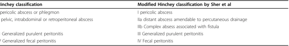
Table 2 Hinchey classification and modified Hinchey classification by Sher et al.
Hinchey classification Modified Hinchey classification by Sher et al
I pericolic abscess or phlegmon I pericolic abscess
II pelvic, intrabdominal or retroperitoneal abscess IIa distant abscess amendable to percutaneous drainage IIb Complex absess associated with fistula
several other classifications have been made (Wasvary et al. in 1999, Kohler et al in 1999, Siewert et al in 1995), among that deserve mention the Hansen/Stock classifica-tion and Kohler et al classificaclassifica-tion, both based on the clinical severity and presentation of disease (Table 3) [31,32]. Finally in 2002 Ambrosetti et al. proposed a sim-plified staging of acute diverticulitis based on CT criteria and showed its prognostic significance in a prospective study: moderate diverticulitis defined by localized sig-moid wall thickening (<5mm) with pericolic fat strand-ing, and severe diverticulitis defined by wall thickening accompanied by abscess, extraluminal air or extraluminal contrast [33]. More recently Klarenbeek et al have pro-posed a new classification arranged in three stages of dif-ferentiating diverticula disease: a) uncomplicated, b) chronic complicated, and c) acute complicated, and according both clinical and radiological findings in addi-tion to treatment modalities, in order to guide the clinical management and form the basis of a practice parameter for diverticular disease [16].
Clinical presentation and treatment
From a clinical point of view, comparing patients with non specific abdominal pain to those with diverticulitis, they are more likely to have a subacute onset of pain (>1 hr), tenderness to palpation only in the left lower quadrant, and raised inflammatory markers; and are less likely to have nausea and vomiting [34]. Other symptoms are: fever, absence of peristalsis, and defence muscularis. Diverticular bleeding and pneumaturia (pathognomic for a colovesical fistula) are rare.
The wide spectrum of diverticular disease requires a dif-ferentiated therapeutic approach to the different manifes-tations. Moderate cases of diverticular disease (phlegmon or small abscess) can be treated conservatively, generally with antibiotics and an easily digestible diet. Large abscesses, if amendable and usually larger than 5 cm, should be good candidates for CT-guided percutaneous drainage [35]. This procedure may relieve symptoms or function as a bridge to (elective) surgery. In cases of fecal peritonitis resulting from a perforation acute surgical intervention should be warranted because it is associated with high morbidity and mortality (10–35%) [36]. It is now thought that after a conservatively treated episode,
diverticular disease usually follows a rather benign course and that complications occur mostly at first presentation [37,38]. Therefore, elective sigmoid resections should be restricted for use in treating complicated disease, such as symptomatic stenosis, fistulas to a hollow organ, or recur-rent diverticular bleeding [39].
Imaging
Imaging may not be necessary in patients with the classic triad of left lower-quadrant pain, fever, and leukocytosis, and in whom uncomplicated diverticulitis is suspected. Imaging also may not be necessary in patients with a his-tory of diverticulitis who present with relatively mild clini-cal symptoms of recurrent disease. However, considering the wide spectrum of diverticular disease , also depending on the age of patients (clinical presentation may differ sig-nificantly in older patients where a reduction in symptom severity causing a misdiagnosis could be present), and the possibile difficult differential diagnosis from other causes of abdominopelvic pain, imaging often plays a definitive role both to realise a differential diagnosis and to guide the therapeutic management [1,8,40]. As reported above, today contrast-enhanced CT has a pivotal role in the clini-cal practice regarding diverticular disease, and because of their superior sensitivity and specificity up to 100%, replaced the most important imaging modality [40]. Espe-cially when an associated abscess is suspected, a CT scan can be very helpful to demonstrate its presence, and also for evaluate the possibility of a percutaneous drainage [41]. In the case of diverticular bleeding, a contrast-enhanced CT examination may demonstrate a contrast blush; and even if it has to be considered that 80% of all diverticular bleeding is self-limiting, occasionally successes of highly selective arterial embolization are described. Although the role of interventional radiology is yet to be determined. Conventional contrast-enhanced MDCT is also highly reliable in excluding other diagnoses of acute abdomen and an underlying carcinoma (Figure 1) [14,15,42-44]. A colonoscopy is indicated when there is doubt about cancer, persisting or recurrent complaints in the left lower quadrant, and suspicion of a stenosis or recurrent blood loss. Follow-up colonoscopy for ruling out malignancy is usually performed 6 weeks after an episode of acute diverticulitis. Moreover the computed
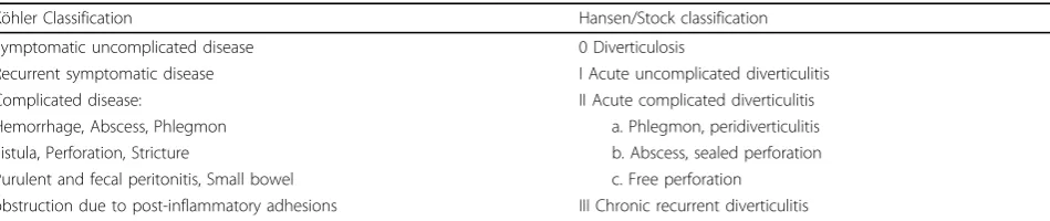
Table 3 Köhler and Hansen/Stock classification.
Köhler Classification Hansen/Stock classification
Symptomatic uncomplicated disease 0 Diverticulosis
Recurrent symptomatic disease I Acute uncomplicated diverticulitis
Complicated disease: II Acute complicated diverticulitis
Hemorrhage, Abscess, Phlegmon a. Phlegmon, peridiverticulitis Fistula, Perforation, Stricture b. Abscess, sealed perforation Purulent and fecal peritonitis, Small bowel c. Free perforation
tomography colonography (CTC) seems to be a reasonable alternative in follow-up of patients with symptomatic diverticular disease [45]. In recent years, magnetic reso-nance imaging (MRI) has gained popularity, because it lacks the ionizing radiation and even if CT remains the modality of choice; however MRI can similarly demon-strate findings of diverticulitis and could be useful in diag-nosis of ischemic colitis [46,47]. Although, colonic diverticulitis is easily diagnosed and classified (graded) by CT than by ultrasound (US), it is important to be aware of the US signs of diverticulitis considering that US is often used as a first modality in the diagnostic approach to the acute abdomen.
US: techinique and findings
US is an imaging modality widely available in the Emer-gency Department. The lower cost and in particular the lack of radiation exposure are the most important advan-tages of US compared to CT. Furthermore US is a real-time dynamic examination and this characteristic conveys dynamic information about bowel motility and changes in position, and to depict blood flow. Another important advantage of US examination is the possibility to correlate the US findings with the point of maximal tenderness. Besides, in not too obese patients, US may be superior to CT, and it is most useful in early, uncomplicated diverticu-litis. From a technical point of view , both curved (3.5– 5.0-MHz) and linear (5.0–12.0-MHz) transducers are most commonly used, in particular the higher frequency probes are useful for detailed investigation of the large bowel wall, identifying its typical haustral pattern. Focal bowel masses, segments of wall thickening, and dilated loops may be evi-dent even at lower frequencies, but high-frequency probes are essential to identify and chracterize changes in the
layers of the bowel wall. At higher frequencies, US images the bowel wall as five alternating bands (gut signature) of high and low echogenicity to produce a characteristic sonographic signature approximating to the concentric layered histologic structure [48]. Visualization of the fine inner and outer bright layers (interface echoes) is highly dependent on the echogenicity of adjacent structures and is most easily seen where there is fluid in the bowel lumen or ascites between bowel loops. Even at lower diagnostic frequencies in loops further from the probe, at least the two most prominent layers are evident due to their relative thickness and high contrast: the bright submucosa (third layer) and the dark muscularis propria (fourth layer) [34]. Doppler scanning demonstrates no signal in normal bowel wall [49,50].
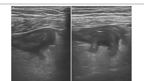
A thinned diverticular wall may be demonstrated at higher probe frequencies with a reduced gut signature due to the absence of muscularis propria. Diverticula appear as bright“ears”out of the bowel wall with acoustic shadow-ing due to the presence of gas or inspissated feces; further-more the neck of diverticulum may be identified as an echogenic band traversing hypoechoic muscularis propria that is often thickened. The diagnostic criteria for diverti-culitis that has been used in US and CT are the same: 1) at least one diverticulum, 2) signs of inflammation of peri-odic fat (dirty fat/stranding) and 3) thickened bowel wall > 4-5 mm [33]. An isolated inflamed diverticulum is identi-fied as an enlarged echo-poor protrusion from the colon wall, with an ill-defined margin surrounded by echogenic noncompressible fat, which represents the inflamed mesentery and omentum‘sealing off’the imminent per-foration. The diverticulum wall signature is lost. A central shadowing echogenicity may indicate the presence of feca-lith (Figure 2) [51]. Often the inflammation will have
Figure 2Sonographic features of uncomplicated diverticulitis: diverticula appear as bright“ear”out of the bowel wall (a); a central shadowing echogenicity may indicate the presence of fecalith (b).
extended into the bowel producing asymmetrical or cir-cumferential hypoechoic mural thickening that may demonstrate hyperemia on Doppler scanning. Diverticuli-tis may progress to an intramural or pericolic abscess indi-cated by an anechoic collection that may contain pockets of air or debris (Figure 3) [4]. Right-sided colonic diverti-culitis in many respects differs from its left-sided cousin. Diverticula of the right colon are usually congenital, soli-tary, true diverticula containing all bowel wall layers. The fecoliths within these diverticula are larger and the diverti-cular neck is wider. There is no hypertrophy of the muscu-laris of the right colonic wall . Puylaert JB in a study of 110 patients with right colonic diverticulitis clearly show, through US and CT, that it invariably has a favorable course and never leads to free perforation or large abscesses. Although relatively rare (left:right = 15:1), it is crucial to make a correct diagnosis since the clinical symp-toms of acute right lower quadrant pain may lead to an unnecessary appendectomy or even right hemicolectomy.
Conclusions
US and CT are both useful in diagnosis of diverticolitis, and their sensibility and specificity are similar. However, CT may be more likely to identify alternative causes of abdominal pain and it is essential for investigating compli-cated diverticular disease especially where there are diffuse signs and clinical suspicion of secondary peritonitis. [52] Instead in most uncomplicated cases the experienced sono-grapher may quickly confirm a diagnosis guided by the clinical signs. US is to be recommended in premenopausal women, and in young people to reduce dose exposure.
Competing interests
The authors declare that they have no competing interests.
Acknowledgements
We thank Ms Julia Hassall for reviewing the english and Dr. Palmino Sacco for helping in the preparation of the images.
Declarations
This article has been published as part ofCritical Ultrasound JournalVolume 5 Supplement 1, 2013: Topics in emergency abdominal ultrasonography. The full contents of the supplement are available online at http://www. criticalultrasoundjournal.com/supplements/5/S1. Publication of this supplement has been funded by the University of Molise, University of Siena, University of Cagliari, University of Ferrara and University of Turin.
Author details
1Department of Medical, Surgical and Neuro Sciences, Section of
Radiological Sciences. Siena, Italy.2University of Bari, Diagnostic Imaging Section, Bari, Italy.3University of Perugia, Thoracic Surgery Unit, Perugia, Italy. 4
University of Foggia, Department of Radiology, Foggia, Italy.5University of Ferrara, Dipartimento di Scienze Chirurgiche, Ferrara, Italy.
Published: 15 July 2013
References
1. Hammond NA, Nikolaidis P, Miller FH:Left lower-quadrant pain: guidelines from the American College of Radiology appropriateness criteria.Am Fam Physician2010,82:766-70, Review.
2. Jacobs DO:Clinical practice. Diverticulitis.N Engl J Med2007,357:2057-66, Review.
3. Laméris W, van Randen A, van Gulik TM, Busch OR, Winkelhagen J, Bossuyt PM, Stoker J, Boermeester MA:A clinical decision rule to establish the diagnosis of acute diverticulitis at the emergency department.Dis Colon Rectum2010,53:896-904.
4. Reginelli A, Pezzullo MG, Scaglione M, Scialpi M, Brunese L, Grassi R: Gastrointestinal disorders in elderly patients.Radiol Clin North Am2008, 46(4):755-71.
5. Hollerweger A, Macheiner P, Rettenbacher T, Brunner W, Gritzmann N: Colonic diverticulitis: diagnostic value and appearance of inflamed diverticula-sonographic evaluation.Eur Radiol2001,11:1956-63. 6. Cuccurullo V, Cioce F, Sica A, Iasiello F, Capasso R, Gatta G, Rubini G:
Gastroenteric diseases in the third millennium: a rational approach to optimal imaging technique and patient selection.Recenti Prog Med2012, 103(11):426-30.
7. Tack D, Bohy P, Perlot I, De Maertelaer V, Alkeilani O, Sourtzis S,
Gevenois PA:Suspected acute colon diverticulitis: imaging with low-dose unenhanced multi-detector row CT.Radiology2005,237:189-96. 8. Horton KM, Corl FM, Fishman EK:CT evaluation of the colon:
inflammatory disease.Radiographics2000,20:399-418.
9. Jang HJ, Lim HK, Lee SJ, Lee WJ, Kim EY, Kim SH:Acute diverticulitis of the cecum and ascending colon: the value of thin-section helical CT findings in excluding colonic carcinoma.Am J Roentgenol2000, 174:1397-402.
10. Sai VF, Velayos F, Neuhaus J, Westphalen AC:Colonoscopy after CT diagnosis of diverticulitis to exclude colon cancer: a systematic literature review.
Radiology2012,263:383-90, Review. Erratum in: Radiology 2012, 264:306. 11. Reginelli A, Mandato Y, Cavaliere C, Pizza NL, Russo A, Cappabianca S,
Brunese L, Rotondo A, Grassi R:Three-dimensional anal endosonography in depicting anal-canal anatomy.Radiol Med2012,117(5):759-71. 12. Reginelli A, Mandato Y, Solazzo A, Berritto D, Iacobellis F, Grassi R:Erros in
the rediological evaluation of the alimentary tract: part II.Semin Ultrasound CT MR2012,33(4):308-311.
13. Angelelli G, Moschetta M, Sabato L, Morella M, Scardapane A, Stabile Ianora AA:Value of“protruding lips”sign in malignant bowel obstructions.Eur J Radiol2011,80(3):681-5.
14. Russo M, Martinelli M, Sciorio E, Botta C, Miele E, Vallone G, Staiano A:Stool Consistency, but Not Frequency, Correlates with Total Gastrointestinal Transit Time in Children.J Pediatr2013.
15. Esposito F, Senese R, Salvatore P, Vallone G:Intrahepatic portal-vein gas associated with rotavirus infection.J Ultrasound2011,14(1):10-3. 16. van Randen A, Laméris W, van Es HW, van Heesewijk HP, van Ramshorst B,
Ten Hove W, Bouma WH, van Leeuwen MS, van Keulen EM, Bossuyt PM, Stoker J, Boermeester MA:OPTIMA Study Group. A comparison of the accuracy of ultrasound and computed tomography in common diagnoses causing acute abdominal pain.Eur Radiol2011,21:1535-45. 17. Laméris W, van Randen A, Bipat S, Bossuyt PM, Boermeester MA, Stoker J:
Graded compression ultrasonography and computed tomography in acute colonic diverticulitis: meta-analysis of test accuracy.Eur Radiol
2008,18:2498-511.
18. Schwerk WB, Schwarz S, Rothmund M:Sonography in acute colonic diverticulitis. A prospective study.Dis Colon Rectum1992,35:1077-84. 19. Painter NS, Truelove SC, Ardran GM, Tuckey M:Segmentation and the localization of intraluminal pressures in the human colon, with special reference to the pathogenesis of colonic diverticula.Gastroenterology
1965,49:169-77.
20. Andersen JC, Bundgaard L, Elbrønd H, Laurberg S, Walker LR, Støvring J: Danish Surgical Society. Danish national guidelines for treatment of diverticular disease.Dan Med J2012,59:C4453.
21. Klarenbeek BR, de Korte N, van der Peet DL, Cuesta MA:Review of current classifications for diverticular disease and a translation into clinical practice.Int J Colorectal Dis2012,27:207-14, Review.
22. Grassi R, Rambaldi PF, Di Grezia G, Mansi L, Cuccurullo V, Cirillo A, Riegler G, Cappabianca S, Rotondo A:Inflammatory bowel disease: value in diagnosis and management of MDCT-enteroclysis and 99mTc-HMPAO labeled leukocyte scintigraphy.Abdom Imaging2011, 36(4):372-81.
23. Lorusso F, Fonio P, Scardapane A, Giganti M, Rubini G, Ferrante A, Stabile Ianora AA:Gatrointestinal imaging with multidetector CT and MRI.
24. Niccoli Asabella A, Renna MA, Stabile Ianora AA, Rubini D, Rubini G:Role of imaging techniques in the evaluation of chyluria.Recenti Prog Med2012, 103(11):555-8.
25. Stabile Ianora AA, Moschetta M, Pedote P, Scardapane A, Angelelli G: Preoperative local staging of colosigmoideal cancer: air versus water multidetector-row CT colonography.Radiol Med2012,117(2):254-67. 26. Ambrosetti P, Gervaz P, Fossung-Wiblishauser A:Sigmoid diverticulitis in
2011: many questions; few answers.Colorectal Dis2012,14:e439-46, Review.
27. Sher ME, Agachan F, Bortul M, Nogueras JJ, Weiss EG, Wexner SD: Laparoscopic surgery for diverticulitis.Surg Endosc1997,11:264-7. 28. Hughes ES, Cuthbertson AM, Carden AB:The surgical management of
acute diverticulitis.Med J Aust1963,50:780-2.
29. Wasvary H, Turfah F, Kadro O, Beauregard W:Same hospitalization resection for acute diverticulitis.Am Surg1999,65:632-5, discussion 636. 30. Köhler L, Sauerland S, Neugebauer E:Diagnosis and treatment of
diverticular disease: results of a consensus development conference. The Scientific Committee of the European Association for Endoscopic Surgery.Surg Endosc1999,13:430-6, Review.
31. Hansen O, Graupe F, Stock W:Prognostic factors in perforating diverticulitis of the large intestine.Chirurg1998,69:443-9, German. 32. Siewert JR, Huber FT, Brune IB:Early elective surgery of acute diverticulitis
of the colon.Chirurg1995,66:1182-9, Review. German.
33. Ambrosetti P, Jenny A, Becker C, Terrier TF, Morel P:Acute left colonic diverticulitis–compared performance of computed tomography and water-soluble contrast enema: prospective evaluation of 420 patients.
Dis Colon Rectum2000,43:1363-7.
34. Rodgers PM, Verma R:Transabdominal ultrasound for bowel evaluation.
Radiol Clin North Am2013,51:133-48, Review.
35. Ambrosetti P, Chautems R, Soravia C, Peiris-Waser N, Terrier F:Long-term outcome of mesocolic and pelvic diverticular abscesses of the left colon: a prospective study of 73 cases.Dis Colon Rectum2005,48:787-91. 36. Oomen JL, Engel AF, Cuesta MA:Mortality after acute surgery for
complications of diverticular disease of the sigmoid colon is almost exclusively due to patient related factors.Colorectal Dis2006,8:112-9. 37. Salem TA, Molloy RG, O’Dwyer PJ:Prospective, five-year follow-up study
of patients with symptomatic uncomplicated diverticular disease.Dis Colon Rectum2007,50:1460-4.
38. Collins D, Winter DC:Elective resection for diverticular disease: an evidence-based review.World J Surg2008,32:2429-33, Review. 39. Ambrosetti P:Acute diverticulitis of the left colon: value of the initial CT
and timing of elective colectomy.J Gastrointest Surg2008,12:1318-20, Review.
40. Le Pennec V, Hourna E, Schmutz G, Pelage JP:Imaging in infections of the left iliac fossa.Diagn Interv Imaging2012,93:466-72, Review.
41. Brandt D, Gervaz P, Durmishi Y, Platon A, Morel P, Poletti PA:Percutaneous CT scan-guided drainage vs. antibiotherapy alone for Hinchey II diverticulitis: a case-control study.Dis Colon Rectum2006,49:1533-8. 42. Mazzei MA, Guerrini S, Cioffi Squitieri N, Imbriaco G, Mazzei FG, Volterrani L:
Non-obstructive mesenteric ischemia after Cardiovascular Surgery: not so uncommon.Ann Thorac Cardiovsc Surg.
43. Mazzei MA, Guerrini S, Cioffi Squitieri N, Genovese EA, Mazzei FG, Volterrani L:Diagnosis of acute mesenteric ischemia/infarction in the era of multislice CT.Recenti Prog Med2012,103:435-7.
44. Mazzei MA, Mazzei FG, Marrelli D, Imbriaco G, Guerrini S, Vindigni C, Civitelli S, Roviello F, Grassi R, Volterrani L:Computed tomographic evaluation of mesentery: diagnostic value in acute mesenteric ischemia.
J Comput Assist Tomogr2012,36:1-7.
45. Hjern F, Jonas E, Holmström B, Josephson T, Mellgren A, Johansson C:CT colonography versus colonoscopy in the follow-up of patients after diverticulitis a prospective, comparative study.Clin Radiol2007,62:645-50. 46. Hammond NA, Miller FH, Yaghmai V, Grundhoefer D, Nikolaidis P:MR
imaging of acute bowel pathology: a pictorial review.Emerg Radiol2008, 15:99-104, Review.
47. Mazzei MA, Guerrini S, Cioffi Squitieri N, Imbriaco G, Chieca R, Civitelli S, Savelli V, Mazzei FG, Volterrani L:MRI: is there a role in clinical management for acute ischemic colitis?World J Gastroenterol2013, 19:1256-63.
48. Kimmey MB, Martin RW, Haggitt RC, Wang KY, Franklin DW, Silverstein FE: Histologic correlates of gastrointestinal ultrasound images.
Gastroenterology1989,96:433-41.
49. Esteban JM, Maldonado L, Sanchiz V, Minguez M, Benages A:Activity of Crohn’s disease assessed by colour Doppler ultrasound analysis of the affected loops.Eur Radiol2001,1:1423-8.
50. Wilson SR, Toi A:The value of sonography in the diagnosis of acute diverticulitis of the colon.Am J Roentgenol1990,154:1199-202. 51. Puylaert JB:Ultrasound of colon diverticulitis.Dig Dis2012,30:56-9,
Review.
52. Marrelli D, Mazzei MA, Pedrazzani C, Di Martino M, Vindigni C, Corso G, Marrelli E, Volterrani L, Roviello F:High accuracy of multislices computed tomography (MSCT) for para-aortic lymph node metastases from gastric cancer: a prospective single-center study.Ann Surg Oncol2011, 18:2265-2272, Epub 2011 Jan 26.
doi:10.1186/2036-7902-5-S1-S5
Cite this article as:Mazzeiet al.:Sigmoid diverticulitis: US findings.
Critical Ultrasound Journal20135(Suppl 1):S5.
Submit your manuscript to a
journal and benefi t from:
7 Convenient online submission 7 Rigorous peer review
7 Immediate publication on acceptance 7 Open access: articles freely available online 7 High visibility within the fi eld
7 Retaining the copyright to your article