© 2012 Ben Azouna et al.; licensee BioMed Central Ltd. This is an open access article distributed under the terms of the Creative
R E S E A R C H
Open Access
Phenotypical and functional characteristics of
mesenchymal stem cells from bone marrow:
comparison of culture using different media
supplemented with human platelet lysate or fetal
bovine serum
Nesrine Ben Azouna
1,2, Faouzi Jenhani
2,3*, Zohra Regaya
3, Lamia Berraeis
3, Tarek Ben Othman
4, Elfi Ducrocq
1and
Jorge Domenech
1*Abstract
Introduction:Mesenchymal stem cells (MSCs) are multipotent cells able to differentiate into several mesenchymal lineages, classically derived from bone marrow (BM) but potentially from umbilical cord blood (UCB). Although they are becoming a good tool for regenerative medicine, they usually need to be expanded in fetal bovine serum (FBS)-supplemented media. Human platelet lysate (HPL) has recently been proposed as substitute for safety reasons, but it is not yet clear how this supplement influences the properties of expanded MSCs.
Methods:In the present study, we compared the effect of various media combining autologous HPL with or without FBS on phenotypic, proliferative and functional (differentiation, cytokine secretion profile) characteristics of human BM-derived MSCs.
Results:Despite less expression of adipogenic and osteogenic markers, MSCs cultured in HPL-supplemented media fully differentiated along osteoblastic, adipogenic, chondrogenic and vascular smooth muscle lineages. The analyses of particular specific proteins expressed during osteogenic differentiation (calcium-sensing receptor (CaSR) and parathormone receptor (PTHR)) showed their decrease at D0 before any induction for MSC cultured with HPL mostly at high percentage (10%HPL). The cytokine dosage showed a clear increase of proliferation capacity and interleukin (IL)-6 and IL-8 secretion.
Conclusions:This study shows that MSCs can be expanded in media supplemented with HPL that can totally replace FBS. HPL-supplemented media not only preserves their phenotype as well as their differentiation capacity, but also shortens culture time by increasing their growth rate.
Introduction
Mesenchymal stem cells (MSCs) represent a rare popu-lation of multipotent progenitors, initially described in bone marrow (BM), giving rise to adipocytes, osteo-blasts, chondrocytes, and vascular smooth muscle (VSM)-like hematopoietic supportive stromal cells [1,2].
These cells are capable of multilineage differentiation from a single cell [3,4] and in vivofunctional reconstitu-tion of injured tissues in preclinical [5] as well in clinical [6] settings. Thereafter, MSCs have been described in virtually all post-natal organs or tissues [7] but also in fetal adnexa, including both umbilical cord blood [8,9] and placenta [7,10,11]. Today, BM remains the principal source of MSCs in studies investigating their potential use in cell therapy, in spite of significant declines in cell proliferation and differentiation capacity with advancing age [12].
* Correspondence: [email protected]; [email protected] 1
Laboratory of Hematopoiesis, UPRES-EA3855, Faculty of Medicine, University François Rabelais, 10 boulevard Tonnellé, 37032 Tours Cedex, France 2
Immunology Research Unit, Faculty of Pharmacy, rue Ibn Sina, 5000 Monastir, Tunisia
Full list of author information is available at the end of the article
MSCs display several common surface antigens, including CD90 (Thy-1), CD166 (SB10/ALCAM), CD73 (SH3), and CD105 (SH2 and endoglin), and lack hema-topoietic (such as CD45 or CD34) and endothelial (such as CD31) markers and HLA-DR. However, significant heterogeneity in the morphology, proliferation, and dif-ferentiation potentials of MSCs has also been observed [13]. Fetal bovine serum (FBS)-based medium is the conventional medium for isolation and expansion of MSCs and has been used in several clinical trials [6]. However, concerns over the safety of FBS-based culture media have been raised because they can trigger adverse responses [14,15]. Accordingly, to culture MSCs, some trials have used media containing animal serum-free substitutes with combinations of growth factors [16]. An alternative is to supplement media with human blood-derived platelet lysates or sera from autologous or allo-genic donors [17-21] to create culture conditions that more closely mimic the human environment. However, it is not yet clear how these culture conditions influence the phenotype and function of the expanded MSC populations.
In the present study, we have tested different media, including combinations of autologous human platelet lysate (HPL) with or without FBS, in order to compare phenotypic, proliferative, and functional characteristics together with the cytokine secretion profile of human MSCs derived from BM. We focused particularly on the mesenchymal differentiation ability of MSCs expanded in these different conditions.
Materials and methods
Mesenchymal stem cell expansion cultures from bone marrow
BM cells (n = 13) were isolated from patients with nor-mal BMs harvested from donors at the Bone Marrow Graft National Center of Tunisia (Tarek, Ben Othman, Tunis) or during orthopedic surgery (Philippe Rosset, Orthopedics Department, Hôpital Trousseau, Tours). The mean age of the donors was 33 ± 2 years (range of 16 to 41 years). Informed consent was obtained from all of the patients (a written consent was provided by the patients from Tours) in accordance with the national ethical guidelines of each country.
Mononuclear cells (MNCs) were isolated from each BM sample by loading onto Ficoll Hypaque solution (d = 1.077). After centrifugation at 800gfor 20 minutes at room temperature, the MNC layer was removed from the interphase and washed twice with phosphate-buf-fered saline (PBS). Then they were seeded into uncoated T25 or T75 flasks (Becton, Dickinson and Company, Franklin Lakes, NJ, USA) at a cell concentration of 1 × 10⁵ cells per square centimeter for BM cells. Cells were cultured in basic growth medium (BGM) consisting of
alpha-minimum essential medium (a-MEM) (Invitrogen Corporation, Carlsbad, CA, USA) with 100 U/mL peni-cillin, 0.1 mg/mL streptomycin (Invitrogen), 2 mM L-glutamine (Invitrogen), 0.025 mg/mL amphotericin B (Invitrogen) with various supplements to obtain the four following expansion culture media: (M1) BGM with 10% (vol/vol) of prescreened FBS (HyClone, Thermo Fisher Scientific Inc., Waltham, MA, USA) and 1 ng/mL fibro-blast growth factor 2 (FGF2) (AbCys, Paris, France), (M2) BGM with 10% FBS and 5% (vol/vol) HPL, (M3) BGM with 10% HPL, and (M4) BGM with 5% HPL. Cultures were maintained in a humidified atmosphere
with 5% CO2 at 37°C and the medium had been
chan-ged twice weekly thereafter. On reaching 60% to 80% confluence, the adherent cells were detached after treat-ment with 0.05% (vol/vol) trypsin/1 mM ethylenediami-netetraacetic acid (EDTA) solution (Invitrogen) and reseeded at 103 cells per square centimeter. All of the studies, except for cell proliferation and CFU-F assays, had been performed after two passages (P2).
Human platelet lysate preparation
HPLs were prepared in accordance with a method pre-viously described [19]. Briefly, they were obtained from different platelet apheresis collections performed at the National Blood Transfusion Center of Tunis (Tunisia). HPLs from at least 10 donors were pooled and, after elimination of remaining platelet bodies by centrifuga-tion (1,400gfor 20 minutes), were frozen at -80°C to be subsequently used as FBS or growth factor substitute or both.
Phenotypic analysis of mesenchymal stem cells by flow cytometry
Mesenchymal differentiation capacity of mesenchymal stem cells
Osteogenic induction
At 50% confluence, the cells were cultured for 14 days in Dulbecco’s modified Eagle’s medium-high glucose (DMEM-HG) (with 4.5 g/L glucose) containing 2% FBS, 0.1 μM dexamethasone (Sigma-Aldrich, St. Louis, MO, USA), 2 mM b-glycerolphosphate (Sigma-Aldrich), and
100 μM ascorbate-2-phosphate (Sigma-Aldrich) with
medium changes every 3 days. After 2 weeks of induc-tion, the cells were stained in accordance with von Kossa and Alizarin red methods to detect the presence of calcium deposition into osteocytes.
Adipogenic induction
The adipogenic induction medium consisted of DMEM-LG (with 1 g/L glucose) supplemented with 20% FBS, 1
μM/L dexamethasone, 0.5 mmol/L
isobutylmethyl-xanthine (Invitrogen), and 60 μmol/L indomethacin
(Sigma-Aldrich). Adipogenic differentiation was evalu-ated after 2 weeks of induction by the cellular accumu-lation of neutral lipid vacuoles that were stained with Nile red (Sigma-Aldrich) and observed by fluorescent microscopy.
Chondrogenic induction
Chondrogenic differentiation was obtained in micropel-lets (3 × 10⁵ cells per pellet) incubated in tubes at 37°C for 21 days in 500μL of chondrogenic medium
consist-ing of DMEM-HG with 0.1μM dexamethasone
(Sigma-Aldrich), 1 mM sodium pyruvate, 170μM ascorbic acid-2-phosphate (Sigma-Aldrich), 350 μM proline (Sigma-Aldrich), 1X insulin-transferrin-selenium (Cambrex, East Rutherford, NJ, USA), and 10 ng/mL transforming growth factor-beta-1 (TGF-b1) (AbCys). Medium was changed every 4 days. Chondrogenic differentiation was evaluated after 21 days of induction. The presence of glycosaminoglycans in cell pellets was revealed by Alcian blue and Toluidine blue staining. On the 21st day, pel-lets were fixed for 48 hours in 4% paraformaldehyde and embedded in paraffin. Sections (4μm thick) were rehydrated in 70%, 90%, and 100% xylene and ethanol. For Alcian blue staining, slides were incubated for 10 minutes in a 1:1 mixture of 3% (vol/vol) Alcian blue
8GX (Sigma-Aldrich) solution in distilled water and 3% (vol/vol) methanol. For Toluidine blue staining, slides were incubated for 10 minutes in a 1% Toluidine blue (Sigma-Aldrich) (vol/vol) solution in 70% ethanol.
Vascular smooth muscle induction
VSM differentiation was obtained in McCoy’s 5A med-ium supplemented with 12.5% FBS, 12.5% horse serum (Invitrogen), 2 mM L-glutamine, 0.8 mM L-serine, 0.15 mM L-asparagine, 1 mM sodium pyruvate, 5 mM sodium bicarbonate, 1μM hydrocortisone and 0.25μg/ mL amphotericin B. Medium was changed every 4 days. VSM differentiation was evaluated after 3 weeks of induction. MSCs were fixed by using 4% parafor-maldehyde and incubated with primary antibodies against rat alpha smooth muscle actin (ASMA) (clone 1A4, mouse anti-human, -mouse, -rat; Sigma-Aldrich) at 4°C for 12 hours. The antigen-antibody reaction was detected by using a specific mouse secondary anti-body coupled to FITC (Alexa Fluor 488; Invitrogen) dyes, and the reaction was visualized by fluorescent microscopy.
Mesenchymal stem cell expression of differentiation markers by RT-PCR and Western blotting
For reverse transcription-polymerase chain reaction (RT-PCR), total RNA was extracted from MSCs at 90% confluence by using TRIZOL reagent (Invitrogen) in accordance with the instructions of the manufacturer. Reverse transcription was carried out by using Prime-Script™RTase (Takara, Kyoto, Japan), and the cDNA fragments were amplified by using RNase Inhibitor (Takara). After denaturation at 65°C for 5 minutes, amplification was carried out by 30 cycles at 30°C for 10 minutes, 42°C for 60 minutes, and 95°C for 5 minutes. Primers used are shown in Table 1. The expression of the following genes was studied: alkaline phosphatase (ALP) nd Runt-related transcription factor 2 (RUNX2) for osteogenic pathway, peroxisome proliferator-acti-vated receptor-gamma (PPARg) and lipoprotein lipase (LPL) for adipogenic pathway, andASMAfor VSM dif-ferentiation. Glyceraldehyde 3-phosphate dehydrogenase (GAPDH) gene expression was used as a control.
Table 1 Sequences of the primers used in reverse transcription-polymerase chain reaction study
Construct Forward sequence Reverse sequence
GAPDH 5’- AATCCCATCACCATCTTCCAGG-3’ 5’-AGAGGCAGGGATGATGTTCTGG-3’
ALP 5’-CTGGACCTCGTTGACACCTG-3’ 5’-GACATTCTCTCGTTCACCGC-3’
RUNX2 5’-AACTTCCTGTGCTCGGTGCTG-3’ 5’-GGGGAGGATTTGTGAAGACGG-3’
LPL 5’-AAAGCCCTGCTCGTGCTGAC-3’ 5’-TAAACCGGGCCACATCCTGT-3’
PPARg 5’-GGAGAAGCTGTTGGCGGAGA-3’ 5’-TCAAGGAGGCCAGCATTGTG-3’
ASMA 5’-TCATGATGCTGTTGTAGGTGGT-3’ 5’-CTGTTCCAGCCATCCTTCAT-3’
Thermocycling was performed with a gradient thermo-cycler (Takara).
For Western blotting, MSCs were harvested at 90% confluence. Cell pellets were resuspended in lysis buffer (8 M urea, 2 M thiourea, 2% CHAPS, 1% NP-40, 2 mM TBP, 16 Protease Inhibitor Mix, 16 Nuclease Mix, 1 mM PMSF, and 2% IPG buffer) and put on ice for 45 minutes. They were then centrifuged at 14,000gfor 15 minutes at 4°C. The supernatant was collected and stored at -80°C. The protein concentration of samples was determined by means of Bradford’s method (Bio-Rad Laboratories, Inc., Hercules, CA, USA). Equal amounts of cell extracts and of conditioned media were separated by SDS-PAGE (12%) and transferred to poly-vinylidene difluoride (PVDF) membranes. Membranes were blocked with 5% skim milk for an hour and left to
react with anti-human b-actin (clone AC-15, mouse
IgG1; Sigma-Aldrich) as a positive control, anti-human leptin (monoclonal; R&D Systems, Inc., Minneapolis, MN, USA, reference number 398), anti-human calcium-sensing receptor (CaSR) (monoclonal; AbCam, Cam-bridge, UK, reference number ab 3513), anti-human parathormone receptor (PTHR) (mouse monoclonal anti-human; Novocastra, part of Leica Microsystems, Wetzlar, Germany), anti-human ASMA (clone 1A4, mouse anti-human, -mouse, -rat; Sigma-Aldrich), and
anti-human transgelin (SM22a) (mouse monoclonal
anti-human; Novocastra). Antibody binding was revealed by incubation with the corresponding HRP-linked IgG (horseradish peroxydase; Bio-Rad Laboratories, Inc.) and the ECL+ Western blotting detection kit (Amersham Biosciences, now part of GE Healthcare, Little Chalfont,
Buckinghamshire, UK). Chemiluminescence was
detected and measured with a Chemi-Smart 2000™ Ima-ger (Vilber Lourmat, Marne-la-Vallée, France).
Clonogenic and proliferative capacities of mesenchymal stem cells
Colony-forming unit-fibroblast assays
CFU-F assays were performed to evaluate the clonal expansion capacity of the MSCs initially cultured in the six different media. For the optimal counting of colo-nies, MNCs derived from BM were seeded at 105 cells per square centimeter in T25 flasks in duplicate. Flasks were incubated in a humidified atmosphere with 5%
CO2 at 37°C. The medium was completely renewed
every 3 days. Cell clusters containing more than 50 cells were scored on day 10 as CFU-F colonies by using an inverted microscope. Cloning efficiency was considered as CFU-F counts per 106MNCs plated.
Population doubling time
To test the role of culture conditions on MSC expan-sion, MSCs were cultured in each of the six expansion media for two passages (P2) before analysis. Population
doubling times (PDTs) were calculated between P1 and P2 as t/n, where t is the duration of culture in days and n is the number of population doublings calculated by using the formulan= (log Nh- log Ni)/log 2, where Nh
is the number of cells harvested at the time of counting at P2 and Ni is the number of cells initially plated at
P1).
Cytokine secretion capacity of mesenchymal stem cells
Cytokines were detected in supernatants of MSCs cul-tured in different expansion media and were collected at P2. Concentrations of IL-6, IL-8, vascular endothelial growth factor (VEGF), FGF2, granulocyte stimu-lating factor (G-CSF), granulocyte-macrophage colony-stimulating factor (GM-CSF), and RANTES (regulated on activation normal T cell expressed and secreted) were simultaneously evaluated by using a commercially available multiplex bead-based immunoassay (Bio-Plex™ cytokine assay system; BD Biosciences).
Statistical analysis
Statistical analysis was performed by using the STAT-GRAPHICS Centurion XVI program version 16.0.08 (StatPoint Technologies Inc., Warrenton, VA, USA). A one-way analysis of variance and Newman-Keuls multi-ple range test were carried out to compare value distri-butions. Significant difference between group values was considered when Pvalue was less than 0.05. Indicated values are mean ± standard error mean.
Results
Phenotypic characteristics of mesenchymal stem cells cultured in various expansion media
Morphological analysis
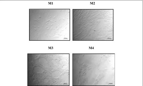
Adherent cell populations from the MNC fraction of human BM samples were generated by expansion cul-ture by using four different media: (M1) BGM with 10% FBS and 1 ng/mL FGF2, (M2) BGM with 10% FBS and 5% HPL, (M3) BGM with 10% HPL, and (M4) BGM with 5% HPL. After 2 weeks of culture, an adherent and stable cell layer was obtained from BM-derived MNCs with all media (n = 13). Figure 1 shows a particular morphology of MSCs cultured with HPL (M3 and M4). In fact, the layer of MSCs appears with many spaces between the cells in comparison with standard medium (M1), in which we have one layer of very confluent MSCs (Figure 1).
Immunophenotype analyzed by flow cytometry
markers CD106, CD73, CD90, and CD105, regardless of the expansion medium used (Figure 2B). For immaturity markers, expression of CD49a was low and CD133 was undetectable without any influence of medium type. Our data did not show any statistical difference between the four culture conditions.
Mesenchymal differentiation capacity of mesenchymal stem cells assessed by specific staining in different culture conditions
The influence of HPL in expansion media on further differentiation potential of MSCs toward osteogenic, adi-pogenic, chondrogenic, and VSM lineages after appro-priate induction was investigated at the second passage (P2) and specific staining (Figure 3). BM cells grown in HPL or FBS deposited an extensive mineralized matrix when cultured for 2 weeks in osteogenic medium, as demonstrated by strong Alizarin red and von Kossa staining. These cells also efficiently differentiated into the adipogenic lineage, as indicated by Nile red staining of lipid droplets in the cytoplasm following culture in adipogenic medium except when a high concentration (10%) of HPL was used (M3). After chondrogenic differ-entiation for 3 weeks, MSCs displayed a typical chon-droblast shape, as indicated by the deposition of specific
glycosaminoglycans seen after Alcian blue and O Safra-nin staiSafra-ning, irrespectively of the expansion media used previously (M1, M2, M3, and M4).
Osteogenic, adipogenic, and vascular smooth muscle gene expression assessed by RT-PCR analysis in mesenchymal stem cells
Gene expression was analyzed by RT-PCR after MSC amplification (P1) in HPL- and FBS-supplemented media before (day 0, or D0) and after differentiation (day 14, or D14) from four different MSCs. We tested the expression of two osteogenic (O)-specific genes (ALP and RUNX2), two adipogenic (A)-specific genes
(PPARg and LPL), and one VSM gene (ASMA) before
differentiation. mRNA expressions of ALP, RUNX2,
PPRg, andASMA were decreased in M2 (10% FBS + 5% HPL) and M3 (10% HPL) media in comparison with standard M1 medium (10% FBS + 1 ng/mL FGF2) or M4 medium containing only 5% HPL (Figure 4A). At D0, HPL seems to decrease the osteogenic markers. Concerning the adipogenic markers,PPARgdecreased at D0 when the HPL was added to the culture medium (M2, M3, and M4) in comparison with standard med-ium (M1). Also, in M2 (10% FBS + 5% HPL), we showed a decrease ofPPARgafter adipogenic induction at D14.
M1 M2
M3
M4
A
B
Control M1 M2 M3 M4
CD34
CD45
CD133 CD14
Control M1 M2 M3 M4
CD49e CD105
CD73
CD90
CD106
Osteogenic, adipogenic, and vascular smooth muscular protein expression assessed by Western blotting in mesenchymal stem cells
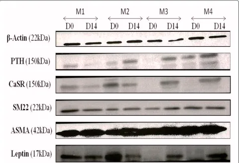
Proteins specific of osteogenic, VSM, and adipogenic lineages were identified in MSCs in different culture conditions. MSCs cultured in 10% FBS + 5% HPL med-ium (M2) displayed the same expression profile for the osteogenic markers CaSR and PTHR as in standard medium (M1) before (D0) and after (D14) differentia-tion. In contrast, the expression of these proteins was clearly decreased in 10% HPL (M3) and 5% HPL (M4) media. We noted the same results with the adipogenic marker (Leptin) (Figure 5). The expression of VSM dif-ferentiation proteins (ASMA and SM22a) seems not to be affected by the type of expansion medium (Figure 5).
Clonogenic and proliferative capacities of mesenchymal stem cells cultured in various expansion media
Colony-forming unit-fibroblast assays
MNCs initially seeded in the four expansion media were assayed for CFU-F. No clear difference was observed in
colony counts for 106 BM MNCs plated when
compar-ing the output in FBS- and HPL-supplemented expan-sion culture media (Figure 6). HPL-supplemented cultures resulted in significantly larger colonies, which appeared densely packed with very small spindle-shaped cells compared with colonies in FBS cultures formed by only loosely connected cells (Figure 3: M2-M3 versus
M1). The growth-promoting effect of HPL on CFU-F was associated with increased density of total cells at confluence.
Population doubling time
Expansion rates of MSCs were very different when cul-tured in media supplemented with or without HPL (Fig-ure 7). Thus, PDTs of MSCs cult(Fig-ured in regular medium containing FBS with FGF2 (M1) remained longer than in HPL-supplemented cultures (M2: 5.6 ± 0.5 days; M3: 5.8 ± 1.03 days; and M4: 3.8 ± 0.7 days; P < 0.0007).
Cytokine expression profile of mesenchymal stem cells
Cytokine expression profile of MSCs from BM was com-pared by using the Bio-Plex™cytokine assay system (BD Biosciences) in accordance with the instructions of the manufacturer. MSC supernatants were collected after 3 to 4 days of culture at P2 with M1, M2, M3, and M4 for BM. This technique allowed the simultaneous mea-surement of concentrations of several cytokines that included FGF2, RANTES, VEGF, IL-6, IL-8, G-CSF, and GM-CSF. In parallel to MSC supernatants, background cytokine concentrations were evaluated in each corre-sponding medium used (M1, M2, M3, and M4). In these growth media, all of the cytokines - except for RANTES, which was present at high concentrations in HPL-containing media (M2, M3 and M4) but was at very low concentrations in standard medium M1 BM Alizarin Red
(O) Von Kossa (O) Alcian Blue (C) Nile Red (A)
ASMA (VSM) D0
ASMA (VSM) D14
M1
M2
M3
M4
(lacking HPL) - were undetectable. MSC supernatants contained IL-6, IL-8, and VEGF at variable levels according to the different expansion culture conditions. Thus, the presence of HPL appeared to increase IL-6 and IL-8 concentrations (Figure 8A, B), while FGF2, G-CSF, and GM-CSF remained undetectable in all culture conditions.
Discussion
This study points out the interest in HPL as a replace-ment for FBS in culture media for expansion of human BM MSCs. Thus, HPL-containing media not only pre-serve their phenotype as well as their differentiation capacity but also shorten culture time by increasing their growth rate. Nevertheless, some differences exist in
γγ
terms of cytokines produced, suggesting functional dif-ferences between MSCs expanded in media supplemen-ted with HPL and FBS. This has to be considered for particular clinical applications.
The possibility to use animal serum-free culture media has been reported in several recent studies by substitut-ing FBS with human-derived supplements such as HPL or human serum [17-25]. In the present study, MSC expansions were performed in three different
HPL-sup-plemented media consisting of BGM (a-MEM) with
(M2) or without (M3 and M4) FBS and compared with the standard medium devoid of HPL (M1). M1 (supple-mented with 10% FBS + 1 ng/mL FGF2) represents the reference medium for MSC expansion in our laboratory. In M2, FGF2 was replaced by 5% HPL (vol/vol). FBS-free M3 and M4 media were supplemented with 10% and 5% HPL, respectively. The four media were used for expansion of BM-derived MSCs to study the influence of HPL on MSC functions. Our study clearly showed
that the presence of HPL is necessary for MSC growth to substitute the standard expansion medium containing the FBS.
The addition of HPL in expansion media did not modify the immunophenotype of MSCs, irrespectively of the amounts used and of the presence or absence of FBS. These cell display a typical feature of MSCs bear-ing CD73, CD90, CD105, and CD106 and lackbear-ing the hematopoietic markers CD45, CD34, and CD14. Imma-turity marker expression did not vary with low levels of CD49a, and undetectable levels of CD133. This lack of consistent immunophenotypic changes of MSCs cul-tured in HPL-supplemented media has been reported [8,19-21,25,26].
the data of previous studies [19,21-23,27]. However, in M3 containing 10% HPL, MSCs displayed a weak adipo-genic differentiation with only a few vesicular adipocytes stained with Nile red.
To assess more precisely the differentiation state of MSCs after expansion in the different media studied and their subsequent differentiation potential, we stu-died the expression of several differentiation genes by
n=6
n=5 n=7
n=5
#
#
#
#
Figure 6Effect of expansion media on colony-forming unit-fibroblast (CFU-F) counts per 106bone marrow mononuclear cells. The media are M1 (10% FBS + FGF2), M2 (10% FBS + 5% HPL), M3 (10% HPL), and M4 (5% HPL). Data shown represent mean ± standard error of the mean from the indicated number of experiments.#No significant difference;P> 0.05.
n=6
n=6
n=4
*
#
#
#
n=6
RT-PCR and of proteins by Western blotting for adipo-genic, osteoadipo-genic, and VSM lineages. These analyses revealed that BM-derived MSCs expanded in all media already express (at both mRNA and protein levels) all of these differentiation markers without any differentiation induction. These data are consistent with those of Delorme and colleagues who demonstrated that
non-differentiated human BM-derived MSCs are character-ized by lineage priming [28].
However, these markers decreased or even seemed to be absent at D0 (before induction) in the media contain-ing the HPL as a supplement (M2, M3, and M4).
This includes early-appearing genes for adipogenic
(PPARg and LPL), osteogenic (RUNX2 and ALP), and
VSM (ASMA) lineages but also specific proteins of adi-pogenic (Leptin), osteogenic (CaSR and PTH), and VSM (ASMA and SM22a) lineages. This HPL-induced differ-entiation defect of MSCs is rather due to an inhibition of adipogenic and osteogenic marker expression than to an expansion of a more immature MSC subpopulation as indicated by immunophenotypic analysis. Neverthe-less, this inhibitory effect had no impact on real multi-potency of MSCs, since they fully differentiated further toward adipogenic, osteogenic, chondrogenic, and VSM lineages.
To complete these phenotypical and functional stu-dies, we investigated clonogenic and proliferative capaci-ties of MSCs according to the presence or absence of HPL in expansion media. Clonogenic efficiency was evaluated within the MNC fraction initially seeded in the four different media from BM. HPL (alone or com-bined with FBS) did not affect the recruitment of the clonogenic MSC fraction with values ranging from 80 to
100 CFU-F per 106 MNCs. This suggests that MSCs
grown in such media are functionally close to those obtained in regular media (devoid of HPL). This paral-lels the lack of immunophenotypic changes for imma-ture populations (CD49a and CD133) known to contain the clonogenic cell population [29,30].
Although HPL-supplemented media did not modify clonogenic efficiency of BM-derived MNCs colonies appeared larger than in regular medium and were formed by small, densely packed, spindle-shaped cells. Such a growth-promoting effect on formed colonies sug-gests an impact of HPL on the proliferative capacity of MSCs. This was confirmed by calculating the PDT of MSCs between passage 1 and passage 2; the PDT was considerably shortened in all of the media containing HPL. This effect can be attributed to HPL itself since it was not enhanced by the presence of FBS. The only replacement of FBS by 5% HPL in BGM led to a maxi-mal effect with about a fourfold reduction of PDT.
Thus, the use of HPL can be of particular interest in clinical applications to shorten human MSC culture duration and then minimize the risk of entering senes-cence and transformation [31]. Higher growth-promot-ing activity of human substitutes for FBS (particularly HPL) on MSCs has been pointed out by several authors [12,18-20,22,24,27,32,33]. Bieback and colleagues [21] showed that only HPL-supplemented medium, in con-trast to human sera- and platelet-enriched plasma, is able to increase the growth rate of MSCs cultured in FBS-supplemented medium. The high levels and num-bers of platelet-derived growth factors contained in HPL are likely to be responsible for the accelerated growth rate of MSCs that could act in synergy with each other. Many growth factors have been found in platelet
con-centrates [27,34]. Among these, TGF-b, PDGF, and
FGF2 have been shown to be critical for both differen-tiation and proliferation of MSCs [35].
In addition, we assessed the cytokine secretion pro-file of MSCs expanded in regular medium (M1) and in three HPL-supplemented media (M2, M3, and M4). We used a multiplex bead-based immunoassay array to simultaneously measure concentrations of seven cyto-kines (FGF2, RANTES, VEGF, IL-6, IL-8, G-CSF, and GM-CSF) in MSC supernatants in comparison with corresponding media. All MSC supernatants contained IL-6, IL-8, and VEGF at variable levels according to the different expansion culture conditions, whereas FGF2, G-CSF, and GM-CSF remained undetectable in supernatants in all culture conditions of MSCs. The presence of HPL increased IL-6 and IL-8 concentra-tions, in particular, at 10%. Variations of RANTES expression cannot be evaluated, because of the high level of this chemokine in HPL-supplemented media, whereas consistent baseline levels were found in super-natants of MSCs cultured in regular medium. In fact, the expression cytokine profile in MSCs cultured in regular medium is now well established. We previously reported by mRNA array that MSCs express a number of tyrosine kinase receptor cytokines, including VEGF, Ang-1, PDGFA, FGF1 and 2, EGF, IGF-1, and HGF, but also a number of CC and CXC chemokines, including RANTES, MCP-1, GRO alpha and beta, IL-8, and SDF-1 [36]. Similar results were found in other studies at the protein level by large-scale analysis [37,38]. Although constitutive expression of the hema-topoietic growth factors G-CSF and GM-CSF, has also been reported in other studies [37,39], these growth factors were not found in the present study. { This may reflect technical differences with lower sensitivity (but higher specificity) of the Bio-Plex™technique, as indicated by the lack of detection of cytokines (except for RANTES) in HPL-containing media. Otherwise, we demonstrated here that secretion of both the pro-inflammatory and hematopoietic cytokine IL-6 and the chemokine IL-8 was increased in MSCs previously expanded in HPL-supplemented medium, as described elsewhere for IL-6 [21]. This indicates that HPL con-tains strong inductors of such cytokines that could minimize the anti-inflammatory activity of MSCs.
Conclusions
useful to shorten expansion culture duration of human MSCs to make their clinical-scale production safer.
Abbreviations
α-MEM: alpha-minimum essential medium;ALP: alkaline phosphatase; ASMA: alpha smooth muscle actin; BGM: basic growth medium; BM: bone marrow; CaSR: calcium-sensing receptor; CFU-F: colony-forming unit-fibroblast; D0: day 0; D14: day 14; DMEM-HG: Dulbecco’s modified Eagle’s medium-high glucose; EDTA: ethylenediaminetetraacetic acid; FBS: fetal bovine serum; FGF2: fibroblast growth factor 2; FITC: fluorescein isothiocyanate; G-CSF: granulocyte colony-stimulating factor; GM-CSF: granulocyte-macrophage colony-stimulating factor; HPL: human platelet lysate; IL: interleukin;LPL: lipoprotein lipase; MNC: mononuclear cell; MSC: mesenchymal stem cell; P1: passage 1; P2: passage 2; PBS: phosphate-buffered saline; PDT: population doubling time; PE: phycoerythrin; PerCP: peridinin chlorophyll protein;PPARγ: peroxisome proliferator-activated receptor-gamma; PTHR: parathormone receptor; RANTES: regulated on activation normal T cell expressed and secreted; RT-PCR: reverse transcription-polymerase chain reaction;RUNX2: Runt-related transcription factor 2; SM22α: transgelin; TGF-β: transforming growth factor-beta; VEGF: vascular endothelial growth factor; VSM: vascular smooth muscle.
Acknowledgements
We gratefully acknowledge the collaborators from the Bone Marrow Graft National Centre of Tunisia (Tunis, Tunisia) for their fundamental cooperation in sample harvesting. We thank Philippe Rosset from the Orthopedics Department (Hôpital Trousseau, Tours, France) for providing us with bone marrow samples and Regaya Zohra, Rayess Lamia, Marie Christine Delos, Julien Gaillard, and Alain Langonné for their technical assistance. We are grateful to Hssine Ben Azouna (University of Letters of Manouba, Tunisia) for checking the manuscript for language mistakes.
Author details
1Laboratory of Hematopoiesis, UPRES-EA3855, Faculty of Medicine, University
François Rabelais, 10 boulevard Tonnellé, 37032 Tours Cedex, France. 2Immunology Research Unit, Faculty of Pharmacy, rue Ibn Sina, 5000
Monastir, Tunisia.3Cell Immunology, Cytometry and Cell Therapy Laboratories, National Blood Centre of Tunisia, Bab Sadoun, 1002 Tunis, Tunisia.4National Centre of Bone Marrow Graft (CNGMO), Bab Sadoun, 1002 Tunis, Tunisia.
Competing interests
The authors declare that they have no competing interests.
Received: 11 January 2011 Revised: 26 September 2011 Accepted: 14 February 2012 Published: 14 February 2012
References
1. Caplan AI:Mesenchymal stem cells.J Orthop Res1991,9:641-650. 2. Prockop DJ:Marrow stromal cells as stem cells for nonhematopoietic
tissues.Science1997,276:71-74.
3. Dennis JE, Merriam A, Awadallah A, Yoo JU, Johnstone B, Caplan AI:A quadripotential mesenchymal progenitor cell isolated from the marrow of an adult mouse.J Bone Miner Res1999,14:700-709.
4. Pittenger MF, Mackay AM, Beck SC, Jaiswal RK, Douglas R, Mosca JD, Moorman MA, Simonetti DW, Craig S, Marshak DR:Multilineage potential of adult human mesenchymal stem cells.Science1999,284:143-147. 5. Pereira RF, Halford KW, O’Hara MD, Leeper DB, Sokolov BP, Pollard MD,
Bagasra O, Prockop DJ:Cultured adherent cells from marrow can serve as long-lasting precursor cells for bone, cartilage, and lung in irradiated mice.Proc Natl Acad Sci USA1995,92:4857-4861.
6. Horwitz EM, Prockop DJ, Fitzpatrick LA, Koo WW, Gordon PL, Neel M, Sussman M, Orchard P, Marx JC, Pyeritz RE, Brenner MK:Transplantability and therapeutic effects of bone marrow-derived mesenchymal cells in children with osteogenesis imperfecta.Nat Med1999,5:309-313. 7. In‘t Anker PS, Scherjon SA, Kleijburg-van der Keur C, de Groot-Swings GM,
Claas FH, Fibbe WE, Kanhai HH:Isolation of mesenchymal stem cells of fetal or maternal origin from human placenta.Stem Cells2004,
22:1338-1345.
8. Erices A, Conget P, Minguell JJ:Mesenchymal progenitor cells in human umbilical cord blood.Br J Haematol2000,109:235-242.
9. Lee OK, Kuo TK, Chen WM, Lee KD, Hsieh SL, Chen TH:Isolation of multipotent mesenchymal stem cells from umbilical cord blood.Blood
2004,103:1669-1675.
10. Yen BL, Huang HI, Chien CC, Jui HY, Ko BS, Yao M, Shun CT, Yen ML, Lee MC, Chen YC:Isolation of multipotent cells from human term placenta.Stem Cells2005,23:3-9.
11. Kern S, Eichler H, Stoeve J, Kluter H, Bieback K:Comparative analysis of mesenchymal stem cells from bone marrow, umbilical cord blood, or adipose tissue.Stem Cells2006,24:1294-1301.
12. Mueller SM, Glowacki J:Age-related decline in the osteogenic potential of human bone marrow cells cultured in three-dimensional collagen sponges.J Cell Biochem2001,82:583-590.
13. Smith JR, Pochampally R, Perry A, Hsu SC, Prockop DJ:Isolation of a highly clonogenic and multipotential subfraction of adult stem cells from bone marrow stroma.Stem Cells2004,22:823-831.
14. Even MS, Sandusky CB, Barnard ND:Serum-free hybridoma culture: ethical, scientific and safety considerations.Trends Biotechnol2006,
24:105-108.
15. Tuschong L, Soenen SL, Blaese RM, Candotti F, Muul LM:Immune response to fetal calf serum by two adenosine deaminase-deficient patients after T cell gene therapy.Hum Gene Ther2002,13:1605-1610.
16. Battula VL, Bareiss PM, Treml S, Conrad S, Albert I, Hojak S, Abele H, Schewe B, Just L, Skutella T, Bühring HJ:Human placenta and bone marrow derived MSC cultured in serum-free, b-FGF-containing medium express cell surface frizzled-9 and SSEA-4 and give rise to multilineage differentiation.Differentiation2007,75:279-291.
17. Stute N, Holtz K, Bubenheim M, Lange C, Blake F, Zander AR:Autologous serum for isolation and expansion of human mesenchymal stem cells for clinical use.Exp Hematol2004,32:1212-1225.
18. Turnovcova K, Ruzickova K, Vanecek V, Sykova E, Jendelova P:Properties and growth of human bone marrow mesenchymal stromal cells cultivated in different media.Cytotherapy2009,11:874-885.
19. Schallmoser K, Bartmann C, Rohde E, Reinisch A, Kashofer K, Stadelmeyer E, Drexler C, Lanzer G, Linkesch W, Strunk D:Human platelet lysate can replace fetal bovine serum for clinical-scale expansion of functional mesenchymal stromal cells.Transfusion2007,47:1436-1446.
20. Capelli C, Domenghini M, Borleri G, Bellavita P, Poma R, Carobbio A, Micò C, Rambaldi A, Golay J, Introna M:Human platelet lysate allows expansion and clinical grade production of mesenchymal stromal cells from small samples of bone marrow aspirates or marrow filter washouts.Bone Marrow Transplant2007,40:785-791.
21. Bieback K, Hecker A, Kocaomer A, Lannert H, Schallmoser K, Strunk D, Ter H:
Human alternatives to fetal bovine serum for the expansion of mesenchymal stromal cells from bone marrow.Stem Cells2009,
27:2331-2341.
22. Muller I, Kordowich S, Holzwarth C, Spano C, Isensee G, Staiber A, Viebahn S, Gieseke F, Langer H, Gawaz MP, Horwitz EM, Conte P, Handgretinger R, Dominici M:Animal serum-free culture conditions for isolation and expansion of multipotent mesenchymal stromal cells from human BM.Cytotherapy2006,8:437-444.
23. Bernardo ME, Avanzini MA, Perotti C, Cometa AM, Moretta A, Lenta E, Del Fante C, Novara F, de Silvestri A, Amendola G, Zuffardi O, Maccario R, Locatelli F:Optimization ofin vitroexpansion of human multipotent mesenchymal stromal cells for cell-therapy approaches: further insights in the search for a fetal calf serum substitute.J Cell Physiol2007,
211:121-130.
24. Lange C, Cakiroglu F, Spiess AN, Cappallo-Obermann H, Dierlamm J, Zander AR:Accelerated and safe expansion of human mesenchymal stromal cells in animal serum-free medium for transplantation and regenerative medicine.J Cell Physiol2007,213:18-26.
25. Reinisch A, Bartmann C, Rohde E, Schallmoser K, Bjelic-Radisic V, Lanzer G, Linkesch W, Strunk D:Humanized system to propagate cord blood-derived multipotent mesenchymal stromal cells for clinical application.
Regen Med2007,2:371-382.
differentiation patterns and neural protein expression.Cytotherapy2009,
11:163-176.
27. Doucet C, Ernou I, Zhang Y, Llense J-R, Begot L, Holy X, Lataillade JJ:
Platelet lysates promote mesenchymal stem cell expansion: a safety substitute for animal serum in cell-based therapy applications.J Cell Physiol2005,205:228-236.
28. Delorme B, Ringe J, Pontikoglou C, Gaillard J, Langonné A, Sensebé L, Noël D, Jorgensen C, Häupl T, Charbord P:Specific lineage-priming of bone marrow mesenchymal stem cells provides the molecular framework for their plasticity.Stem Cells2009,27:1142-1151. 29. Deschaseaux F, Gindraux F, Saadi R, Obert L, Chalmers D, Herve P:Direct
selection of human bone marrow mesenchymal stem cells using an anti-CD49a antibody reveals their CD45med, low phenotype.Br J Haematol2003,122:506-517.
30. Tondreau T, Meuleman N, Delforge A, Dejeneffe M, Leroy R, Massy M, Mortier C, Bron D, Lagneaux L:Mesenchymal stem cells derived from CD133-positive cells in mobilized peripheral blood and cord blood: proliferation, Oct4 expression, and plasticity.Stem Cells2005,
23:1105-1112.
31. Rubio D, Garcia-Castro J, Martín MC, de la Fuente R, Cigudosa JC, Lloyd AC, Bernad A:Spontaneous human adult stem cell transformation.Cancer Res
2005,65:3035-3039.
32. Blande IS, Bassaneze V, Lavini-Ramos C, Fae KC, Kalil J, Miyakawa AA, Schettert IT, Krieger JE:Adipose tissue mesenchymal stem cell expansion in animal serum-free medium supplemented with autologous human platelet lysate.Transfusion2009,49:2680-2685.
33. Chevallier N, Anagnostou F, Zilber S, Bodivit G, Maurin S, Barrault A, Bierling P, Hernigou P, Layrolle P, Rouard H:Osteoblastic differentiation of human mesenchymal stem cells with platelet lysate.Biomaterials2010,
31:270-278.
34. van den Dolder J, Mooren R, Vloon APG, Stoelinga PJW, Jansen JA: Platelet-rich plasma: quantification of growth factor levels and the effect on growth and differentiation of rat bone marrow cells.Tissue Eng2006,
12:3067-3073.
35. Ng F, Boucher S, Koh S, Sastry KS, Chase L, Lakshmipathy U, Choong C, Yang Z, Vemuri MC, Rao MS, Tanavde V:PDGF, TGF-beta, and FGF signaling is important for differentiation and growth of mesenchymal stem cells (MSCs): transcriptional profiling can identify markers and signaling pathways important in differentiation of MSCs into adipogenic, chondrogenic, and osteogenic lineages.Blood2008,112:295-307. 36. Ponte AL, Marais E, Gallay N, Langonne A, Delorme B, Herault O,
Charbord P, Domenech J:Thein vitromigration capacity of human bone marrow mesenchymal stem cells: comparison of chemokine and growth factor chemotactic activities.Stem Cells2007,25:1737-1745.
37. Potian JA, Aviv H, Ponzio NM, Harrison JS, Rameshwar P:Veto-like activity of mesenchymal stem cells: functional discrimination between cellular responses to alloantigens and recall antigens.J Immunol2003,
171:3426-3434.
38. Schinköthe T, Bloch W, Schmidt A:In vitrosecreting profile of human mesenchymal stem cells.Stem Cells Dev2008,17:199-206.
39. Haynesworth SE, Baber MA, Caplan AI:Cytokine expression by human marrow-derived mesenchymal progenitor cellsin vitro: effects of dexamethasone and IL-1 alpha.J Cell Physiol1996,166:585-592.
doi:10.1186/scrt97