UNIVERSITÀ DEGLI STUDI DI TRENTO
Sede Amministrativa: Università degli Studi di Trento
CIMeC Centro Mente/Cervello
Scuola di Dottorato in Cognitive and Brain Sciences
Cognitive Neuroscience Program
CICLO XXII
MULTIPLE SPATIAL REPRESENTATIONS
FOR HAPTIC PERCEPTION
Head of the School: Prof. Roberto Cubelli
Supervisor: Prof. Francesco Pavani
Doctoral Candidate: Luigi Tamè
DATA CONSEGNA TESI
Estratto
Nella vita di tutti i giorni, il nostro corpo entra in contatto con stimolazioni tattili multiple provenienti dal mondo esterno. Come il nostro sistema somatosensoriale identifica e localizza questi stimoli multipli sulla nostra superficie corporea è il tema generale all’interno del quale si inquadrano le ricerche della presente tesi. Gli stimoli tattili sul nostro corpo, possono essere codificati e rappresentati spazialmente attraverso l’utilizzo di molteplici sistemi di riferimento. All’inizio il tocco è codificato in uno spazio sensoriale (sensory-space) basato sulla mappa somatotopica, successivamente livelli superiori di elaborazione si occupano di identificare spazialmente l’evento tattile in riferimento alla struttura complessiva del nostro corpo (body-space) ed al mondo esterno (external-space). Nel presente lavoro abbiamo svolto da una parte una serie di esperimenti comportamentali volti a verificare quali sistemi di rappresentazione spaziale siano utilizzati in un contesto di doppia stimolazione tattile simultanea (DSS). Dall’altra, attraverso l’utilizzo della risonanza magnetica funzionale (fMRI), abbiamo cercato di delineare quali siano le basi neurali dei processi cognitivi sottostanti l’elaborazione e la rappresentazione spaziale di stimoli tattili presentati alle dita in successione. In un primo studio comportamentale, utilizzando il paradigma di DSS, abbiamo definito il codice di codifica utilizzato in maniera preponderante, quando stimoli tattili sono presentati su dita differenti (i.e., intra- vs. inter-mano) con le mani poste in diverse posture (mani con palmo verso il basso vs. mano ruotata). Successivamente, in un secondo lavoro abbiamo testato l’influenza della visione (es., visione delle mani) e dei conflitti di natura visuo-propriocettiva sulla rappresentazione spaziale del tocco. Siamo passati poi ad indagare il ruolo giocato da un cambiamento a livello visivo della struttura morfologica della mano nella rappresentazione dello stimolo tattile. Infine, tramite l’utilizzo di un paradigma di fMRI adaptation con stimolazione tattile alle dita, abbiamo cercato di definire le basi neurali della percezione tattile durante un contesto di stimolazione ripetuta. In particolare, abbiamo verificato come interagiscono fra loro stimoli tattili localizzati su parti del corpo lontane in termini di struttura dello corpo (es., indice sinistro e destro), ma vicine in termini di rappresentazioni neurali (per effetto di alcuni aspetti bilaterali delle rappresentazioni somatotopiche).
Abstract
In everyday life, our body gets in contact with multiple tactile stimuli from the outside world. How our somatosensory system identifies and localises these multiple stimuli entering in contact with our body surface, is the general framework to which the researches of the present thesis belong. Tactile stimuli on our body can be spatially coded and represented by using multiple reference frames. Touch is initially encoded into a sensory-space within primary somatosensory map and then further stages of processing can represent the location of tactile event with respect to the overall body structure (body-space) or to the outside world (external-space). In the present thesis we report first a series of behavioural experiments aimed at investigating which spatial reference frame is adopted in a special context of sensory stimulation, namely the double simultaneous stimulation (DSS). Then, we used functional magnetic resonance (fMRI) as a tool for delineating the neural bases of the cognitive processes sub-serving the elaboration and representation of concurrent stimuli for conscious tactile perception. In a first behavioural study using the tactile DSS paradigm, we defined the spatial coding used by observers when tactile stimuli are delivered with different fingers combinations (i.e., within vs. between hands) and hand postures (i.e., hands palm-down vs. palm-up). In a second behavioural work we tested the influence of different visual modulations (e.g., seeing body parts or objects) and visual-proprioceptive conflict (e.g., seeing body parts in a different position with respect to one adopted by the participant) on the spatial representation of touch. Furthermore, we investigated the effect of changes to the visual structural morphology of a body part on the spatial representation of touch. Finally, using a fMRI adaptation paradigm for touches at the fingers, we aimed to define the neural bases of tactile perception in a repeated stimulations context. In particular, we assessed the mutual interaction between tactile stimuli located at body parts that are clearly distinct in terms of the body-space (e.g., left and right index fingers), but proximal in terms of neural representations (due to some bilateral responses of the somatosensory cortices).
Table of contents
Estratto ... 3
Abstract ... 4
1
Introduction ... 7
1.1 Multiple spatial representation for touch ... 10
1.2 Visuo/tactile interaction in the spatial coding of touch ... 14
1.3 Spatial representation of touch in neuropsychological patients ... 20
2
Results ... 26
2.1 Overview of the behavioural studies ... 26
2.1.1 Study 1: Spatial coding in a Double Simultaneous tactile Stimulation (DSS) ... 26
2.1.2 Study 2: Assessing the role of vision on tactile DSS ... 33
2.2 Overview of imaging study ... 41
2.2.1 Study 3: Neural correlates of tactile coding, an fMRI adaptation paradigm ... 41
3
Papers ... 46
3.1 Double simultaneous tactile stimulation within and between hands: insights for spatial coding of touch at the fingers ... 46
3.2 When vision does not affect touch: A limited role for vision in tactile double simultaneous stimulation ... 87
3.3 Multiple spatial representation of touch: an fMRI adaptation paradigm ...149
4
General Discussion ... 189
4.1 Discussion of the behavioural results ...189
4.1.1 Interference and facilitation effects in a DSS tactile task ...190
4.1.2 Influence of visual inputs on tactile DSS ...191
4.3 Conclusions ...195
1
Introduction
In the last decades an increasing amount of researches have investigated
the multiple spatial representations in which sensory stimuli can be coded in
the brain (e.g., Aglioti, Smania & Peru, 1999; Avillac, Denève, Olivier, Pouget
& Duhamel, 2005; Colby, 1998; Kappers, 2004). We code sensory stimuli
coming from the external world using multiple reference frames, based on
egocentric coordinate systems that are eye-centered, head-centered, or
arm-centered. In addition, allocentric reference frames can also be adopted in
object- or external- space coordinates. These multiple coding endow our brain
with remarkably flexible representations for acting in the environment (Colby
& Duhamel, 1996). At the same time they pose constant computational
challenges for our spatial perception. For instance, if we look towards our
hand and we rotate it (e.g., palm-down vs. palm-up) our retinal coordinates
remain constants, while the hand-centered coordinates are radically changed.
In the same way, if we keep our hand in front of us but move the eyes to
fixate a point to the left or to the right with respect to the hand, we change the
eye-centered coordinates of the hand, while the body-centered coordinates are
kept constants (i.e., hand is in the same position with respect to the body).
When this issue is considered across sensory modalities it complicates even
further. Different sensory modalities code stimuli in space according to
different reference frames, in the respective primary sensory brain areas.
However, in associative brain areas coherent representations of the stimuli in
framework for the present thesis is precisely this multiplicity of spatial
representations in the specific context of tactile perception.
In the tactile modality stimuli can be represented using multiple spatial
coding as a function of task demands (e.g., Serino & Haggard, 2009). These
different coding likely occur at different stages of the information process. A
low level representation is the well known somatotopic map in primary
somatosensory cortex (SI) (Disbrow, Roberts & Krubitzer, 2000; Krubitzer,
2000; Kurth, Villringer, Mackert, Schwiemann, Braun, Curio, Villringer &
Wolf, 1998; Penfield & Rasmussen, 1950), whereas higher representational
stages are those based on body-parts coordinates, such as the head or the
hands (e.g., Schicke & Röder, 2006), or on the overall structural representation
of the body. Finally, the structural representation of the body has to be linked
with coordinates in external space for us to determine where in the
environment was the even that come into contact with our body (e.g., Azañón
& Soto-Faraco, 2008; Maravita, 2006). Whenever we code a tactile stimulus on
the skin one of these reference frames win likely dominate (i.e., it will receive
higher relative weighting) when we detect, identify or localize tactile stimuli
on our body surface and in the environment.
As we can easily observe from everyday life experience the different
sensory modalities constantly work together to create a coherent
representation of the external environment. Single sensory system functioning
alone cannot represent the incredible richness of the outside world. In the
representation of tactile stimuli on our body surface can largely be influenced
by information from the visual modality. For instance, looking at the body
part where a tactile event occurs can modulate performance in the tactile task,
likely affecting early representational stage of the elaboration process
(Taylor-Clarke, Kennett & Haggard, 2002). This inter-modal interaction, between
vision and touch has been named visual enhancement of touch (VET; Tipper,
Lloyd, Shorland, Dancer, Howard & McGlone, 1998). The peculiarity of this
effect is that it occurs even when the visual input is completely
non-informative and therefore unnecessary for tactile stimulus coding (e.g.,
Kennett, Taylor-Clarke & Haggard, 2001). A number of other paradigms and
effects have now documented interaction between tactile, visual and
proprioceptive input in the spatial coding of touch (for reviews see Macaluso,
2006; Macaluso & Driver, 2005; Dijkerman & de Haan, 2007; Driver &
Noesselt, 2008; Pears & Jackson, 2004; Spence, Pavani, Maravita & Holmes,
2004).
This introductory chapter of the thesis will contain a brief overview of the
literature regarding the multiple spatial representation of touch, the
multisensory nature of these representations with particular reference to the
role of vision on coding of touch, and finally a brief account of the
neuropsychological evidence supporting this fractionation of spatial
1.1
Multiple spatial representation for touch
The most basic spatial representation of touch in our brain is the one
available in primary somatosensory cortex, which contains a topographic
representation of the skin surface (e.g., Blankenburg et al., 2003; Penfield &
Rasmussen, 1950). At this primary level of spatial coding, however, the body
is not categorically differentiated into parts (de Vignemont Tsakiris &
Haggard, 2006) and the spatial relationships between body-parts differ with
respect to the actual organisation of the body. For instance, hand and face are
proximal in primary somatosensory cortex, but distant in terms of body space
(e.g., Farnè, Roy, Giraux, Dubernard & Sirigu, 2002). Furthermore, the relative
size of body parts is distorted in the somatotopic map. For instance, fingers
and face are highly represented in the cortex when compared to other body
parts like elbows or thighs (Penfield & Rasmussen, 1950). Because of this
relative distortion in the cortical representation, fingers and face are
magnificated and the extent of this sovra representation is termed
“magnification factor” (Sur, Merzenich & Kaas, 1980). Therefore we have a
somatotopic disproportion in the sensory homunculus for the hands, directly
proportional to the receptors number on the skin. The cortex dimension for
the different part of the body are directly proportional to afferent sensory
projection density, and also to the importance of sensory input received from
the specific body areas (Sur et al., 1980). Finally, at this representational stage
even the distinction which body side is stimulated may be difficult. Although
from the controlateral body side, stimulations from ipsilateral body parts can
also reach this sensory region. Interactions between body sides at this level of
body representation have been documented by neurophysiological studies in
Brodmann area 2 of the monkey (Iwamura, Taoka & Iriki, 2001; Iwamura,
Tanaka, Iriki, Taoka & Toda, 2002), and they likely originate from direct
projections from the thalamus as well as trans-callosal connections between
homologous regions in the two hemispheres (Killackey, Gould, Cusick, Pons
& Kaas, 1983). At the behavioural level, interaction between body sides have
also been documented in a number of tactile tasks. For instance, errors in
tactile localisation at the hands in humans is affected both by stimulation of
adjacent fingers of the same hand (Schweizer, Braun, Fromm, Wilms &
Birbaumer, 2001) and by concurrent stimulation on the opposite hand (Braun,
Hess, Burkhardt, Wühle & Preissl, 2005).
Higher level of representation for touch are obtained when the spatial
coding of touch takes into account the overall structure of the body (i.e.,
where was the tactile stimulation on the body). This higher-order spatial
representation has been termed ‘body space’ (de Vignemont et al., 2006) or
‘Mental Body Representation’ (Serino & Haggard, 2009). Moreover, a
intermediate representation between SI and the MBRs, representing different
body parts and the relation between them, as been proposed (Rusconi,
Gonzaga, Adriani, Braun & Haggard, 2009). This stage has been named Body
Structural Representations (BSRs) and has some particular properties. For
number and the order of the fingers on the hand. Moreover, BSRs seems to be
stable along time, independent of the postural changes and present even
when the body is not stimulated. Furthermore, spatial coding of touch can
also occur with respect to the external environment (i.e., where the tactile
event was in external space; for reviews see Holmes & Spence, 2004; and
Maravita, 2006). During our dynamic interactions with the environment, the
body assumes many different postures depending on the behavioural
demands. To map tactile stimuli with respect to the external space our brain
needs to take into account where the touch was on the body, and where the
body was in space. The time course for this remapping has recently been
documented by Azañón and Soto-Faraco (2008) who showed that an
unconscious image of the tactile sensation in somatosensory space prevails
until 60 ms after stimulus onset. By contrast, a representation of tactile
sensation in external space is consolidated in the interval ranging from 180 to
360 ms after stimulus onset.
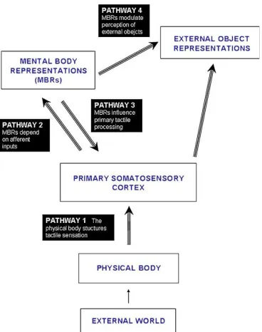
Recently, Serino & Haggard (2009), proposed a functional model
suggesting the main nodes and pathways involved in the representation of
tactile stimuli detected on the body surface (see Figure 1). The authors first
describe the information flow from the external environment to the primary
somatosensory cortex (SI), then hypothesise the existence of abstract mental
body representations (MBRs). A final stage referred to the external object
Figure 1: Analytic model of the relation between touch and the body proposed by Serino & Haggard (2009).
In their model the authors suggest that the different nodes are connected
with one another through mainly unilateral information flow, even though
feedback from MBRs to SI are also expected. As argued above, SI contains a
spatial representation that follow the organization of the somatotopic map of
the body surface. The mental body representations is defined as additional
brain areas that contain an abstract representation of the body derived from
sensory input, but it can be dissociated from it (body image and body schema
are considered both part of this stage). In addition, this representation is
considered a multimodal rather than a unimodal processing stage, in which
the actual state with respect to the dynamic nature of the body is represented
possible. Finally, MBRs can also influence upper areas for perception of
objects in the external space (external object representations).
1.2
Visuo/tactile interaction in the spatial coding of touch
Multisensory integration allows our brain to create a coherent and rich
representation of the external environment (Calvert, Spence & Stein, 2004;
Stein & Meredith, 1993). Different sensory modalities, such as vision and
touch reciprocally, interact even when the task can in principle be solved
using one sensory system in isolation. Recently, a great amount of studies
have investigated the role played by vision on touch perception (e.g.,
Botvinick & Cohen, 1998; Ernst & Banks, 2002; Folegatti , de Vignemont,
Pavani, Rossetti & Farnè, 2009; Honma, Koyama & Osada, 2009; Làdavas &
Farnè, 2004; Macaluso & Driver, 2005; Macaluso & Maravita, 2009; Mancini,
Bricolo & Vallar, 2009; Pavani, Spence & Driver, 2000; Serino & Haggard,
2009).
A classical phenomenon of this type of interaction is “visual enhancement
of touch” (VET), in which non-informative vision of a body part results in
responses to touch that are faster with respect to when the visual information
is absent (e.g., Tipper, Lloyd, Shorland, Dancer, Howard & McGlone, 1998),
and because even faster for familiar body parts (Tipper, Phillips, Dancer,
Lloyd, Howard & McGlone, 2001). In addition, enhancement of tactile acuity
first work documenting this phenomenon Tipper and colleagues (1998) used a
simple detection task and showed that mere vision of a body part (i.e.,
without proprioception) can influence tactile perception. In their work they
asked three separate groups of observers to detect as fast as possible a
predefined target vibration delivered to the thenar muscle (i.e., base of the
thumbs) of each participant’s hand under three different visual conditions.
One group looked at a video in front of them where, depending of the
experimental trial, their own right or left hand, was projected (vision without
proprioception). The second participant’s group were instructed to orient
their head and eyes towards one of the two own hands occluded from view
(proprioception without vision). Finally, the last participants’ group did a task
identical to the previous one with the only exception that they also viewed
their hands (vision plus proprioception). The authors found that responses
were faster when participants looked at their own hands and, more
important, that vision alone was sufficient to produce there faster responses
to the tactile stimulation (Tipper et al., 1998). Kennett and colleagues (2001)
tested two-point tactile discrimination thresholds (2PDTs) on the forearm,
while modulating visual input by presenting conditions in which the arm was
visible or, instead, a neutral object (i.e., cylinder) was visible. Tactile spatial
resolution was better when the arm was seen and even better when it was
magnified in size. By contrast, performance was not improved when the
neutral object was shown. The authors interpreted this result as direct
possible explanation proposed by the authors is that feedback modulation to
unimodal areas from multimodal areas (e.g., posterior parietal cortex, where
there are neurons that respond both at visual an tactile stimuli, Graziano, Yap,
& Gross, 1994), can pre-activate the somatosensory cortex, thus resulting in
enhanced tactile discrimination. Taylor-Clarke and colleagues (2002) using the
same paradigm (2PDTs) found a modulation of the somatosensory cortex
activity by vision of the arm, as measured by event-related encephalography.
When a visual input (i.e., participant’s own arm) was presented, a modulation
of the cortical activity in the somatosensory cortex was registered using
somatosensory event-related potentials (ERPs) (Taylor-Clarke et al., 2002). In
a further work Press, Taylor-Clarke, Kennett and Haggard (2004) tested VET
while participants perform different tactile tasks: detection or discrimination,
with or without spatial components. These experiments were done in order to
verify whether the VET effect described in previous experiments was a
generic effect on tactile perception or it occurred just under specific spatial
conditions and task demand. What they found is that visual enhancement of
touch was present only for difficult discrimination task that included spatial
components. In the difficult discrimination task two tappers were applied on
the left forearm in a spatial separation close to the 2PDT. Participants were
instructed to discriminate the activated tapper (one was silent), far and near
with respect to the elbow, as fast as possible. Response was given by pressing
two keys with the right hand. Only task with those specific characteristics
performance when participants saw their own arm with respect to when they
saw an object. The authors attributed this effect to a feedback signal from
multimodal to somatosensory areas that modify tactile receptive fields size
(RFs), decreasing their dimension and improving spatial sensitivity. On the
contrary, when participants performed an easy spatial discrimination task or
a difficult non spatial discrimination task, there was a decrement in
performance in viewing the arm with respect to viewing an object. No specific
explanation about this last result was provided by the authors (Press et al.,
2004). Interestingly, Serino and colleagues (2007) tested brain damage patients
and found that visual enhancement of touch was present only in subjects with
poor tactile acuity. This evidence has been interpreted as an intervention of
visual input when the tactile domain is not sufficiently efficient in solving a
specific spatial task (Serino, Farnè, Rinaldesi, Haggard & Làdavas, 2007).
Furthermore, evidences for modulation of RFs size in primary somatosensory
cortex by visual input has been recently documented, both behaviourally
(Haggard, Christakou & Serino, 2007) and by using the transcranial magnetic
stimulation (TMS) technique (Fiorio & Haggard, 2005). Haggard and
colleagues (2007) used vibrotactile maskers presented with orthogonal
arrangement with respect to the tactile target in a close or far spatial
proximity on the participant’s forearm. Participants were instructed to
perform two-alternative forced choice (2AFC) spatial discriminations to
localize the targets as proximal (i.e., closer to elbow) or distal (i.e., closer to
representation of the target only if they fall into their RFs. The authors found
that viewing the body made far maskers less effective, while made near
maskers more effective. This result has been taken as evidence that tactile
receptive fields size was reduced when participants viewed the body
(Haggard et al., 2007). A parallel line of research showed that application of a
single-pulse TMS over SI, but not over SII, produced a suppression of the VET
effect (Fiorio & Haggard, 2005). Additional evidences that VET may come at
the SI level are provided by a recent work by Serino, Padiglioni, Haggard and
Làdavas (2009), in which the authors tested whether VET can spread from
body parts which are adjacent in terms of somatotopy. They verified that VET
can indeed extend from one body part (i.e., hand) to another (i.e., cheek), but
this spreading occurs only between parts that are close represented in the
somatotopic map (i.e., cheek and hand, but not hand and foot).
In summary, studies on VET suggest a modulation of a non-informative
visual input on the perception of a pure tactile stimulus. This modulation is
unlikely to reflect an effect of spatial attention, because in the typical control
condition of the VET studies participants see an object instead of a body part,
but both kept in the same spatial position. Consequently attention is always
overtly fixed to the same locus. In addition, the effect is not related to a
particular visual information as all the studies refer to a non-informative
visual input, therefore cross-modal integration of specific cues cannot explain
the effect. As we described above, VET seems to derived from a top-down
somatosensory cortex. These projections could affect touch by modulating the
relative dimension of the tactile receptive fields of the stimulated body part
(Serino & Haggard, 2009).
Some authors have recently proposed that VET could derive, at least in
part, from a response bias boosting the propensity to respond when a body
part, instead of an object, is seen (Johnson, Burton & Ro, 2006). Johnson and
colleagues tested systematically this account in a series of experiments on the
influence of a light on a finger on tactile perception at that same finger. Data
were analysed by using Signal Detection Theory (SDT: Macmillan &
Creelman, 1991) and showed an increase in the near-threshold tactile
perception level. However, a shift in the response bias was also documented,
when touch and visual stimuli were simultaneously presented. Participants
were more prone to respond (i.e., less conservative) when tactile an visual
stimuli were both presents. This bias has been interpreted as a possible
consequence of multisensory experience that occur in the interaction with the
external environment. Sensory information available from the external world,
is synchronised in space and time the majority of the time when it originates
from the same object (Johnson et al., 2006). Since the majority of work on VET
did not used SDT procedure for analysing the data (e.g., Kennett et al., 2001) it
is possible that some VET finding could reflect a shift in the response
criterion. However, some study that reported VET used the two alternative
forced-choice paradigm (e.g., Taylor-Clarke, Kennett & Haggard, 2004), which
close to chance level; Longo, Cardozo & Haggard, 2009). In summary, a shift
in the criterion can partially explain the performance change in the visual
enhancement of touch, but cannot completely account for this effect.
In the present section we described the VET in details as one of the effect
resulting from visuo-tactile interaction. However, in literature there are many
others phenomenon that documented multisensory effect on tactile
perception (e.g., Calvert, Spence & Stein, 2004; Maravita & Iriki, 2004). For
instance the rubber hand illusion (e.g, Folegatti et al., 2009), in which a visual
proprioceptive conflict affects touch performance, or influence of auditory
inputs on touch (e.g., Soto-Faraco & Deco, 2009). Conscious of the large
amount of visual effects on touch, here, we primarily described VET effect
because some of our studies mainly focused on the effects of visual inputs
related to body parts on tactile spatial representation processing.
1.3
Spatial representation of touch in neuropsychological patients
A series of effects, derived particularly from neuropsychological
conditions, revealed how different representational level can be used for
spatial coding of touch on the body. These types of phenomenon includes, for
instance, tactile extinction (patients that extinguish contralesional tactile
stimulation during concurrent bilateral stimulation: e.g., Moscovitch and
Behrmann, 1994), synchiria (patients with unilateral brain damaged that
& Rapp, 2008) or finger agnosia (patients that make errors in identifying
which finger is stimulated: e.g., Kinsbourne & Warrington, 1962).
Tactile extinction is a condition in which unilateral brain damaged patients
fail to report a controlesional touch when this is presented together with an
ipsilateral one (Bender, 1952). Moscovitch and Behrmann (1994) tested tactile
extinction in 10 right brain-damaged patients, by delivering double
simultaneous touches to the opposite side of the wrist of the right or left hand,
when the hands were palm-down or palm-up. Regardless of hands posture
the missed tactile stimulus was systematically the one that occupied the
leftmost location in external space (see Figure 2A panel “c”). These results
demonstrate that patients coded tactile stimuli using a representational stage
at an high level of tactile information processing. Indeed, if patients would
code stimuli using a reference frame based on a lower stage of spatial
representation processing (e.g., somatotopic map), they should extinguish the
stimulus on the same region of the skin regardless of the hands posture in
external space. This finding is compatible with the results of a recent fMRI
study showing that primary somatosensory cortex is always activated
bilaterally in a right brain-damaged patient showing tactile extinction,
suggesting that the competition leading to extinction occurs after the afferent
tactile stimuli are processed by the primary somatosensory cortex
(Beversdorf, Hughes & Heilman, 2008). Moreover, Valenza and colleagues
(2004) found that patients with right parietal brain damage and visual neglect
positioned in the contralesional (affected) hemispace, and a concurrent
stimulation occurred on the elbow. On the contrary, extinction was not
present when the same hand was positioned in the ipsilesional hemispace (see
Figure 2B for the experimental set-up). The same test was repeated using
functional magnetic resonance (fMRI), with the purpose of defining the neural
bases of this spatial modulation of extinction. The authors found that when
the right hand was positioned in the contralesional hemispace, there was a
reduction in the blood-oxygenation level dependent (BOLD) responses to
tactile stimuli in the primary somatosensory cortex of the intact hemisphere
(Valenza, Seghier, Schwartz, Lazeyras & Vuilleumier, 2004). This finding was
considered by the authors evidence that limb position affect elaboration
process occurring at the level of the primary somatosensory cortex. Thus,
considering these studies it seems not completely clear if the neural correlates
of tactile extinction derive fully from higher stages of spatial representation
processing or instead low stages can be partially responsible for the effect.
Furthermore, a single-case electrophysiological study on a patient with right
hemisphere brain damage showing tactile extinction revealed neuronal
activity in the somatosensory cortex of the impaired hemisphere (Eimer,
Maravita, Van Velzen, Husain & Driver, 2002). The authors suggested that
this result is an evidence of residual unconscious processing of extinguished
tactile stimulation. Moreover, the same activity has been registered in the
injured hemisphere also with unilateral stimulation, even though attenuated
extinction can be related not to elimination of a sensory stimulus, but only to
an attenuation of the brain response of it.
Figure 2. Experimental conditions with different postures assumed by the participants in Moscovitch and Behrmann (1994) (panel A) and Valenza and colleagues (2004) (panel B) studies.
Some patients with unilateral brain damaged report to perceive bilateral
sensations after unilateral stimulation: a particular condition known as
synchiria (Medina & Rapp, 2008). Medina and Rapp (2008) tested a patient
with a left hemispheric brain damage and found that synchiria was affected
by the position of the hand decreasing when the hand was moved from the
contralesional to the ipsilasional hemispace in trunk- and head-centred
coordinates. In addition, it was not present with crossed hands (Medina &
Rapp, 2008). The authors suggested that these results imply the use of
multiple stages in the spatial representation of the tactile stimuli. These two
effects (i.e., extinction and synchiria), briefly described, gave opposite
behavioural results (i.e., unprocessed or additional process of a tactile
stimulus at the level of awareness). However, both cases provide evidence
about multiple representational stages involved in tactile stimuli perception.
Indeed, both neurological conditions derived from a unilateral brain damage
with patients that fail to report the correct perceptual experience. However, in
extinction the deficit produce a suppression at the level of awareness of a
stimulus that was physically present. Instead, in synchiria patients report to
perceive two stimuli after single stimulation, experiencing an additional
percept at the level of awareness, failing to represent at some stage of the
information processing the correct perceptual sensory input.
Another deficit revealed in individuals with a selective brain damage is
finger agnosia. In this neuropsychological condition patients with left parietal
lobe damage are not able to clearly name which specific finger has been
stimulated. This neurological impairment cannot permit patients to separate
identity of the fingers (Kinsbourne & Warrington, 1962). Recently, Rusconi
and colleagues (2009) suggested that finger agnosia should be referred to a
deficit in the connection between SI and the BSRs. Body structural
representation is defined as a stage, of the elaboration process, where the
body parts order (e.g., number of fingers) and their relationship are
represented. Therefore, this stage is clearly differentiated from the
representation present in the somatotopic map in SI (for more details on the
BSRs see Rusconi et al., 2009).
All these neuropsychological evidences, that we have briefly described,
by the mediation of multiple spatial representational stages. Moreover,
individuals with brain damages clearly show selective impairments at one or
2
Results
Considering the actual state of the art about spatial representation of
touch, we performed a series of behavioural experiments to investigate which
spatial reference frame is adopted in a special context of sensory stimulation,
namely double simultaneous stimulation (DSS). This was investigated in
details in Study 1 through posture manipulations, and examined in relation to
the role of vision in Study 2. Moreover, we used a neuroimaging technique
(fMRI) in order to delineate the main neural pathways sub-serving these
representational processes in Study 3. This section will briefly summaries the
methods and the results of the behavioural experiments and the preliminary
data of the imaging study. An extended description of each study is available
in section 3 of this Thesis.
2.1
Overview of the behavioural studies
2.1.1
Study 1: Spatial coding in a Double Simultaneous tactile
Stimulation (DSS)
In this first study we adapted the double simultaneous tactile stimulations
paradigm (DSS) for stimuli delivered within as well as between hands to
examine the role of multiple body representations in spatial coding of touch.
In addition, we investigated the relative contribution of the different spatial
representation for touch by manipulating hands posture. Unlike previous
the target and the masker (e.g., Craig & Evans, 1995; Craig, 1982), in Study 1
we modulated the relative position of the stimuli on the fingers. In the first
experiment, we used tactile stimuli at threshold level, with the hands always
resting in the same position (i.e., both hands palm-down). In the second
experiment, we used supra-threshold stimulation with hands assuming
different spatial positions across blocks (i.e., one hand palm-down and one
palm-up).
EXPERIMENT 1
A series of tactile stimuli at threshold level were delivered to the index
and/or middle fingers by using four stimulators. We asked participants to
detect whether a pre-specified target finger was tactually stimulated or not.
Across blocks, the target finger was either the index or the middle finger of
the right or left hand and it could be stimulated alone or together with a
non-target finger. DSS stimulation was delivered within the same hand (e.g.,
Figure 2.1b) or between hands (e.g., Figure 2.1c). We expected that DSS would
lead to slower and less sensitive detection of the target (i.e., tactile
interference) with respect to the condition in which the target finger was
stimulated alone. More critical for the issue of spatial coding of touch, our
experimental set-up lead to substantially different predictions of interference
pattern as a function of the adopted spatial code. If interference occurs in
somatosensory space it should be maximal when target and non-target fingers
cortical territories in primary and secondary somatosensory cortices which
can inhibit the adjacent territories activated by the target.
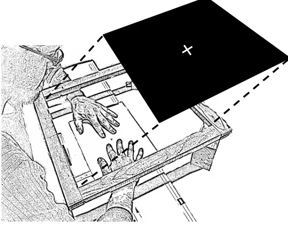
Figure 2.1. Schematic drawing of the experimental setup. Note that hands and tactile stimulators are shown here only for illustrative purposes, as they were in fact occluded under the horizontal computer display throughout the experiment.
In addition, because cortical territories neighbouring to the target can
also be activated through inter-hemispheric transfer (Harris et al., 2001;
Iwamura et al., 1994, 2001, 2002), substantial interference effects should also
emerge when the non-target stimulation occurs at the finger of the other hand
which is non-homologous with respect to the target. By contrast, a body space
representation of touch should lead to strong tactile interference mainly
within hands, with little or no tactile interference between hands. This (a) T-trials
(b) T+DFSH (c) T+SFDH (d) T+DFDH
because filtering of irrelevant stimulation between hands should be easier
whenever the target hand is clearly specified by a structural body
representation.
Finally, if participants solve the task entirely based on the location of
touches in external space, comparable interference should emerge when the
non-target finger is on the same hand as the target or is the homologous
finger of the other hand. This because distance in external space was identical
in these two experimental conditions (see experimental set-up in Figure 2.2).
Figure 2.2. Schematic drawing of the experimental setup. Note that hands and tactile stimulators are shown here only for illustrative purposes, as they were in fact occluded under the horizontal computer display throughout the experiment.
Results and discussion
Results showed significant interference effects only in terms of reaction
times and not for sensitivity. These were not affected by the specific pairings
effect in RTs were numerically smaller when the non-target finger was
homologous to the target (T+SFDH) with respect to the other conditions. We
speculated that one potential reason for the weak tactile interference
documented in this first experiment was that tactile stimulators were not
entirely reliable when driven at voltages closer to threshold levels. This could
have produced an uncontrolled inter-finger variability and could have
changed the stimulation ratio between the different target and non-target
finger in DSS trials, making the between finger competition less effective.
(This experiment was considered preliminary and is not reported in full in the
extended manuscript of Study 1 that appears in Section 3).
EXPERIMENT 2
In this second experiment we made all tactile stimuli clearly
supra-threshold. In addition, we examined the role of hands posture to assess the
potential involvement of any spatial coding of touch beyond body
representations. We asked participants to perform the same task of
Experiment 1, adopting two different hand postures across blocks. In half of
the blocks, both hands were palm down (as in Experiment 1). In the
remaining blocks, one hand was palm down while the other hand was palm
up (as shown in Figure 2.3, in which the hand rotated palm-up is shown as
darker for illustrative purposes only).
The logic of this manipulation is the following: if any between-hand
body-space (rather than external-body-space), tactile interference should remain
unchanged across hands posture. By contrast, if tactile interference operates
on an external space reference frame it should change as a function of the
adopted hands posture.
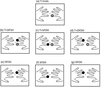
Figure 2.3. Example of the stimulation conditions with one hand palm-down and the other rotated palm-up, when the target finger is the right index finger. Unfilled circles indicate the stimulation at the target finger; filled black circles indicate stimulation at the non-target finger. a) target only trial; b-d) DSS trials; e-g) catch trials.
Results and discussion
The results of the present experiment revealed interference effects of
DSS stimulation compared to target only trials. It emerged reliably with our
clearly suprathreshold stimulation both in terms of sensitivity and RTs. This (a) T-trials
(g) DFDH (f) SFDH
(e) DFSH
predicted interference effect on sensitivity was manifest only within the same
hand (see Figure 2.4). This pattern of results were confirmed by RTs that, in
addition, showed interference also between hands, particularly for distractors
delivered to fingers non-homologous with respect to the target (e.g., the left
middle finger when the target was the right index). Importantly, these
interference effects within and between hands were not modulated as a
function of hand posture, supporting the notion that within and between
interference effects may be solved at low stage of body representation. This
interference is compatible with DSS competition occurring in somatotopic
space, because at this low representational stage the differentiation between
the two hands is less clearly defined and stimulation delivered to the
non-homologous finger of the other hand can reach cortical territories ipsilateral to
the target (Braun et al., 2005; Iwamura et al., 2001, 2002; Killackey et al., 1983).
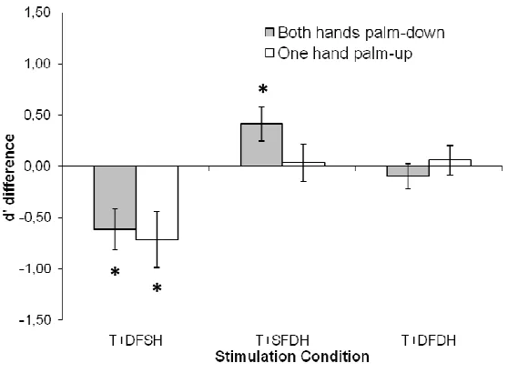
The results of Experiment 2 also revealed an unexpected increase in
tactile sensitivity that occurred between hands and was strictly dependant
upon hand posture (see Figure 2.4). This effect emerged when the target
finger was stimulated together with the homologous fingers of the other hand
and both hands were palm down. Instead, it disappeared when either hand
was rotated palm-up. This posture dependent modulation rules out the
possibility that this increased tactile sensitivity emerged at a low
representational stage. Instead, this phenomenon reveals the use of a spatial
representation for touch which takes into account the overall structure of the
Figure 2.4. Sensitivity difference (d’ difference) computed by subtraction between single and DSS trials as a function of Stimulation Condition. Error bars represent the Standard Errors (SE). “T+DFSH” represent DSS trial in which target finger and the non-homologous finger of the same hand were stimulated, “T+SFDH” target finger and the homologous finger of the opposite hand with respect to the target were stimulated and “T+DFDH” represent the condition in which target finger and non-homologous finger of opposite hand were stimulated.
2.1.2
Study 2: Assessing the role of vision on tactile DSS
In this second study, we tested the sensitivity of tactile DSS paradigm with
stimuli delivered within and between-hands to different levels of
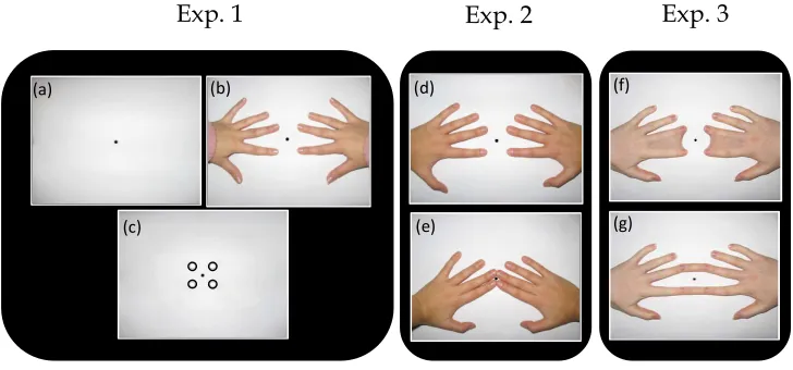
hand-related visual inputs (see Figure 2.5). In Experiment 1, we examined the role
of seeing vs. not seeing the hands. In Experiment 2, we examined the role of a
visual/proprioceptive conflict by showing images of participant’s own hands
that either matched or not matched their unseen hand posture. Finally, in
Experiment 3 we introduced a novel manipulation of visual hand-morphing
(i.e., merging of fingers), to determine whether different types of visual
*
structural distortions of the hands could affect the low level stage of the tactile
processing at which DSS interference occurs.
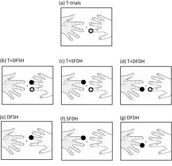
Figure 2.5. Illustrations of the possible Visual Conditions for Experiment 1, 2 and 3 are presented.
EXPERIMENT 1
The aim of the first experiment was to investigate whether vision of a body
part (i.e., participant’s own hands) can modulates detection of a target tactile
stimulus presented with a concurrent non-target stimulation on another
finger (tactile DSS). Across blocks participants saw: 1) a fixation cross in the
middle of the screen; 2) an image of their own hands exactly reproducing the
fingers as positioned under the screen; 3) four empty circles, each vertically
aligned with the first phalanx of each fingers just below the screen. If vision of
a body part affect tactile perception under DSS type of interference, one
should expect better performance when the participant’s own hands (see
Figure 2.5b), compared to circles (see Figure 2.5c), are displayed.
Exp. 1 Exp. 2 Exp. 3
(e)
(d) (f)
(g) (a)
(c)
Participants performed a go/no-go task to detect whether a tactile
stimulus was delivered to a pre-specified target finger (e.g., right index),
which could be stimulated alone or simultaneously with a non-target finger,
either on the same hand as the target (e.g., right middle finger), or on the
other hand (at homologous or non-homologous finger with respect to the
target finger; e.g., left index or left middle finger, respectively) (for the same
type of methodology see Study 1 of the present thesis).
Results and discussion
The results showed reliable interference effect of DSS trials with respect to
target only stimulations. Similar to our previous study (see Study 1 of the
present thesis) significant tactile interference emerged for conditions in which
the non-target finger stimulation was on the same hand as the target and also
when it was on the non-homologous finger on the non-target hand. These
findings imply, as we previously suggested, that DSS interference is driven by
competition being solved at a relatively low stage of touch representation
(Study 1). Also consistent with our previous work, we found that DSS
interference was significantly reduced, if not absent, when homologous
fingers across hands were stimulated (i.e., T and T+SFDH conditions did not
differ).
Although interference effect in tactile domain was clear, vision of the
hands did not affect tactile DSS performance. A significant enhancement of
hands, were added to the visual scene. We suggest that this finding is
compatible with the circles allowing for a better focusing of selective spatial
attention on the spatial regions above which the tactile stimuli were delivered
(Spence, Pavani & Driver, 2000). However, catch trials showed that
mislocalisazation of single target within the same hand as the target was
modulated by vision to some extent. Finally, examination of the criterion
revealed a change in the participants responses tending to be more
conservative in the tactile conditions that were more difficulties (i.e., when the
distractor finger was the homologous finger of the same hand or the
non-homologous finger of the opposite hand). In addition, we found a more liberal
responses criterion when participants saw their own hands with respect to
fixation only, possibly suggesting a tendency in favour of the ‘go’ response
when a body part is seen (see Johnson et al., 2006).
Taken together these data suggest that, even though tactile DSS paradigm
proved particularly sensible to low stage of tactile spatial representation
processing, it seems not to be affected by VET.
EXPERIMENT 2
It is possible that the tactile interference reported under DSS in
Experiment 1, although not sensitive to visual manipulation that would have
improved performance, would still be worsened by conflicting
visual-proprioceptive information. Indeed, while VET would have produced
may affect touch perception, namely by hampering tactile performance (e.g.,
Folegatti et al, 2009). To test this possibility, in Experiment 2 we modulated
the spatial congruency between the seen and felt hand posture. Participants
were shown a visual scene in which their hands were positioned with fingers
placed close to each others (see Figure 2.5e), thus creating a conflict between
the visual and proprioceptive hand position (i.e., a visual-proprioceptive
conflict). In another condition, participants’ hands were visually displayed in
a congruent position as the proprioceptive one (see Figure 2.5d). Note that the
latter condition is identical to Experiment 1. If such a conflict between vision
and proprioception is effective in modulating the tactile interference under
DSS, one should expect better performance for intermodal congruent as
compared to the incongruent and conflicting condition.
Methods were identical to Experiment 1, with the following exceptions.
We changed the visual condition by adopting two different images of the
participant’s own hands with fingers placed at dissimilar positions (congruent
vs. incongruent).
Results and discussion
Similar to Experiment 1 significant tactile interference emerged in terms of
sensitivity, only for the conditions in which the non-target finger was
stimulated on the same hand as the target and when it was the
non-homologous finger of the other hand. Regarding of the RTs an interference
We revealed also a general decrement of tactile sensitivity when we
presented the incongruent hands image with respect to the congruent hands
image. This visual effect can be referred to the spatial incompatibility between
seen hands and the real ones, similarly to what has been reported recently by
Folegatti and colleagues in a single detection task approach (Folegatti et al,
2009). However, similarly to Experiment 1, there was no significant
modulation of the pattern of tactile interference effect (DSS trials conditions)
by visual-proprioceptive incongruence. Finally, analysis on the criterion
revealed a change in the participants responses, which tended to be more
conservative in DSS trials with respect to Target only trials and more liberal
when they saw their own hands in the congruent, as compared to the
incongruent position.
EXPERIMENT 3
As we showed in Experiment 2, a visual/proprioceptive conflict can
affect touch (see also Folegatti et al., 2009), but not DSS modulation. In this
final experiment, we tried to alter visually the structural morphology of the
body part from which proprioceptive information could be derived, to see if it
can play a role in shaping the interference effects under DSS. To the best of
our knowledge this is an entirely novel manipulation in literature on the
influence of vision on touch perception. To this aim, in Experiment 3 we
introduced visual changes in the structural morphology of the hands (i.e.,
which their own hands seen were modified in their morphology (i.e., fingers
webbed). A recent study provided evidence that real fingers webbing affect
perception at a low stage of the information processing (e.g., Stavrinou, Della
Penna, Pizzella, Torquati, Cianflone, Franciotti, Bezerianos, Romani &
Rossini, 2006).
Methods were identical to Experiment 1 and 2, with the following
exceptions. The first visual morphing condition was characterised by webbing
index and middle finger of either hand (i.e., intra-hand morphing) (see Figure
6f). The second visual morphing condition was done exactly like the previous
one except that in this case we merged the homologous fingers of either hands
(left and right index fingers and left and right middle fingers) (i.e., inter-hands
morphing) (see Figure 6g).
Results and discussion
Similar to the previous Experiments a cost for DSS trial with respect to
single touch condition (interference effect) was revealed, confirming again the
stability and constancy of our basic effect. Also the interference was strongly
present at the intra-hand level and at the inter-hand level only for the
non-homologous finger, following the exact same pattern described in the
previous experiments. However, the visual structural morphing, did not
affect tactile DSS or the grade of interference between fingers. Finally, data on
the criterion revealed a change in the participants responses, who become
note that compared to the previous Experiments we did not find changes in
the response criterion when participants saw their own hands with intra- or
inter-hand morphing.
Overall conclusions from the behavioural studies
Taken together the behavioural experiments have shown that DSS
stimulation can produce interference effects when stimuli are delivered both
within and between hands. In addition, we documented an increase in target
sensitivity during between-hand DSS at homologous fingers which may relate
to a redundancy of spatial codes for the concurrent tactile events. Only the
latter phenomenon was affected by changes in hand posture. In keeping with
the notion that touch can be spatially coded in different frames of reference
we suggested that tactile DSS interference is resolved at a low
representational stage (somatotopic), whereas increased tactile sensitivity in
this task relies on a higher representational stage which takes into account the
layout of the body in space. This conclusion was further strengthened by our
second behavioural study. Non-informative visual inputs about the
stimulated body parts did not affect DSS tactile interference. Thus, the DSS
paradigm seems to be largely immune to matching or conflicting vision from
the stimulated body part, suggesting that DSS interference may occur within
the somatosensory system, and possibly prior to any modulations of vision on
2.2
Overview of imaging study
2.2.1
Study 3: Neural correlates of tactile coding, an fMRI adaptation
paradigm
In the previous behavioural studies we examined how multiple spatial
representation can serve tactile spatial coding of touch, in the special context
of DSS. In the present work, we used an fMRI adaptation paradigm (for a
review see Grill-Spector & Malach, 2001; Krekelberg, Boynton & van Wezel,
2005) to probe the possible neural basis of these multiple spatial coding. The
adaptation effect is a typical physiological response of the neurons that results
from the successive repetition of a feature to which neurons are selective.
Following the logic of this physiological effect when two tactile events are
repeated on exactly the same region of skin, all neurons that have a strictly
somatotopic response should reduce their activity. These neurons should
instead show no reduction of activity if the stimulation repeats over two
distinct regions of skin. The crucial question, in relation to the issue of
reference frames for touch, is whether some population of neurons in the
brain can adapt to stimulation that repeats over distinct region of skin, when
some other aspect of spatial coding is in fact identical. For instance when the
repeated stimulation is delivered to homologous body parts (e.g., indexes of
either hands). In that a case the region of the skin would differ, but the
identity of the body part would stay the same. Our expectation is to find a
different grade of fMRI adaptation to these finger pairing specifically in SI
over the same region of skin (i.e., same finger stimulated twice), because in
this sensory area the dominant representation of touch should be primarily
contralateral. By contrast, we predicted that SII could adapt to stimulation
that repeats over the same finger (i.e., indexes of either hands), because in this
sensory area bilateral representations of touch have been extensively
documented (e.g., Blatow, Nennig, Durst, Sartor & Stippich, 2007).
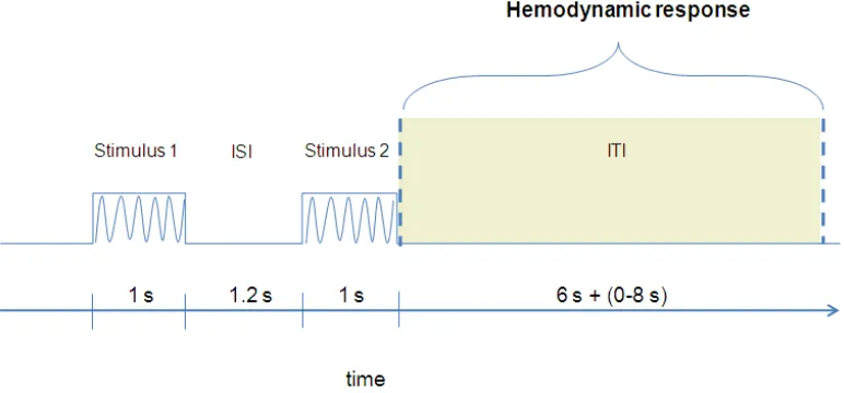
We used the fMRI adaptation paradigm with two successive
vibrotactile stimuli (see Figure 2.6) delivered to the first phalanx of the index
and middle fingers of either hands. These produced four different finger pairs
stimulation conditions: (1) Left index stimulated twice (Li – Li); (2) Left
middle and index fingers (Lm – Li); (3) Right and left indexes (Ri – Li); Right
middle and left index fingers (Rm – Li). The experiment consisted of 4
event-related fMRI adaptation scans. Participants were instructed to pay attention
always to the left index finger throughout the experiment.
We calculated individual functional maps for each participant. Maps
for the right and left hemisphere were functionally defined as all voxels that
were significant in the omnibus test (fixed effects analysis; FFX) with four
regressors corresponding to the experimental conditions (i.e., Li - Li, Lm - Li,
Ri - Li, Rm - Li). On the resulting maps we identify four Patches of Interests
(POIs), separately for each participant, consisting in the primary and
secondary somatosensory cortex of both hemispheres on the basis of brain
anatomy and functional response (see Figure 2.7).
Figure 2.7. A paradigmatic example of the four Patches of Interests (POIs) of subject 2 defined from the fixed effect analysis with the four conditions as regressors.
For each POIs, we generated a correspondent Region of Interests
(ROIs) in 3D space. On the ROIs we calculated the Beta values, on the L
R
SI
SII
SI
hemodynamic response. Within these ROIs we analysed the Beta values by
executing a repeated measure Analysis of Variance (ANOVA) with Area (SI,
SII), Hemisphere (Right, Left), Hand (within, between) and Finger
(homologous, non-homologous) as within participants variables.
Results and discussion
The results revealed more adaptation effect when homologous as
compared with non-homologous fingers were stimulated. Remarkably this
occurred regardless of the hemisphere and of the somatosensory cortical area
(i.e., SI, SII) (see Figure 2.8). Therefore, adaptation occurred for stimuli
delivered on the same region of the skin (i.e., left index stimulated twice) and
for stimuli delivered on homologous fingers of different hands (i.e., right and
left indexes) regardless of the somatosensory area (i.e., SI and SII). The activity
difference between homologous and non-homologous stimulations show that
at low stage of spatial representation processing (SI) segregation of Finger
identity (i.e., which finger was stimulated within the same hand, index or
middle finger), is clearly establish. However, when homologous fingers of
opposite hands are stimulated, side identification seems not to be entirely
unambiguous, even in SI.
Finally, we revealed a main effect of area that indicates more activation
in SII as compared with SI. This latter result can derived from a disproportion
represented) with respect to other body parts (see Maldjian, Gottschalk, Patel,
Detre & Alsop, 1999).
Figure 2.8. BOLD amplitude (Beta Values) for homologous and non-homologous fingers regardless of the body side (RH, LH) and areas (i.e., SI, SII). Error bars reflect the standard error of the mean (SE).
Overall conclusion of the imaging study
The imaging results revealed the usefulness of the fMRI adaptation
paradigm to investigate the neural basis of touch, with particular regards to
the same and to different body sides. We reported evidence in favour of the
existence of bilateral representation of tactile stimuli delivered at the fingers
in both primary and secondary somatosensory areas. Importantly, the present
imaging data support the previous behavioural results on DSS interference
within and between hands (Study 1; Study 2).
Homologous Non - homologous
3
Papers
3.1
Double simultaneous tactile stimulation within and between
hands: insights for spatial coding of touch at the fingers
Double simultaneous tactile stimulation within and between
hands:
Insights for spatial coding of touch at the fingers
Luigi Tamè1, Alessandro Farnè2,3 Francesco Pavani1,4
1. Center for Mind/Brain Sciences, University of Trento, Rovereto, Italy;
2. INSERM UMR-S 864 “Espace et Action”, Bron, F-69500, France;
3. Université Claude Bernard Lyon I, Lyon, F-69000, France
4. Dep. of Cognitive Sciences and Education, University of Trento, Rovereto,
Italy
Address for correspondence:
Francesco Pavani
Center for Mind/Brain Sciences, University of Trento
Corso Bettini 31, 38068 Rovereto, Italy
Phone: +39 0464 808674. Fax: +39 0464 808602.
E-Mail: [email protected]
Abstract
Introduction
During everyday life, we localise somatosensory stimuli on our body
surface almost without effort. However, this seemingly simple task hides the
existence of multiple spatial representations of the tactile event in our brain
(e.g., de Vignemont, Tsakiris & Haggard, 2006; Gallace & Spence, 2008;
Haggard, Kitadono, Press & Taylor-Clarke, 2006). In a recently proposed
flow-chart of sensory representations for touch (Serino & Haggard, 2009),
touch is initially encoded into a sensory space within the primary
somatosensory map (Blankenburg, Ruben, Meyer, Schwiemann & Villringer,
2003; Penfield & Rasmussen, 1950), but the location of the tactile event is
coded also with respect to other frames of reference in further processing
stages. Tactile sensation can be mapped in a mental body representation, to
localise tactile events with respect to body-parts and body-side (e.g., Schicke
& Röder, 2006), or in egocentric/allocentric representations of external space,
to localise tactile events in the outside world (e.g., Azañón & Soto-Faraco,
2008; Brozzoli, Ishihara, Göbel, Salemme, Rossetti & Farnè, 2008). In the
present work, we adapted a paradigm of double simultaneous tactile
stimulation on the fingers to investigate at which representation level the
competition between concurrent tactile stimuli is resolved, and infer which
spatial representation of touch may be dominant while solving this task. In
addition, we assessed to what extent manipulations of hand posture can
Multiple spatial representations of touch
The most basic spatial representation of touch in the cortex is the one
available in primary somatosensory cortex, which contains a topographic
representation of the skin surface (Blankenburg et al., 2003; Penfield &
Rasmussen, 1950). At this primary level of spatial representation, however,
the body is not categorically differentiated into parts (de Vignemont et al.,
2006) and the spatial relationships between body-parts differ with respect to
the actual organisation of the body. For instance, hand and face are adjacent
in primary somatosensory cortex, but