REVIEW
Current status and perspectives
of genome editing technology for microalgae
Seungjib Jeon
1,2, Jong‑Min Lim
3, Hyung‑Gwan Lee
4, Sung‑Eun Shin
5, Nam Kyu Kang
1, Youn‑Il Park
6,
Hee‑Mock Oh
4, Won‑Joong Jeong
3, Byeong‑ryool Jeong
1,2*and Yong Keun Chang
1,2*Abstract
Genome editing techniques are critical for manipulating genes not only to investigate their functions in biology
but also to improve traits for genetic engineering in biotechnology. Genome editing has been greatly facilitated by
engineered nucleases, dubbed molecular scissors, including zinc‑finger nuclease (ZFN), TAL effector endonuclease
(TALEN) and clustered regularly interspaced palindromic sequences (CRISPR)/Cas9. In particular, CRISPR/Cas9 has
revolutionized genome editing fields with its simplicity, efficiency and accuracy compared to previous nucleases.
CRISPR/Cas9‑induced genome editing is being used in numerous organisms including microalgae. Microalgae have
been subjected to extensive genetic and biological engineering due to their great potential as sustainable biofuel
and chemical feedstocks. However, progress in microalgal engineering is slow mainly due to a lack of a proper trans‑
formation toolbox, and the same problem also applies to genome editing techniques. Given these problems, there
are a few reports on successful genome editing in microalgae. It is, thus, time to consider the problems and solutions
of genome editing in microalgae as well as further applications of this exciting technology for other scientific and
engineering purposes.
Keywords:
Genetic engineering, Microalgae, Genome editing, CRISPR/Cas9, Biofuels, GMO conflicts
© The Author(s) 2017. This article is distributed under the terms of the Creative Commons Attribution 4.0 International License (http://creativecommons.org/licenses/by/4.0/), which permits unrestricted use, distribution, and reproduction in any medium, provided you give appropriate credit to the original author(s) and the source, provide a link to the Creative Commons license, and indicate if changes were made. The Creative Commons Public Domain Dedication waiver (http://creativecommons.org/ publicdomain/zero/1.0/) applies to the data made available in this article, unless otherwise stated.
Background
Targeted genome modifications are crucial for genetic
analyses and genetic engineering in all aspects of biology
and related biotechnological fields. Different from
ran-dom integration of cloned genes for overexpression,
spe-cific alterations of the eukaryotic genome have been great
challenges for all biologists and biotechnologists. Gene
targeting (GT) was initially developed in recombinogenic
lower eukaryotes by introducing a homologous transgene
into the cell, and by utilizing homologous recombination
(HR), scientists were able to knockout or replace genes
of interest [
1
]. GT has been successfully demonstrated
in animals [
2
,
3
] and plants [
4
]. However, GT in these
higher organisms has been very difficult, in part, because
they are not recombinogenic [
5
]. Newly developed
techniques, including genome editing techniques, have
bypassed this hurdle by engineered nucleases, dubbed
“molecular scissors,” and the subsequent repair of DNA
strand breaks results in mutations or replacements of the
genes of interest [
6
].
Engineered nucleases include zinc-finger nucleases
(ZFNs), transcription activator-like effector nucleases
(TALENs), and clustered regularly interspaced
palindro-mic sequences (CRISPR)/CRISPR-associated protein 9
(Cas9) [
7
]. These three, in particular CRISPR/Cas9, will
be described for microalgal genome editing in this review
even though there have been other nucleases
includ-ing meganucleases and group II intron-based targetrons
adopted for genome editing in other organisms [
8
]. These
sequence-specific nucleases have enabled researchers to
cleave genomic DNA and to obtain mutations of a gene
resulting from faulty repair of the cleaved DNA.
Microalgae have emerged as important platforms for
the production of biofuels and other biomolecules, and
genetic engineering of microalgae is, thus, one of the
fastest growing biotechnology fields [
9
]. In addition to
Open Access
*Correspondence: [email protected]; [email protected]; [email protected]
overexpression of genes of interest, genome editing is
essential for the suppression of genes interfering with the
production of target molecules. However, progress in this
field has been hampered by multiple layers of difficulties
inherent to microalgae. This review will describe what
has been achieved in microalgal genome editing and
examine in detail the problems associated with
micro-algal genome editing and suggest possible solutions.
Genome editing has many applications that have been
shown in other organisms, and the possibility of these
applications in microalgae will also be evaluated. Because
the overexpression of genes shares the same
techni-cal problems, we will also include a brief description of
microalgal transformation technology.
Introduction to genetic engineering
Genetic engineering, by definition, requires the delivery
of genetic material to the genome resulting in genetic
modifications. Since reports of successful transfection of
animal cells with isolated viral DNAs in the early 1970s
[
10
,
11
], it took more than 10 years to achieve the
trans-formation of plants using Agrobacteria that are capable
of transforming plants in nature [
12
]. As usual,
transfor-mation of the model microalga
Chlamydomonas
came
much later in the late 1980s [
13
,
14
]. The long delay in
achieving microalgae transformation was not simply due
to a smaller microalgal community but also to technical
problems inherent to microalgae. The same delay was
seen for genome editing techniques in microalgae. As
pointed out lately, successful genome editing should be
based on solid transformation techniques for an
organ-ism [
15
]. We, thus, summarize the historical perspectives
of plant and microalgal transformation, which can
pro-vide clues to genome editing in microalgae.
Transformation of plants lagged behind animals
prob-ably due to the presence of the protective cell wall, and
initial attempts of a workaround with protoplasts did not
provide much success [
16
]. Instead, plant geneticists
devel-oped alternative techniques using the natural
transforma-tion machinery of Agrobacteria, which circumvented the
removal of the cell wall [
12
]. In addition to Agrobacteria,
particle bombardment also gained popularity, which can
avoid the host range limitations of Agrobacteria [
17
]. Later
on, the removal of the plant cell wall improved, and
trans-formation of protoplasts via polyethylene glycol (PEG) or
electroporation was also established in plants [
18
,
19
].
Transformation of the model microalga
Chla-mydomonas reinhardtii
was achieved by the
transfor-mation techniques used for plants, which was done for
nuclear [
13
,
14
] and chloroplast genomes [
20
]. Later,
trans-formation by glass bead agitation was uniquely developed
for
C. reinhardtii
, where protoplast cells were agitated
vig-orously with glass beads [
21
]. These pioneering works led
to the development of the transformation toolbox in
Chla-mydomonas
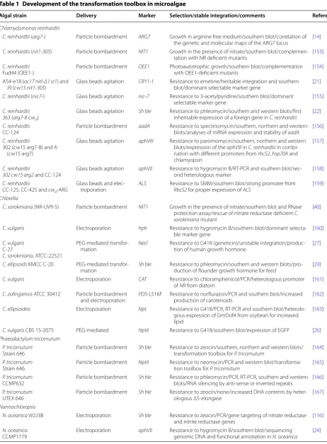
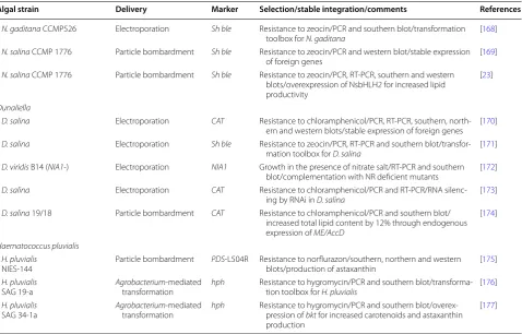
and other microalgae summarized in Table
1
.
In general, delivery of genetic material in microalgae is
considered much less efficient compared to that of plants.
On the other hand, once transformants are obtained,
microalgae are mostly single cells and can be maintained
somatically, not requiring the tedious (and recalcitrant in
some species) regeneration steps in plants [
15
].
The development of the transformation toolbox in
microalgae, excluding
Chlamydomonas
, presented
another layer of difficulties mainly due to the paucity of
selection markers that have been developed for plants
and other microbes (Table
1
). This is probably caused
by their evolutionary divergence, where their cellular
machineries have been differentiated so much that
exist-ing herbicides and antibiotics cannot be used for
selec-tion purposes. This is particularly true for the model
“industrial microalgae”
Nannochloropsis
that are
toler-ant to most toler-antibiotics and herbicides, which leaves only
phleomycin and its derivative Zeocin for selection by the
ble
gene from
Streptoalloteichus hindustanus
(
Shble
) [
22
,
23
]. Exceptionally,
N. oceanica
CCMP1779 is sensitive
to hygromycin that is selectable with
aphVII
[
24
].
How-ever, green algae sharing lineages with plants offers more
options of selection, including hygromycin and other
herbicides and antibiotics, similar to that of plants [
25
].
Microalgal transformation still suffers from an
extremely low transformation efficiency, even compared
to plants, which calls for drastically improved
transfor-mation techniques. Lately, cell wall removal and
PEG-mediated protoplast transformation have been reported
in
Chlorella
and
Cyanidioschyzon
[
26
–
29
].
PEG-medi-ated protoplast transformation in plants achieves a
higher transformation efficiency without the concerns
of the host range of Agrobacteria or expensive
equip-ment [
30
].
Chlorella
is gaining popularity in many global
consortia of algal biofuel production, and it is, thus,
interesting to see how this technique develops not only
in
Chlorella
but also in other microalgae. Another
tech-nique that can be considered is bacterial conjugation for
delivery of episomal vectors into microalgal cells, which
is claimed to be more efficient than conventional
trans-formation techniques in diatoms [
31
]. Optimized
tech-niques of these might provide solutions for the current
problems of delivery techniques in microalgae, which
may be used for genome editing in microalgae.
Table 1 Development of the transformation toolbox in microalgae
Algal strain Delivery Marker Selection/stable integration/comments References
Chlamydomonas reinhardtii
C. reinhardtii (arg7‑) Particle bombardment ARG7 Growth in arginine free medium/southern blot/correlation of the genetic and molecular maps of the ARG7 locus [14] C. reinhardtii (nit1‑305) Particle bombardment NIT1 Growth in the presence of nitrate/southern blot/complemen‑
tation with NR deficient mutants [153] C. reinhardtii
Fud44 (OEE1‑) Particle bombardment OEE1 Photoautotrophic growth/southern blot/complementation with OEE1‑deficient mutants [154] A54‑e18 (ac17 nitl‑Δ1 sr1) and
J9 (cw15 nit1‑305) Glass beads agitation CRY1‑1 Resistance to emetine/heritable integration and southern blot/dominant selectable marker gene [21] C. reinhardtii (nic7‑) Glass beads agitation nic‑7 Resistance to 3‑acetylpyridine/southern blot/dominant
selectable marker gene [155]
C. reinhardtii
363 (arg7‑8 cwd) Glass beads agitation Sh ble Resistance to phleomycin/southern and western blots/first inheritable expression of a foreign gene in C. reinhardtii [22] C. reinhardtii
CC‑124 Particle bombardment aadA Resistance to spectinomycin/southern, northern and western blots/analyses of mRNA expression and stability of aadA [156] C. reinhardtii
302 (cw15 arg7‑8) and A (cw15 arg7)
Glass beads agitation aphVIII Resistance to paromomycin/southern, northern and western blots/expression of the aphVIII in C. reinhardtii in combi‑ nation with different promoters from rbcS2, hsp70A and chlamyopsin
[157]
C. reinhardtii
302 cw15 arg2 and CC‑124 Glass beads agitation aphVII Resistance to hygromycin B/RT‑PCR and southern blot/sec‑ond heterologous marker [158] C. reinhardtii
CC‑125, CC‑425 and cwd‑ARG
Glass beads and elec‑
troporation ALS Resistance to SMM/southern blot/strong promoter from RbcS2 for proper expression of ALS [159] Chlorella
C. sorokiniana (NR‑UV9‑5) Particle bombardment NIT1 Growth in the presence of nitrate/southern blot and RNase protection assay/rescue of nitrate reductase deficient C. sorokiniana mutant
[40]
C. vulgaris Electroporation hph Resistance to hygromycin B/southern blot/dominant selecta‑
ble marker gene [160]
C. vulgaris C‑27
C. sorokiniana, ATCC‑22521
PEG‑mediated transfor‑ mation
Neor Resistance to G418 (geneticin)/unstable integration/produc‑ tion of human growth hormone [27]
C. ellipsoids KMCC C‑20 PEG‑mediated transfor‑ mation
Sh ble Resistance to phleomycin/southern and western blots/pro‑ duction of flounder growth hormone for feed [29] C. vulgaris Electroporation CAT Resistance to chloramphenicol/PCR/heterologous promoter
of NR from diatom [161]
C. zofingiensis ATCC 30412 Particle bombardment
and electroporation PDS‑L516F Resistance to norflurazon/PCR and southern blot/increased production of carotenoids [162] C. ellipsoidea Electroporation Npt Resistance to G418/PCR, RT‑PCR and southern blot/heterolo‑
gous expression of GmDof4 from soybean for increased lipid
[163]
C. vulgaris CBS 15‑2075 PEG‑mediated NptII Resistance to G418/southern blot/expression of EGFP [26] Phaeodactylum tricornutum
P. tricornutum
Strain 646 Particle bombardment Sh ble Resistance to zeocin/southern, northern and western blots/transformation toolbox for P. tricornutum [164] P. tricornutum
Strain 646 Particle bombardment NptII Resistance to neomycin/PCR and western blot/transforma‑tion toolbox for P. tricornutum [165] P. tricornutum
CCMP632 Particle bombardment Sh ble Resistance to phleomycin/PCR, RT‑PCR, southern and western blots/RNA silencing by anti‑sense or inverted repeats [166] P. tricornutum
UTEX 646 Particle bombardment Sh ble Resistance to zeocin/none/increased DHA contents by heter‑ologous Δ5‑elongase [167] Nannochloropsis
N. oceanica W2J3B Electroporation Sh ble Resistance to zeocin/PCR/gene targeting of nitrate reductase and nitrite reductase genes [116] N. oceanica
CCMP1779 Electroporation
aphVII Resistance to hygromycin B/southern blot/sequencing
(siRNA) and microRNAs (miRNAs) in
C. reinhardtii
and in other microalgae [
24
,
33
–
38
]. In addition to the
RNA-based knockdown techniques, gene targeting via
HR was also introduced in
Chlamydomonas
and
Chlo-rella
[
39
,
40
]; however, these results were not
reproduc-ible probably due to the non-recombinogenic nature of
microalgae. Similar difficulties were also observed in
most of higher eukaryotes including animals and plants
[
2
–
4
] because gene targeting was originally developed
in recombinogenic yeasts [
1
]. Difficulties and/or
ineffi-ciencies of the above reverse genetic techniques called
for a more efficient and precise modification of DNA,
which led to genome editing, also known as genome
engineering, using engineered nucleases. This review
will further discuss genome editing, mainly focusing on
microalgae, including difficulties and possible solutions.
Genome editing using engineered nucleases
Genome editing uses recombinant nucleases engineered
to recognize and cleave specific sequences in the genome,
resulting in double strand breaks (DSBs). DSBs are
repaired mostly by a homology-independent and
error-prone DNA repair mechanism, called non-homologous
end joining (NHEJ), resulting in mutations at the
cleav-age site [
41
–
46
]. Nucleases include ZFN, TALEN and
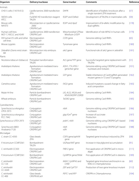
CRISPR/Cas9 as summarized in Fig.
1
a, and Table
2
lists
representative cases of genome editing in plants and
ani-mals and all known cases of microalgal genome editing.
Unfortunately, attempts of genome editing in microalgae
have had limited success with only a handful of reports.
Other endonucleases have been used for genome editing
in other organisms, including meganucleases and group
II intron-based targetrons as summarized in Fig.
1
b [
8
,
47
,
48
]. It would be interesting to find out how these
nucleases work in microalgae.
ZFN and TALEN appeared as an alternative to gene
tar-geting via HR and have been used for targeted
modifica-tion of genomes [
49
–
52
]. They are fusion proteins of the
restriction enzyme
Fok
I [
53
] and their respective DNA
binding proteins of zinc finger [
54
–
58
] and transcription
activator-like effector (TALE) [
59
], summarized in Fig.
1
a.
The resulting DSBs induced by
Fok
I are repaired mostly by
Table 1 continued
Algal strain Delivery Marker Selection/stable integration/comments References
N. gaditana CCMP526 Electroporation Sh ble Resistance to zeocin/PCR and southern blot/transformation
toolbox for N. gaditana [168]
N. salina CCMP 1776 Particle bombardment Sh ble Resistance to zeocin/PCR and western blot/stable expression
of foreign genes [169]
N. salina CCMP 1776 Particle bombardment Sh ble Resistance to zeocin/PCR, RT‑PCR, southern and western blots/overexpression of NsbHLH2 for increased lipid productivity
[23]
Dunaliella
D. salina Electroporation CAT Resistance to chloramphenicol/PCR, RT‑PCR, southern, north‑ ern and western blots/stable expression of foreign genes [170] D. salina Electroporation Sh ble Resistance to zeocin/PCR, RT‑PCR and southern blot/transfor‑
mation toolbox for D. salina [171] D. viridis B14 (NIA1‑) Electroporation NIA1 Growth in the presence of nitrate salt/RT‑PCR and southern
blot/complementation with NR deficient mutants [172] D. salina Electroporation CAT Resistance to chloramphenicol/PCR and RT‑PCR/RNA silenc‑
ing by RNAi in D. salina [173]
D. salina 19/18 Particle bombardment CAT Resistance to chloramphenicol/PCR and southern blot/ increased total lipid content by 12% through endogenous expression of ME/AccD
[174]
Haematococcus pluvialis H. pluvialis
NIES‑144 Particle bombardment PDS‑L504R Resistance to norflurazon/southern, northern and western blots/production of astaxanthin [175] H. pluvialis
SAG 19‑a Agrobacteriumtransformation‑mediated hph Resistance to hygromycin/PCR and southern blot/transforma‑tion toolbox for H. pluvialis [176] H. pluvialis
SAG 34‑1a Agrobacteriumtransformation‑mediated hph Resistance to hygromycin/PCR and southern blot/overex‑pression of bkt for increased carotenoids and astaxanthin production
[177]
aadA, aminoglycoside 3′-adenyltransferase; ALS, acetolactate synthase; aphVII, aminoglycoside phosphotransferase; aphVIII, aminoglycoside 3′-phosphotransferase;
ARG7, argininosuccinate lyase; AccD, acetyl CoA carboxylase; bkt, beta carotene ketolase; CAT, chloramphenicol acetyltransferase; CRY1-1, ribosomal protein S14; DHA, docosahexaenoic acid; hph, hygromycin phosphotransferase; ME, malic enzyme; Neor, neomycin phosphotransferase; nic-7, quinolinate synthetase; NIT1, NIA1,
the error-prone repair mechanism, NHEJ, in most
eukar-yotes, and mutations can be created at the cleavage sites
in the form of small insertions or deletions (INDELs). A
donor DNA can be included in the mutagenesis process
and can be inserted at the DSB site via NHEJ or HR, which
is called a knock-in [
60
]. A knock-in can be used for more
efficient disruption of the target gene or stable
expres-sion of a gene at a specific location of the genome [
45
,
61
],
which will be discussed in more detail.
CRISPR/Cas9 has gained much attention not only
from biologists who are actually working on it but also
from the social media including the economic, legal, and
industrial sectors [
62
], which is reflected by the heated
legal battles for the patent of CRISPR/Cas9 [
63
,
64
]. This
unprecedented attention is mainly due to its excellent
potential as the next generation genome editing
tech-nique. CRISPR/Cas9 is simple, accurate and efficient
compared to other editing techniques [
7
,
61
]. In
addi-tion, recombinant Cas9 protein can be assembled with
single guide RNA (sgRNA) and delivered as
ribonucleo-proteins (Cas9 RNPs) into the cells [
60
,
65
]. Delivery of
Cas9 RNPs can minimize off-targeting and thus
cytotox-icity, and avoid the hassles of cloning markers and
sgR-NAs. More importantly, the Cas9 nuclease activity can be
assessed prior to the lengthy transformation process [
60
,
65
,
66
]. Cas9 RNPs, in contrast to vector-driven
expres-sion of Cas9 and sgRNAs, may also avoid conflicts from
genetically modified organisms (GMOs) depending on
the different legal systems [
67
–
69
]. Given the advantage
of CRISPR/Cas9, this review will focus on it as the choice
of genome editing techniques in microalgae. Lately many
variations of different classes and types of CRISPR/
Cas9 have been reported [
70
], and thus, CRISPR will be
reserved for the general term for all or any variations.
Biology and application of CRISPR
The CRISPR locus was first identified as short direct
repeats interspaced with short sequences in
E. coli
[
71
]
and later in other bacteria and even in mitochondria
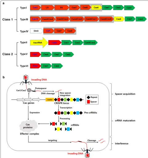
and giant viruses, as summarized in Fig.
2
a [
72
,
73
].
The CRISPR systems are adaptive immune systems that
provide sequence-specific protection against invading
viruses or conjugative plasmids [
70
,
74
–
76
]. It should
be noted that there is another type of immunity in
bac-teria called restriction–modification systems [
75
,
77
], for
which the restriction enzymes revolutionized molecular
biology resulting in the Nobel Prize in 1978. CRISPR is
also revolutionizing all aspects of biology and
biotech-nology and may be nominated for a Noble Prize [
78
].
The CRISPR immunity is divided into three stages
(Fig.
2
b): spacer acquisition (or adaptation), CRISPR RNA
(crRNA) biogenesis, and interference stages. During the
spacer acquisition stage, a target DNA sequence, known
as a protospacer, is excised and inserted at the 5
′
end of
the CRISPR array producing a new spacer. The
subse-quent crRNA biogenesis includes transcription and
pro-cessing of the CRISPR array into mature crRNAs. At the
final interference stage, crRNAs guide the effector
com-plex to the target site and cleave the DNA producing DSBs
in the re-invading viruses. There are excellent reviews on
the biology of CRISPR [
70
,
74
,
76
]. This review will focus
on the effectors including endonucleases because these
nucleases are used for genome editing [
79
].
The CRISPR systems can be classified into two classes
and six types, summarized in Fig.
2
a. Class 1 CRISPR
systems contain effectors composed of multi-subunit
proteins, while those of class 2 contain a single
effec-tor with multi-domain such as Cas9 or Cpf1. Class 1
is divided into types I, III and IV, and class 2 includes
types II, V and VI, for which the types were numbered
based on their order of discovery [
74
,
80
]. The CRISPR
systems are very diverse, and Fig.
2
depicts only a
rep-resentative composition of genes at the CRISPR loci.
It is estimated that CRISPR is present in about 50% of
bacteria and ~ 90% of archaea [
80
]. For the purpose of
technological applications, class 2 effector nucleases
are mainly used due to their convenience in cloning
and delivery into host cells. Class 2 nucleases are also
diverse in their structural and functional aspects, and
this diversity heralds a new age of genome editing that
can be customized for individual research projects [
74
].
Use of CRISPR for genome editing
Since the initial finding of the mysterious repeats of the
CRISPR array in
E. coli
in 1987 and in many other
bac-teria in 2000 [
71
,
72
,
81
], CRISPR/Cas9 has been shown
(See figure on previous page.)Table 2 Genome editing in microalgae and other organisms
Organism Delivery/nuclease Gene/marker Comments References
Animals
CHO‑S cells (11619‑012) Lipofectamine 2000 transfection
ZFN DHFR Identification of biallelic knockouts after a single transient ZFN treatment [57] SKOV3 cells
ATCC FuGENE HD transfection reagentTALEN EGFP and DsRed Development of TALENs in mammalian cells [59] Mouse cells
NIH3T3 Microinjection or Lipofectamine 2000 CRISPR/Cas9
EGFP and ApoE Improvement of bi‑allelic modification by
dual sgRNAs [178]
Human cell lines
MCF‑7, NSCLC and A549 Lipofectamine 2000 transfectionCRISPR/Cas9 Mitochondrial GTPase Mitofusin‑2/GFP Identification of role MFN2 in human cells [179] H9 human ES cells and other
cell lines ElectroporationCRISPR/Cas9 CCR5 gene Genome editing Cas9 RNPs [65] Mouse zygotes Electroporation
CRISPR/Cas9 Tyrosinase gene Genome editing Cas9 RNPs [180] Zebrafish (Danio rerio) strain
AB Microinjection into embryosCRISPR/Cas9
akt2 gene Functional role of akt2 gene in zebrafish [181]
Plants
Nicotiana tabacum (tobacco) Protoplast transformation
TALEN ALS gene/YFP gene Successful targeted gene replacement with TALENs [51] Arabidopsis thaliana Agrobacterium‑mediated trans‑
formation CRISPR/Cas9
ADH1, TT4, RTEL1
gene/bar gene Genome editing using CRISPR/Cas9‑based nucleases and nickases [101]
Arabidopsis thaliana Agrobacterium‑mediated trans‑ formation
CRISPR/Cas9
GFP gene Stable inheritance of Cas9/sgRNA‑generated mutant genes in T2 and T3 progeny [182]
Camelina sativa Agrobacterium‑mediated trans‑ formation
CRISPR/Cas9
FAD2 gene Induced mutations caused change in fatty
acid composition [183]
Maize Hi‑line Particle bombardment
CRISPR/Cas9 LIGMS45, ALS2/MOPAT, MS26 and ‑DSREB Genome editing Cas9 RNPs [184] Wheat embryos Particle bombardment
CRISPR/Cas9 TaGW2 gene Genome editing Cas9 RNPs [185]
Cyanobacteria
Synechococcus elongatus
UTEX 2973 ConjugationCRISPR/Cas9 nblA Genome editing using CRISPR/Cas9‑based nucleases [186] Synechococcus elongatus
PCC 7942 ConjugationCRISPR/Cas9
glgc/GmR gene Increase of succinate
Production [187]
Synechococcus UTEX 2973 Conjugation
CRISPR/Cpf1 psbA1, nblA Genome editing using CRISPR/Cpf1‑based nucleases [188] Synechocystis 6803
Anabaena 7120 ConjugationCRISPR/Cpf1 nblAnifH Genome editing using CRISPR/Cpf1‑based nucleases [188] Microalgae
C. strain CC‑4350 Glass beads ZFN
COP3 gene/aphVIII
gene Targeted gene knockout induced by ZFN [58] P. tricornutum CCMP2561 Bombardment
TALEN UGPase/NAT gene Increase in triacylglycerol accumulation [91] C. reinhardtii CC503 Electroporation
CRISPR/Cas9 FKB12 gene First application of CRISPR/Cas9 in micro‑algae [111] P. tricornutum CCMP2561 Bombardment
CRISPR/Cas9 CpSRP54 gene/
Shble First application of CRISPR/Cas9 in diatoms [189]
C. reinhardtii
CC‑124 ElectroporationCRISPR/Cas9 MAA7ChlM, CpSRP43 and Targeted gene knockout and knock‑in via NHEJ in Chlamydomonas [60] C. reinhardtii
CC‑4349 ElectroporationCRISPR/Cas9 ZEP and CpFTSY Production of two‑gene knockout mutant [112] C. reinhardtii
CC‑400 Glass beadsCRISPR/dCas9
to target DNA specifically in vitro resulting in DSBs [
82
,
83
]. This led to the first reports of genome editing in
eukaryotic cells in 2013 [
84
,
85
], and then, an explosive
number of reports followed [
79
] (Table
2
).
The advantages of the CRISPR system for genome
editing reside in the effector nucleases, for which the
nucleases do not require the tedious and labor-intensive
cloning of DNA binding domains for targeting
speci-ficity. In contrast to the predecessor nucleases ZFN
and TALEN, the DNA specificity of CRISPR nucleases
is provided by a guide RNA composed of crRNA and
trans-activating crRNA (tracrRNA), which were further
simplified by the sgRNA [
7
]. This simple design and
prep-aration of CRISPR enables multiplexed mutagenesis by
simply adding up sgRNAs [
86
,
87
]. In addition, CRISPR
nucleases appear to be more efficient and more precise
compared to predecessors [
88
].
Another advantage of CRISPR is the diverse nucleases
which can be customized for individual needs. Cas9 from
Streptococcus pyogenes
(SpCas9) was initially used for
CRISPR-mediated genome editing in animal cells [
82
,
83
]. Lately, nucleases including CRISPR from
Prevotella
and
Francisella
1 (Cpf1) and CRISPR from
Campylobac-ter jejuni
(CjCas9) have been introduced for genome
edit-ing with improved efficiency and specificity [
41
,
89
,
90
].
New class 2 CRISPR systems are being reported, and the
number is increasing [
74
], and different types of
nucle-ases will offer customization of the editing technique for
individual research projects.
Best of all, CRISPR appears to be most efficient in
microalgal genome editing based on the number of
papers reported so far, even though it debuted last in
the genome editing field (Table
2
). ZFN and TALEN
have been used for mutagenesis of
Chlamydomonas
and
a diatom, respectively [
58
,
91
], and to the best of our
knowledge, there have not been any follow-up reports.
Fortunately, CRISPR is gaining a strong foothold in
microalgal genome editing, which may provide the
possibility of practical and efficient genome editing in
microalgae.
However, CRISPR technology still has some
limita-tions, which requires improvements for proper use
in genome editing. CRISPR-induced mutations occur
randomly depending on the repair of DSBs mostly via
NHEJ [
92
,
93
]. Currently precision genome
engineer-ing is emergengineer-ing for better management of mutagenesis,
including gene replacement, multiple cleavage and base
correction [
46
], some of which will be further described
in “
Applications of the CRISPR system
” section.
Off-tar-geting can still be an issue for medical and agricultural
purposes, even though CRISPR is considered as the most
accurate genome editing technique [
94
]. Fortunately,
CRISPR offers a variety of nucleases with improved
ver-satility and/or fidelity, which can also provide optional
PAM sequences [
41
,
89
,
90
,
95
]. These improvements will
benefit both biology and biotechnology fields,
particu-larly for microalgal community.
Technical aspects of CRISPR‑mediated genome editing
A key to successful genome editing is efficient
deliv-ery of genetic materials, and it has been the main
bot-tleneck in transformation of microalgae. In general, the
cell wall is considered as the most significant barrier for
the introduction of macromolecules into plant cells [
16
].
To avoid this problem, the cell wall is removed, and the
resulting protoplasts are subjected to PEG-mediated
transfection, which appears to be very effective without
the need for expensive supplies and equipment [
18
,
30
,
96
]. With the proper removal of the cell wall, this
tech-nique can result in a transfection efficiency of up to 70%
in cassava mesophyll protoplasts [
97
], which may offer
an opportunity to improve microalgal transformation.
In fact, as summarized in Table
1
, there have been a few
reports on PEG-mediated transformation of microalgae
including
Chlorella
[
26
,
27
,
29
] and
Cyanidioschyzon
[
28
]. These attempts did not result in a greatly improved
Table 2 continued
Organism Delivery/nuclease Gene/marker Comments References
N. oceanica IMET1 Electroporation
CRISPR/Cas9 Nitrate reductase gene/HygR Targeted gene knockout in Nannochloropsis [113] N. gaditana CCMP1894 Electroporation
Cas9 Editor line ZnCys TFBSD Knockout and attenuation of ZnCys in nochloropsis Nan- [114]
ADH1, alcohol dehydrogenase 1; akt2, AKT serine/threonine-protein kinases 2; ALS, acetolactate synthase; aphVIII, aminoglycoside 3′-phosphotransferase; ApoE, apolipoprotein E; bar, herbicide bialaphos; BSD, blasticidin S deaminase; CCR5, C–C motif chemokine receptor 5; ChlM, Mg-protoporphyrin IX S-adenosyl methionine
O-methyl transferase; COP3, light-gated proton channel rhodopsin; CpFTSY, signal recognition particle receptor protein, chloroplast; CpSRP43, chloroplast signal recognition particle 43; CpSRP54, chloroplast signal recognition particle 54; DHFR, dihydrofolate reductase; DsRed, red fluorescent protein; EGFP, green fluorescent protein; FAD2, fatty acid desaturase 2; FKB12, peptidyl-prolyl cis–trans isomerase; glgc, glucose-1-phosphate adenylyl transferase; GmR, gentamycin-resistance gene;
transformation efficiency; however, this technique can
be improved by complete and/or efficient removal of
the cell wall, which may also improve genome editing in
microalgae.
sequence-specific DNA cleavage [
74
]. The founding
member of such nucleases is SpCas9, and its homologs
have been identified in many bacterial strains, and they
use two RNA molecules (crRNA and tracrRNA) or
one sgRNA for binding to the protospacer, the target
sequence on DNA. SpCas9 has the protospacer adjacent
motif (PAM) as an additional sequence specificity that
provides security minimizing off-target effects. PAM
for SpCas9 is mainly NGG but sometimes NAG and is
located directly 3
′
to the protospacer [
7
,
79
].
Cas9 homologs are equipped with two endonuclease
domains producing DSBs, namely the RuvC and HNH
domains. These domains are modular enabling individual
engineering for different purposes. RuvC was originally
identified as part of the
RuvABC
operon (for
“resist-ance to UV light”) and is the endonuclease involved in
the resolution of the Holliday junction during UV repair
[
98
]. The HNH domain (named for the histidine,
aspara-gine and histidine residues critical for the nuclease
activ-ity) is found in many endonucleases including restriction
enzymes and meganucleases [
99
]. Both RuvC and HNH
domains are required for producing DSBs. However,
their catalytic sites can be modified to produce a
nick-ase by making an individual mutation of either D10A or
H840A, respectively [
79
,
100
]. Such Cas9 nickases are
not efficient for inducing mutations but can be used for
enhanced knock-in of expression cassettes via HR [
101
].
In addition, both catalytic sites can be mutated to
pro-duce the dead Cas9 (dCas9), and this can be used for
variants of CRISPR/Cas9 techniques including CRISPR
interference (CRISPRi), where dCas9 is used as a
sequence-specific DNA binding protein leading to
inter-ference of transcription of the target gene [
102
–
104
].
The CRISPR system also offers different types of
nucle-ases, such as Cpf1 and C2c1, which is considered the
biggest advantage compared to other genome editing
techniques [
74
]. Cpf1 was initially identified as a type V
CRISPR effector from
Prevotella
and
Francisella
, which
shows endonucleolytic activities different from Cas9.
The differences include a T-rich PAM site and
stag-gered cleavage of DNA located 3
′
to PAM [
95
]. Cpf1
from
Acidaminococcus
sp. (AsCpf1) and
Lachnospiraceae
bacterium
(LbCpf1) have been used for genome
edit-ing in animals and plants [
66
,
89
,
95
]. These Cpf1s do
not require tracrRNA, which offers simpler preparation
of the guide RNA. In addition, it appears to be more
efficient and accurate than Cas9 [
66
,
89
] and thus has
emerged as the next generation nuclease for genome
edit-ing. Additional type V nucleases, but less studied, include
C2c1 and C2c3. These type V effectors are further
classi-fied as subtype V-B and V-C, while Cpf1 belongs to the
V-A subtype [
74
]. These subtypes are characterized by
different domain structures, which can be used for
cus-tomized genome editing purposes.
Interestingly, there is another type of CRISPR
sys-tem containing endonucleases that cleave RNA targets
instead of DNA. These belong to the type VI, and the
sequence-specific RNases include C2c2 and many
oth-ers [
74
,
105
]. The type VI CRISPR system is reminiscent
of the eukaryotic RNA silencing mechanism involving
the RNA-induced silencing complex (RISC), in which
Argonaute (AGO) and Dicer carry out guide RNA (i.e.,
siRNA)-based sequence-specific identification and
cleav-age of the target RNA, respectively [
106
–
108
]. However,
C2c2 carries both functions [
109
] revealing another
bac-terial ingenuity of simplicity, in contrast to the bulky,
complex and elaborate eukaryotic counterparts. RNAi
in eukaryotic systems is not very reliable in the
suppres-sion of gene expressuppres-sion particularly in microalgae [
110
],
and the type VI CRISPR systems may provide a better
alternative.
Different from any gnome editing techniques, the
CRISPR system enables the delivery of preassembled
Cas9 or Cpf1 RNP with the cognate guide RNAs in vitro
[
65
,
89
]. Compared to vector-driven expression of the
nucleases and guide RNAs, the RNP system is simple
and convenient without the need for the laborious and
time-consuming cloning process and thus obtains results
faster. There are other benefits of the RNP approach
including the pre-test of the nuclease activity in vitro.
There was a correlation between in vitro and in vivo
activities of SpCas9 in
Chlamydomonas
for different
tar-get sites in the same gene [
60
]. In addition, RNP delivery
can minimize off-target effects and possible toxicity from
the continuous expression of a nuclease [
60
,
65
,
111
]. It
does not introduce any artificial DNA including markers
and heterologous genes and may avoid GMO conflicts
[
67
]. Best of all, it has been successful in the recalcitrant
model algae
Chlamydomonas
[
60
,
112
]. This may provide
interesting opportunities to deal with the difficulties in
microalgal genome editing.
other microalgae including
Nannochloropsis
[
116
] and
Guillardia
[
117
]. On the other hand, haploids do not
allow the knockout of essential genes, which should be
considered before making a gene list for potential
outs. In this case, one can consider the CRISPR
knock-down approaches such as CRISPRi and attenuation of
gene expression by targeting UTRs that will be described
in more detail [
118
,
119
].
Applications of the CRISPR system
The simplicity of the CRISPR systems has led to a sudden
increase in variant technologies, which was difficult with
previous techniques of genome editing or any reverse
genetic techniques. First, the Cas9 nuclease can be
eas-ily manipulated to create nickases or dCas9, and these
variants can be used for additional genome manipulation
including knock-in and CRISPRi [
7
,
60
,
102
]. Second,
multiple sites can be targeted simultaneously by simply
adding guide RNAs, for which two sites can be targeted
to obtain a chromosomal deletion, inversion or
transloca-tion [
7
]. The numbers can be increased to target multiple
genes at the same time [
86
] or even to create a barcoded
CRISPR mutant library [
87
]. It should also be noted that
anti-CRISPRs have been identified in bacteriophages as
an arms race against their hosts [
120
,
121
], which
pro-vides an interesting possibility that they can be used in
genome editing, such as a conditional knockout. There is
a long list of applications for the CRISPR systems [
119
,
122
,
123
], and some of these that are relevant or
applica-ble to microalgae will be described in this review.
dCas9 for CRISPRi and manipulation of gene expression
Cas9 contains well defined endonuclease domains that
can be modified to create a nickase or even dCas9, and
these mutants can also be used for new exciting
tech-niques. In particular, dCas9 can bind to the target site
without cleaving DNA, and this can interfere with
cel-lular processes including transcription. This CRISPRi
technique has been shown in bacteria and even in
Chla-mydomonas
[
102
,
104
,
119
,
124
]. In bacteria, dCas9
inter-feres with the expression of target gene(s) by providing
steric hindrance to RNA polymerase or transcription
fac-tors, depending on the location of target sites [
102
,
124
].
In
Chlamydomonas
, CRISPRi is shown to knockdown the
expression of phosphoenolpyruvate carboxylase (PEPC)
[
104
], albeit less effective possibly due to the difference
between prokaryotic and eukaryotic transcription
mech-anisms, where eukaryotic transcription is more tolerant
to DNA binding proteins including chromatin.
dCas9 can also be repurposed for other functions by
fusion of domains involved in transcriptional activation
(CRISPRa), repression (CRISPRi), and epigenetic
regula-tion (Fig.
3
). It should be noted that CRISPRi is used for
simple interference without any fused proteins [
102
,
124
]
and for active interruption with repressor domains [
119
],
which may be resolved in the future. For CRISPRa, the
multiple repeats of the herpes simplex VP16 activation
domain (VP64) and the nuclear factor-κB transactivating
subunit activation domain (p65AD) have commonly been
used as activator domains in eukaryote systems [
125
–
127
]. These subunits are fused to the N or C terminus of
dCas9 as a single or multiple units. After it is shown that
having more activators improve the activation efficiency,
several units including different activator domains can be
added. For example, the ‘SunTag’ array consists of 10
cop-ies of a small peptide epitope each linked with VP64 and
sfGFP by scFV [
128
]. As another example, the synergistic
tripartite activation method (VPR) uses a tandem fusion
of three transcription activators, VP64, p65 and Rta
[
129
]. The synergistic activation mediator (SAM) is fused
to VP64, and two MS2 RNA aptamers added to the
tetra-loop and second stem-tetra-loop of the sgRNA recruit p65Ad
and heat shock factor1 (HSF1) through MCP [
130
,
131
].
Repressors that have been used in CRISPRi include
MAX-interacting protein 1 (MXI1) from yeast,
Krüppel-associated box (KRAB) domain of Kox1, the CS domain
of HP1α, the WPRW domain of Hes1, or four
concate-nated mSin3 domains (SID4X) which are fused either to
the amino or carboxyl terminus [
125
,
132
,
133
]. However,
it appears that this field has some room for
improve-ments. Epigenetic regulation is critical for proper
expres-sion of genes, which can be achieved by histone and
DNA modifications. The Lys-specific histone
demethyl-ase 1 (LSD1) fusion protein, the catalytic core of histone
acetyltransferase (p300), or DNMT3A, a DNA
methyl-transferase, has been tested with dCas9 [
134
–
136
]. These
can be used as epigenome editing tools to reveal
inter-actions between the epigenome and regulatory elements
and their epigenetic mechanism of gene expression [
137
,
138
] not only in higher eukaryotes but also in microalgae.
Knock‑ins with CRISPR
knock-in events have been reported in zebrafish and
Chlamydomonas
[
60
,
142
].
Knock-in events can be used in targeted integration of
transgenes at certain locations on the genome. Random
integration of transgenes suffers from position effects and
transgene silencing [
143
,
144
]. Such detrimental effects
are also known in microalgae including
Chlamydomonas
[
33
,
34
,
145
–
147
] and can influence the stable expression
of transgenes. These problems can be solved in part by
integrating transgenes at transcriptionally favorable
loca-tions on the genome. Actually, such “safe-harboring” has
been shown; the knock-in of transgenes at
transcription-ally active sites, e.g., near the rDNA cluster, increases the
expression of transgenes [
46
,
148
,
149
]. Currently,
func-tional genomic data including RNA-Seq and
epigenom-ics mapping of histone modifications can offer candidates
of transcriptionally active locations. These locations
can be targeted for the integration of expression
cas-settes. In microalgae, fortunately, cloning of the flanking
homologous sequences may be not necessary because the
knock-in via NHEJ can occur in
Chlamydomonas
and
Nannochloropsis
[
60
,
114
].
Perspectives of genome editing in microalgae
Microalgae and CRISPR are relatively new additions to
biotechnology fields, which are expected to contribute to
biomaterial production and genome editing techniques,
respectively. Combination of the two quintessential
com-ponents is potentially the key to solve the
environmen-tal problems associated with usage of fossil fuels. Such
example has been reported recently, where
CRISPR-induced knockout or attenuation of a regulatory gene
can increase lipid accumulation in industrial microalgae
Nannochloropsis
[
114
].
Different from previous editing techniques, CRISPR
allows systemic, albeit labor-intensive, screening of
knockout mutations due to its simplicity and convenience
[
113
,
114
]. This is reflected by the number of reports,
in which ZFN and TALEN-induced mutagenesis for
only one for each techniques since 2013 [
58
,
91
].
How-ever, successful genome editing with CRISPR alone has
been documented three times in 2016 as summarized in
Table
2
. This success heralds new and improved genome
editing field in microalgae, which attracts great interests
of academic and industrial biology and biotechnology.
not possible, in which attenuation should be considered.
For example, attenuation can be achieved by targeting
outside of coding sequence such as untranslated regions
or by CRISPRi [
114
,
119
].
Problems and possible solutions with CRISPR application
in microalgae
Microalgae are still difficult to manipulate genes
pos-sibly due to their multitude of problems. Firstly, it is
hard to deliver genetic materials into the cells, probably
because they have unique cell wall and surface structures
that reflect their complex taxonomic lineages [
151
]. This
diversity hinders development of standardized protocols
for transformation. To avoid such problems, one can
remove the cell wall and employ protoplast
transforma-tion, which has been demonstrated for a few microalgae
including
Chlorella
as summarized in Table
1
[
26
,
27
,
29
]. Protoplasts are in general easier to transform, which
may improve efficiencies of not only transformation but
also genome editing. Secondly, microalgae may have very
efficient silencing systems against introduced genetic
materials including DNA and RNA at the transcriptional
and post-transcriptional levels. Such silencing systems
have been reported for the model algal
Chlamydomonas
[
33
,
145
], and are expected to exist in other microalgae
[
34
]. Temporary knockdown of one of the silencing
com-ponents may improve transformation efficiency [
34
].
Permanent mutations of silencing components are not
recommended, because they are also involved in genome
stability [
145
].
Cas9 RNP appears to be more efficient than
vector-driven Cas9 in
Chlamydomonas
[
60
,
111
,
112
], and is
advantageous if heterogeneous genetic material should
not be introduced, particularly in areas where GMOs are
prohibited. However, high quality non-toxic recombinant
Cas9 protein is not easy to prepare or is expensive to
pur-chase from a company. However, for research purposes,
a stable line of Cas9 or equivalent nucleases can be
con-structed for efficient gene editing. For example, the Cas9
Editor line has been successfully employed to produce 18
mutations in
Nannochloropsis
[
114
].
Precision genome editing technologies require precise
mutagenesis without producing off-targeting events,
which has not been well established in microalgae. Such
precision is crucial for certain applications of CRISPR
particularly for gene therapy in human, and is well
estab-lished in animals and plants [
152
]. Fidelity of genome
editing can be improved by Cas9 RNP in animal cells
[
65
,
66
], and Shin et al. reported no off-targeting events
in
Chlamydomonas
using Cas9 RNP [
60
]. Other than the
latter, off-targeting has not been examined in microalgae,
where such efforts should improve safety and consistency
of genome editing in microalgae.
Conclusions
Genome editing is essential for obtaining mutations of
target genes enabled by recombinant nucleases with
sequence specificity. The latest nucleases found in the
CRISPR systems are far better than the predecessors in
terms of their simplicity, accuracy and efficiency. This
improved CRISPR technology can be used in the
cor-rection of mutations, replacement of genes, and
tar-geted integration of overexpression cassettes. It can also
be used for many other purposes including attenuation
of gene expression, removal of transgenic markers, etc.,
and the list is getting longer. The microalgal community
is catching up with this new and exciting technology but
is lagging behind the main stream technical
develop-ments in animals and plants. We need to first solve the
fundamental problems in microalgae, which is the
inef-ficient delivery of genetic materials into the cell. Given
such a tremendous barrier, many more papers have been
reported with CRISPR compared to the previous
tech-niques, which may herald a new age of genome editing in
microalgae.
Abbreviations
amiRNAs: artificial microRNAs; Cas9 RNPs: Cas9/gRNA ribonucleoproteins; Cpf1: CRISPR from Prevotella and Francisella 1; CRISPR/Cas9: clustered regularly interspaced palindromic sequences/CRISPR‑associated protein 9; CRISPRa: CRISPR activation; CRISPRi: CRISPR interference; crRNA: CRISPR RNA; dCas9: dead Cas9; DSBs: double strand breaks; GMO: genetically modified organism; GT: gene targeting; HDR: homology‑dependent recombination; HR: homolo‑ gous recombination; INDELs: insertions or deletions; miRNAs: microRNAs; NHEJ: non‑homologous end joining; PAM: protospacer adjacent motif; PEG: polyethylene glycol; RNAi: RNA interference; sgRNA: single guide RNA; siRNA: small interfering RNAs; SpCas9: Cas9 from Streptococcus pyogenes; TALEN: TAL effector endonuclease; tracrRNA: trans‑activating crRNA; VP64: VP16 activation domain; ZFN: zinc‑finger nuclease.
Authors’ contributions
YKC, B‑rJ, H‑MO, W‑JJ and Y‑IP conceived of and outlined this review; J‑ML, Y‑IP and W‑JJ contributed to “Introduction to genetic engineering” section and “Genome editing using engineered nucleases” section; H‑GL and H‑MO contributed to “Applications of the CRISPR system” section; SJ, S‑ES, KNK and B‑rJ contributed to “Background” section, “Genome editing using engineered
nucleases” section, “Biology and application of CRISPR” section, and “Perspec‑
tives of genome editing in microalgae”; YKC, SJ and B‑rJ organized and drafted
the whole manuscript. All authors read and approved the final manuscript.
Author details
1 Advanced Biomass Research and Development Center (ABC), 291 Daehak‑ro, Yuseong‑gu, Daejeon 34141, Republic of Korea. 2 Department of Chemi‑ cal and Biomolecular Engineering, Korea Advanced Institute of Science and Technology (KAIST), 291 Daehak‑ro, Yuseong‑gu, Daejeon 34141, Republic of Korea. 3 Plant Systems Engineering Research Center, Korea Research Insti‑ tute of Bioscience and Biotechnology (KRIBB), 125 Gwahak‑ro, Yuseong‑gu, Daejeon 34141, Republic of Korea. 4 Cell Factory Research Center, Korea Research Institute of Bioscience and Biotechnology (KRIBB), 125 Gwahak‑ro, Yuseong‑gu, Daejeon 34141, Republic of Korea. 5 LG Chem, 188 Munji‑ro, Yuseong‑gu, Daejeon 34122, Republic of Korea. 6 Department of Biological Sciences, Chungnam National University, Daejeon 34134, Republic of Korea.
Acknowledgements
Planning (ABC‑2011‑0031350, ABC‑2010‑0029728, ABC‑2011‑0031343, ABC‑ 2010‑0029723 and NRF‑2015M3A6A2065747).
Competing interests
The authors declare that they have no competing interests.
Availability of data and materials
Not applicable.
Consent for publication
Not applicable.
Ethics approval and consent to participate
None.
Publisher’s Note
Springer Nature remains neutral with regard to jurisdictional claims in pub‑ lished maps and institutional affiliations.
Received: 4 August 2017 Accepted: 4 November 2017
References
1. Hinnen A, Hicks JB, Fink GR. Transformation of yeast. Proc Natl Acad Sci USA. 1978;75(4):1929–33.
2. Doetschman T, Gregg RG, Maeda N, Hooper ML, Melton DW, Thompson S, Smithies O. Targetted correction of a mutant HPRT gene in mouse embryonic stem cells. Nature. 1987;330(6148):576–8.
3. Thomas KR, Capecchi MR. Site‑directed mutagenesis by gene targeting in mouse embryo‑derived stem cells. Cell. 1987;51(3):503–12. 4. Lee KY, Lund P, Lowe K, Dunsmuir P. Homologous recombination in
plant cells after Agrobacterium‑mediated transformation. Plant Cell. 1990;2(5):415–25.
5. Puchta H, Fauser F. Gene targeting in plants: 25 years later. Int J Dev Biol. 2013;57(6–8):629–37.
6. Tan W, Proudfoot C, Lillico SG, Whitelaw CB. Gene targeting, genome editing: from Dolly to editors. Transgenic Res. 2016;25(3):273–87. 7. Kim H, Kim JS. A guide to genome engineering with programmable
nucleases. Nat Rev Genet. 2014;15(5):321–34.
8. Guha TK, Wai A, Hausner G. Programmable genome editing tools and their regulation for efficient genome engineering. Comput Struct Biotechnol J. 2017;15:146–60.
9. Jeon S, Jeong B‑r, Chang YK. Chemicals and fuels from microalgae. In: Lee SY, editor. Consequences of microbial interactions with hydrocar‑ bons, oils, and lipids: production of fuels and chemicals., Handbook of hydrocarbon and lipid microbiologyCham: Springer International Publishing; 2017. p. 1–21.
10. Nicolson MO, McAllister RM. Infectivity of human adenovirus‑1 DNA. Virology. 1972;48(1):14–21.
11. Graham FL, van der Eb AJ. A new technique for the assay of infectivity of human adenovirus 5 DNA. Virology. 1973;52(2):456–67.
12. Tepfer D. Transformation of several species of higher plants by Agrobac-terium rhizogenes: sexual transmission of the transformed genotype and phenotype. Cell. 1984;37(3):959–67.
13. Fernández E, Schnell R, Ranum LP, Hussey SC, Silflow CD, Lefebvre PA. Isolation and characterization of the nitrate reductase struc‑ tural gene of Chlamydomonas reinhardtii. Proc Natl Acad Sci USA. 1989;86(17):6449–53.
14. Debuchy R, Purton S, Rochaix JD. The argininosuccinate lyase gene of Chlamydomonas reinhardtii: an important tool for nuclear transforma‑ tion and for correlating the genetic and molecular maps of the ARG7 locus. EMBO J. 1989;8(10):2803–9.
15. Altpeter F, Springer NM, Bartley LE, Blechl AE, Brutnell TP, Citovsky V, Conrad LJ, Gelvin SB, Jackson DP, Kausch AP, et al. Advancing crop trans‑ formation in the era of genome editing. Plant Cell. 2016;28(7):1510–20.
16. Finer JJ. Plant nuclear transformation. In: Kempken F, Jung C, editors. Genetic modification of plants: agriculture, horticulture and forestry. Berlin: Springer; 2010. p. 3–21.
17. Klein TM, Wolf ED, Wu R, Sanford JC. High‑velocity microprojectiles for delivering nucleic‑acids into living cells. Nature. 1987;327(6117):70–3. 18. Lazzeri PA, Brettschneider R, Lührs R, Lörz H. Stable transformation of barley via PEG‑induced direct DNA uptake into protoplasts. Theor Appl Genet. 1991;81(4):437–44.
19. Shimamoto K, Terada R, Izawa T, Fujimoto H. Fertile transgenic rice plants regenerated from transformed protoplasts. Nature. 1989;338(6212):274–6.
20. Boynton JE, Gillham NW, Harris EH, Hosler JP, Johnson AM, Jones AR, Randolph‑Anderson BL, Robertson D, Klein TM, Shark KB, et al. Chloro‑ plast transformation in Chlamydomonas with high velocity micropro‑ jectiles. Science. 1988;240(4858):1534–8.
21. Nelson JA, Savereide PB, Lefebvre PA. The CRY1 gene in Chlamydomonas reinhardtii: structure and use as a dominant selectable marker for nuclear transformation. Mol Cell Biol. 1994;14(6):4011–9.
22. Stevens DR, Rochaix JD, Purton S. The bacterial phleomycin resistance gene ble as a dominant selectable marker in Chlamydomonas. Mol Gen Genet. 1996;251(1):23–30.
23. Kang NK, Jeon S, Kwon S, Koh HG, Shin SE, Lee B, Choi GG, Yang JW, Jeong B‑r, Chang YK. Effects of overexpression of a bHLH transcrip‑ tion factor on biomass and lipid production in Nannochloropsis salina. Biotechnol Biofuels. 2015;8:200.
24. Vieler A, Wu G, Tsai CH, Bullard B, Cornish AJ, Harvey C, Reca IB, Thornburg C, Achawanantakun R, Buehl CJ, et al. Genome, functional gene annotation, and nuclear transformation of the heterokont oleaginous alga Nannochloropsis oceanica CCMP1779. PLoS Genet. 2012;8(11):e1003064.
25. León R, Fernández E. Nuclear transformation of eukaryotic microalgae: historical overview, achievements and problems. Adv Exp Med Biol. 2007;616:1–11.
26. Yang B, Liu J, Liu B, Sun PP, Ma XN, Jiang Y, Wei D, Chen F. Development of a stable genetic system for Chlorella vulgaris‑A promising green alga for CO2 biomitigation. Algal Res. 2015;12:134–41.
27. Hawkins RL, Nakamura M. Expression of human growth hormone by the eukaryotic alga, Chlorella. Curr Microbiol. 1999;38(6):335–41. 28. Ohnuma M, Misumi O, Fujiwara T, Watanabe S, Tanaka K, Kuroiwa T.
Transient gene suppression in a red alga, Cyanidioschyzon merolae 10D. Protoplasma. 2009;236(1–4):107–12.
29. Kim DH, Kim YT, Cho JJ, Bae JH, Hur SB, Hwang I, Choi TJ. Stable integra‑ tion and functional expression of flounder growth hormone gene in transformed microalga, Chlorella ellipsoidea. Mar Biotechnol (NY). 2002;4(1):63–73.
30. Masani MY, Noll GA, Parveez GK, Sambanthamurthi R, Prüfer D. Efficient transformation of oil palm protoplasts by PEG‑mediated transfection and DNA microinjection. PLoS ONE. 2014;9(5):e96831.
31. Karas BJ, Diner RE, Lefebvre SC, McQuaid J, Phillips AP, Noddings CM, Brunson JK, Valas RE, Deerinck TJ, Jablanovic J, et al. Designer diatom episomes delivered by bacterial conjugation. Nat Commun. 2015;6:6925.
32. Merchant SS, Prochnik SE, Vallon O, Harris EH, Karpowicz SJ, Witman GB, Terry A, Salamov A, Fritz‑Laylin LK, Marechal‑Drouard L, et al. The Chlamydomonas genome reveals the evolution of key animal and plant functions. Science. 2007;318(5848):245–50.
33. Wu‑Scharf D, Jeong B‑r, Zhang C, Cerutti H. Transgene and transposon silencing in Chlamydomonas reinhardtii by a DEAH‑box RNA helicase. Science. 2000;290(5494):1159–62.
34. Kim EJ, Ma X, Cerutti H. Gene silencing in microalgae: mechanisms and biological roles. Bioresour Technol. 2015;184:23–32.
35. Cerutti H, Johnson AM, Gillham NW, Boynton JE. Epigenetic silencing of a foreign gene in nuclear transformants of Chlamydomonas. Plant Cell. 1997;9(6):925–45.
36. Yamasaki T, Kim EJ, Cerutti H, Ohama T. Argonaute3 is a key player in miRNA‑mediated target cleavage and translational repression in Chla-mydomonas. Plant J. 2016;85(2):258–68.
38. Molnár A, Schwach F, Studholme DJ, Thuenemann EC, Baulcombe DC. miRNAs control gene expression in the single‑cell alga Chlamydomonas reinhardtii. Nature. 2007;447(7148):1126–9.
39. Nelson JA, Lefebvre PA. Targeted disruption of the NIT8 gene in Chla-mydomonas reinhardtii. Mol Cell Biol. 1995;15(10):5762–9.
40. Dawson HN, Burlingame R, Cannons AC. Stable transformation of Chlorella: rescue of nitrate reductase‑deficient mutants with the nitrate reductase gene. Curr Microbiol. 1997;35(6):356–62.
41. Zhang K, Raboanatahiry N, Zhu B, Li M. Progress in genome editing technology and its application in plants. Front Plant Sci. 2017;8:177. 42. Wijshake T, Baker DJ, van de Sluis B. Endonucleases: new tools to edit
the mouse genome. Biochim Biophys Acta. 2014;1842(10):1942–50. 43. Zaidi SS, Tashkandi M, Mansoor S, Mahfouz MM. Engineering plant
immunity: using CRISPR/Cas9 to generate virus resistance. Front Plant Sci. 2016;7:1673.
44. Seol JH, Shim EY, Lee SE. Microhomology‑mediated end joining: good, bad and ugly. Mutat Res. 2017. doi:10.1016/j.mrfmmm.2017.07.002. 45. Lieber MR. The mechanism of double‑strand DNA break repair by
the nonhomologous DNA end‑joining pathway. Annu Rev Biochem. 2010;79:181–211.
46. Salsman J, Dellaire G. Precision genome editing in the CRISPR era. Biochem Cell Biol. 2017;95(2):187–201.
47. Smith J, Grizot S, Arnould S, Duclert A, Epinat JC, Chames P, Prieto J, Redondo P, Blanco FJ, Bravo J, et al. A combinatorial approach to create artificial homing endonucleases cleaving chosen sequences. Nucleic Acids Res. 2006;34(22):e149.
48. Lambowitz AM, Zimmerly S. Group II introns: mobile ribozymes that invade DNA. Cold Spring Harb Perspect Biol. 2011;3(8):a003616. 49. Gaj T, Gersbach CA, Barbas CF 3rd. ZFN, TALEN, and CRISPR/
Cas‑based methods for genome engineering. Trends Biotechnol. 2013;31(7):397–405.
50. Bibikova M, Golic M, Golic KG, Carroll D. Targeted chromosomal cleav‑ age and mutagenesis in Drosophila using zinc‑finger nucleases. Genet‑ ics. 2002;161(3):1169–75.
51. Zhang Y, Zhang F, Li X, Baller JA, Qi Y, Starker CG, Bogdanove AJ, Voytas DF. Transcription activator‑like effector nucleases enable efficient plant genome engineering. Plant Physiol. 2013;161(1):20–7.
52. Carlson DF, Tan W, Lillico SG, Stverakova D, Proudfoot C, Christian M, Voytas DF, Long CR, Whitelaw CB, Fahrenkrug SC. Efficient TALEN‑ mediated gene knockout in livestock. Proc Natl Acad Sci USA. 2012;109(43):17382–7.
53. Sugisaki H, Kanazawa S. New restriction endonucleases from Flavo-bacterium okeanokoites (FokI) and Micrococcus luteus (MluI). Gene. 1981;16(1–3):73–8.
54. Smith J, Bibikova M, Whitby FG, Reddy AR, Chandrasegaran S, Carroll D. Requirements for double‑strand cleavage by chimeric restriction enzymes with zinc finger DNA‑recognition domains. Nucleic Acids Res. 2000;28(17):3361–9.
55. Cathomen T, Joung JK. Zinc‑finger nucleases: the next generation emerges. Mol Ther. 2008;16(7):1200–7.
56. Carroll D. Progress and prospects: zinc‑finger nucleases as gene therapy agents. Gene Ther. 2008;15(22):1463–8.
57. Santiago Y, Chan E, Liu PQ, Orlando S, Zhang L, Urnov FD, Holmes MC, Guschin D, Waite A, Miller JC, et al. Targeted gene knockout in mam‑ malian cells by using engineered zinc‑finger nucleases. Proc Natl Acad Sci USA. 2008;105(15):5809–14.
58. Sizova I, Greiner A, Awasthi M, Kateriya S, Hegemann P. Nuclear gene targeting in Chlamydomonas using engineered zinc‑finger nucleases. Plant J. 2013;73(5):873–82.
59. Feng Y, Zhang S, Huang X. A robust TALENs system for highly efficient mammalian genome editing. Sci Rep. 2014;4:3632.
60. Shin SE, Lim JM, Koh HG, Kim EK, Kang NK, Jeon S, Kwon S, Shin WS, Lee B, Hwangbo K, et al. CRISPR/Cas9‑induced knockout and knock‑in mutations in Chlamydomonas reinhardtii. Sci Rep. 2016;6:27810. 61. Bortesi L, Fischer R. The CRISPR/Cas9 system for plant genome editing
and beyond. Biotechnol Adv. 2015;33(1):41–52.
62. Chang K. These foods aren’t genetically modified but they are ‘edited’. In: The New York Times. Internet: The New York Times; 2017. p. D1. 63. Cohen J. CRISPR patent ruling leaves license holders scrambling. Sci‑
ence. 2017;355(6327):786.
64. Ledford H. Broad Institute wins bitter battle over CRISPR patents. Nature. 2017;542(7642):401.
65. Kim S, Kim D, Cho SW, Kim J, Kim JS. Highly efficient RNA‑guided genome editing in human cells via delivery of purified Cas9 ribonucleo‑ proteins. Genome Res. 2014;24(6):1012–9.
66. Kim D, Kim J, Hur JK, Been KW, Yoon SH, Kim JS. Genome‑wide analysis reveals specificities of Cpf1 endonucleases in human cells. Nat Biotech‑ nol. 2016;34(8):863–8.
67. Kim J, Kim JS. Bypassing GMO regulations with CRISPR gene editing. Nat Biotechnol. 2016;34(10):1014–5.
68. Woo JW, Kim J, Kwon SI, Corvalan C, Cho SW, Kim H, Kim SG, Kim ST, Choe S, Kim JS. DNA‑free genome editing in plants with preassembled CRISPR–Cas9 ribonucleoproteins. Nat Biotechnol. 2015;33(11):1162–4. 69. Editorial Nature. Gene editing in legal limbo in Europe. Nature.
2017;542:392.
70. Choi KR, Lee SY. CRISPR technologies for bacterial systems: cur‑ rent achievements and future directions. Biotechnol Adv. 2016;34(7):1180–209.
71. Ishino Y, Shinagawa H, Makino K, Amemura M, Nakata A. Nucleotide sequence of the iap gene, responsible for alkaline phosphatase isozyme conversion in Escherichia coli, and identification of the gene product. J Bacteriol. 1987;169(12):5429–33.
72. Mojica FJ, Díez‑Villaseñor C, Soria E, Juez G. Biological significance of a family of regularly spaced repeats in the genomes of Archaea, Bacteria and mitochondria. Mol Microbiol. 2000;36(1):244–6.
73. Levasseur A, Bekliz M, Chabriere E, Pontarotti P, La Scola B, Raoult D. MIMIVIRE is a defence system in mimivirus that confers resistance to virophage. Nature. 2016;531(7593):249–52.
74. Shmakov S, Smargon A, Scott D, Cox D, Pyzocha N, Yan W, Abudayyeh OO, Gootenberg JS, Makarova KS, Wolf YI, et al. Diversity and evolution of class 2 CRISPR–Cas systems. Nat Rev Microbiol. 2017;15(3):169–82. 75. Deveau H, Garneau JE, Moineau S. CRISPR/Cas system and its role in phage‑bacteria interactions. Annu Rev Microbiol. 2010;64:475–93. 76. Jiang F, Doudna JA. CRISPR–Cas9 structures and mechanisms. Annu Rev
Biophys. 2017 (in press).
77. Nathans D, Smith HO. Restriction endonucleases in the analysis and restructuring of DNA molecules. Annu Rev Biochem. 1975;44:273–93. 78. Cohen J. The birth of CRISPR Inc. Science. 2017;355(6326):680–4. 79. Hsu PD, Lander ES, Zhang F. Development and applications of CRISPR–
Cas9 for genome engineering. Cell. 2014;157(6):1262–78.
80. Wright AV, Nunez JK, Doudna JA. Biology and applications of CRISPR systems: harnessing nature’s toolbox for genome engineering. Cell. 2016;164(1–2):29–44.
81. Mojica FJ, Juez G, Rodriguez‑Valera F. Transcription at different salinities of Haloferax mediterranei sequences adjacent to partially modified PstI sites. Mol Microbiol. 1993;9(3):613–21.
82. Jinek M, Chylinski K, Fonfara I, Hauer M, Doudna JA, Charpentier E. A programmable dual‑RNA‑guided DNA endonuclease in adaptive bacterial immunity. Science. 2012;337(6096):816–21.
83. Gasiunas G, Barrangou R, Horvath P, Siksnys V. Cas9–crRNA ribonu‑ cleoprotein complex mediates specific DNA cleavage for adaptive immunity in bacteria. Proc Natl Acad Sci USA. 2012;109(39):E2579–86. 84. Cong L, Ran FA, Cox D, Lin S, Barretto R, Habib N, Hsu PD, Wu X, Jiang W, Marraffini LA, et al. Multiplex genome engineering using CRISPR/Cas systems. Science. 2013;339(6121):819–23.
85. Mali P, Yang L, Esvelt KM, Aach J, Guell M, DiCarlo JE, Norville JE, Church GM. RNA‑guided human genome engineering via Cas9. Science. 2013;339(6121):823–6.
86. Wang H, Yang H, Shivalila CS, Dawlaty MM, Cheng AW, Zhang F, Jaenisch R. One‑step generation of mice carrying mutations in multiple genes by CRISPR/Cas‑mediated genome engineering. Cell. 2013;153(4):910–8.
87. Wong AS, Choi GC, Cui CH, Pregernig G, Milani P, A