http://www.sciencepublishinggroup.com/j/ijovs doi: 10.11648/j.ijovs.20170204.15
Statins Effects on Diabetic Retinopathy Among Patients
with Type 2 Diabetes Mellitus
Mohamed Anwar Hammad
1, *, Dzul Azri Mohamed Noor
1, Syed Azhar Syed Sulaiman
1,
Tarek Mohamed Elsayed
21
Department of Clinical Pharmacy, School of Pharmaceutical Sciences, Universiti Sains Malaysia, Penang, Malaysia
2
Department of Pharmacy Practice, Kulliyyah of Pharmacy, International Islamic University Malaysia, Kuantan, Malaysia
Email address:
m_anwaaar@hotmail.com (M. A. Hammad), dzulazri@usm.my (D. A. M. Noor), sazhar@usm.my (S. A. S. Sulaiman), tarek@iium.edu.my (T. A. Elsayed)
*
Corresponding author
To cite this article:
Mohamed Anwar Hammad, Dzul Azri Mohamed Noor, Syed Azhar Syed Sulaiman, Tarek Mohamed Elsayed. Statins Effects on Diabetic Retinopathy Among Patients with Type 2 Diabetes Mellitus. International Journal of Ophthalmology & Visual Science.
Vol. 2, No. 4, 2017, pp. 106-114. doi: 10.11648/j.ijovs.20170204.15
Received: July 30, 2017; Accepted: August 21, 2017; Published: September 21, 2017
Abstract:
Diabetic Retinopathy (DR) is the leading cause of vision loss in adults aged 20–74 years. DR classified as the fifth most common cause of preventable blindness and fifth furthermost common cause of moderate to severe visual impairment. A retrospective cohort study was performed to evaluate the prevalence of DR, and the effect of statins on the DR, among outpatients with Type 2 diabetes mellitus. The study was done at the endocrine clinic in Hospital Pulau Pinang, Malaysia. Two cohorts of 717 diabetic outpatients (559 statins user and 158 statins non-users) were investigated for demographic data and diagnosis of DR. Findings were presented as descriptive statistics. The age of 717 subjects was (55.2±14.9) years and females 367 (51.2%). About 166 (23.2%) patients had DR with age (58.2±14.9) years. From 559 (78%) outpatients were statins-user, 143 (25.6%) had DR with age (58.5±14.9) years. While the control group 158 (22%) participants, only 23 (14.6%) had DR with age (56.3±14.9) years. The relative risk (RR) for DR in the statin-user group for DR is 1.75 and excessive relative risk (ERR) 75%. The absolute risk (AR) is 11% and number need to harm (NNH) is 9. Nearly one-quarter of the subjects had diabetic retinopathy. The risk of diabetic retinopathy incidence is higher in the statins user group than statins non-user cohort.Keywords:
Eye, Malaysia, Diabetic Retinopathy, Statins, Type 2 Diabetes Mellitus1. Background
Diabetic Retinopathy (DR) is a microvascular complication of diabetes subsequent for hyperglycemia and glucose-related hyperosmolarity, and diabetic macular edema is a prominent cause of severe vision loss in patients with DR [1 - 4]. While the fundamental elements of progression of diabetic retinopathy and diabetic macular edema are known to be concomitant with the duration of diabetes and hypertension, dyslipidemia is considered a risk factor for DR and diabetic macular edema too [5, 6, 7].
Diabetic Retinopathy (DR) is the primary reason of vision damage in individuals aged 20–74 years [8]. From 1990– 2010, DR graded as the fifth greatest corporate root of preventable loss of sight and fifth supreme communal cause of moderate to severe visual impairment [9]. In 2010, of an
assessed, 285 million persons global with diabetes, over one-third had signs of DR, and a one-one-third of these are suffering from vision-threatening diabetic retinopathy (VTDR), defined as severe nonproliferative DR or proliferative DR (PDR) or the presence of diabetic macular edema (DME) [10]. These estimates are expected to rise further due to the increasing prevalence of diabetes, aging of the population and growing of life expectancy of those with diabetes [11 - 15].
countrywide revision in Denmark reported that statin users had a lesser cumulative occurrence of diabetic neuropathy and DR before the diagnosis of diabetes [22]. On the other hand, Mansi et al. [23] described that statin use was accompanied with an augmented risk of diabetic complications in their investigation of propensity score-matched statin users and non-users. Although these individual revisions presented different outcomes, the American College of Cardiology (ACC) and the American Heart Association (AHA) lately suggested statins for all persons with diabetes who have low-density lipoprotein (LDL) cholesterol ≥ 70 mg/dL and amongst the ages of 40 and 75 years-old [24]. However, the consequence of statin use on the increase of diabetic macular edema and progression of DR in individuals with pre-existing Type 2 diabetes is unknown.
The role of statins in the development of microvascular disease in patients with diabetes is uncertain. In this project, the hypothesis that statins use effect on the risk of diabetic retinopathy was verified in individuals with Type 2 diabetes mellitus.
2. Objectives
To explore the prevalence of diabetic retinopathy and determine the association between systemic statins use and diabetic retinopathy among Malaysian outpatients with Type 2 diabetes mellitus.
3. Methods and Study Design
A retrospective cohort study was performed at the endocrine clinic in Hospital Pulau Pinang, Malaysia. Two groups of 717 diabetic outpatients (559 statins user and 158 statins non-users) were reviewed. Patients with Type 2 diabetes were checked retrospectively to determine the effect of statins usage on diabetic retinopathy. Demographic data, ophthalmologist diagnosis of DR and the clinical data were collected from the patients’ medical records and the corresponding medical team.
3.1. Inclusion Criteria
Outpatients with Type 2 diabetes mellitus, age 18 years and above were included in the study. Patients who had eye investigation to diagnose the diabetic retinopathy. Patients started statin therapy from ≥6 months.
3.2. Exclusion Criteria
Patients with cancer, HIV, pregnancy, blindness, below 18 years old, patients with Type 1 diabetes were excluded from the study, and Patients started statin therapy from <6 months.
3.3. Ethical Consideration
From the ethical standpoint, the project tracked the Clinical Research Centre (CRC) processes of the registering in Hospital Pulau Pinang. Also, the project had registry
number in the National Medical Research Register (NMRR ID: NMRR-15-1068-25700) [25]. Also, all subjects have signed an informed consent form, and all of the study steps were done under the supervision of experts. All aspects of the study proposal, containing access to and the use of clinical and demographic statistics of the subjects, was authorized by the institutional medical ethics committee and the local health authorities before the beginning of this study. Information on patients was strictly protected and used for clinical research only. The dignity and confidentiality of the subjects are protected in the future study and publication.
Figure 1. The flowchart of the study, AIDS: acquired immune deficiency syndrome.
3.4. Data Collection
The primary researcher collected data. An established, validated data collection scheme was used for collecting clinical, patients’ demographics and laboratory data. Demographic features include age, gender, height, weight, and ethnicity. The associated clinical variables include DR, co-morbidities, statins medications, drugs dosage, and their period. Data were collected from patients, patients’ medical records, and medical team. “Figure 1” provides the flowchart of the project presented in this paper.
3.5. Statistical Analysis
modeling and multinomial logistic regression test were used to monitor the influence of confounders. A CI: 95% and p-value at the level of 0.05 were considered statistically significant.
4. Results
The age of 717 subjects was (55.2±14.9) years and females 367 (51.2%). The ethnicity distribution was 268 (37.4%) Malay, 255 (35.6%) Chinese and 194 (27%) Indian. About 166 (23.2%) cases had diabetic retinopathy with mean of age (58.2±14.9) years. From 559 (78%) subjects were
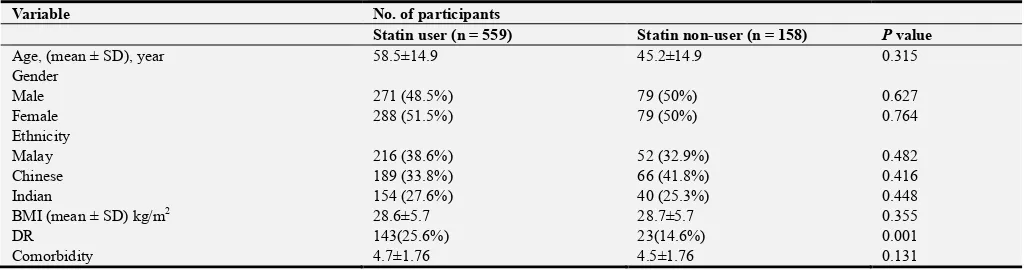
statins-user, 143 (25.6%) had DR with age (58.5±14.9) years. The control group 158 (22%) cases, only 23 (14.6%) had DR with age (56.3±14.9) years. Correlations tests showed a significant correlation between statins use and DR (p-value: 0.004) as in Table 1 and Table 2. The relative risk (RR) for DR in the statin-user cohort for DR is 1.75 and excess relative risk (ERR) 75%. The absolute risk (AR) is 11% and number need to harm (NNH) is nine as shown in Table 3. While Kruskal Wallis Test indicated a statistical significance of the relation of age, history of diabetes (diabetic period), hypertension and statin on DR as described in Table 4.
Table 1. Comparison of baseline characteristics of DR with statin user and statin non-user patients.
Variable No. of participants
Statin user (n = 559) Statin non-user (n = 158) P value
Age, (mean ± SD), year 58.5±14.9 45.2±14.9 0.315
Gender
Male 271 (48.5%) 79 (50%) 0.627
Female 288 (51.5%) 79 (50%) 0.764
Ethnicity
Malay 216 (38.6%) 52 (32.9%) 0.482
Chinese 189 (33.8%) 66 (41.8%) 0.416
Indian 154 (27.6%) 40 (25.3%) 0.448
BMI (mean ± SD) kg/m2 28.6±5.7 28.7±5.7 0.355
DR 143(25.6%) 23(14.6%) 0.001
Comorbidity 4.7±1.76 4.5±1.76 0.131
Correlation is significant at the p-value < 0.05 level (2-tailed). BMI: Body Mass Index, DR: Diabetic Retinopathy, SD: Standard Deviation.
Table 2. Correlations among DR incidence and statins.
Correlations test DR Statins
Kendall's tau_b
DR
Correlation Coefficient 1.000 0.108
Sig. (2-tailed) . 0.004
N 717 717
Statins
Correlation Coefficient 0.108** 1.000
Sig. (2-tailed) 0.004 .
N 717 717
Spearman's rho
DR
Correlation Coefficient 1.000 0.108**
Sig. (2-tailed) . 0.004
N 717 717
Statins
Correlation Coefficient 0.108 1.000
Sig. (2-tailed) 0.004 .
N 717 717
Correlation is significant at the p-value < 0.05 level (2-tailed). DR: Diabetic Retinopathy.
Table 3. Contingency table of the effect of statin on diabetic retinopathy.
DR Without DR Total Risk %
Statin user 143 416 559 0.256
Statin non-user 23 135 158 0.146
DR: Diabetic Retinopathy
Table 4. Kruskal Wallis Test.
Variables my effect on DR Chi-Square df P-value.
Age 7.569 1 0.006
Diabetic Period 52.455 1 0.000
Ethnicity 0.208 1 0.648
Gender 0.288 1 0591
HbA1c 1.73 1 0.188
Hypertension 5.715 1 0.017
Medicine adherence 0.024 1 0.878
Statin 8.403 1 0.004
Multivariable Cox proportional hazard regression modeling documented a statistically significant risk of statins, HbA1c control, hypertension, diabetic period and medications adherence for diabetic retinopathy incidence (p-value < 0.001) as illustrated in Table 5. On the other hand, glycated hemoglobin HbA1c value, gender, and ethnicity showed non-significant association with the incidence of diabetic retinopathy (p-value > 0.05).
Table 5. Multivariable Cox proportional hazard regression modeling.
Variables my
effect on DR B SE Wald df p-value
Statins -0.661 0.102 41.994 1 0.000
HbA1c 0.039 0.027 1.995 1 0.158
Gender 0.121 0.075 2.598 1 0.107
Ethnicity -0.072 0.050 2.035 1 0.154
HbA1c_Control 0.676 0.116 34.165 1 0.000
HTN -0.409 0.095 18.414 1 0.000
Diabetic_Period -0.051 0.005 97.148 1 0.000
Adherence 0.273 0.082 11.077 1 0.001
HAZ_1 0.181 0.081 5.002 1 0.025
P-value is statistically significant at the level of 0.05level (2-tailed). DR: Diabetic Retinopathy, HbA1c: Glycated hemoglobin A1c, HAZ: Hazard, HTN: Hypertension
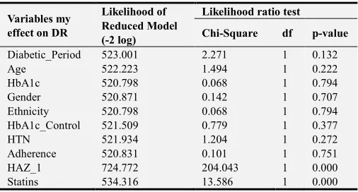
Multinomial logistic regression confirmed the statistical significance impact of statins on diabetic retinopathy incidence (p-value < 0.001) as described in Table 6.
Table 6. Multinomial logistic regression.
Variables my effect on DR
Likelihood of Reduced Model (-2 log)
Likelihood ratio test
Chi-Square df p-value
Diabetic_Period 523.001 2.271 1 0.132
Age 522.223 1.494 1 0.222
HbA1c 520.798 0.068 1 0.794
Gender 520.871 0.142 1 0.707
Ethnicity 520.798 0.068 1 0.794
HbA1c_Control 521.509 0.779 1 0.377
HTN 521.934 1.204 1 0.272
Adherence 520.831 0.101 1 0.751
HAZ_1 724.772 204.043 1 0.000
Statins 534.316 13.586 1 0.000
P-value is statistically significant at the level of 0.05level (2-tailed). DR: Diabetic Retinopathy, HbA1c: Glycated hemoglobin A1c, HAZ: Hazard, HTN: Hypertension
5. Discussion
Diabetic retinopathy (DR) is a common microvascular problem of diabetes mellitus (DM) and is measured as the principal source of visual damage in working-age adults universally. Dyslipidemia has been connected with DR, but not with development of the proliferative form of DR although the precise part in the pathogenesis of DR and diabetic macular edema (DME) rests uncertain. As a consequence, a rational query raising is whether adjustment of dyslipidemia may modify the progression of DR. Statins do not seem to affect DR development. On the other hand, fenofibrate has been established to significantly decrease the
degree of advance of DR in persons with pre-found mild DR, although it has no effort on individual’s vision nor the avoidance of DR expansion in Type 2 diabetic individuals without DR. A fascinating point that requirements further assessment is why patients without DR or those with extreme DR look to have no advantage from fenofibrate treatment [26, 27].
Statins are a category of medications prescribed for dyslipidemia and atherosclerotic illnesses. Later of their appearance, many adverse events have been associated with their use. Ocular complaints are rare but thoughtful side-effects of statins (blurred vision, hypermetropia, decreased visual acuity, visual disorders, myopia, visual field deficiency, astigmatism, and presbyopia) which might be concomitant with liver or muscle problems by investigating the frequency of ocular adverse events among the documented adverse drug reactions from the Food and Drug Administration (FDA) and Adverse Drug Reactions Advisory Committee (ADRAC) data. FDA between 1988 and 2013 and ADRAC between 1988 and 2011, among 131,755 cases of patients taking statins in the Food and Drug Administration, there were 2325 cases reported ocular adverse events after using statins (1.8%). The most highly reported ocular adverse reactions associated with statins were blurred vision (48.4%) and visual impairment (25.7%). A bigger proportion of the adverse events for cases taking atorvastatin (2.1%). Of the 1.8%, visual side effects mostly happened alone (60.9%), tracked by 30.3% where muscle adverse events also were tangled. The Adverse Drug Reactions Advisory Committee data held 136 patients of statins related ocular adverse events (47 patients stated blurred vision and 64 documented vision impairment) [28, 29].
Spontaneous documentations from the National Registry of Drug-Induced Ocular Side Effects, the Food and Drug Administration and the World Health Organization (WHO) were collected on statins and ptosis, diplopia, and ophthalmoplegia. Two hundred fifty-six case reports of ptosis, diplopia, and ophthalmoplegia concomitant with statins were reported, comprising 91 females, 143 males, and 22 case reports where the sex was not identified. The average age was 64.5+/-10 years. Average time from the beginning of therapy to the appearance of the ADR was 8.3+/-1.5 months (range, 1 day-84 months). Seven patients were taking two statin drugs, and 5 also were taking gemfibrozil. Nine patients had diabetes mellitus. A total of 23 case reports described complete ophthalmoplegia. Ptosis was reported alone 8 times and in conjunction with diplopia 18 times. According to WHO criteria, the association between statin treatment and diplopia, ptosis, or ophthalmoplegia is possible. Plausible mechanism myositis of the extraocular muscles, the levator palpebrae superioris muscles, or both [30].
Registry of Drug-Induced Ocular Side Effects, the WHO, and the FDA, are summarized with the classification of this visual side effect according to WHO criteria. After 300 days with 11 positive dechallenge reports and two positive rechallenge cases. Some patients also received medicines known to rise hemorrhage times. Ocular hemorrhage is "possibly" related to statin treatment, according to the collected data, [31].
In 2014, Mohammad Asghar et al., determine the frequency of retinopathy and its different grades among Type 2 diabetic patients with metabolic syndrome. This research was performed in the Department of Medicine, Lady Reading Hospital Peshawar from March 2011 to August 2011. Through a descriptive cross sectional study strategy, a total of 201 patients with diabetes mellitus having metabolic syndrome were selected consecutively from the outpatient department and fundoscopy was done to determine and grade DR and results were recorded. The average age of participants was 39 ± 12.2 years with 54.7% female gender. On fundoscopy, DR was established in 35 (17.4%) of patients with the utmost of them with retinopathy found in older age group, i.e., 34.6% in the age group ≥ 60 years old and 20% in the age group 50-59 years. On grading of diabetic retinopathy, 40% were in the mild to moderate nonproliferative diabetic retinopathy (NPDR) group, 37.1% in the severe nonproliferative diabetic retinopathy (NPDR) group and 22.9% were in the proliferative diabetic retinopathy (PDR) group which agrees with our findings. Diabetic retinopathy is a common sequela of diabetes in patients with metabolic syndrome with proliferative diabetic retinopathy less prevalent than nonproliferative diabetic retinopathy. It necessitates steady track up of these patients to prevent the increase of proliferative disease and its difficulties. More studies are recommended before making commendations for amendments in principles of its management [32, 33].
Jameel Nasser et al., 2014 recognize risk features for DR among outpatients with diabetes attending primary care health centers and to measure the level of control. Case control study was conducted in twenty-two health centers. The medical archives of outpatients with diabetes who were checked for retinopathy throughout the year 2011 have been investigated. The following were reported: age, diabetes duration, gender, glycated hemoglobin (A1C), blood pressure (BP), lipid profile, smoking status, absence or presence of chronic renal failure. Also, Guardian drugs [Angiotensin Converting Enzyme Inhibitors (ACEIs), Angiotensin Receptor Blockers (ARBs), Statins and Aspirin] were reported. Also, patients with diabetes who were reviewed as normal (no DR) from 4 health centers were randomly selected, and their medical records were examined to compare the risk mentioned above factors between those with and those without DR. A total of 1,508 retinal examining forms were revised, 112 patients have identified with DR. A total of 263 reviewed but had no DR were inspected in the designated four health centers. In DR, uncontrolled A1C was existed in 81 (72.3%) cases, high BP in 69 (61.6%) and
Low-Density Lipoprotein in 81 (72.3%). There was statistically significant relationship between HbA1c ≥53mmol/mol (P=0.000), augmented diabetes period (P=0.000), LDL ≥2.6mmol/l (P=0.002), total cholesterol ≥5.2mmol/l (P=0.008), and the incidence of DR. There was no significant association between age, gender, triglycerides level ≥1.7mmol/l, blood pressure and occurrence of DR. The use of, ARBs, aspirin, fibrates, and statins was significantly higher in patients with DR [34, 35, 36] which tallies with our results.
Jie Zhang and Gerald McGwin examine whether statins prevent the development of diabetic retinopathy. They conducted a nested case-control study among patients at the Birmingham Veterans Affairs Medical Center, Birmingham, Alabama. Within a study population of male patients with diabetes mellitus (n = 6441), they recognized incident cases of diabetic retinopathy identified between January 1, 1997, and December 31, 2001 (n = 114). Control cases were designated using incidence density sampling and were matched for diabetes period. Information concerning filled statin prescriptions was obtained for controls and cases. Cases and controls did not vary about overall statin use in the crude investigation (odds ratio, 1.01; 95% confidence interval, 0.64-1.59) and multivariate analyses adjusted for age, ethnicity, and comorbidities (odds ratio, 1.00; 95% confidence interval, 0.60-1.67). The outcomes of this study do not support an association between statins and diabetic retinopathy [37].
A criticism of the literature demonstrates that the influence of age on the dominance and severity of diabetic retinopathy is still vague, and differs with the people being examined. In the United Kingdom Prospective Diabetic Study [38], Stratton et al. stated that elder age (>58 years) was a risk feature for the increase of DR (RR 2.1, 95% CI 1.5 to 2.7) but not for its occurrence [39]. In 2010, Tan et al. reviewed persons with DR in Singapore and found that subjects aged 65 years and older had a more risk of preexisting diabetic retinopathy at presentation (multivariate OR 2.2, p<0.001). Other studies have also identified younger age as a risk factor for diabetic retinopathy. The Wisconsin Epidemiological Study of Diabetic Retinopathy [40] described that harshness of DR was correlated with younger age at diagnosis. The same revision stated that the 10-year incidence of retinopathy, the progression of retinopathy, and progression of proliferative retinopathy were highest in the group diagnosed before 30 years of age [41, 42]. A clinic-based cross-sectional study in Singapore reported that younger age was a risk factor for vision-threatening DR (multivariate OR 0.97, p=0.00) [43] which supported by Raman et al., study in 2011 [44]. They believed that the differences in the impact of age on diabetic retinopathy in various studies might be explained by confounders such as variations in genetic, environmental or lifestyle factors, and the type of patients screened (population-based vs. clinic-based).
(T2DM) who were enrolled in a large US managed-care network. In this study of adolescents aged ≤21 years with newly diagnosed T1DM or T2DM who were under ophthalmic surveillance. They described the occurrence and timing of diabetic retinopathy beginning. Kaplan–Meier survival curves evaluated the onset of diabetic retinopathy diagnosis for patients. Multivariable Cox proportional hazard regression modeling recognized factors accompanied with the risk of emerging diabetic retinopathy. Model predictors were age, ethnicity and onset diabetes mellitus diagnosis, gender, net worth, and glycated hemoglobin (HbA1c) fraction (HbA1c). Hazard ratios (HRs) with confidence intervals (CIs) 95% for rising diabetic retinopathy. Among the 2240 youths with Type 1 diabetes mellitus (T1DM) and 1768 youths with Type 2 diabetes mellitus (T2DM), 20.1%, and 7.2% developed diabetic retinopathy over a median follow-up time of 3.2 and 3.1 years, correspondingly. Survival curves demonstrated that youths with T1DM developed diabetic retinopathy earlier than adolescences with T2DM (P < 0.0001). For every single 1-point growth in HbA1c, the hazard for DR rose 20 % (HR: 1.20; 95% CI: 1.06–1.35) and 30% (HR: 1.30; 95% CI: 1.08–1.56) amongst early stages of life with Type 2 or Type 1 diabetes mellitus, in that order. Current guidelines recommend that ophthalmic investigation begins 3 to 5 years after initial diabetes mellitus diagnosis, at which point in their study, >18% of youths with T1DM had already received ≥1 DR diagnosis. Adolescences with Type 1 or Type 2 diabetes mellitus display a significant risk for diabetic retinopathy and should undertake regular examination by eye-care professionals to safeguard timely diabetic retinopathy diagnosis and limit progression to vision-threatening disease [45].
In 2017, Chung et al., investigated the association between statin use and hypertriglyceridemia with diabetic macular edema in patients with Type 2 diabetes and diabetic retinopathy. Chung et al., found that diabetic retinopathy developed in 23% of statin users and 18% of non-users (p = 0.506), but diabetic macular edema was observed in 23% of statin users and 48% of non-users (p = 0.008). Statins decreased low-density lipoprotein cholesterol levels in patients with and without diabetic macular edema (p = 0.043 and p = 0.031, respectively). Hypertriglyceridemia at 6 months before the progress of macular edema was significantly accompanying with central retinal thickness (OR: 1.52; 95% CI: 1.14-2.02, p: 0.005) [46].
Statistical tests as Kendall's tau_b, Spearman's and Kruskal Wallis Test showed a significant correlation between statins use and DR. While Kruskal Wallis Test also indicated a statistical significance of the relation of age, period of diabetes, hypertension on DR. Multivariable Cox proportional hazard regression modelling reported a significant statistical risk of statins, HbA1c control, hypertension, diabetic time and medications adherence for diabetic retinopathy occurrence. Multinomial logistic regression test was done to rule out the covariates which confirmed the statistically significant effect of statins on the probability of diabetic retinopathy incidence with statins
therapy.
Prevalence of DR in this study (23.2%) agrees with the finding of systemic review by (Williams et al., 2004). This systemic review covered 359 studies from 50 countries which published in 100 various journals. Williams and his colleagues reported the prevalence of DR (6.7% – 30.2%) among the patients with Type 2 diabetes [47]. Another review by (Davis et al., 2017) documented the prevalence of DR which was 20% among 145 234 patients [48].
On the other hand, through 215,725 person-years of follow-up, 2866 patients established diabetic retinopathy, 1406 developed DR, 1248 had diabetic nephropathy (DN), and 2392 suffer from gangrene of the foot. Statin users settled an inferior cumulative occurrence of DR (hazard ratio (HR): 0·60, 95% CI: 0·54-0·66; p <0·0001), and the foot gangrene (HR:0·88, CI: 0·80-0·97; p: 0·010), and DN (0·66, 0·57-0·75; p<0·0001), but not DN (HR: 0·97, CI: 0·85-1·10, p=0·62), Compared with non-statin users. These consequences were similar after amending for the competing risk of death, after matching for a propensity score, after amending for visits to the doctor of family, and by covariates stratification. The corresponding multivariable adjusted hazard ratio for risk of diabetes mellitus in the total population was 1·17 (95% CI 1·14-1·21; p<0·0001) [49]. Systemically administered pravastatin efficiently treats diabetic retinopathy in the rats without central nervous system adverse effects. The pravastatin efflux transport mechanism from the blood brain barrier has already been illuminated [50]. Furthermore, randomized clinical trials are required to confirm these findings in the human being.
6. Conclusion
Study Limitation
This study has been done in a single hospital, where the time was narrowed, and the research was constrained by data availability in Hospital Pulau Pinang.
Funding
Universiti Sains Malaysia (USM) Fellowship.
Conflicts of Interest
All the authors do not have any possible conflicts of interest.
References
[1] F. Bandello, R. Lattanzio, I. Zucchiatti and C. Del Turco, “Pathophysiology and treatment of diabetic retinopathy,” Acta. Diabetol., vol. 50, issue 1, 2013. doi: 10.1007/s00592-012-0449-3.
[2] L. Wu, P. Fernandez-Loaiza, J. Sauma, E. Hernandez-Bogantes and M. Masis, “Classification of diabetic retinopathy and diabetic macular edema,” World J. Diabetes, vol. 4, pp. 290–294, 2013. doi: 10.4239/wjd.v4.i6.290.
[3] R. Madonna, G. Giovannelli, P. Confalone, F. V. Renna, Y. J. Geng and R. De Caterina, “High glucose-induced hyperosmolarity contributes to COX-2 expression and angiogenesis: implications for diabetic retinopathy,” Cardiovasc. Diabetol., vol. 15, issue 18, 2016. doi: 10.1186/s12933-016-0342-4.
[4] M. A. Hammad, A. A. Khamis, K. M. Al- Akhali, T. M. Elsayed, A. M. Alasmri, E. M. Al-Ahmari, et al. “Evaluation of Drug Dosing in Renal Failure,” IOSR-JPBS, vol. 11, issue 5, suppl, 3, pp. 39-50, 2016. DOI: 10.9790/3008-1105033950.
[5] R. Benarous, M. B. Sasongko, S. Qureshi, E. Fenwick, M. Dirani, T. Y. Wong and E. L. Lamoureux, “Differential association of serum lipids with diabetic retinopathy and diabetic macular edema,” Invest. Ophthalmol. Vis. Sci., vol. 52, pp. 7464–7469, 2011. doi: 10.1167/iovs.11-7598.
[6] R. Crosby-Nwaobi, I. Chatziralli, T. Sergentanis, T. Dew, A. Forbes and S. Sivaprasad, “Cross talk between lipid metabolism and inflammatory markers in patients with diabetic retinopathy,” J. Diabetes Res., vol. 2015, issue 191382, 2015. doi: 10.1155/2015/191382.
[7] Y. R. Chung, S. W. Park, S. Y. Choi, S. W. Kim, K. Y. Moon, J. H. Kim and K. Lee, “Association of statin use and hypertriglyceridemia with diabetic macular edema in patients with Type 2 diabetes and diabetic retinopathy,” Cardiovasc. Diabetol., vol. 16, issue 4, 2017. http://doi.org/10.1186/s12933-016-0486-2.
[8] N. Cheung, P. Mitchell and T. Y. Wong, “Diabetic retinopathy,” Lancet. Vol. 376, issue 9735, pp. 124-36, 2010. doi: 10.1016/S0140-6736(09)62124-3.
[9] R. R. Bourne, G. A. Stevens, R. A. White, J. L. Smith, S. R. Flaxman, H. Price, et al., “Causes of vision loss worldwide, 1990–2010: a systematic analysis,” Lancet Glob Health, vol. 1, issue 6, pp. e339–49, 2013. doi:
10.1016/S2214-109X(13)70113-X.
[10] J. W. Yau, S. L. Rogers, R. Kawasaki, E. L. Lamoureux, J. W. Kowalski, T. Bek, et al., “Global prevalence and major risk factors of diabetic retinopathy,” Diabetes Care, vol. 35, issue 3, pp. 556–64, 2012. doi: 10.2337/dc11-1909.
[11] R. Lee, T. Y. Wong, and C. Sabanayagam, “Epidemiology of diabetic retinopathy, diabetic macular edema and related vision loss,” Eye and Vision, vol. 2, issue 17, 2015. doi:10.1186/s40662-015-0026-2.
[12] P. Chous, “New strategies to assess the risk of diabetes-related vision loss,” Diabetes. Optometry Times, vol. 9, issue 6, pp. 20-27, Jun 2017.
[13] M. A. Hammad, D. A. Mohamed Noor, S. A. Sulaiman, M. A. Al-Mansoub and M. Qamar, “A Retrospective Study on the Age of Onset for Type 2 Diabetes Diagnosis,” World Academy of Science, Engineering and Technology, International Science Index, Pharmacological and Pharmaceutical Sciences, vol. 4, issue 8, p. 982, 2017. DOI: 10.13140/RG.2.2.24224.69124.
https://www.accp.com/docs/meetings/abstracts/2013_virtual.p df.
[14] M. A. Hammad, B. Tangiisuran, N. Abd El Aziz and Y. Hassan, “A prospective study of uncontrolled glycaemia secondary to drug-drug interactions in Type 2 diabetes mellitus patients at Penang general hospital in Malaysia” Pharmacotherapy, vol. 33, issue 5, pp. E50-E50, 2013. DOI: 10.13140/RG.2.1.2049.1281,
https://www.accp.com/docs/meetings/abstracts/2013_virtual.p df.
[15] M. A. Hammad, D. A. Mohamed Noor, S. A. Syed Sulaiman, N. A. Aziz and Y. Elsobky, “A prospective study of
prevalence of uncontrolled glycaemia in Type 2 diabetes mellitus outpatients” Pharmacotherapy, vol. 36, pp. e83–e138, 2016. DOI: 10.13140/RG.2.1.4848.0247/1.
https://www.accp.com/docs/meetings/abstracts/2016_virtual.p df.
[16] J. G. Robinson, “Statins and diabetes risk: how real is it and what are the mechanisms?” Curr. Opin. Lipidol., vol. 26, pp. 228–235, 2015. doi: 10.1097/MOL.0000000000000172.
[17] S. Simsek, C. G. Schalkwijk and B. H. Wolffenbuttel, “Effects of rosuvastatin and atorvastatin on glycaemic control in Type 2 diabetes—the CORALL study,” Diabet. Med., Vol. 29, pp. 628–631, 2012. doi: 10.1111/j.1464-5491.2011.03553.x.
[18] R. V. Shah and A. B. Goldfine, “Statins and risk of new-onset diabetes mellitus, ”Circulation., Vol. 126, pp. e282–e284, 2012. doi: 10.1161/CIRCULATIONAHA.112.122135.
[19] H. Cederberg, A. Stancakova, N. Yaluri, S. Modi, J. Kuusisto and M. Laakso, “Increased risk of diabetes with statin treatment is associated with impaired insulin sensitivity and insulin secretion: a 6-year follow-up study of the METSIM cohort” Diabetologia, vol. 58, pp. 1109–1117, 2015. doi: 10.1007/s00125-015-3528-5.
[21] M. A. Hammad, D. A. Mohamed Noor, S. A. Syed Sulaiman, A. Kharshid, N. A. Aziz and T. M. Elsayed, “A Prospective Study on the Evaluation of Statins Usage on HbA1c Control among Type 2 Diabetes Mellitus in an Outpatients Setting” World Academy of Science, Engineering and Technology, International Science Index, Pharmacological and Pharmaceutical Sciences, vol. 4, issue 8, p. 978, 2017. DOI: 10.13140/RG.2.2.27914.36806
[22] S. F. Nielsen and B. G. Nordestgaard, “Statin use before diabetes diagnosis and risk of microvascular disease: a nationwide nested matched study” Lancet Diabetes Endocrinol., vol. 2, pp. 894–900, 2014. doi: 10.1016/S2213-8587(14)70173-1.
file:///C:/Users/USER/Downloads/Comparison%20of%20Stati
ns%20Dose%20Intensity%20on%20HbA1c%20-%20Paper%20(1).pdf
[23] Mansi, C. R. Frei, C. P. Wang and E. M. Mortensen, “Statins and new-onset diabetes mellitus and diabetic complications: a retrospective cohort study of US healthy adults,” J. Gen. Intern. Med., vol. 30, pp. 1599–1610, 2015. doi: 10.1007/s11606-015-3335-1.
[24] N. J. Stone, J. G. Robinson, A. H. Lichtenstein, C. N. Bairey Merz, C. B. Blum, R. H. Eckel, A. C. Goldberg, D. Gordon, D. Levy, D. M. Lloyd-Jones, et al., “2013 ACC/AHA guideline on the treatment of blood cholesterol to reduce atherosclerotic cardiovascular risk in adults: a report of the American College of Cardiology/American Heart Association Task Force on Practice Guidelines,” Circulation, vol. 129, pp. S1–S45, 2014. doi: 10.1161/01.cir.0000437738.63853.7a.
[25] National Medical Research Register, National Institute of Health NIH guideline. (updated 11-Jun-2017) Available at: https://www.nmrr.gov.my/fwbLoginPage.jsp?fwbPageId=NM RR_Home (Accessed: 7- Jun -2017).
[26] I. P. Chatziralli, “The Role of Dyslipidemia Control in the Progression of Diabetic Retinopathy in Patients with Type 2 Diabetes Mellitus,” Diabetes Ther., vol. 8, issue 2, pp. 209-212, 2017.
[27] E. Ioannidou, V. S. Tseriotis and K. Tziomalos, “Role of lipid-lowering agents in the management of diabetic retinopathy,” World J. Diabetes, vol. 8, issue 1, pp. 1-6, 2017.
[28] V. Mizranita and E. H. Pratisto, “Statin-associated ocular disorders: the FDA and ADRAC data,” Int J Clin Pharm., vol. 37, issue 5, pp. 844-50, 2015.
[29] A. P. Khawaja, M. P. Chan, D. C. Broadway, D. F. Garway-Heath, R. Luben, J. L. Yip, S. Hayat, et al., “Systemic medication and intraocular pressure in a British population: the EPIC-Norfolk Eye Study,” Ophthalmology, vol 121, issue 8, pp. 1501-7, Aug 2014.
[30] F. W. Fraunfelder and A. B. Richards, “Diplopia, blepharoptosis, and ophthalmoplegia and 3- hydroxy-3-methyl-glutaryl-CoA reductase inhibitor use,” Ophthalmology, vol. 115, issue 12, pp. 2282-2285, 2008.
[31] F. W. Fraunfelder, “Ocular hemorrhage possibly the result of HMG-CoA reductase inhibitors,” J. Ocul. Pharmacol. Ther., vol. 20, issue 2, pp. 179-82, 2004.
[32] M. Asghar, M. Rehman, M. Z. Khan, M. Abdur Rehman and M. Z. Tahir, “Frequency of Retinopathy and its Different Grades among Type II Diabetic Patients with Metabolic Syndrome in our population,” Pak. J. Ophthalmol., vol. 30, issue 4, pp. 219-223, 2014.
[33] C. K. Kramer, T. C. Rodrigues, L. H. Canani, J. L. Gross and M. J. Azevedo, “Diabetic Retinopathy Predicts All-Cause Mortality and Cardiovascular Events in Both Type 1 and 2 Diabetes: Meta-analysis of observational studies. Diabetes Care,” vol. 34, issue 5, pp. 1238–44, 2011. doi:10.2337/dc11-0079
[34] J. Nasser, F. Habib, B. Al Tajer, M. Al Tajer, F. Juma and M. Almohri, “Risk Factors for Diabetic Retinopathy in Patients Attending Primary Care Settings. Bahrain Medical Bulletin,” vol. 36, issue 1, pp. 1-9, 2014.
[35] E. Y. Chew, W. T. Ambrosius and M. D. Davis, “Effects of Medical Therapies on Retinopathy Progression in Type2 Diabetes,” N Engl. J. Med., vol. 363, issue 3, pp. 233-44, 2010.
[36] Q. Mohamed, M. C. Gillies and T. Y. Wong, “Management of Diabetic Retinopathy. A Systematic Review,” JAMA, vol, 298, issue 8, PP. 902-16, 2007.
[37] J. Zhang and G. McGwin, “Association of Statin Use with the Risk of Developing Diabetic Retinopathy” Arch. Ophthalmol., vol. 125, issue 8, PP. 1096-1099, 2007.
[38] M. Stratton, E. M. Kohner, S. J. Aldington, R. C. Turner, R. R. Holman, S. E. Manley, et al. “UKPDS 50: risk factors for incidence and progression of retinopathy in Type II diabetes over 6 years from diagnosis,” Diabetologia, vol. 44, pp. 156– 63, 2001.
[39] C. S. H. Tan, E. M. Q. Gay and W. K. Ngo, “Is age a risk factor for diabetic retinopathy?” Br J Ophthalmol., vol. 94, issue 9, p. 1268, 2010.
[40] R. Klein, B. E. Klein, S. E. Moss, M. D. Davis and D. L. DeMets, “The Wisconsin Epidemiologic Study of diabetic retinopathy III. Prevalence and risk of diabetic retinopathy when age at diagnosis is 30 or more years,” Arch. Ophthalmol., vol. 102, issue 4, pp. 527–32, 1984. doi:10.1001/archopht.1984.01040030405011.
[41] R. Klein, E. K. Klein, S. E. Moss and K. J. Cruickshanks, “The Wisconsin Epidemiologic Study of diabetic retinopathy XIV. Ten-year incidence and progression of diabetic retinopathy,” Arch. Ophthalmol., vol. 112, issue 9, pp. 1217– 28, 1994. doi:10.1001/archopht.1994.01090210105023.
[42] K. Hietala, V. Harjutsalo, C. Forsblom, P. Summanen and P-H. Groop on behalf of the FinnDiane Study Group, “Age at Onset and the Risk of Proliferative Retinopathy in Type 1 Diabetes” Diabetes Care, vol. 33, issue 6, pp. 1315-1319, 2010. doi:10.2337/dc09-2278.
[43] L. L. Lim, S. Y. Lee, C. L. Cheng, D. W. Wong, S. G. Ong, C. L. Ang, et al., “Diabetic retinopathy in diabetics referred to a tertiary centre from a nationwide screening programme,” Ann. Acad. Med. Singapore., vol. 37, pp. 753-9, 2008. PMID: 18989491.
[44] R. Raman, K. Vaitheeswaran, K. Vinita and T. Sharma, “Is Prevalence of Retinopathy Related to the Age of Onset of Diabetes? Sankara Nethralaya Diabetic Retinopathy Epidemiology and Molecular Genetic Report No. 5” Ophthalmic. Res., vol. 45, pp. 36–41, 2011. DOI: 10.1159/000314720.
[46] Y. R. Chung, S. W. Park, S. Y. Choi, S. W. Kim, K. Y. Moon, J. H. Kim and K. Lee, “Association of statin use and hypertriglyceridemia with diabetic macular edema in patients with type 2 diabetes and diabetic retinopathy,” Cardiovasc. Diabetol., vol. 16, issue 1, suppl. 4, 2017.
[47] R. Williams, M. Airey, H. Baxter, J. Forrester, T. Kennedy-Martin and A. Girach, “Epidemiology of diabetic retinopathy and macular oedema: a systematic review” Eye, vol. 18, pp. 963–983, 2004. Doi:10.1038/sj.eye.6701476.
[48] A. Davis, A Baldwin, M. Hingorani, A. Dwyer and D. Flanagan, “A review of 145 234 ophthalmic patient episodes
lost to follow-up,” Eye, vol. 31, pp. 422–429, 2017. Doi:10.1038/eye.2016.225.
[49] S. F. Nielsen and B. G. Nordestgaard. Statin use before diabetes diagnosis and risk of microvascular disease: a nationwide nested matched study. Lancet Diabetes Endocrinol., vol. 2, issue 11, pp. 894-900, 2014.