Introduction
Migraine with aura [1] (classic migraine [2]) is probably the first recognised headache. The prominent position of the visual phenomena has also probably led to the term: “ophthalmic migraine” [3].
Scintillating scotoma and obscuration (“dimness”/ foggy vision) are the frequently occurring visual phenom-ena in migraine [e.g., 4–6]. The typical sequence of events has been observed to be: visual phenomena; then a symp-tom-free period; and finally, the pain phase of the attack [1]. Others have observed that the sequence of events more than just occasionally may be reversed: the pain
Migraine with aura: visual disturbances
and interrelationship with the pain phase.
Vågå study of headache epidemiology
O. Sjaastad
Department of Neurology, St. Olavs Hospital,
Trondheim University Hospitals (NTNU), Trondheim, Norway
O. Sjaastad
Vågå Communal Health Centre, Vågåmo, Norway
L.S. Bakketeig
Nasjonalt folkehelseinstitutt, EPLE, Nydalen, Oslo, Norway H.C. Petersen
University of Southern Denmark, Institute of Public Health, Epidemiology, Odense, Fyn, Denmark
O. Sjaastad () Gautes gate 12,
N-7030 Trondheim, Norway e-mail: tora.rui@ntnu.no Tel.: +47-73-525276 Fax: +47-73-551539
Ottar Sjaastad Leiv S. Bakketeig Hans C. Petersen
Abstract In the Vågå study of headache epidemiology, 1838 or 88.6% of the available 18–65-year-old inhabitants of the commune were included. Everyone was ques-tioned and examined personally by the principal investigator (OS). There were 178 cases of various types of visual disturbances during the migraine attack, which corre-sponds to 9.7% of the study group. The prevalence among females was 11.9% and among males 7.4%; female/male ratio was 1.70, as against 1.05 in the total Vågå study population. By far the most fre-quently occurring visual distur-bance pattern was (A) 1. Visual dis-turbances →2. pain-free interlude
→3. pain phase (in 78% of the cases). Other frequent patterns were: (B). Visual disturbances, but no pain phase (24%); and: (C) 1. Pain phase →2. visual disturbances (23%). Evidently, in the solitary
case, there might be more than one visual disturbance pattern. The most frequently occurring solitary visual disturbances were: scintillat-ing scotoma (62%) and obscuration (33%); but also more rare ones were identified, like anopsia, autokinesis (movement of station-ary objects), tunnel vision and micropsia. Among the non-visual aura disturbances, paraesthesias and speech disturbances were the most frequent ones. The prevalence of migraine with aura seemed to be considerably higher than in similar studies. This also includes studies that have been carried out with a face-to-face interview technique.
Key wordsMigraine with aura • Migraine •Scintillating scotoma • Obscuration •Anopsia
Received: 20 February 2006
phase comes first and then the visual phenomena [7–9]. Generally, however, it is asserted that although this sequence has been observed, the visual-phenomena-first type is the more frequent pattern [1, 3, 7].
There is, however, at least one prominent exception to these overall concepts. Klee [4], in his studies concerning ‘severe migraine’, maintains that the visual disturbances appear at the pain maximum in the majority of cases. Klee studied 150 migraineurs: 50 extremely carefully (“prima-ry series”) and 100 in a somewhat more curso(“prima-ry fashion (“secondary series”). “At least one type of visual symp-tom” during attacks was found in 76% and 47% of the cases, respectively (Table 6, IV [4]). It is remarkable that in 62% and 31% in the “primary” and “secondary” series, respectively, visual disturbances occurred during the pain phase. Also, Selby and Lance [10] frequently found pain-phase visual phenomena, i.e., in 22 of 59 cases (37%).
Klee’s study concerned “severe migraine”. Detailed information is available concerning the aura phase in migraine, in general. However, little is known about the visual disturbance pattern at the grass-roots level. Is the pain maximum/visual disturbance combination less fre-quent than in “severe migraine”? Klee also described macropsia and micropsia in his “severe” cases, as well as movements of stationary objects in the visual fields (so-called autokinesis) [11]. How frequent would such phe-nomena be in an unselected, grass-roots population?
These items, as well as the prevalence of migraine with aura per seat the grass-roots level, are the main topics of this communication.
Material and methods
During a two-year period, 1995–1997, every 18–65-year-old inhabitant in the Vågå commune in the mountainous area of southern Norway was invited to participate in this study of headache prevalence. The study comprised 1838 parishioners (F: 942; M: 896); this represents 88.6% of the available individuals [12]. Each parishioner was subjected to a face-to-face interview and a physical/neurological examination, totalling 60–75 min. In problem cases, a new appointment would be allotted. The struc-tured interview was based upon a questionnaire, filled in by the principal investigator, OS. The study was approved by the regional ethics committee and State Data Inspectorate, and all participants signed a participation approval.
The categorisation of the visual phenomena as being migrainous was done at the examination, and – accordingly – it was based upon the IHS criteria in current use at the time [1].
Which specified visual phenomena did the patients experi-ence? A propagating “crescent” of the homonymous type (scin-tillating scotoma); obscuration only; anopsia (dimness of vision, without “positive” visual phenomena); “unformed flashes of light”/star-shaped figures (or photopsias); changing patterns.
Rare visual phenomena were also a topic, such as: macropsia; micropsia; “tunnel vision”; deficiency as regards distance esti-mation; autokinesis and metamorphopsia. Not all of these ques-tions could invariably be satisfactorily answered in this setting.
The sequence of the events was detailed: which came first, the visual phenomena (and similar phenomena) or the pain? How frequent were visual phenomena without any accompanying pain? The duration of the pain-free interval after the visual phe-nomena – if any – was roughly estimated. The other aura symp-toms were likewise searched for: localised sensibility distur-bances, motor weakness and speech disturbances.
We have adopted Klee’s [4] definition of obscuration: during obscuration, the affected individual can just barely see – and identify – some visual field structures. With anopsia, no struc-tures whatsoever can be seen.
Headache intensity was graded according to a 0–6+ system, described elsewhere [13]: 1+=“minimal unpleasantness”; 2+=“heaviness”/“discomfort”; 3+=“mild”; 4+=“moderate”; 5+=“intense” (“severe”) headache and 6+=“excruciatingly severe headache”. Stages 3–5+ correspond to the IHS stages [1]. Pain per se was not a main item in the present communication.
The figures given herein represent lifetime prevalence. The estimates given reflect what the migraineurs felt was the usual
pattern(s). Cases of what we considered as definite migraine with aura were included.
So-called jabs were also searched for, and “CF” or “features indicative of cervical abnormality” was calculated [14].
The exact percentage of absence/presence of visual distur-bance during attacks, intraindividually, was not considered an appropriate item in this retrospective study.
Results
Prevalence of the visual disturbances
There were 178 parishioners with visual disturbances, most of them combined with a migrainous pain. Based on the actual figures, the prevalence was 9.7% (178 out of 1838) (Table 1). There were 112 females and 66 males, the gender ratio being 1.70 as against 1.05 in the total Vågå study material. The prevalence among females was 11.9% (112 out of 942) and among males 7.4% (66 out of 896). The latter figures differ statistically (p=0.0012; Fisher’s exact test). The mean age at examination, both sexes, was 43.2 years. Those with scintillating scotoma only constituted 6.1% of the series.
The panorama of major visual phenomena is set forth in Table 1. If the rare and partly bizarre visual distur-bances are added (Table 2), there would be a total of 233 visual manifestations. Thus, several parishioners had more than one type of visual disturbance.
parishioner as stemming from the one eye. Few had tried to close the one eye and then the other. Such migraineurs were generally not sent off for self-observation, as much feed-back could not be expected. The combination of the slowly moving crescent and one visual field location almost cer-tainly indicated a homonymous phenomenon. The error induced by adopting this point of view should be minimal.
Scintillating scotoma, obscuration and photopsias
Scintillating scotoma and obscuration were by far the two most frequently occurring phenomena (Table 1). In sever-al cases, the visusever-al disturbance seemed to sever-alternate between scintillating scotomas and obscurations, from one attack to another. Photopsias were present in 30 cases (Table 1). Deleting photopsias entirely would not alter the situation much, because in every case except eight, pho-topsias coexisted with scintillating scotomas and/or obscuration, at least occasionally. In two out of the eight individuals that lacked other visual symptoms, there were sensory phenomena during the initial phase of attacks.
Moreover, in those with solitary photopsia, it appeared prior to the pain phase and not at other times.
Anopsia
Anopsia had been noted by four parishioners (Table 1). Three of them had at some time experienced scintillating scotoma or obscurations, lasting ≥20 min. In only one of the four cases anopsia was the only visual disturbance, the anopsia lasting 10–15 min; there was no subsequent pain. In this case, the link to migraine as such is probably rela-tively weak. This case could accordingly have been reject-ed as a migraine+aura. Anopsia nevertheless coexistreject-ed with dysphasia in the latter case.
The anopsia episode appeared at the peak of pain in one of them; generally, there seemed to have been one solitary episode (with one exception). There may be a transition, back and forth, between obscuration and anop-sia during the solitary attack. Whether one or the other of these two manifestations is expressed may be a question of degree and not necessarily of nature.
Table 1 The most commonly occurring visual disturbances
Type of visual disturbance N Per centa F M F/M ratio
Scintillating scotoma 112 62 70 42 1.64
Obscuration 58 33 33 25 1.32
Photopsia 30 16 19 11 1.73
Anopsia 4 2 2 2 1
Total 204
aPer cent of the total no. of migraineurs with visual disturbances (n=178)
In 178 migraineurs, a total of 204 visual disturbance patterns were likely to have existed. Other, rare visual disturbances are mentioned in Table 2
Table 2 Rare and partly bizarre visual disturbances in migraine with aura
Type of visual disturbance N Male Female Ratio F/M
Autokinesisa 8 5 3 0.6
Depth vision failure 5 4 1 0.25
Tunnel vision 4 2 2 1.–
“Half” persons/half visual field 4 0 4 –
Micropsia 4 2 2 1.–
Macropsia 1 0 1 –
Metamorphopsiab 2 2 0 –
Diplopia 1 1 0 –
Total 29
aApparent movement of stationary objects
bObjects were perceived as having distorted or blurred contours [4]
Types of visual disturbances (total number): 204 (Table 1)+29=233
The combination obscuration-anopsia is the reason why anopsia is included in Table 1.
Rare visual disturbances
A number of rare, visual disturbances were present, among them well-known ones, such as micropsia, macrop-sia and tunnel vision (Table 2). Most of the rare visual phenomena appeared prior to pain, but in one of the cases of tunnel vision and one of the micropsia cases, respec-tively, these phenomena generally appeared during the pain phase (see later). In all cases, except two cases of micropsia, there were other, concurrent visual distur-bances. Autokinesis and failing depth vision were the two “most frequently occurring ones of the rare phenomena” (Table 2). For the sake of completeness, a case of diplop-ia is also mentioned. This 52-year-old male was fairly cer-tain that the diplopia was additional to the obscuration, and there were multiple episodes within 30 min. At other times, he had fairly typical migraine without aura attacks.
Sensory changes, dysphasia, motor disturbances and other non-visual phenomena
As such phenomena were not infrequently temporally connected with the visual disturbances, they are also detailed. A minor delay between the various components could, of course, not be timed exactly [15].
Paraesthesias – maybe occasionally also with slight sensory deficits – were the most frequent, non-visual phe-nomena (Table 3). A face/upper extremity location seemed
to be the most frequent one (Table 4). The sum of the soli-tary sensory phenomena exceeded the number of cases by far; thus, in the solitary case, there might be more than one location of the phenomena (Table 4).
The speech disturbance encountered most frequently seemed to be difficulty with finding words and wrong usage of words: usage of words related to, but different from, the desired one, phonetically and/or semantically, i.e., “paraphasias” [16–18]. There may possibly have been dysarthria in one case. Of course, we may have erred in the distinction between dysphasia and dysarthria, but probably not to a large extent.
The rarely noted motor complaints (n=2) generally seemed to appear more or less simultaneously with the visual complaints, typical scintillating scotomas, sensory symptoms and dysphasia. Motor phenomena could appar-ently also appear repetitively during a given attack (n=1). The motor complaints were mild – face and arm (but no hemiplegia) – and outlasted the visual complaints clearly in the one case, by 30–90 minutes. The last-mentioned, long-lasting episodes occurred only during pregnancy (see Table 3). For this reason, it was not rubricated under hemi-plegic migraine. Nor were there any familial cases.
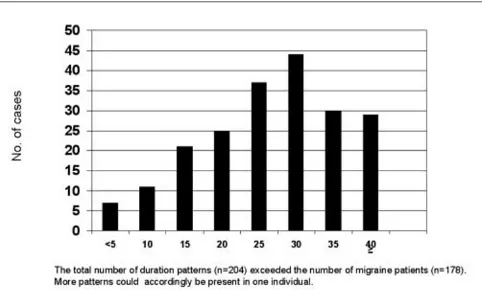
Duration, visual phenomena
The duration of the visual phenomena varied considerably, i.e., from <5 min up to one hour or more. Most frequently, the visual phenomena seemed to last from >15 to ≤35 min (67% of the cases; Fig. 1). In a few cases, the scintillating scotoma could last <5 min. An interesting pattern is no. II (Fig. 2), where multiple, about 2-min-long visual phenom-ena – “mini-teichopsias” finally might spark a seemingly regular, but particularly severe, migraine attack. When summed up, the total duration of the visual disturbance in these patients might equal the duration of a “regular” tei-Table 3 Sensory, speech, motor disturbances and other
distur-bances
Group of symptoms N
Paraesthesias 34
Dysphasia 17
Dysphasia during pregnancy only 1a
Dysarthria 1 (?)
Motor disturbances 2
Feeling of unreality/delusion 1
Memory defect 1b
In addition, there was one case of near-fainting during the pain phase There may also have been another – even less certain – case of dysarthria
aThis may have been a “vascular event” during pregnancy, in a
migraineur; bDefect memory for events taking place during the
chopsia. In contrast, visual phenomena could last 60–120 min, or longer. In most of the long-lasting ones, there were scintillating scotoma (n=5) or obscuration (n=3). In one of the latter ones, there might also be “half-figures”/“half per-sons” and paraesthesias (one half of the tongue).
Visual disturbances without a pain phase
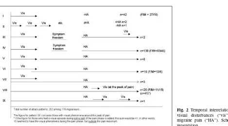
This subgroup (Fig. 2, pattern I) is the second largest group of all patterns. The various types of visual distur-bances encountered in this subgroup are set forth in Table 5. Apparently, in many of the cases, the picture had not been uniform through the years. In childhood/adoles-cence, there might have been the more traditional picture, with subsequent headache (which conceivably might have
been of a relatively low degree). With age, pain tended to disappear. No exact information exists as regards the fre-quency of this development.
Scrutiny also showed that among those who initially contended that they had no pain, a minor fraction admit-ted that they occasionally could have “minimal unpleas-antness” (stage I, scale 0–6+, see Methods). They even could have “heaviness/discomfort”, i.e., stage II, the first stage of head pain, and with procrastination. Usually, however, there was “no pain”. It has been a notion that this pattern is most common among men. In the present series, however, there was a female/male ratio of 1.8. If the visu-al phase only consists of a hard-to-evvisu-aluate phenomenon, e.g., micropsia, with no subsequent headache phase at that, then the link to migraine may be at stake.
Visual disturbances at the peak of pain
Visual disturbance at the peak of the pain phase (Table 6) was the third largest group (n=26, i.e., 15%; pattern VIII, Fig. 2). If all those are included that had visual distur-bances during the pain phase, but not necessarily at the peak of the pain curve, the number of cases would be 41, i.e., 23% (Fig. 2). The most frequently encountered visual disturbances were scintillating scotoma and obscuration (Table 6). The duration of visual disturbances varied largely – from “a few minutes” to 0.5–1 h. In three soli-tary cases, the disturbances could last “for hours”, “to the end of the attack”, “the whole day” (all obscurations).
Fig. 2 Temporal interrelationship: visual disturbances (“vis”) and migraine pain (“HA”). Schematic presentation
Table 4 Localisation of sensory changes during attack
Paraesthesias in No.
Face 17
Arm 14
Finger 10
Lip 8
Tongue (1/2) 6
Total 55
The array of visual disturbances, both when the pain phase was lacking (Table 5) and when the visual distur-bances appeared during the pain phase as such (Table 6), bore considerable resemblance to the panorama of visu-al disturbances during regular migraine with aura attacks (Table 1).
Interrelationship between duration of visual disturbance phase and the subsequent pain-free interval
As already detailed, there was considerable variation as regards duration of visual phenomena, i.e., from ≤5 min to >1 h (Fig. 1). The symptom-free interval prior to the pain phase also seemed to vary considerably in length. Could the aura and pain-free interval mutually influence each other as regards duration?
Long-lasting visual disturbance has in this context been defined as a disturbance, lasting ≥20 min. “Short interlude” has been defined as no pause or even overlap between visual disturbances and pain. There might be a tendency to an inverse correlation between the duration of the visual phase and pain-free interval (short visual aura phase being linked to a long-lasting pain-free interval and
vice versa). It should, nevertheless, be emphasised that although there seemed to be a tendency, there were excep-tions to this coarse rule. Also, the interlude may be less important in the patients’ estimation than the impressive visual disturbances and the hard-to-cope-with pain phase. The duration of the interlude may have been more super-ficially treated by the patients than the two other phases. Moreover, the fact that the attacks, in part, were far back in time, adds further uncertainty. The viewpoint forward-ed by the patients on whether or not there really was a pain-free interlude between the visual and pain phases comes into quite another category of likelihood.
Temporal interrelationship between visual phenomena and pain
Theoretically, there are multiple alternative ways in which the relationship between visual disturbances and pain can “be arranged” (Fig. 2). In real life, the most frequent pat-tern, by far, was the “classic” one: visual phenomena → pain-free interval → headache (pattern IV), which was observed in 138 of 178 cases, i.e., in 78%. Other frequent patterns were: visual phenomena and then subsequently no pain (pattern I) and visual phenomena during the pain phase (patterns VIII and IX). In 26 cases, the visual episode appeared at the pain maximum. If those with visu-al phenomena at some time during the pain phase are added, then the number would be 41, i.e., 23% (pattern VIII), or actually 42, if pattern IX is also added. It is strik-ing that for the “big groups”, I and IV, there was a mean F/M ratio of 2.4 while, for the third big group, no VIII, i.e., visual disturbances at the peak of the pain curve, the F/M ratio was 0.73.
In most of the patterns, the visual phenomena appeared first (no. of patterns=6); in three patterns, the pain appeared as early as or earlier than the visual disturbances (Fig. 2).
Validation
The migraine with aura part of the questionnaire was val-idated with two different tests [12]. For both, sufficient time had been allowed to elapse to hinder any memory from the first examination on the part of the principal investigator (OS). Recheck of 100 consecutive records (investigation A), was carried out in a blinded fashion (blinded as to name, sex, family history and diagnoses). Five participants were diagnosed as migraine+aura on examination I and on examination II, rendering a high kappa value.
Table 5 Types of visual disturbance in those with no ensuing headachea
Type of visual disturbance N
Scintillating scotoma 30
Obscuration 12
Photopsia 3
Anopsia 1
Micropsia 1
Total 47b
aPattern I, Figure 2
bThere were 42 individuals (24%) in this subgroup
Table 6 Types of visual disturbances at the peak of paina
Type of visual disturbance N
Scintillating scotoma 14
Obscuration 10
Photopsia 5
Anopsia 1
Micropsia 1
Tunnel vision 1
Total 32b
aPattern VIII, Figure 2
In the second test (investigation B), 41 parishioners were rechecked in an absolutely blinded way with regard to migraine with aura, on average 14.8 months after exam-ination I. Five participants were diagnosed as migraine+aura cases on examination I. All these five were re-diagnosed as migraine with aura on examination II. However, we were astonished to learn that two other citi-zens also proved to have had visual disturbances at the time of investigation I – and before that. They did, how-ever, not respond positively to our inquiry about visual disturbances on examination I, but did so on investigation II. The questions asked were the same on both occasions. Two out of seven patients accordingly failed to notify us: 29%. The kappa value would accordingly be much lower than for the observation on records (A). The second investigation pinpoints the weaknesses of investigation A. As only the records were checked with method A, there may also among these have been individuals who “forgot” to tell about their visual abnormalities. Only what is already in the record can be checked by method A.
One more individual forgot to tell about his visual dis-turbances during examination A. In this case, we were told about it right after examination A. This made such an impression that there would actually be no test of pattern recognition on investigation B. This patient was accord-ingly excluded in this context.
Other findings
Jabs were present in 44.9% of the cases. The mean CF value (features indicative of cervical abnormality) was 0.91+; mean Vågå series 0.79+ on a scale 0–5.0+, see Methods). One 18-year-old male noticed red and hot ears bilaterally during attacks (spontaneous information).
Discussion
Prevalence of visual disturbances in migraine
In the present study, there was a high prevalence of migraine with aura. The prevalence of visual disturbances in migraine is, to some extent, influenced by two indepen-dent factors:
1. The eagerness/experience of the investigator in uncov-ering such symptoms.
2. The assessment whether some visual phenomena real-ly are migrainous in nature.
As regards (1), in the validation section of the present study, failure to detect visual disturbances has been
described (n=2, out of 41 cases). This certainly can derive from some type of negligence on the part of the investigator. As regards (2), are, for instance, micropsias as such indicative?/pathognomonic? of migraine? Even dark spots in the visual field [4, 6] (“mouches volantes” or myodes-opsia [19]) have in some contexts been considered signif-icant in migraine. Dark spots have been totally disregard-ed in the present context, even though some migraineurs claim that they only occurred before the attack pain.
Heyck [3] puts it this way: “However, sometimes metamorphopsia and primitive visual hallucinations are considered to be migraine aura”. His statement is open to interpretations. Photopsias belong to another category. The IHS [1, 20] does not use this term, but uses: “star shaped figures”. Both Lance [8] and Klee [4] use photop-sia (“flashes of light”/”stars”) as a term for migrainous visual disturbances. In the present work, photopsia has been accepted as being migrainous in nature (see Results). Alvarez [6] (“light spots”) and Peatfield and Rose [21] (“flashes as “circles””) have produced evidence that such phenomena appear in anoptic migraineurs. The IHS [1] also mentions “scotoma without positive phenomena”, in other words pure obscurations. Also, Hachinski et al. [5] found that among 100 migraine children with visual dis-turbances, 77% had: “transient blindness, blurring of vision, and varied scotomas.”
In the present context, inclusion or not of e.g., photop-sias as the sole visual manifestations would not influence the prevalence figures appreciably, due to the small number of cases with photopsia as the solitary visual phenomenon. In spite of the reservations already made, the figures obtained herein should be regarded as being minimum fig-ures. Three individuals in the control series (B) did not inform us about their visual symptoms. These are hardly exceptional cases; such cases would probably be present in the rest of the series as well.
Lifetime prevalence of visual disturbances in migraine would seem to be around 9.7%. This is a figure consider-ably higher than in two comparable studies, based on a face-to-face interview technique, i.e., 1.6% [22] and 6% [23], respectively. One of these values is 3.8 times higher than the other, although both were based on IHS criteria, in itself an astonishing fact. In the literature, there is a highly varying prevalence. Such studies have mostly been done by questionnaire technique or telephone interviews, and not by personal contact. They have also partly been performed prior to the advent of the IHS migraine criteria. Moreover, the Vågå population is largely untreated/not regularly treated for the migraine, and this not only con-cerns specific medication, but also – to a certain extent – non-specific painkillers.
with Rasmussen’s results from Copenhagen [24], which showed a female/male ratio of 1.67.
Migraine aura has been specifically studied in a metic-ulous way in several investigations in recent years [e.g., 25, 26]. Many of the facets studied in the present context have been illuminated in such studies. Space does not allow a detailed treatise of all the observations and a com-parison with the present observations. There is a striking difference regarding the frequency of migraine with aura, respectively 4.1% [26] and 9.7% (present study). The vast differences in design may, at least partly, explain these differences as regards observations. For further detail, the reader is referred to these communications [25, 26].
Groups of interrelationship between visual disturbances and headache
Visual disturbances during the pain phase were the third most frequent pattern (Fig. 2). These results concur with those of Klee [4] and Selby and Lance [10]. But the fre-quency was rather less than that reported by Klee [4] (see Introduction). There may be various, alternative explana-tions for this: Klee’s study [4] was more profound and meticulous in this respect than the present one. Moreover, Klee selected the most “severe” cases [4]. Our series – and other series – were not selected in that way. In other words, he had no cases of aura+/pain–. A more correct approach would, therefore, probably be to compare his cases with our cases of pain+/aura+, i.e., 178 – 43=135 cases. Then, a figure for visual disturbances during pain phase would be 30%, a result closer to that of Klee.
Mean intensity of attacks, as assessed in the present series, according to our grading system, 0–6+ [13], was
approximately 4.0+, in other words a “moderate” intensity. This figure also comprised those with visual disturbances, but without pain (a total of 24%), so that the average pain in those with a pain phase would be appreciably higher. Klee [4] used a particular terminology to describe the visu-al phenomena, thus “corona phenomenon”, “flickering”, “unclear vision” and metamorphopsia (distorted contours). These terms may largely correspond to scintillating sco-toma, but he does not use the latter term [4]. Differing nomenclature might influence the visual disturbances sub-grouping, but not necessarily the grouping of visual phe-nomena into the “prior to headache”/“during headache” groups. We have no adequate explanation for the difference between the Klee data and our own, as regards the fre-quency of visual disturbances during the pain phase.
Jabs in migraine with aura
Raskin and Schwartz [27] found a high prevalence of jabs in migraine: 42%. Interestingly, a similar prevalence of jabs was found among migraine plus aura patients in Vågå: 45%. The prevalence of jabs in the entire series was 35.2% [12]. Thus, the prevalence of jabs in migraine plus aura may seem to be somewhat higher than in controls, also in Vågå.
Acknowledgements We are indebted to GlaxoSmithKline of Norway, Pharmacia & Upjohn, and the Alf Harborg Foundation, Department of Neurology, St. Olavs Hospital, Trondheim, Trondheim University Hospitals, for generous support during the various phases of the investigation. The authors are also grateful to the personnel at the Vågå Health Centre at Vågåmo for their aid. Last, but not least, we thank the inhabitants of the Vågå commune for their collaboration.
References
1. Headache Classification Committee of the International Headache Society (1988) Classification and diagnostic criteria for headache disorders, cranial neuralgia and facial pain. Cephalalgia 8[Suppl 7]:1–96
2. – (1962) The Ad Hoc Committee on Classification of Headache. Arch Neurol 6:173–176
3. Heyck H (1981) Headache and facial pain. Georg Thieme Verlag, Stuttgart, pp 10–20
4. Klee A (1968) A clinical study of migraine with particular reference to the most severe cases. Thesis, Munksgaard, Copenhagen
5. Hachinski V, Porchawka J, Steele JC (1973) Visual symptoms in the migraine syndrome. Neurology 23:570–579
6. Alvarez WC (1960) The migrainous scotoma as studied in 618 persons. Am J Ophthalmol 49:489–504
7. Selby G (1983) Migraine and its vari-ants. Adis Health Science Press, Sydney
8. Lance JW (1993) Mechanism and man-agement of headache, 5th edn.
Butterworth-Heinemann, Oxford 9. Queiros L, Rapoport A, Weeks R,
Sheftell F, Siegel S, Baskin S (1997) Characteristics of migraine visual aura. Headache 37:137–141
10. Selby G, Lance JW (1960)
11. Klee A, Willanger R (1966) Disturbances of visual perception in migraine. Acta Neurol Scand 42:400–415
12. Sjaastad O, Pettersen H, Bakketeig LS (2001) The Vågå study; epidemiology of headache. I. The prevalence of ultra-short paroxysms. Cephalalgia
21:207–215
13. Sjaastad O, Fredriksen TA, Petersen HC, Bakketeig L (2002) Grading of headache intensity. A proposal. J Headache Pain 3:117–127
14. Sjaastad O, Fredriksen TA, Petersen H, Bakketeig L (2003) Features indicative of cervical abnormality. A factor to be reckoned with in clinical headache work and research? Funct Neurol 18:195–203
15. Lord GDA (1986) Clinical characteris-tics of the migrainous aura. In: Amery WK, Wauquier A (eds) The prelude to the migraine attack. Baillière Tindall, London, pp 87–98
16. Monrad-Krohn GH (1954) The clinical examination of the nervous system, 10th Edn. HK Lewis & Co, London 17. Mayo Clinic and Mayo Foundation
(1963) Clinical examinations in neurol-ogy. WB Saunders, Philadelphia 18. Scherwin I, Geschwind N (1978)
Normal substrates of behavior. In: Nicholi AM Jr (ed). The Harvard guide to modern psychiatry. The Belknap Press of Harvard University Press, Cambridge, MA, pp 59–80 19. Malling B (1953) Sykdommer I
glasslegemet [Diseases of the corpus vitreum]. In: Rosengren B (ed.) Nordisk Lärebok i oftalmiatrik. Bonniers, Stockholm, pp 229–233 20. Headache Classification
Sub-Committee of the International Headache Society (2004) The International Classification of Headache Disorders, 2nd edn. Cephalalgia 24[Suppl 1]:1–160
21. Peatfield RC, Rose FC (1981) Migrainous visual symptoms in a woman without eyes. Arch Neurol 38:466
22. Pereira Monteiro JM (1995) Cefaleias. Estudo epidemiologico e clinico de uma população urbana. Thesis, Porto 23. Rasmussen BK (1994) Epidemiology
of headache. Thesis. Köbenhavns Universitet, Copenhagen
24. Rasmussen BK (1995) Epidemiology of headache. Cephalalgia 15:45–68 25. Russell MB, Iversen HK, Olesen J
(1994) Improved description of the migraine aura by a diagnostic aura diary. Cephalalgia 14:107–117 26. Russell MB, Olesen J (1996) A
noso-graphic analysis of the migraine aura in a general population. Brain
119:355–361