R E S E A R C H
Open Access
Muscle function and fatigability of trunk
flexors in males and females
Rita E. Deering, Jonathon W. Senefeld, Tatyana Pashibin, Donald A. Neumann and Sandra K. Hunter
*Abstract
Background:Optimal function of the abdominal muscles is necessary for several life functions including lifting and carrying tasks. Sex differences in strength and fatigability are established for many limb muscles and back extensor muscles, but it is unknown if sex differences exist for the abdominal muscles despite their functional importance. Methods:Eighteen females (24.3 ± 4.8 years) and 15 males (24.1 ± 6.6 years) performed (1) isometric trunk flexion maximal voluntary contractions (MVCs) in a range of trunk positions to establish a torque-angle curve and (2) submaximal (50% MVC), intermittent isometric contraction (6 s on, 4 s off) until task failure to determine fatigability of the trunk flexor muscles. Dual X-ray absorptiometry quantified body fat and lean mass. Physical activity levels were quantified with a questionnaire. Torque-angle curves, electromyography (EMG), MVC torque, and torque steadiness were compared with repeated measures ANOVA with sex as a between-subjects factor.
Results:For the torque-angle curve, MVC torque was reduced as the trunk angle increased toward flexion (p< 0. 001). Males had greater MVC torque than females at the extended positions (31% difference), with no sex differences in torque in upright sitting (p >0.05). Time-to-task failure for the submaximal fatigability task in upright sitting was similar between males and females (12.4 ± 7 vs 10.5 ± 6 min). Time-to-task failure was positively associated with strength (r= 0.473,p= 0.005) and self-reported physical activity (r= 0.456,p= 0.030). Lean mass in the trunk was positively associated with trunk flexor strength (r= 0.378,p= 0.011) and self-reported physical activity (r= 0.486, p= 0. 007). Finally, torque steadiness [coefficient of variation of torque (CV)] during submaximal isometric contractions decreased with contraction intensity and was similar for males and females across all intensities.
Conclusions:Unlike many limb muscle groups, males and females had similar fatigability and torque steadiness of the trunk flexor muscles during isometric contractions. Stronger individuals, however, exhibited less fatigability. Lower self-reported physical activity was associated with greater fatigability of trunk flexor muscles. The relationship between strength and fatigability of the trunk flexor muscles and physical activity supports the importance of abdominal muscle strengthening to offset fatigability in both males and females.
Keywords:Trunk flexors, Sex differences, Fatigability, Strength, Abdominal muscles
Background
Optimal function of the abdominal muscles is important for functional mobility, including lifting and carrying tasks [1]. While the abdominal muscles are the prime movers of trunk flexion [2], this muscle group performs multiple other key functions. For example, the abdominal muscles, along with the diaphragm and pelvic floor muscles, regu-late intra-abdominal pressure (IAP) [3]. Through this regulation of IAP, the abdominal muscles also provide
postural support and stability of the lumbar spine, while allowing transfer of loads from the extremities to the trunk (and vice versa) [1, 3–5]. The abdominal muscles also play a role in breathing and continence [1, 6–8], through synergistic action with the diaphragm and pelvic floor muscles. The abdominal muscles are also isometric-ally active during movements of the upper and lower ex-tremities [4, 5, 9]. Due to the need for abdominal muscle activation during nearly all functional tasks, these muscles are often active isometrically and at submaximal levels during sustained contractions such as during a carrying task, or repetitive contractions, as during lifting tasks.
* Correspondence:Sandra.Hunter@marquette.edu
Department of Physical Therapy, Marquette University, PO Box 1881, Milwaukee, WI 53201, USA
Given the importance of optimal abdominal muscle function and the lack of knowledge on the function of this muscle group, a more thorough understanding of the strength, fatigability, and force control of these mus-cles is required. Sex differences in strength and fatigabil-ity have been identified in the upper and lower limb muscles, with females typically demonstrating lower strength but decreased fatigability compared with males [10, 11]. Smidt et al. [12] showed decreased fatigability of the trunk flexor and extensor muscles in females dur-ing a maximal, reciprocal, dynamic fatigudur-ing protocol; however, this study did not assess the contribution of the hip flexor muscles to the trunk flexion task. Simi-larly, females were less fatigable in the back extensor muscles compared with males for a sustained submaxi-mal isometric contraction at 50% maxisubmaxi-mal voluntary iso-metric contraction (MVC) [13]. Intermittent isoiso-metric contractions of the abdominal muscles may be more functionally relevant, because postural stabilization is often achieved with isometric contractions, and many activities of daily living are repetitive in nature. The use of intermittent contractions also removes the confound-ing factor of the reduced blood flow experienced durconfound-ing sustained contractions. It is not known if there are sex differences in strength or fatigability for the trunk flexor muscles for a submaximal intermittent, isometric task.
An important aspect of force control that can affect functional performance is the steadiness of a contrac-tion, which can be measured as the magnitude of force (or torque) fluctuations [14, 15]. During isometric con-tractions of limb muscles, the fluctuations in force are quantified as the standard deviation (SD) about a target force of a sustained contraction or as the coefficient of variation (CV) of torque when normalized to mean force produced [15]. In limb muscles, force fluctuations are primarily explained by oscillations in common drive to the motor neuron pool and the motor unit discharge rate variability [15–17] resulting in larger force fluctua-tions at higher contraction intensities when calculated as the SD of force. The force fluctuations expressed as the CV, however, are reduced at the higher intensities of contraction, creating an inverse relationship between force fluctuations and contraction force [18–20]. Whether this relationship between steadiness and con-traction intensity is also present during trunk flexion contractions is unknown, particularly given that activa-tion of the motor neuron pool occurs from multiple spinal levels during trunk flexion [2]. In addition to the input of common drive onto the motor neuron pool, other factors may influence the shape of this relationship for the abdominals. Additionally, in limb muscles, force fluctuations will increase throughout a fatiguing contrac-tion [21, 22], and it is also unknown if this is true during fatiguing exercise of the trunk flexor muscles in males
and females. Generally, at low intensities, females exhibit greater force fluctuations than males. This has been ob-served in the elbow flexors, first dorsal interosseous (fin-ger abduction) and knee extensor muscles [23–25], but it is not known if sex differences in force steadiness exist for the trunk flexor muscles. A better understanding of the force control of the abdominal muscles would be beneficial because the abdominal muscles play a major role in regulation of intra-abdominal pressure and the stiffness of the spine [26], so it is possible that large fluc-tuations in abdominal muscle force could cause fluctua-tions in IAP, thus impacting spinal stiffness.
This study determined if there were sex differences in isometric trunk flexion for MVC torque across a range of trunk flexion angles, fatigability, and torque steadiness in young, healthy males and females. We hypothesized that males would generate greater peak isometric trunk flexion torque because males typically have a greater muscle mass than females, especially in the upper body [27]. Due to the critical role of the abdominal muscles in postural support and as accessory muscles of ventilation [2], we also hypothesized that, although females would be less fatigable than males, the sex differences would be small compared to those observed in limb muscles (e.g., elbow flexors) [11, 21, 28].
Methods
to provide frequency, quantity, and intensity of activities over the previous 12-month period. METs were also able to be estimated for occupational physical activity based on occupational history and questions. Thus, the weekly metabolic equivalents (MET h week−1) was calculated from the occupational and leisure physical activity. La-boratory measurements of isometric trunk flexion MVC torque, submaximal torque steadiness, and fatigability were made using a Biodex System 4 dynamometer (Biodex, Shirley, New York), as described below.
Trunk flexion torque
The participants were seated in a back flexion-extension attachment for a Biodex dynamometer (Fig. 1a) such that the right anterior superior iliac spine was aligned with the axis of rotation of the dynamometer. A scapular roll (15-cm diameter) was positioned at the level of the scapular spine, and the head rest of the device was adjusted to participant comfort. The pelvis was stabilized with a sacral pad posteriorly and two tightly fastened straps
anteriorly. A strap was also used to restrain the thighs. Ver-tical straps were placed on the anterior aspect of each shoulder to restrain the upper body, and these straps were joined at the midline of the chest with a buckle. The partic-ipants were instructed to flex their trunk, as though per-forming an abdominal curl up, without allowing their legs to lift off of the seat. All trunk flexion attempts were visu-ally assessed by the investigator (a physical therapist), and feedback was provided to the participants if compensatory movement patterns were observed.
Trunk flexion torque was recorded online using a Power 1401 A-D converter and Spike2 software [Cambridge Electronics Design (CED), Cambridge, UK]. Torque sig-nals were digitized at 500 Hz and displayed on a 48-cm monitor placed ~150 cm in front of the participant.
Electromyography
Electromyography (EMG) signals were obtained for the right rectus abdominis, left external oblique, and right rectus femoris using two 8-mm silver chloride surface
a
b
recording electrodes (Coulbourn Instruments, Whitehall, PA) arranged in a bipolar configuration according to rec-ommended placements [30]. EMG signals were ampli-fied (1000×) and band-pass (13–1000 Hz) and Notch (60 Hz) filtered with Coulbourn modules (Coulbourn In-struments, Allentown, PA). Signals were recorded online using a Power 1401 A-D converter (CED) and were digi-tized at 2000 Hz.
Experimental protocol
All participants were instructed to refrain from caffeine for at least 2 h and alcohol, pain medication, and anti-inflammatory medications for at least 12 h prior to ex-perimental sessions.
Session one Body composition
A DXA scan was performed to estimate fat mass, lean muscle mass, and bone mineral density. The participants were asked to remove all metal before the scan. The partic-ipants were positioned on the scanner bed in a supine pos-ition with forearms in neutral pospos-ition. Legs were bound just superior to the knees and the ankles with straps to pre-vent external rotation of the hips during the scan. The par-ticipants were asked to lie as still as possible and to not talk, unless there was a problem, during the scan.
Muscle thickness
Ultrasonography was used to determine thickness of the right rectus abdominis muscle. The participants were positioned in a supine position on a plinth with their shirt removed. Muscle thickness measurements of the right rectus abdominis [31] were taken at 2.5 cm above and below the umbilicus. The full medial-lateral width of the rectus abdominis was scanned at each of these po-sitions and the measurement was taken, while the par-ticipant held their breath at end expiration, in the region that visually appeared to be the thickest.
Torque-angle curve
In order to assess MVC torque at varying muscle lengths and establish a torque-angle curve for the trunk flexor muscles, the participants were placed in six different positions within the back flexion/extension attachment (Biodex). A calibrated digital angle gauge (Wixey WR300 Digital Angle Gauge, Barry Wixey Develop-ment, Sanibel, FL) was used inferior to the sternal notch to ensure that each participant was at the same position. Upright sitting was identified as zero degrees. MVC isometric torque was evaluated at 20° of flexion, upright sitting, and 10°, 20°, 30°, and 40° of extension, in a randomized order.
Maximal voluntary contractions
The participants performed at least three isometric trunk flexion MVCs for ~3 s at each position. MVCs were separated by at least 1 min of rest, in order to limit fatigability. MVCs were performed, with verbal encour-agement, until the participant was able to perform two contractions where torques were within 5% of each other. The higher of these two contractions was used as the MVC. For each MVC, the participant was asked to flex the trunk forward, as though curling the shoulders down toward the hips without engaging the lower extrem-ities. The participants were closely examined while per-forming trunk flexion MVCs in order to identify evidence of movements involving other muscle groups (e.g., legs elevating slightly off of chair due to activation of hip flexor muscles), and trials were only included in analysis if cor-rect form was performed by the participant. On average, four MVCs were performed at each trunk angle, with a range of 3 to 6 MVCs for most participants. One partici-pant did require seven trials at one position but was able to successfully perform the correct trunk flexion maneu-ver in 3–5 attempts for the remaining trunk angles. The participants were also cued to not hold their breath.
Although flexion of the upper trunk is primarily per-formed by the rectus abdominis muscles [2], EMG of the rectus femoris muscle in the lower limb was mea-sured to provide quantifiable evidence that lower ex-tremity muscles, particularly those that contribute to hip flexion, were not being excessively utilized during the trunk flexion contractions. In order to normalize EMG of the rectus femoris during trunk flexion contractions, knee extension MVCs were performed to obtain max-imal EMG. Three trials were performed at each position, with at least 1 min of rest in between each contraction. To perform MVCs of the knee extensors, an adjustable strap was placed around the shank of each participant to stabilize the limb.
Steadiness (torque fluctuations)
Submaximal isometric contractions at five different in-tensities (5, 10, 20, 50, and 70% MVC) were performed among a subgroup of participants (9 females, 11 males) in order to assess torque fluctuations (steadiness). The participants were positioned upright (0°) and a computer monitor provided visual feedback of a target line at the respective trunk flexion torque. The participants were instructed to trace the line as steady as possible for 6 s. Two trials were performed at each intensity, with inten-sities performed in a random order.
Session two
Intermittent submaximal fatiguing protocol
upright position (0°), as many postural tasks are per-formed in upright positions, at an intensity of 50% MVC torque. Prior to the fatiguing exercise, the participants performed baseline MVCs for trunk flexion and knee extension, as previously described. For the fatiguing exercise of the trunk flexor muscles, a target line repre-senting 50% MVC was displayed on a computer screen in front of the participant. Vertical cursors were dis-played to cue the participants when to contract (6 s) and when to relax (4 s). The participants were instructed to trace the target line as accurately as possible during each separate contraction. A trunk flexion MVC was per-formed every minute and sustained for 6 s, in order to match the contraction/relaxation cycle of the fatiguing task. Every 60 s, the participants verbally rated their per-ceived exertion during the 50% MVC fatiguing task (modified Borg scale, 0–10 scale) [32].
Each participant was verbally encouraged to continue the fatiguing task as long as possible. Task failure was defined as inability to maintain target torque (50% MVC) for 3 of the 6 s of a contraction or an MVC≤50% of baseline MVC. If task failure was reached during a submaximal contraction, an MVC was performed as the next contraction and then the fatiguing task was termi-nated. Representative torque and EMG activity of a fa-tiguing exercise bout is shown in Fig. 1b. To measure recovery, MVCs of the trunk flexor muscles were per-formed 10 and 20 min after the end of the intermittent submaximal fatiguing protocol.
Data analysis
Data obtained from the Biodex (MVC torque, steadiness of contraction, submaximal torque) was analyzed offline using Spike 2 software (CED). The MVC torque during trunk flexion contractions was determined by averaging the force over a 0.5-s interval around the peak torque during the MVC. Time-to-task failure for the intermittent submaxi-mal fatigue task was calculated from the onset of the first submaximal contraction to the end of the final MVC.
Torque steadiness was quantified as the standard devi-ation (SD) of torque during submaximal contractions and during the fatiguing protocol. Because the amplitude of the torque fluctuations is dependent on the absolute torque [15], steadiness was also quantified as the coeffi-cient of variation (CV) of torque, calculated as (SD of torque/mean torque) × 100%. During the sets of submax-imal isometric contractions (5, 10, 20, 50, and 70% MVC), torque steadiness was quantified over a 3-s inter-val of a 6-s contraction. In order to represent changes in the control of force during the intermittent submaximal fatigue task, torque steadiness was calculated as the average torque fluctuations from three submaximal con-tractions at each quartile of the exercise protocol (begin-ning, 25%, 50%, 75%, and 100% of time-to-task failure).
The maximal EMG activity of each muscle during trunk flexion MVCs was quantified as the root mean square (RMS) value during the same 0.5-s interval as the MVC torque. During the intermittent submaximal fatigue task, EMG was quantified as the average RMS of the EMG signal from three submaximal contractions and was obtained at the same intervals of the same sub-maximal contractions as torque and steadiness were calculated. MVCs for knee extension were also per-formed during each test session to obtain maximal EMG from the rectus femoris. Rectus abdominis and external oblique EMG during submaximal contractions was nor-malized to the RMS of the maximum EMG signal of each respective muscle obtained during trunk flexion MVCs in each test session. A 30-Hz high-pass filter was applied to rectus abdominis and external oblique EMG to remove ECG artifact [33]. EMG of rectus femoris dur-ing trunk flexion contractions (MVCs and submaximal contractions) was normalized to the RMS of the max-imum EMG signal obtained from knee extension MVCs at each respective trunk position.
Ultrasound images were analyzed using the GE Vivid e ultrasound machine. Thickness of the right rectus ab-dominis muscle was measured from the inferior aspect of the superior fascial border to the superior aspect of the inferior fascial border [31].
Statistical analysis
Data within the text and tables are presented as means ± SD and in figures as means ± standard error of the mean (SEM). Independent samples ttests were used to compare sex differences (males and females) for the fol-lowing variables: subject characteristics, self-reported physical activity levels, rectus abdominis muscle thick-ness, MVC torque of trunk flexor muscles prior to fa-tiguing exercise, and time-to-task failure of the fafa-tiguing task. Repeated measures analysis of variance (ANOVA) was used to compare across conditions with sex as a be-tween subject factor for the following variables: torque steadiness (SD of torque and CV of torque) across time during the fatiguing task and across intensities (% MVC) for submaximal contractions, MVC torque at each trunk position (torque-angle curve), and MVC torque from task failure through recovery. Pearson correlation was performed to determine the associations between dependent variables, with only significant correlations reported. Statistical analysis was performed on SPSS ver-sion 24 (IBM, Armonk, NY, USA). Significant differences were defined asp≤0.05.
Results
Baseline measures
taller (t31=−3.7, p= 0.001), weighed more (t31=−2.0, p
= 0.049), and had lower body fat (t31= 7.4, p< 0.001)
than females. Males also had greater lean mass in the trunk (t31=−6.4,p< 0.001) than females, even when trunk
lean mass was normalized to height (t31=−6.1,p< 0.001).
Thickness of the right rectus abdominis muscle belly was 1.4 times greater in males than females when measured at 2.5 cm above the umbilicus (t31=−3.48,p= 0.003) and 1.3
times thicker in males than females at 2.5 cm below the umbilicus (t31=−3.7,p= 0.002). See Table 1.
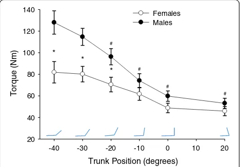
Torque-angle curve
Both males and females generated larger MVC torque in extended positions (−40°, −30°, −20°) relative to more flexed positions (−10°, 0°, 20°; position:F5, 27= 25.4,p<
0.001, ηp2= 0.825; Fig. 2). Pairwise comparison indicated
all positions, with the exception of −30°, were statisti-cally different (p< 0.05) than the position of peak torque (−40°). However, males had greater isometric torque than females (sex: F1, 31= 7.5, p= 0.01, ηp2= 0.194), but
not for all positions (position × sex:F2, 4= 6.9,p= 0.001,
ηp2= 0.182). Post hoc testing (t tests with adjusted α<
0.025) demonstrated sex differences in strength for the extended positions (−40°, t31=−3.0, p= 0.006; −30°, t31
=−3.2, p= 0.003; −20°, t31=−2.5, p= .022) with males
generating greater torque than females in these posi-tions. No sex differences in MVC strength were present in the−10°, 0°, and 20° positions.
EMG for torque-angle curve
EMG activity of the rectus femoris did not differ across trunk position (position:F5, 27= 1.6, p= 0.199,ηp
2
= 0.227) and was not different between sexes (sex: F1, 31= 1.0,
p= 0.319, ηp2= 0.032), suggesting that the differences in
torque across positions was not a result of contributions from the hip flexor muscles.
Fluctuations in torque (steadiness)
Torque steadiness was quantified for contraction intensities ranging between 5 and 70% MVC in the upright sitting position (0°). Torque produced at each target intensity in-creased for both males and females (intensity:F4, 15= 60.4,
p< 0.001, ηp 2
= 0.941; intensity × sex:F4, 15= 1.2,p= 0.363,
ηp2= 0.238), with no difference in absolute or relative torque
(% MVC) between the sexes (sex:F1, 18= 2.4,p= 0.142,ηp2
= 0.116 andF1 ,18= 2.0,p= 0.205,ηp 2
= 0.087, respectively). Standard deviation of torque was greater at high inten-sities compared with low inteninten-sities (intensity: F4, 15=
6.6, p= 0.003, ηp 2
= 0.639), for both males and females (intensity × sex:F4, 15= 1.3,p= 0.299, ηp2= 0.264; Fig. 3).
CV of torque was highest at a target intensity of 5% MVC and declined as target torque increased (intensity: F4, 15= 21.4, p< 0.001, ηp2= 0.851) for both males and Table 1Subject characteristics
Females (n= 18) Males (n= 15)
Age Years 24.3 ± 4.8 24.1 ± 6.6
Weight kg 65.2 ± 12.6 73.1 ± 8.8*
Height cm 166.6 ± 8.4 176.8 ± 7.6*
Body mass index kg m−2 23.3 ± 3.5 23.1 ± 2.3
Body fat % 32.5 ± 5.1 18.6 ± 5.7*
Lean mass in trunk kg 20.0 ± 3.6 27.0 ± 3.2*
Trunk lean mass/height kg/cm 0.12 ± 0.02 0.15 ± 0.02*
Self-reported physical activity over the preceding 12 months (data from 16 females and 14 males)
Met h week−1 44.7 ± 27.4 60.1 ± 39.9
Rectus abdominis muscle thickness (above umbilicus) cm 1.0 ± 0.18 1.4 ± 0.36*
Rectus abdominis muscle thickness (below umbilicus) cm 0.97 ± 0.13 1.3 ± 0.35*
kgkilograms,cmcentimeters,mmeters,METmetabolic equivalents
*p≤0.05
Trunk Position (degrees)
-40 -30 -20 -10 0 10 20
Torque (Nm)
20 40 60 80 100 120
140 Females
Males
* *
* #
#
#
#
females (intensity × sex:F4, 15= 2.5,p= 0.085,ηp 2
= 0.402; Fig. 3). There were no sex differences in SD or CV of torque (sex:F1, 18= 0.339,p= 0.568,ηp2= 0.018 andF1, 18
= 0.001,p= 0.977,ηp 2
< 0.001, respectively).
Fatigability and recovery
Time-to-task failure and MVC torque
Time-to-task failure for the isometric intermittent fatigue task did not differ between males and females (sex:t31= −0.78,p= .440; Table 2). MVC torque was not different be-tween males and females at baseline (57.3 ± 23.8 vs 49.5 ± 22.2, respectively; p= 0.336) and declined during the fa-tiguing exercise so that at task failure, the relative reduction in MVC torque from baseline was similar for the males and females (−30.64 ± 18.6% and −29.4 ± 13.7%, respect-ively; sex: t31= 0.18, p= 0.862). MVC torque increased in
recovery similarly for males and females (time × sex:F2, 30
= 0.6, p= 0.571, ηp2= 0.037) with no difference between
males and females (sex: F1, 31= 1.1, p= 0.313, ηp2= 0.033).
By 20 min post exercise, MVC torque was fully recovered and similar between sexes (t31= 1.0,p= 0.315; Table 2).
Torque and steadiness during the fatiguing task
Average torque (Nm) and relative torque (% MVC) pro-duced during the submaximal contractions was similar between the sexes (sex:F1, 31= 0.58,p= 0.454,ηp
2
= 0.018 and F1, 31= 0.07, p= 0.797, ηp2= 0.002, respectively)
and declined over time (time: F4, 28= .7, p< 0.001, ηp2
= 0.604 and F4, 28= 12.4, p< 0.001, ηp 2
= 0.638, respect-ively) for both males and females (time × sex: F4, 28
= 0.43, p= 0.789, ηp2= 0.057 and F4, 28= 0.72, p= 0.584,
ηp 2
= 0.094, respectively).
SD of torque increased during the fatiguing task (time: F4,28= 6.1, p= 0.001, ηp2= 0.467) for both males and
females (time × sex: F4, 28= 0.64, p= 0.642, ηp 2
= 0.083; Fig. 4a). CV of torque also increased throughout the fa-tiguing protocol (time:F4, 28= 6.4,p= 0.001,ηp2= 0.476) for
both males and females (time × sex:F4, 28= 0.94,p= 0.456,
ηp2= 0.118; Fig. 4b). There was no sex difference in force
fluctuations when measured with SD or CV of torque (sex: F1, 31= 2.0, p= 0.168, ηp
2
= 0.060 and F1, 31= 3.0,
p= 0.094, ηp2= 0.088, respectively).
EMG activity during the fatiguing task
EMG activity (% MVC) of the rectus abdominis (Fig. 5) and external oblique muscles increased throughout the fatiguing protocol (time:F4, 28= 5.7,p= 0.002,ηp2= 0.449
and F4, 28= 5.9,p= 0.001, ηp2= 0.457, respectively),
simi-larly for males and females (time × sex: F4, 28= 0.615,p
= 0.432, ηp2= 0.123 and F4, 28= 0.615, p= 0.655, ηp2=
0.081, respectively). There was no sex difference of rec-tus abdominis or external oblique EMG activity during the fatigue task (sex: F1, 31= 0.02, p= 0.899, ηp2= 0.001
andF1, 31= 2.9,p= 0.096,ηp2= 0.087, respectively).
Rectus femoris EMG remained low (<12% of maximal EMG) throughout the fatiguing protocol for both males and females (time × sex:F4, 28= 1.6,p= 0.190,ηp2= 0.191),
Contraction Intensity (% MVC)
0 10 20 30 40 50 60 70
SD of Torque (Nm)
0.0 0.5 1.0 1.5 2.0 2.5 3.0
CV of Torque (%)
0 2 4 6 8 10 12 14 16 18 Females SD
Males SD Males CV Females CV
Fig. 3Torque steadiness of trunk flexion at different intensities of contraction. Mean (±SEM) torque fluctuations for males and females represented as the standard deviation (SD) of torque and the coefficient of variation (CV) of torque, at 5, 10, 20, 50, and 70% of maximal voluntary contraction (MVC) in upright sitting. Torque steadiness differed with contraction intensity for SD and CV, but there were no differences between males and females
Table 2Muscle function and fatigability characteristics
Females (n= 18) Males (n= 15)
Baseline MVC at zero position Nm 49.5 ± 22.2 57.3 ± 23.8
Baseline MVC normalized to trunk lean mass/height from DXA Nm/kg cm−1 1.6 ± 0.6 1.5 ± 0.7
Baseline MVC normalized to abdominal muscle thickness below umbilicus from US Nm cm−1 51.0 ± 22.0 48.5 ± 20.3
Time-to-task failure min 10.6 ± 5.5 12.4 ± 7.4
Rating of perceived exertion at task failure 0–10 scale 6.9 ± 2.0 6.0 ± 2.4
MVC at task failure % baseline MVC 70.6 ± 13.7% 69.6 ± 18.6%
MVC at 10 min recovery % baseline MVC 98.9 ± 23.7 90.5 ± 18.8
MVC at 20 min recovery % baseline MVC 102.2 ± 18.7 94.2 ± 26.3
MVCmaximal voluntary contraction,USultrasound,DXAdual X-ray absorptiometry,minminutes
with no effect of time (time: F4, 28= 1.7, p= 0.175, ηp2=
0.197) or sex (sex:F1, 31= .2,p= 0.275,ηp 2
= 0.038).
Associations between variables
Trunk flexor MVC torque in upright sitting (0° trunk flexion) was positively, linearly correlated with fatigability of the trunk flexor muscles (r= 0.473,r2= 0.223,p= 0.005; Fig. 6a). Trunk flexor MVC torque was also positively cor-related with lean mass in the trunk, and this correlation was strongest at the−40° position, where both sexes gen-erated the greatest peak torque (−40°,r= 0.595,r2= 0.354, p< 0.001; 0°,r= 0.378,r2= 0.143,p= 0.03, Fig. 6b). Longer
time to failure of the trunk flexor muscles was associated with greater self-reported physical activity over the previ-ous 12 months (r= 0.456, r2= 0.208, p= 0.011; Fig. 6c). Greater self-reported physical activity over the previous 12 months was also associated with greater lean mass in the trunk (r= 0.486,r2= 0.236,p= 0.007; Fig. 6d).
Discussion
There were several novel findings in this study. First, there was no sex-related difference in fatigability or steadiness of the trunk flexor muscles. Second, men were stronger than women (MVC torque), but this was only at the more extended positions. Accordingly, males had more lean mass than females (measured with DXA scan) and greater rectus abdominis muscle thickness (measured with ultrasonography). The strength of the re-lationship between lean mass and strength was strongest in the extended positions (−40,−30,−20), where a sex dif-ference in strength was also observed. However, there was no sex difference in fatigability or in strength in the up-right and flexed positions (−10, 0, 20). Third, MVC torque and fatigability (time-to-task failure) of the trunk flexor muscles, both performed in upright sitting, were positively correlated, such that stronger individuals were less fatig-able, and this is in contrast to several other muscle groups, such as the elbow flexor muscles [28]. Physical activity levels (self-reported) were associated with fatigability, demonstrating that more physically active people were less fatigable.
The relationship between torque steadiness and con-traction intensity that we observed is consistent with that seen in other muscle groups such as the plantar flexors, dorsiflexors, finger abductors, and elbow flexor muscles, such that the SD of torque increased and CV
b
Time (% Task Failure)
0 25 50 75 100
CV of Torque (%)
3 4 5 6 7 8 9 10
a
Time (% Task Failure)
0 25 50 75 100
SD of Torque (Nm)
0.8 1.0 1.2 1.4 1.6 1.8 2.0 2.2
Females Males
Fig. 4Steadiness of submaximal contractions during the fatiguing task. Steadiness of submaximal contractions performed during the intermittent isometric trunk flexion fatiguing protocol. Standard deviation (SD) of torque (a) and coefficient of variation (CV) of torque (b) are shown as the mean ± SEM of three submaximal contractions at each quartile (beginning, 25%, 50%, 75%, and end) of the total time-to-task failure. Fluctuations in torque increase over time for both males and females (p< 0.05) with no sex difference in steadiness between males and females
Time (% task failure)
0 20 40 60 80 100 120
RMS EMG (%)
35 40 45 50 55 60 65
Females Males
of torque decreased as contraction intensity increased [18, 20]. However, there was no sex difference in the torque steadiness during trunk flexion contractions while in the upright sitting. For both sexes, however, the CV of torque of the trunk flexor muscles (15%) was higher than that typically seen in other muscle groups at low contraction intensities (<10% MVC) (Fig. 3), such as the first dorsal interosseous (~4%), elbow flexors (~2%), and quadriceps muscles (~1.5%) [34–36]. As for other muscles, common drive to the motor units and their dis-charge rates of the trunk flexor muscles impact the steadiness of contraction [16], probably explaining the similarity in the shape of the CV-force intensity curve between the abdominals and limb muscles.
There are several possible explanations for these muscle group differences in torque steadiness amplitude, i.e., the greater CV of torque of the trunk flexor muscles. First, the abdominal muscles are innervated from several spinal levels (T7–L1) [2]. The activation of many alpha motor neurons from multiple spinal levels is required to control torque generated by the multiple large muscles that comprise the trunk flexors [2]. The neurological complexity of this task may contribute to the large fluc-tuations in torque. The large CV of torque of the trunk
flexor muscles may also be impacted by the relatively long and massive trunk, which may make this body ment more difficult to control than smaller limb seg-ments, like the forearm or index finger. Ventilation may also impact torque steadiness, as the active abdominal muscles must accommodate the rhythmic expansion and contraction of the thorax and abdomen (trunk) during strength testing. This movement of the rib cage may also reduce the stability of the proximal attachments of the abdominal muscles [37]. There may be minor contribu-tions in the force output from chest and shoulder mus-cles, such as the upper trapezius musmus-cles, because during the task, the trunk was restrained by two large straps that contact the superior aspects of the shoulders. Lastly, during contraction, the summation of forces from multiple motor units is influenced by the interaction be-tween contractile tissue and connective tissue [38]. Thus, the presence of multiple tendinous intersections within the rectus abdominis [2], and the fascial attachments of the internal and external obliques, may impact the sta-bility of the force generated by the muscle fibers and transferred across the connective tissue, possibly influen-cing the magnitude of the torque fluctuations during trunk flexion. The contribution of the mechanical and
Physical Activity (MET hrs/wk)
0 30 60 90 120 150
Lea
n
M
a
ss (
k
g)
12 16 20 24 28 32 36 MVC (Nm)
20 40 60 80 100 120
Lea
n
M
a
ss (
k
g)
12 16 20 24 28 32 36
Time to Task Failure (s)
0 400 800 1200 1600
P
h
ys
ic
a
l A
c
ti
vi
ty
(M
E
T
h
rs
/w
k
)
0 30 60 90 120 150
Time to Task Failure (s)
0 400 800 1200 1600
M
VC (
N
m)
20 40 60 80 100 120
Females Males
a
b
c
d
anatomical features of this unique muscle group, and the influence of discharge rate variability of the motor units from multiple muscles originating from common drive, is yet to be explored.
There was no sex-related difference in fatigability of the trunk flexor muscles for strength-matched males and females during the submaximal, intermittent isometric fa-tiguing protocol. This finding is in contrast to other mus-cles, such as the elbow flexors, where males demonstrated greater fatigability compared with strength-matched females [21]. The lack of sex difference in fatigability may be due to the fact that the abdominal muscles are a pos-tural and ventilatory muscle group and thus may be de-signed to be especially fatigue resistant in both sexes. Häggmark and Thorstensson [39] showed that the ab-dominal muscles of males and females are comprised of approximately 55–58% type I muscle fibers, which are fa-tigue resistant relative to other fibers (type II). In other muscle groups, females tend to have a greater proportion of type I muscle fibers than males, which may contribute to females being more fatigue resistant than males [10, 11]. However, in muscle groups that have a high proportion of type I fibers in both males and females, such as the tibialis anterior, the sex difference in fatigability is diminished or absent [10, 40], which is consistent with our findings. Furthermore, ratings of perceived exertion at task failure were not different between sexes in our study, suggesting that males and females gave similar effort dur-ing the fatigudur-ing exercise task (Table 2). Ratdur-ings of per-ceived exertion at task failure were not, on average, at maximal levels, because some participants reported feeling as though they could continue the task if allowed to utilize compensatory movement strategies. However, all partici-pants met the criteria for failure of the fatiguing task. Im-portantly, our study showed that physical activity was more a determinant of fatigability than the sex of the indi-vidual, as shown by the significant correlation between time-to-task failure and self-reported physical activity.
Strength and fatigability of the trunk flexor muscles were positively correlated. This is in contrast to most other muscle groups, where weaker individuals are more fatigue resistant, such as for sustained isometric contrac-tions of the elbow flexor and hand grip muscles in young adults [28, 34, 41], where occlusion of blood flow is the primary mechanism responsible for the inverse relation-ship between strength and fatigability. The current study utilized an intermittent isometric protocol for which oc-clusion of blood flow is not a primary mechanism, thus making it less likely that strength-related blood flow dif-ferences between participants would influence fatigabil-ity. The role of the abdominal muscles in stability of the lumbar spine and pelvis, and as accessory muscles of ventilation [2], may explain the physiological need for a positive correlation between strength and fatigability in
order to minimize injury risk and to avoid possible im-pairments with breathing during exercise. While these mechanisms were not tested in this study, it is possible that some combination of neural input from multiple large abdominal muscles, architecture of the muscle and connective tissue [42], blood flow [43], and sympathetic drive [44] to this muscle group, may contribute to people with stronger abdominal muscles exhibiting greater fa-tigue resistance.
The positive correlation between strength and fatig-ability in this study may provide insight into the lack of a sex difference in fatigability. Females are typically weaker than males and demonstrate greater resistance to fatigue but there was no sex difference in strength for the trunk flexor muscles in upright sitting, and this was the position for the test of fatigability. In strength-matched males and females who performed an intermit-tent, isometric submaximal fatiguing protocol with the elbow flexors, also at 50% of MVC torque, women were less fatigable than men [21]. We did not observe this sex difference in fatigability of the trunk flexors in the males and females in this study, who did not differ in strength in upright sitting. While this study did not examine mechanisms responsible for fatigability, we hypothesize that several factors may contribute to the lack of a sex difference in fatigability. However, future research is needed to identify the mechanisms responsible for the relationship between strength and fatigability in this muscle group. This association also supports the import-ance of“core”strengthening.
Conclusions
This study shows that there are no sex differences in fat-igability or force control during isometric trunk flexion contractions. These findings of minimal differences in fatigability for the trunk muscles is in contrast to other studies that show clear differences in fatigability of other muscle groups, such as the elbow flexors and knee ex-tensors [11]. Furthermore, although men were stronger than females in the extended trunk positions of sitting, there was a minimal difference in maximal strength in upright and flexed sitting positions. Stronger males and females during upright sitting, however, were less fatig-able than weaker individuals, and both strength and fat-igability may be modulated by physical activity levels. The relationship between strength and fatigability of the trunk flexor muscles and physical activity sup-ports the importance of abdominal muscle strength-ening to offset fatigability.
Abbreviations
contraction; Nm: Newton meters; RMS: Root mean square; SD: Standard deviation; SEM: Standard error of the mean
Acknowledgements
The authors would like to thank Hugo Pereira, PT, Ph.D., for his assistance with the design of the steadiness testing procedures; Andrew Starsky, PT, Ph.D., for his consultation on the design of the torque-angle curve testing procedures; and Jordan Lemens for assistance with the data analysis and entry.
Funding
The first author was supported by funding from the Marquette University Clinical and Translational Rehabilitation Health Science Graduate Program; by an American Dissertation Fellowship from the American Association of University Women; and by the National Center for Advancing Translational Sciences, National Institutes of Health, through Grant Numbers UL1TR001436 and 1TL1TR001437 (publication contents are solely the responsibility of the authors and do not necessarily represent the official views of the NIH).
Availability of data and materials
The datasets used and/or analyzed during the current study are available from the corresponding author on reasonable request.
Authors’contributions
RD participated in the study design, subject recruitment, data collection, data analysis, and manuscript preparation. JS participated in the study design, piloting, development of the sampling and analysis programs, and manuscript preparation. TP participated in the data collection and analysis. DN participated in the study design, biomechanical consultation, and manuscript preparation. SH participated in the study design, interpretation of results, and manuscript preparation. All authors read and approved the final manuscript.
Competing interests
The authors declare that they have no competing interests.
Consent for publication Not applicable.
Ethics approval and consent to participate
This study was approved by the Institutional Review Board at Marquette University. All participants provided written informed consent prior to participating in the study.
Publisher’s Note
Springer Nature remains neutral with regard to jurisdictional claims in published maps and institutional affiliations.
Received: 17 November 2016 Accepted: 5 April 2017
References
1. Lee DG, Lee LJ, McLaughlin L. Stability, continence and breathing: the role of fascia following pregnancy and delivery. J Bodyw Mov Ther. 2008;12:333–48. 2. Neumann DA. Kinesiology of the musculoskeletal system: Foundations for
rehabilitation. Third ed. St. Louis: Mosby Elsevier; 2017.
3. Hodges PW, et al. Intervertebral stiffness of the spine is increased by evoked contraction of transversus abdominis and the diaphragm: in vivo porcine studies. Spine. 2003;28(23):2594–601.
4. Hodges PW, Richardson CA. Relationship between limb movement speed and associated contraction of the trunk muscles. Ergonomics. 1997;40(11): 1220–30.
5. Hodges PW, Richardson CA. Contraction of the abdominal muscles associated with movement of the lower limb. Phys Ther. 1997;77(2):132–42.
6. Sapsford RR, et al. Co-activation of the abdominal and pelvic floor muscles during voluntary exercises. Neurourol Urodyn. 2001;20(1):31–42.
7. Sapsford RR, et al. Pelvic floor muscle activity in different sitting postures in continent and incontinent women. Arch Phys Med Rehabil. 2008;89(9):1741–7. 8. Neumann P, Gill V. Pelvic floor and abdominal muscle interaction: EMG activity
and intra-abdominal pressure. Int Urogynecol J Pelvic Floor Dysfunct. 2002;13(2):125–32.
9. Hodges PW, Richardson CA. Altered trunk muscle recruitment in people with low back pain with upper limb movement at different speeds. Arch Phys Med Rehabil. 1999;80(9):1005–12.
10. Hunter SK. Sex differences in human fatigability: mechanisms and insight to physiological responses. Acta Physiol (Oxf). 2014;210(4):768–89.
11. Hunter SK. The relevance of sex differences in performance fatigability. Med Sci Sports Exerc. 2016;48(11):2247–56.
12. Smidt G, et al. Assessment of abdominal and back extensor function. A quantitative approach and results for chronic low-back patients. Spine (Phila Pa 1976). 1983;8(2):211–9.
13. Clark BC, et al. Gender differences in skeletal muscle fatigability are related to contraction type and EMG spectral compression. J Appl Physiol (1985). 2003;94(6):2263–72.
14. Almuklass AM, et al. Force steadiness as a predictor of time to complete a pegboard test of dexterity in young men and women. J Appl Physiol (1985). 2016;120(12):1410–7.
15. Enoka RM, et al. Mechanisms that contribute to differences in motor performance between young and old adults. J Electromyogr Kinesiol. 2003;13(1):1–12.
16. Farina D, Negro F. Common synaptic input to motor neurons, motor unit synchronization, and force control. Exerc Sport Sci Rev. 2015; 43(1):23–33.
17. Hunter SK, Pereira HM, Keenan KG. The aging neuromuscular system and motor performance. J Appl Physiol (1985). 2016;121:982–995.
18. Tracy BL. Visuomotor contribution to force variability in the plantarflexor and dorsiflexor muscles. Hum Mov Sci. 2007;26(6):796–807.
19. Tracy BL, et al. Aging, visuomotor correction, and force fluctuations in large muscles. Med Sci Sports Exerc. 2007;39(3):469–79.
20. Jesunathadas M, et al. Discharge properties of motor units during steady isometric contractions performed with the dorsiflexor muscles. J Appl Physiol (1985). 2012;112(11):1897–905.
21. Hunter SK, et al. Men are more fatigable than strength-matched women when performing intermittent submaximal contractions. J Appl Physiol (1985). 2004; 96(6):2125–32.
22. Hunter SK, Enoka RM. Changes in muscle activation can prolong the endurance time of a submaximal isometric contraction in humans. J Appl Physiol (1985). 2003;94(1):108–18.
23. Clark BC, et al. Sex differences in muscle fatigability and activation patterns of the human quadriceps femoris. Eur J Appl Physiol. 2005;94(1-2):196–206. 24. Christou EA, et al. The 1- to 2-Hz oscillations in muscle force are
exacerbated by stress, especially in older adults. J Appl Physiol (1985). 2004; 97(1):225–35.
25. Brown RE, Edwards DL, Jakobi JM. Sex differences in force steadiness in three positions of the forearm. Eur J Appl Physiol. 2010;110(6):1251–7. 26. Hodges P, et al. Intervertebral stiffness of the spine is increased by evoked
contraction of transversus abdominis and the diaphragm: in vivo porcine studies. Spine (Phila Pa 1976). 2003;28(23):2594–601.
27. Janssen I, et al. Skeletal muscle mass and distribution in 468 men and women aged 18-88 yr. J Appl Physiol (1985). 2000;89(1):81–8.
28. Hunter SK, Enoka RM. Sex differences in the fatigability of arm muscles depends on absolute force during isometric contractions. J Appl Physiol (1985). 2001;91(6):2686–94.
29. Kriska AM, et al. Development of questionnaire to examine relationship of physical activity and diabetes in Pima Indians. Diabetes Care. 1990;13(4): 401–11.
30. Cram JR. Cram’s introduction to surface electromyography. 2nd ed. Sudbury: Jones and Bartlett; 2011. p. 412.
31. Whittaker JL, Warner MB, Stokes M. Comparison of the sonographic features of the abdominal wall muscles and connective tissues in individuals with and without lumbopelvic pain. J Orthop Sports Phys Ther. 2013;43(1):11–9.
32. Borg GA. Psychophysical bases of perceived exertion. Med Sci Sports Exerc. 1982;14(5):377–81.
33. Redfern M, Hughes R, Chaffin D. High-pass filtering to remove electrocardiographic interference from torso EMG recordings. Clin Biomech (Bristol, Avon). 1993;8(1):44–8.
34. Hunter SK, Critchlow A, Enoka RM. Influence of aging on sex differences in muscle fatigability. J Appl Physiol (1985). 2004;97(5):1723–32.
36. Welsh SJ, Dinenno DV, Tracy BL. Variability of quadriceps femoris motor neuron discharge and muscle force in human aging. Exp Brain Res. 2007; 179(2):219–33.
37. Hunter SK, et al. Task differences with the same load torque alter the endurance time of submaximal fatiguing contractions in humans. J Neurophysiol. 2002; 88(6):3087–96.
38. Taylor AM, Christou EA, Enoka RM. Multiple features of motor-unit activity influence force fluctuations during isometric contractions. J Neurophysiol. 2003;90(2):1350–61.
39. Häggmark T, Thorstensson A. Fibre types in human abdominal muscles. Acta Physiol Scand. 1979;107(4):319–25.
40. Avin KG, Law LA. Age-related differences in muscle fatigue vary by contraction type: a meta-analysis. Phys Ther. 2011;91(8):1153–65.
41. Hunter SK, et al. Active hyperemia and vascular conductance differ between men and women for an isometric fatiguing contraction. J Appl Physiol (1985). 2006;101(1):140–50.
42. Gracovetsky S. Is the lumbodorsal fascia necessary? J Bodyw Mov Ther. 2008;12(3):194–7.
43. Manohar M. Blood flow to the respiratory and limb muscles and to abdominal organs during maximal exertion in ponies. J Physiol. 1986;377:25–35. 44. Derchak PA, et al. Effects of expiratory muscle work on muscle sympathetic
nerve activity. J Appl Physiol (1985). 2002;92(4):1539–52.
• We accept pre-submission inquiries
• Our selector tool helps you to find the most relevant journal
• We provide round the clock customer support
• Convenient online submission
• Thorough peer review
• Inclusion in PubMed and all major indexing services
• Maximum visibility for your research
Submit your manuscript at www.biomedcentral.com/submit