IJBCP
International Journal of Basic & Clinical Pharmacology
Original Research Article
Evaluation of wound healing activity of Hibiscus rosa sinensis linn in
albino rats
Monil Yogesh Neena Gala*, Swanand S. Pathak
INTRODUCTION
Wound and wound healing is a matter of great concern for all the medical practitioners ever since the dawn of civilization and rise of modern medicine.1 Irrespective of
the inciting injury, similar healing process takes place, i.e., the lost tissue is not regenerated but is replaced by a fibrous tissue.2
Knowledge of wound healing and various promoters of wound healing enable the doctors to manipulate the wound
to achieve an optimal result in a rapid period. Infinite literatures on solution of wound healing suggest that the problem is not so simple and straightforward to tackle.
During time of invent of asepsis and dawn of antibiotic era, wound care had achieved milestones, but the widespread development of antibiotic-resistant bacteria is a major roadblock.3
In India where majority of population is from poor economic background, it is difficult for many patients to
ABSTRACT
Background: From debridement to the appearance of healthy granulation tissue is the maximum time during which the patient suffers more economically and psychologically. Due to high cost expenditure in hospitalisation, dressing material cost and loss of daily earnings during this period which puts a psychological pressure over patient and family especially in India where majority of the population is still below poverty line. Also access to quick treatment is yet not possible to majority of population living in rural with no access to basic healthcare.
Methods: It was an experimental study wherein Wistar rat models were used to check for the wound healing property of Hibiscus Rosa Sinensis (HRS) compared with one of the common standard drugs of treatment for available today, betadine. To observe the wound healing property of the HRS flower extract, the experiment was divided into 2 parts i.e. excision wound model and incision wound model. The groups were treated with respective medication along with a control group of rats.
Results:
At the end of the experiment, it was observed that HRS flower extract
increased the wound breaking strength as compared to control but not as effectively as compared to the betadine ointment.Conclusions: HRS flower extract helps in early epithelization and helps in decreasing the wound size. Betadine is associated with adverse events like life threatening allergic reactions, water retention, etc.
Keywords: Animal, Betadine, Hibiscus, Skin, Flower, Wound healing, Wistar DOI: http://dx.doi.org/10.18203/2319-2003.ijbcp20192194
Department of Pharmacology, Jawaharlal Nehru Medical College, DMIMS, Sawangi, Meghe, Wardha, Maharashtra, India
Received: 22 March 2019 Accepted: 11 April 2019 Revised: 03 May 2019
*Correspondence to:
Dr. Monil Yogesh Neena Gala, Email: ynmvgala@gmail.com
afford costly wound care modalities. For these patients
being bedridden is a curse as majority of them earn livelihood on daily wages basis.4
Pharmaceutical companies are taking advantages of these complexities to introduce multiple remedies, staking high claims for commercial interest. Numerous remedies are available today to counter the various detrimental factors to the natural wound healing process.5 Considering these
factors, it is necessary to incorporate a simple, cheap, easily available, effective modality that can also be used without any medical supervision to treat wound healing.4
Hibiscus rosa sinensis (HRS) extract was found to be such a great modality in better wound care as it is cheap, easily available, had no side effects and can be applied without any assistance.6
There are very few studies present in literature comparing the efficacy of HRS extracts with modern medicines. Due to this reason, present study aimed at studying the effectiveness of HRS flower extract as a wound healing agent as compared to betadine ointment.
The aim was to study the wound-healing activity of
Hibiscus rosa-sinensis in albino rats. The objective was to evaluate natural wound healing process in (control) group of rats, to evaluate wound healing property of Hibiscus rosa sinensis flower on (test) group of rats, to evaluate wound healing property of betadine topical ointment on (standard) group of rats and to compare the wound healing property of Hibiscus rosa sinensis flower and betadine topical ointment.
METHODS
The present study was conducted in the Department of Pharmacology, Jawaharlal Nehru Medical College, Sawangi (Meghe), Wardha, Maharashtra, India. The study was carried out for a period of 24 months from November 2016 to November 2018.
The approval from Institutional Animal Ethical Committee of the research protocol was approved by the Institutional Animal Ethical Committee (no: DMIMSDU/IAEC/2016-17/12), Jawaharlal Nehru Medical College, Sawangi, Meghe, Wardha, Maharashtra, India.
Material
Hibiscus rosa sinensis (HRS) flower
Fresh HRS flower were collected from area near by JNMC, Sawangi, Wardha, Maharashtra, India.
Authentication of the plant
The plants were identified and authenticated by the taxonomist at Mahatma Gandhi Ayurveda College, Wardha, Maharashtra, India.
Purchase of chemicals
Betadine topical ointment (10%), ketamine, xylazine and petroleum jelly (ointment base) was obtained from
Maharashtra Scientific Medical Store (Wardha,
Maharashtra, India).
Animals
The study was conducted using 36 Wistar Albino rats, of either sex weighing 150-250 g from institutional animal house, Sawangi (Meghe), Wardha, Maharashtra, India.
Inclusion criteria
• Sex: male and female rats,
• Weight: 150-250 gm,
• Age: 02 months to 14 months.
Exclusion criteria
• Pregnant female rats,
• Age: more than 14 months,
• Unhealthy/diseased rats.
[image:2.595.313.551.414.535.2]About 7 grams of reddish-brown semisolid extract was obtained from 50 grams of dried powder of flowers (Figure 1 and Figure 2).
[image:2.595.313.556.577.733.2]Figure 1: Preparation of plant material in powdered form.
Figure 2: Preparation of crude ethanolic extract.
Flower of Hibiscus Rosa Sinensis were shade dried.
They were then powdered in a blender.
Then, they were stored in air-tight container until further use.
Powder was extracted in soxhlet apparatus using 95% ethanol at 60-80 degree celsius for 12 hours
.
Extracts were filtered.
Ointment formulations
About 5% (w/w) of HRS extract was determined to be the optimum dose to promote wound healing according to Raduan S et al, and Meena AK et al.8,9 Ointment
formulations was prepared from the extract 5% (w/w), where 5 gram of the extract was incorporated into 100 grams of petroleum jelly. Betadine topical ointment (10% w/w) was used as a standard drug for comparing the wound healing potential of the extract.
Acclimatization
Rats were only used after 7 day acclimatization period (12:12 hours, light:dark cycle) to the laboratory environment. They were housed under the standard nutritional and environmental conditions of light, temperature and humidity. They were fed with standard laboratory chow and provided water ad libitum. Post experimental study, the rats were returned to the animal house after rehabilitation.
Primary skin irritation test
The skin irritation test was done by a method based on the test described by Suraj et al.10 A 2 cm2 dorsal area was
trimmed and then shaved. After cleaning the area with surgical spirit, 5% w/w of extract was applied topically to observe any adverse reaction. Similarly, 10% w/w betadine ointment was applied topically to observe any adverse reaction. Animals did not show any adverse effects and thus the prepared extract was considered safe for topical application along with the standard drugs.
Preoperative preparation
Weight of the rats was taken. Pre-emptive analgesia was considered and ophthalmic ointment to the eye was administered following induction of anesthesia to prevent corneal drying. Body hair was removed from the surgical site and surgical scrub alternating between a disinfectant and alcohol was carried out. Warm fluid bag was used to prevent heat loss. Isotonic fluid was kept ready in case of excessive fluid loss.
Induction of anesthesia
Injection ketamine (50 mg): 0.4 ml and injection xylazine (2%): 0.4 ml was given intra-peritoneal.11 Since, it is a
dissociative anaesthetic, it interrupts the neuronal traffic between the cortex and thalamus. This effect is characterized by sustained reflex movements, stiff muscle, unconsciousness and open eyes. Absence of response to painful stimuli indicates complete induction of anaesthesia.
Post-operative management
Isotonic fluid was supplied along with warm fluid bag or gloves as required. Rats were frequently checked every
10-15 mins during recovery from anesthesia. Food and water intake were observed and also analgesic were administered.
Excision wound7
Rats were inflicted with excision wound by cutting away a 500 mm2 full thickness of skin on the back as described by
Morton JJ et al, under anaesthesia.12
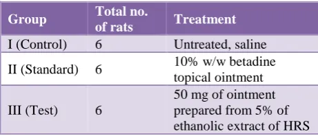
[image:3.595.311.545.256.353.2]The wound was left undressed to open environment. The rats were divided into following groups and received the following treatment:
Table 1: Grouping for excision wound model.
Group Total no. of rats Treatment
I (Control) 6 Untreated, saline
II (Standard) 6 10% w/w betadine
topical ointment
III (Test) 6
50 mg of ointment prepared from 5% of ethanolic extract of HRS
• The ointment was topically applied once a day,
starting from the day of the operation, till complete epithelization. This model was used to monitor wound contraction and wound closure time.
• Wound contraction was calculated as percentage
reduction in wound area. The progressive change in wound area was monitored by tracing the wound margin on graph paper every alternate day.
Percentage (%) wound closure= (Wound area on Day 0-Wound area on Day 'n')/ (0-Wound area on Day 0) * 100
Incision wound6,13
After inducing anaesthesia, a 2 cm long paravertebral incision was made through the full thickness of the skin on left side (flank) of the vertebral column of the rats back as described by Ehrlich HP et al.14 The wound was closed
with interrupted sutures (ethilion/nylon).
The rats were divided into following groups and received the following treatment:
Table 2: Grouping for incision wound model.
Group Total no.
of rats Treatment
I (Control) 6 Untreated, saline
II (Standard) 6 10% w/w betadine
topical ointment
III (Test) 6
[image:3.595.312.543.654.753.2]• The ointment was topically applied once a day. The
sutures were removed on the 7th day. Wound-breaking strength (wound healing) was measured in anesthetized rats on the 10th day after wounding using a tensiometer.
Measurement of wound breaking strength15
Tensile strength, the force required to open a healing skin wound, was used to measure healing. The instrument used for this measurement is tensiometer. It consisted of animal operation table serving as the base. On one side of the operation table, an IV stand was fixed. It served as a fix point to which one Kelly’s forceps was fixed at one end.
[image:4.595.311.544.70.275.2]Another Kelly’s forceps were tied to a piece of fishing line (20-lb test monofilament) to which 500 ml polyethylene bottle was tied at the end. The grips of the Kelly’s forceps were then attached to the either side of the incised wound and was to adjust so that the polyethylene bottle was freely hanging in the air.
Figure 3: Tensiometer arrangement (self-made).
Table 3: Comparison of wound area (mm2) in three groups by applying descriptive statistics.
Group N Mean SD SE
95% confidence interval for mean
Minimum Maximum
Lower
bound Upper bound
Day 0
Control 06 508.66 11.57 4.72 496.52 520.80 490.00 520.00
Standard 06 513.66 07.73 03.15 505.54 521.78 500.00 520.00
Test 06 505.00 08.94 03.65 495.61 514.38 490.00 515.00
Day 2
Control 06 462.00 06.60 02.69 455.07 468.92 452.00 470.00
Standard 06 423.16 08.40 03.42 414.35 431.98 417.00 440.00
Test 06 460.50 09.37 03.82 450.66 470.33 446.00 470.00
Day 4
Control 06 398.33 09.89 04.03 387.95 408.71 384.00 414.00
Standard 06 351.83 16.22 06.62 334.80 368.86 321.00 364.00
Test 06 407.66 08.52 03.48 398.72 416.61 396.00 420.00
Day 6
Control 06 364.00 22.76 09.29 340.10 387.89 320.00 380.00
Standard 06 263.16 17.64 07.20 244.64 281.68 231.00 280.00
Test 06 364.50 10.34 04.22 353.63 375.36 348.00 375.00
Day 8
Control 06 310.83 08.01 03.27 302.42 319.23 297.00 320.00
Standard 06 191.66 15.95 06.51 174.91 208.41 173.00 217.00
Test 06 304.83 13.94 05.69 290.19 319.47 285.00 320.00
Day 10
Control 06 283.66 12.04 04.91 271.02 296.30 264.00 297.00
Standard 06 138.66 16.76 06.84 121.07 156.26 120.00 164.00
Test 06 248.66 14.94 06.10 232.97 264.35 224.00 270.00
Day 12
Control 06 254.33 11.75 04.80 241.99 266.67 240.00 264.00
Standard 06 89.16 14.20 05.79 74.26 104.07 75.00 110.00
Test 06 183.83 05.77 02.35 177.77 189.89 175.00 192.00
Day 14
Control 06 237.33 09.50 03.87 227.36 247.30 226.00 248.00
Standard 06 44.50 12.38 05.05 31.49 57.50 32.00 60.00
Test 06 143.00 08.48 03.46 134.09 151.90 130.00 150.00
Day 16
Control 06 214.16 11.17 04.56 202.43 225.89 198.00 228.00
Standard 06 16.83 07.85 03.20 08.58 25.08 09.00 28.00
Test 06 97.66 10.61 4.33 86.52 108.80 86.00 110.00
Day 18
Control 06 202.33 11.96 04.88 189.78 214.88 186.00 218.00
Standard 06 01.66 01.96 00.80 -00.39 03.73 00.00 04.00
[image:4.595.56.545.346.754.2]Water added to the polyethylene bottle was weighed and
considered as the tensile strength of the wound (Figure 3).
The endpoint was achieved as soon as the wound gapping was seen. Tensile strength of the healed wound for an individual animal was calculated by the mean determination of the tensile strength of the 2 paravertebral incision.
Statistics
The data was subjected to statistical evaluation by applying the tests of significance as Paired t-test, One-way ANOVA and multiple comparison test: post hoc Tukey test. The software used in the analysis were SPSS 17.0 version and GraphPad Prism 5.0 and p<0.05 is considered as level of significance.
RESULTS
Table 3 shows the increasing mean difference of wound area between rats treated with HRS flower extract (test), betadine ointment (standard) and saline treated (control).
On Day 0, the mean wound area of control, standard and test group were 508.66±11.57 mm2, 513.66±07.73 mm2
and 505.00±08.94 mm2 respectively. On Day 18, the
readings recorded were 202.33±11.96 mm2, 01.66±01.96
mm2 and 47.33±12.04 mm2 respectively (Table 3).
[image:5.595.311.546.141.318.2]Figure 4: Comparison of wound area (mm2) in three groups.
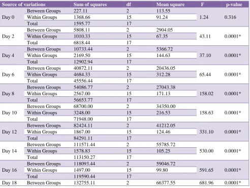
Table 4: Comparison of wound area (mm2) in three groups by applying One Way ANOVA.
Source of variations Sum of squares df Mean square F p-value
Day 0
Between Groups 227.11 2 113.55
1.24 0.316
Within Groups 1368.66 15 91.24
Total 1595.77 17
Day 2
Between Groups 5808.11 2 2904.05
43.11 0.0001*
Within Groups 1010.33 15 67.35
Total 6818.44 17
Day 4
Between Groups 10733.44 2 5366.72
37.10 0.0001*
Within Groups 2169.50 15 144.63
Total 12902.94 17
Day 6
Between Groups 40872.11 2 20436.05
65.44 0.0001*
Within Groups 4684.33 15 312.28
Total 45556.44 17
Day 8
Between Groups 54086.77 2 27043.38
158.02 0.0001*
Within Groups 2567.00 15 171.13
Total 56653.77 17
Day 10
Between Groups 68700.00 2 34350.00
158.63 0.0001*
Within Groups 3248.00 15 216.53
Total 71948.00 17
Day 12
Between Groups 82424.11 2 41212.05
331.10 0.0001*
Within Groups 1867.00 15 124.46
Total 84291.11 17
Day 14
Between Groups 111571.44 2 55785.72
530.00 0.0001*
Within Groups 1578.83 15 105.25
Total 113150.27 17
Day 16
Between Groups 118093.44 2 59046.72
591.65 0.0001*
Within Groups 1497.00 15 99.80
Total 119590.44 17
[image:5.595.53.546.387.756.2]From Figure 4, it is evident that betadine had the most
efficacious wound healing activity followed by the HRS flower extract (Figure 4). The difference in the wound healing property observed in comparing the 3 groups was statistically significant (p-value=0.0001) (Table 4 and Table 5).
[image:6.595.53.545.176.580.2]Table 6 illustrates the wound area over a period of 18 days. While the standard group shows the most effective healing, the test group shows a slow but effective healing as well over a period of 18 days.
Table 5: Comparison of wound area (mm2) in three groups by applying multiple comparisons: post hoc Tukey test.
Time
period Group
Mean
diff. (I-J) Std. error p-value
95% confidence interval
Lower bound Upper bound
Day 0 Control
Standard -5.00 5.51 0.645 -19.32 9.32
Test 3.66 5.51 0.787 -10.65 17.99
Standard Test 8.66 5.51 0.288 -5.65 22.99
Day 2 Control
Standard 38.83 4.73 0.0001* 26.52 51.14
Test 1.50 4.73 0.946 -10.80 13.80
Standard Test -37.33 4.73 0.0001* -49.64 -25.02
Day 4 Control
Standard 46.50 6.94 0.0001* 28.46 64.53
Test -9.33 6.94 0.394 -27.36 8.70
Standard Test -55.83 6.94 0.0001* -73.86 -37.79
Day 6 Control
Standard 100.83 10.20 0.0001* 74.33 127.33
Test -0.50 10.20 0.999 -27.00 26.00
Standard Test -101.33 10.20 0.0001* -127.83 -74.8
Day 8 Control
Standard 119.16 7.55 0.0001* 99.54 138.78
Test 6.00 7.55 0.712 -13.61 25.61
Standard Test -113.16 7.55 0.0001* -132.78 -93.54
Day 10 Control
Standard 145.00 8.49 0.0001* 122.93 167.06
Test 35.00 8.49 0.002* 12.93 57.06
Standard Test -110.00 8.49 0.0001* -132.06 -87.93
Day 12 Control
Standard 165.16 6.44 0.0001* 148.43 181.89
Test 70.50 6.44 0.0001* 53.76 87.2308
Standard Test -94.66 6.44 0.0001* -111.39 -77.93
Day 14 Control
Standard 192.83 5.92 0.0001* 177.44 208.21
Test 94.33 5.92 0.0001* 78.94 109.71
Standard Test -98.50 5.92 0.0001* -113.8 -83.11
Day 16 Control
Standard 197.33 5.76 0.0001* 182.35 212.3
Test 116.50 5.76 0.0001* 101.51 131.48
Standard Test -80.83 5.76 0.0001* -95.81 -65.85
Day 18 Control
Standard 200.66 5.69 0.0001* 185.87 215.46
Test 155.00 5.69 0.0001* 140.20 169.79
[image:6.595.56.547.610.750.2]Standard Test -45.66 5.69 0.0001* -60.46 -30.87
Table 6: Percentage of wound healing.
Day (Duration) Control Standard Test
Day 0 0% 0% 0%
Day 2 9% 18% 9%
Day 4 22% 32% 19%
Day 6 28% 49% 28%
Day 8 39% 63% 40%
Day 10 44% 73% 51%
Day 12 50% 83% 64%
Day 14 53% 91% 72%
Day 16 58% 97% 81%
Table 7: Comparison of skin breaking strength (g) in three groups by applying descriptive statistics.
Groups N Mean SD Std.
Error
95% Confidence Interval for
Mean Minimum Maximum
Lower Bound Upper Bound
Control 06 15.76 1.12 0.46 14.58 16.94 14.00 17.00
Standard 06 41.36 2.04 0.83 39.22 43.51 38.00 44.00
[image:7.595.48.547.202.254.2]Test 06 26.50 2.16 0.88 24.22 28.77 24.00 30.00
Table 8: Comparison of skin breaking strength (g) in three groups by applying One-way ANOVA.
Source of variations Sum of Squares df Mean Square F p-value
Between Groups 1983.16 2 991.58
292.98 0.0001*
Within Groups 50.76 15 3.38
[image:7.595.52.545.298.364.2]Total 2033.93 17
Table 9: Comparison of skin breaking strength (g) in three groups by applying multiple comparisons: post hoc Tukey Test.
Groups Mean Difference
(I-J) Std. Error p-value
95% Confidence Interval
Lower Bound Upper Bound
Control Standard -25.60 1.06 0.0001* -28.35 -22.84
Test -10.73 1.06 0.0001* -13.49 -7.97
Standard Test 14.86 1.06 0.0001* 12.10 17.62
Percentage reduction in wound area recorded on Day 18 in control, standard and test were 60%, 100% and 91% respectively (Table 6).
Table 7 and Figure 5 shows the comparison of skin breaking strength observed in the three groups. The skin breaking strength of control, standard and test group were 15.76±1.12g, 41.36±2.04g and 26.50±2.16g respectively. The group of rats treated with betadine topical ointment showed the highest tensile strength followed by the HRS flower extract (Table 7) (Figure 5).
[image:7.595.52.287.573.732.2]The difference in the skin breaking strength observed in comparing the 3 groups was statistically significant (p-value=0.0001) (Table 8 and Table 9).
Figure 5: Comparison of skin breaking strength (g) in three groups.
DISCUSSION
The present study was undertaken to evaluate wound healing activity of Hibiscus rosa sinensis linn in albino rats and comparing it with standard treatment. This was considered as the aim of the study. The finding and results from this study is similar to several other studies that have included extracts of HRS flower.6,13,16
Betadine is a formulation based on povidone-iodine. Povidone iodine is an effective antiseptic with bactericidal property against gram positive and gram-negative organism, that does not impede wound healing.17-20
However, several studies have reported side effects such as life-threatening allergic reactions, visible water retention, boy temperature fluctuations etc.21
Bhaskar A et al, had reported similar findings regarding the skin breaking strength in their study by the 12th day.13
Shivananda NB et al, reported higher collagen content in HRS flower treated rats. They demonstrated an increase in hydroxyproline, an amino acid which is a major component of collagen.6
Although HRS flower as such has no specific antimicrobial activity of its own, the wound healing promoting effect can be attributed to its various hormonal activities which include hypoglycaemic, androgen like, antioxidant and anticonvulsant activities.6 The increased tensile strength,
Shen H et al, demonstrated that N-butyl alcohol extract of
HRS (NHRS) flower is far more potent than other forms of HRS extract. They also suggested that NHRS increases secretion of vascular endothelial growth factor (VEGF) which accelerates wound healing and accelerates the regeneration of epidermal layer by upregulation of transforming growth factor (TGF-ß1). They concluded that NHRS elevates levels of VEGF and TGF-ß1 which promotes faster wound repair via increased angiogenesis, collagen fibre deposition and as well as increasing the activity of macrophages.16
Bhaskar A et al, reported the presence of tannins, saponins, flavonoids and terpenoids in their phytochemical evaluation of HRS flower extract which highlighting flavonoids as the active substance in promoting wound healing. However, they have reported an increase in the hexosamine and uronic acid molecules which are responsible for the synthesis of extracellular matrix as the
primary method of wound healing.13
Factors that delay the healing example diabetes, obesity, stress, and other are not taken purposefully to establish that HRS improves wound healing when no other deteriorating factors are present.
Recommendations
The experiment needs further evaluation in other animals such as dogs, monkeys etc. Clinical evaluation needs to be carried out. Cellular level and molecular level studies are required to conclude regarding mechanism of action.
ACKNOWLEDGEMENTS
Authors would like to thank Department of Pharmacology, Animal House, Central Research Laboratory and Animal Ethical committee of Jawaharlal Nehru Medical College, Sawangi, Wardha, Maharashtra, India, Mahatma Gandhi Ayurveda College, Selu, Maharashtra, Indian Council of Medical Research, New Delhi, India for support during study.
Funding: No funding sources Conflict of interest: None declared
Ethical approval: The study was approved by the Institutional Animal Ethical Committee, Jawaharlal Nehru Medical College, Sawangi, Meghe, Wardha, Maharashtra, India (no: DMIMSDU/IAEC/2016-17/12)
REFERENCES
1. Forrest RD. Early history of wound treatment. J R Soc
Med. 1982;75(3):198-205.
2. Shield HealthCare. How wounds heal: the 4 main
phases of wound healing, 2015. Available at: http://www.shieldhealthcare.com/community/wound/ 2015/12/18/how-wounds-heal-the-4-main-phases-of-wound-healing/. Accessed 21 September 2018.
3. Boateng JS, Matthews KH, Stevens HNE, Eccleston
GM. Wound healing dressings and drug delivery systems: a review. J Pharm Sci. 2008;97(8):2892-923.
4. Gupta N, Gupta SK, Shukla VK, Singh SP. An Indian
community-based epidemiological study of wounds. J Wound Care. 2004;13(8):323-5.
5. Daunton C, Kothari S, Smith L, Steele D. A history of
materials and practices for wound management. Wound Pract Res. 2012;20:174-86.
6. Shivananda NB, Raju SS, Orette FA, Rao CAV.
Effects of Hibiscus rosa sinensis L (Malvaceae) on wound healing activity: a preclinical study in a sprague dawley rat. Int J Low Extrem Wounds. 2007;6(2):76-81.
7. Bhaskar A, Nithya V. Evaluation of the
wound-healing activity of Hibiscus rosa sinensis L
(Malvaceae) in Wistar albino rats. Indian J Pharmacol. 2012;44(6):694-8.
8. Meena AK, Jain A, Pandey K, Singh RK. Acute
toxicity and genotoxic activity of Hibiscus rosa-sinensis flower extract. Am J Phytomed Clin Therapeutics. 2014;2(4):524-9.
9. Raduan SZ, Abdul Aziz MW, Roslida AH, Zakaria
ZA, Zuraini A, Hakim MN. Anti-inflammatory effects of Hibiscus rosa-sinensis L. and Hibiscus rosa-sinensis var. alba ethanol extracts. Int J Pharm Pharmaceut Sci. 2013;5(4):754-62.
10. Davis SV, Shenoi SD, Prabhu S, Shirwaiker A,
Balachandran C. Clinical evaluation of patients patch tested with plant series: a prospective study. Indian J Dermatol. 2011;56(4):383-8.
11. Injectable Anesthesia. Research at Penn State.
Available at:
https://www.research.psu.edu/arp/anesthesia/injectabl e-anesthesia.html. Accessed 27 August 2018.
12. Morton JJ, Malone MH. Evaluation of vulneray
activity by an open wound procedure in rats. Arch Int Pharmacodyn Ther. 1972;196(1):117-26.
13. Bhaskar A, Nithya V. Evaluation of the
wound-healing activity of Hibiscus rosa sinensis L
(Malvaceae) in Wistar albino rats. Ind J Pharmacol. 2012;44(6):694–8.
14. Ehrlich HP, Hunt TK. Effects of cortisone and vitamin A on wound healing. Ann Surg. 1968;167(3):324-8. 15. Lodhi S, Jain AP, Rai G, Yadav AK. Preliminary
investigation for wound healing and
anti-inflammatory effects of Bambusa vulgaris leaves in rats. J Ayurveda Integr Med. 2016;7(1):14-22. 16. Shen HM, Chen C, Jiang JY, Zheng YL, Cai WF,
Wang B, et al. The N-butyl alcohol extract from
Hibiscus rosa-sinensis L. flowers enhances healing potential on rat excisional wounds. J Ethnopharmacol. 2017;198:291-301.
17. Bigliardi PL, Alsagoff SAL, El-Kafrawi HY, Pyon
J-K, Wa CTC, Villa MA. Povidone iodine in wound healing: a review of current concepts and practices. Int J Surg. 2017;44:260-8.
18. Burks RI. Povidone-iodine solution in wound
19. Durani P, Leaper D. Povidone-iodine: use in hand
disinfection, skin preparation and antiseptic irrigation. Int Wound J. 2008;5(3):376-87.
20. Fleischer W, Reimer K. Povidone-iodine in antisepsis-state of the art. Dermatol. 1997;195(2):3-9.
21. McDowell S. Are we using too much Betadine? RN.
1991;54(7):43-5.