27
The Effects of Delayed Ischemic Preconditioning
on Performance and Recovery in Repeated Cycling Bouts
Chantelle A. Robitaille1, Akemi G. King1, Christina A. Buchanan1, Lance C. Dalleck1 1High Altitude Exercise Physiology Program, Western Colorado University, Gunnison, CO, USA
Introduction
The popularity of endurance events, and in particular, multi-day events, continues to increase. With the growing popularity of events such as the Tour de California, Ride the Rockies, along with multi-day destination cycling trips, it is important for athletes to focus not only on their
performance, but also their recovery in order to complete these multi-day events. Such events are particularly challenging as athletes need not only have the ability to recover between multiple climbs and sprints during each stage, they must also be able to recover from each day’s challenges to perform well in the subsequent stage. In short, during these longer events, the ability
ABSTRACT
Introduction: Ischemic preconditioning (IPC) is a technique of systematically occluding and reperfusing blood vessels that has shown to induce a biochemical cascade that increases the availability of nitric oxide (NO) and adenosine and enhances blood flow, endothelial function, and ATP regeneration. IPC has two physiological windows of protection; the first (immediate) window take place within the first 12 hours of treatment, while the second (delayed) takes place 24-72 hours post-treatment. The aim of this study was to evaluate its effects cyclists performing repeated incremental cycling bouts. Methods: In a randomized, crossover study, subjects received a 4 x 5min bilateral IPC treatment to the thigh. 24 and 48 hours after treatment, they performed an incremental cycling trial, followed by a 15-min recovery period, and then repeated the same cycling trial and recovery period. Time to fatigue (TTF) was recorded for each trial and heart rate (HR), blood lactate (La-), and blood Results: pressure (BP) were recorded throughout the recovery period. Despite
individual variability, delayed IPC improved TTF (p<0.05), decreased performance decrement between trials, lowered HR and BP (p<0.05), which allowed cyclists to work at higher lactate levels (p<0.05). Conclusions: These results suggest that delayed IPC may be an effective performance and recovery strategy for endurance athletes.
28 to recover has a large impact on an athlete’s
ability to perform.
During a day of competitive cycling, athletes need to perform at high and medium intensities, with short bouts at lower intensities to recover and usually, refuel. Adenosine Triphosphate (ATP) is the body’s energy molecule and must be present for muscles to contract and for any degree of force to be exerted. ATP must also be continuously regenerated to allow work to continue. All three energy systems will be called into action during an endurance cycling event; the phosphagen system during short sprints at an all-out effort (maximum 10-12 seconds), the glycolytic system during high and medium-intensity, long-duration climbs, and oxidative system during lower-intensity, long-duration efforts1. When the glycolytic system is in
action, ATP is produced by breaking down carbohydrates (CHO) from dietary glucose or from stored glycogen in the muscles. Aside from ATP, pyruvate and NADH+H+ are
produced. At higher intensities, pyruvate can further be converted to lactate by combining with NADH and H+. Lactate can act as a
buffer or as a fuel source when it is oxidized and releases NAD+ to sustain glycolysis2.
However, there comes a point when the demand for ATP exceeds the body’s ability to remove H+ and as they begin to accumulate,
this leads to a decrease in pH and, eventually, to metabolic acidosis. In a state of metabolic acidosis, ATP production and enzyme action are inhibited, as are troponin and tropomyosin, because these protons
compete with Ca2+ binding to interfere with
muscle contraction, and motor recruitment and motor pathways are impaired. Hyperventilation may also occur to help buffer the change in pH, however this comes with a greater O2 cost that can compete with
the O2 available to the exercising skeletal
muscle.
At higher intensities, lactate is formed as a result of accumulated protons and can actually act as a buffer to protect from metabolic acidosis. The body’s ability to sustain a high level of intensity exercise depends largely on the body’s ability to buffer or consume protons in order to maintain pH balance and stave off metabolic acidosis2,3,4. Thus, the concurrent presence
of lactate and acidosis should be a positive sign that the body is working hard to maintain a sustainable pH level and can also indicate that the body is in a state of high glycolytic flux5.
A number of studies have shown that an increase in muscular blood flow assists with the efflux of H+ and lactate and therefore
that exercise helps with the removal of these energy byproducts3,6,7. One method that has
shown promise in increasing blood flow is a
treatment known as Ischemic
Preconditioning, or IPC.
IPC is a technique of systematically occluding and reperfusing blood vessels with a blood pressure cuff8,9. IPC was first used clinically
as a means of protecting vital organs from
29
cumulative metabolic deficits and
myocardial ischemia10 during certain types
of surgical procedures.
IPC has two physiological windows of protection. The first window, known as the immediate response, takes place during the first 12 hours of treatment. The second window, or delayed response, takes place 24-72 hours post treatment11,12.
IPC has been shown to induce a biochemical cascade that increases the availability of nitric oxide (NO) and adenosine, and enhances blood flow, endothelial function, and ATP regeneration13,14. This naturally led
exercise physiologists to explore whether the benefits from this biochemical cascade could be harnessed to enhance athletic performance.
Several studies performed on cyclists found that IPC does indeed appear to have an ergogenic effect by increasing time to fatigue, improving endothelial function; increasing O2 consumption, stroke volume,
and substrate delivery; improving the clearance of metabolites15,16,17 as well as an
improvement in cycling performance17,18,19.
Although researchers are not entirely certain of the exact mechanisms at play, Salvador et al., (2016a) determined through an extensive meta-analysis that the beneficial effect of IPC was observed mainly during times of high metabolic demand, whereas sprint and power performance did not seem to be influenced. Their analysis suggests that results are dependent on aerobic pathways
and perhaps also an increase in central motor drive. The central effects of fatigue locomotor muscles can lead to an inhibition of central motor drive, and this feedback can place limitations on endurance efforts21.
It should be noted that most studies related to IPC and athletic performance have been immediately following treatment and there is very little research examining the delayed effects of IPC on performance11,12,22. There
also have been no published studies to date examining the effects on IPC on lactate recovery kinetics in multiple exercise bouts. Much of the literature supports that IPC does enhance submaximal and maximal aerobic performance, although the delayed response of IPC on performance and recovery has not yet been determined.
Despite the lack of specific research on IPC and lactate recovery kinetics, there is a clear connection between lactate and H+
consumption and blood flow dynamics with respect to metabolic acidosis. It was hypothesized that since IPC improves blood flow and tissue oxygenation, it would have a positive effect not only on performance and time to fatigue, but would have also a positive effect on subsequent recovery.
Methods
Participants30 Table 1. Descriptive Characteristics of Participants, Mean ± SD
Baseline Parameter Males (n=5) Females (n=5)
Age (years) 37.6 ± 3.0 36.6 ± 3.9
Height (cm) 174.0 ± 9.9 169.4 ± 8.1
Weight (kg) 76.1 ± 6.2 66.9 ± 13.2
BMI 25.1 ± 1.1 23.2 ± 2.8
Systolic BP (mmHg) 114.4 ± 6.1 109.4 ± 8.8
Diastolic BP mmHg) 70.8 ± 6.9 72.0 ± 6.0
Total Cholesterol 204.2 ± 22.48 193.6 ± 47.01
LDL Cholesterol 114.2 ± 12.6 95.2 ± 31.0
HDL Cholesterol 69.2 ± 13.2 84.8 ± 18.0
Triglycerides 100.2 ± 62.2 89.8 ± 38.9
Glucose 90.6 ± 6.1 86.8 ± 3.3
VO2max 45.6 ± 5.5 36 ± 5.2
Subjects were blinded to the effect of IPC on exercise performance and recovery and were not informed about the purpose of the study. Subjects were also screened for cardiovascular and metabolic risk factors,
which can possibly influence the
effectiveness of IPC treatment23. All subjects
were acclimatized to the elevation of the High Altitude Performance (HAP) Lab, located at 2350 meters. All subjects were fully informed of the study procedures and associated risks before completing informed consent, physical activity readiness (PAR-Q), and medical history questionnaires. The Institutional Review Board (IRB) at Western State Colorado University reviewed and approved this research project [HRC2017-02-03R53].
Experimental Design
Subjects reported to the HAP Lab for a total of eight visits. Visit 1 involved lipids and glucose testing in a fasted state and
recording of anthropomorphic
31 of seven days after Visit 5 for males and four
weeks later for females to control for menstrual cycle. This visit involved the opposite IPC treatment of Visit 3. Visits 7 and 8 took place 24 hours and 48 hours later,
respectively, and were replications of Visits 4 and 5. The lead researcher as well as study subjects were blinded to the IPC and CON treatments. Figure 1 provides an overview of the visits.
Figure 1. Experimental Flow Chart. GLU (glucose), BP (blood pressure), SpO2 (blood oxygen saturation),
VO2max (maximal oxygen uptake), IPC (ischemic preconditioning treatment), CON (placebo treatment), RPE
(rate of perceived exertion), HR (heart rate), La (blood lactate concentration). *BP (Blood pressure) was taken at the end of each recovery period.
Protocols
Height and Weight
Subjects’ weights and heights were measured to the nearest 0.1kg and 0.5cm, respectively, using a medical grade scale and stadiometer (Tanita, Tokyo, Japan). Participants were instructed to wear light clothing and to remove shoes for the purpose of the measurements. BMI was also calculated to ensure healthy body composition. The equation used to calculate BMI was: BMI= kg/m2.
Blood Pressure, Resting Heart Rate, and Blood-Oxygen Saturation
Participants were requested to sit
comfortably and quietly for a minimum of five minutes before measurements were taken. RHR and SpO2 were measured using a
Gurin Products Fingertip Pulse Oximeter (Tustin, CA). BP was measured using a sphygmomanometer (Medline, Mundelein, IL) around the brachial artery and a stethoscope for Korotkoff sounds. Three measurements were taken and averaged for validity. All resting measurements were taken by the primary investigator to ensure consistency.
Fasting Lipids and Blood Glucose
32 caffeine within this fasting period.
Participants washed and warmed their hands once they arrived at the HAP Lab. An alcohol swab was used to clean the finger and dried with gauze before puncturing with a lancet. A fingerstick sample was collected with a heparin-coated 40µl capillary tube without milking the finger. The sample was immediately dispensed onto a test cassette for analysis in a Cholestech LDX System device (Alere, Inc., Waltham, MA). This device measured low-density lipoprotein (LDL) cholesterol, total cholesterol (TC), high- density lipoprotein (HDL) cholesterol, triglycerides (TG), and blood glucose (GLU). Studies have revealed that this system of analysis shows reliability with standard
clinical laboratory measurements of plasma proteins and lipoproteins24.
Ischemic Preconditioning
The IPC treatment was performed in a supine position and the occlusion cuff was placed around the upper thigh and inflated to 220mmHg for five minutes. The cuff was removed and then immediately placed on the opposite thigh for 5 minutes of occlusion. This process was repeated a total of four times over a 40-minute period as shown in Figure 2. For the placebo treatment, the same protocol was followed, but the cuff was instead inflated to 40mmHg.
33
Recovery Measurements
Each incremental cycling test was followed by a 15-minute active recovery at 20% of maximal W. During each active recovery period, fingerprick blood samples were taken from fingertips at minutes 0, 5, 10, and 15. Blood was collected on a test strip and lactate concentration was measured using the Lactate Plus device (Nova Biomedical, MA). HR was recorded at these same time points. BP was also taken at the 15-minute mark. Resting, baseline measurements were also taken for blood lactate, HR, and BP in the same methods described above.
Statistical Analyses
Measurements were analyzed using the Statistical Package for Social Sciences (SPSS) Version 24.0 (IBM Corporation, Armonk, NY). All variables were initially checked for normality of variance using the Kolmogorov-Smirnov test. Measures of centrality and spread are presented as frequency and mean ± standard deviation (SD) and Eta squared for effect size (ES). Two-way repeated measures ANOVA was used to compare time to fatigue between the IPC treatment and CON groups. Repeated measures ANOVA was used to compare the change in lactate kinetics, HR, and BP between the IPC treatment groups. The alpha level of statistical significance was set at p< 0.05 for all analyses.
Results
All IPC treatments and testing sessions were well tolerated by all subjects and 10 out of 10 subjects completed the full experiment. One of the female subjects experienced some bruising to her upper thighs 24 hours after receiving IPC treatment, but there was no discomfort reported from any other subject.
Performance- Time to Fatigue
34 Table 2. Performance time in seconds (mean ± SD).
Trial 1A Trial 1B Change Trial 2A Trial 2B Change
(n=10) (n=10) A to B (n=10) (n=10) A to B
TTF Placebo
(sec) 174.1 ± 71.8 126.0 ± 53.2* -49.2 ± 69.5 162.8 ± 75.6 136.8 ± 61.7* -21.4 ± 34.6 TTF IPC (sec) 196.8 ± 101.5† 164.2 ± 69.3*† -32.6 ± 58.1 200.7 ± 86.8 158.4 ±68.7*†‡ -42.3 ± 33.1‡
Time to fatigue (TTF). Ischemic preconditioning (IPC). *Significant main effect of trial bout on TTF. † Significant main effect of treatment on TTF. ‡ Post-hoc testing revealed significant difference between time point and previous time point.
As demonstrated in Figure 3, performance times varied among individual subjects. In each of the trials, between six and eight
individuals experienced a greater
improvement in their TTF after receiving the IPC treatment. The IPC group demonstrated a mean performance time in Trial 2B that was on par with the Placebo group Trial 2A.
Overall, five individuals achieved greater TTF in all IPC trials, one individual achieved greater TTF in three IPC trials, two individuals had greater TTF in two of the IPC trials, and one individual improved during one of the IPC trials. There was only one individual who did not improve TTF during IPC trials.
Figure 3. Performance time (TTF) mean responses for Placebo and IPC groups for each time trial (1A, 1B, 2A,
35
treatment, despite individual response.
Blood Lactate Recovery
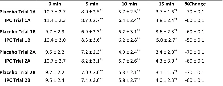
A significant main effect of treatment (p<0.05) was found at all time points during all trials (ES 0.93, 0.877, 0.894, and 0.901 during trials 1A, 1B, 2A, and 2B, respectively). A significant effect main of time (p<0.05) was also found at all time
points during all trials (ES 0.678, 0.636, 0.781, and 0.797 during trials 1A, 1B, 2A, and 2B, respectively, see Table 3). Although not statistically significant, blood lactate was consistently higher in all IPC trials when compared to the Placebo trials and appeared to recover at a similar rate.
Table 3. Mean blood lactate measurements SD for each recovery time point for all trials following placebo and IPC treatment.
0 min 5 min 10 min 15 min %Change
Placebo Trial 1A 10.7 ± 2.7 8.0 ± 2.5*† 5.7 ± 2.5*† 3.7 ± 1.6*† -70 ± 0.1 IPC Trial 1A 11.4 ± 2.3 8.7 ± 2.7*† 6.4 ± 2.4*† 4.8 ± 2.4*† -60 ± 0.1
Placebo Trial 1B 9.7 ± 2.9 6.9 ± 3.3*† 5.2 ± 3.1*† 3.6 ± 2.3*† -60 ± 0.1 IPC Trial 1B 10.4 ± 3.0 8.3 ± 3.6*† 6.2 ± 2.8*† 5.0 ± 2.7* -50 ± 0.1
Placebo Trial 2A 9.5 ± 2.2 7.2 ± 2.3*† 4.9 ± 2.4*† 3.4 ± 2.0*† -70 ± 0.1 IPC Trial 2A 10.7 ± 2.7 8.2 ± 3.1*† 5.7 ± 2.6*† 4.3 ± 3.0*† -60 ± 0.1
Placebo Trial 2B 9.2 ± 2.2 7.0 ± 3.0*† 5.3 ± 2.1*† 3.1 ± 1.5*† -70 ± 0.1 IPC Trial 2B 9.5 ± 2.4 7.4 ± 3.0*† 5.8 ± 2.7*† 4.0 ± 2.3*† -60 ± 0.1
*Significant change from time point to immediately post-exercise. † Significant change from time
point to previous time point.
Heart Rate Recovery
A significant main effect of treatment (p<0.05) was found at all time points during all trials (ES 0.994, 0.99, 0.995, and 0.995 during Trials 1A, 1B, 2A, and 2B, respectively. A significant main effect of time (p<0.05) was also found at all time points during all trials (ES 0.578, 0.600, 0.771, and 0.671 during
36 Table 4. Mean HR measurements ± SD for each recovery time point for all trials following placebo and IPC treatment.
0 min 5 min 10 min 15 min %Change
Placebo Trial 1A 146.1 ± 13.0 119.7 ± 13.4*† 118.8 ± 12.7* 119.0 ± 10.2* -18 ± 0.9 IPC Trial 1A 146.8 ± 15.7 120.4 ± 12.4*† 121 ± 11.6* 119.7 ± 12.2* -18 ± 0.1 Placebo Trial 1B 150.5 ± 20.14 118.5 ± 14.4*† 117.6 ± 13.2* 117.3 ± 11.1* -21 ± 0.1 IPC Trial 1B 148.5 ± 19.6 122.0 ± 13.8*† 120.8 ± 12.5* 121.2 ± 12.2* -18 ± 0.9 Placebo Trial 2A 142.9 ± 10.0 115.9 ± 9.5*† 116.0 ± 9.3* 114.0 ± 9.1* -20 ± 0.8 IPC Trial 2A 145.6 ± 17.2 120.3 ± 11.3*† 119.2± 9.8* 117.2 ± 10.9* -19 ± 0.1 Placebo Trial 2B 146.4 ± 15.1 117.0 ± 11.6*† 118.3 ± 10.4* 117.1 ± 11.7* -19 ± 0.9 IPC Trial 2B 147.9 ± 17.7 120.3 ± 9.7*† 118.9 ± 10.9* 119.5 ± 10.9* -18 ± 0.9
*Significant change from time point to immediately post-exercise. † Significant change from time
point to previous time point.
Mean Arterial Pressure Change
A significant main effect of treatment (p<0.05) was found at all time points during all trials (ES 0.998, 0.999, 0.998, and 0.998 during trials 1A, 1B, 2A, and 2B,
respectively). MAP only increased
significantly at one time point in the IPC group at the end of the recovery period. Although not statistically significant, MAP was lower at the start of all IPC trials when compared to the Placebo trials (see Table 5).
Table 5. Mean Arterial Pressure (MAP, mmHg) measurements ± SD taken at the end of each recovery time point for all trials following placebo and IPC treatment.
Baseline 15 min %Change
Placebo Trial 1A 88.3 ± 5.0 91.9 ± 6.6 4 ± 0.1 IPC Trial 1A 83.9 ± 6.5 91.0 ± 5.2 * 8 ± 0.1
Placebo Trial 1B 88.3 ± 5.0 88.6 ± 5.3 0.3 ± 0.1
IPC Trial 1B 83.9 ± 6.5 88.7 ± 3.7 6 ± 0.1
Placebo Trial 2A 85.8 ± 6.0 89.4 ± 5.9 4 ± 0.1
IPC Trial 2A 83.9 ± 7.8 87.7 ± 4.5 4 ± 0.1
Placebo Trial 2B 85.8 ± 6.0 89.8 ± 5.4 5 ± 0.1
37
Discussion
To our knowledge, this is the first study to examine the second window of protection of IPC and its effects on repeated cycling performance. We also believe it is the first investigation to examine how the second window of protection of IPC impacts recovery. The main findings of the study were: 1) there was a significant main effect of IPC treatment on TTF, and despite only finding significance between IPC trials 2A and 2B, mean performance time was
greater and mean performance
decrement was reduced with IPC despite varied individual performance, and that; 2) there was a significant main effect of IPC treatment on blood lactate levels, and subjects reached higher lactate levels in all trials with IPC treatment when compared to placebo. Lactate also appeared to recover at a similar rate with both
treatments. Interesting secondary
outcomes from the study were the positive effects of IPC treatment on heart rate and MAP. Post-trial heart rates with IPC treatment were on par with those in the placebo trials, despite higher workloads. The baseline MAP was lower at all IPC time points compared to placebo and, with the exception of one time point, MAP was also consistently lower after each of the IPC trials.
Performance- Time to Fatigue
The TTF results of the current study parallel results of a study performed by Kido et al., (2015), in which IPC improved
TTF by 4% in an incremental cycling test. They also found that lactate levels trended higher in the IPC group and that HR responses were on par with placebo, despite higher workload. They also found no difference in O2 uptake, lactate, or RPE,
but they did observe a 5.4± 4.8% greater maintenance of tissue saturation index in the IPC treatment group.
In a related crossover study26, researchers
had male cyclists perform a repeated 60-second supramaximal test, with a 45-minute period of passive recovery. They used a similar IPC protocol as the current study with 220 mmHg for the IPC treatment condition and 20 mmHg for the control condition. They found a 2% performance improvement, with no difference in VO2 response. They also
measured lactate throughout the passive recovery period and found that there was a significant change in the IPC treatment group when compared to the control trial. They concluded that an increased skeletal muscle activation and higher anaerobic contribution were enhanced by IPC in short-term exercise performance.
This is consistent with the results in a recently published study by Paradis-Deschênes et al. (2016), where they
examined the performance and
38 placebo (20 mmHg) administered to both
thighs, but subjects were tested at simulated low (FIO2 0.180, ~1200 m) and moderate (FIO2 0.154, ~2400 m) altitudes. At the low altitude, the IPC group had improved 5 km time trial performance by 1.1% and at the moderate altitude, they improved their 5 km time trial performance by 1.5%. Similar to the current study, they also observed some variability, with some responders and non-responders. They also found that an increase in HR, SV and Q was driven by IPC treatment. They suggested the possibility that IPC acted directly on the cardiovascular response and that it may alter vasodilatory pathways and they concluded that IPC might be an effective strategy to enhance high-intensity endurance performance at a moderate altitude. Interestingly, the reported
performance improvements in the
aforementioned studies are comparable to the effects of a one-month ‘live high — train low’ altitude training program for elite runners28.
However, these results conflict slightly with a study by Patterson et al. (2015), who performed a single-blind crossover study on male cyclists. The cyclists performed a series of 12 x 6-second sprints after receiving IPC (220 mmHg) or placebo (20 mmHg) treatment. They found that IPC treatment elicited a 2.4-3.7% increase in peak power for the first three sprints, but lead to no further change in the subsequent sprints. They also found no differences between trials for blood lactate, O2 uptake, or RPE at
any of the time points. They determined that
IPC might be beneficial for sprint activity, but not for endurance. This study is different in the cycling trial design, which might explain the difference in results when compared to the current study. As a single-blind study, perhaps there was also some bias on behalf of the subjects if there were aware of the treatment being received and therefore did not rate their perception of effort differently. A study by Crisafulli et al. (2011) also showed some conflicting results in their study on cyclists when compared to the present study. They had subjects perform an all-out supramaximal cycling bout following an IPC treatment, and at another time, without IPC treatment. Subjects were asked to perform at 130% of their established maximal W until exhaustion. Researchers found no difference in TTF between the treatments.
39 performance, researchers found that active
recovery was more beneficial for
maintaining endurance performance and sustaining power output30. They also found
that active recovery works best when performed at a moderate intensity (80- 90% or VT2, or 55-60% of heart rate reserve) to promote greater blood lactate removal and sustain endurance performance.
Based on the variability of individual responsiveness to IPC, the current study considered the importance of healthy endothelial function on IPC response, and the importance of active recovery to promote enhanced repeat performance. Looking at limitations from past studies, we controlled for risk factor burden through pre-screening and subject selection and controlled for the amount of training outside of the study, which knowingly can impact endothelial function. We also selected trained cyclists who were similarly fit, which might explain the potency of the results in this novel research into the delayed phase of IPC and its effects on performance and recovery. There was one male subject who performed better in the placebo trials and one female subject who performed similarly in the placebo and IPC trials. The male subject became a new parent just weeks before the IPC trials and was suffering from sleep deprivation and stress, which we believe impacted his performance. When he came back for the placebo trial four weeks later, he reported being better rested and that he’d been out biking in the preceding few weeks, which could also explain a better
performance. The female subject reported discomfort and bruising from the IPC treatment, which could have negatively impacted her performance.
Recovery- Lactate, HR, and MAP
There was a main effect of IPC treatment found at all time points and lactate levels were consistently higher in the IPC trials. Although subjects worked harder for a longer duration, their blood lactate levels appeared to recover similarly to the placebo, despite reaching higher levels at the end of the exercise bout. This is in keeping with the results found post-exercise in a study performed by Cruz et al. (2016) where they observed significantly higher lactate levels in the IPC treatment group. These results suggest that IPC treatments enhance the ability to clear, or to shuttle, lactate. A likely mechanism could be explained by an increased expression of monocarboxylate transporter proteins 1 1) and 4 (MCT-4). Research by Juel et al. (2003) lead to the observation that these transporter proteins were expressed in erythrocytes following oxygen deprivation. MCT-1 proteins are found on Type I muscle fibres and favour the uptake of lactate as a fuel source for ATP production. MCT-4 proteins are found on Type II muscle fibres, which favour lactate efflux so that it can be better oxidized and used as fuel for glycolysis. These proteins play an important role in shuttling lactate to where it can be better utilized for fuel, and since lactate is shuttled on a 1:1 ratio with H+, this action also serves as a buffer,
40 acidosis and subsequent fatigue. Since IPC
exerts ischemic stress, it is plausible that MCTs are also expressed following IPC and have an impact on the lactate levels reached. With an increased expression of MCT-4, lactate could effectively be shuttled out to allow a high rate of glycolytic flux, while the MCT-1 allowed Type I fibres to take up lactate as a fuel and H+ are shuttled into
mitochondria where they can be used to regenerate ATP in the electron transport chain. This proton consumption not only helps with ATP regeneration, but also effectively maintains pH balance and allows subjects to perform for a longer duration. Hypoxia also turns on hypoxia-inducible factor-1 alpha (HIF-1α), which is the master regulator of cellular and developmental response to hypoxia32. It controls the
metabolic and pH-regulating pathways, causing cells to respond to hypoxia by upregulating glucose transporters GLUT-1 and GLUT-4. HIF-1 α also inhibits pyruvate from entering the TCA cycle, which leads to lactate conversion so that glycolysis can continue.
There was also a main effect of IPC treatment on heart rate and MAP at all time points. Indeed, HR responses were on par with the placebo trials, despite higher workload and higher blood lactate levels. The recovery rate was also similar across all trials. MAP increased significantly at only one point in the recovery period after Trial 1A, but was similar or lower than the placebo at all time points. The baseline MAP was also lower 24 and 48 hours after IPC treatment.
IPC has been known to lower HR and BP25,33,
which was certainly apparent in the current study. Although we did not measure stroke volume, we can infer that stroke volume increased as a result of IPC’s influence on HR and vascular resistance. Since IPC is a known vasodilator due to stimulating the release of adenosine, bradykinin, and nitric oxide, it is likely that subjects experienced an increase in stroke volume, which permitted the heart rate values to remain comparable to placebo values despite higher workloads. Our subjects were able to perform for longer with IPC treatment and we can infer that with less vascular resistance, more oxygenated blood was able to circulate to skeletal muscle with each heartbeat.
Our hypothesis was that delayed IPC would be beneficial to both performance and recovery and our results show that IPC was indeed beneficial. IPC is a safe technique that can enhance performance and recovery, with no serious side effects. In this study, subjects experienced a 21% mean improvement in time to fatigue. These results with delayed IPC are better than other studies outlined above that noted a 3-5% improvement. This is on par, if not better than, the improvement demonstrated in other known ergogenic aids such as caffeine and erythropoietin (EPO)34,35.
41 performance with caffeine ingestion was 3.2
± 4.3% in endurance athletes. Since individual athletes vary in terms of caffeine metabolism, it is not a substance that all athletes can tolerate due to increased heart rate and digestive issues.
In comparison, there has been a lot of controversy over erythropoietin (EPO), which is a banned substance in competition. In one of the most notable studies on EPO and endurance performance, a small sample of non-elite cyclists were able to improve their time to fatigue by 54% due to a 12% increase in VO2max after a 14-day EPO
protocol35.
Since these were fit, but non-elite athletes, it was extrapolated that in an elite population, that performance increase might be closer to 5%. In contrast, a more recent study on elite athletes published in The Lancet36 showed that although peak
power and VO2max increased in the subjects
who received EPO, their performance in a race setting did not differ significantly (0-3% time improvement). IPC shows good promise for safely enhancing performance and recovery, although there is much more to learn about effective dosage, individual response, and the protective window of delayed IPC.
Limitations
To our knowledge, this is the only study looking at the delayed phase of IPC and how it effects performance and recovery. We
controlled for menstrual cycle, activity level, caffeine intake, and also pre-screened our subjects to exclude those with any risk factor burden. Since the mechanisms behind individual variability and IPC responsiveness are still somewhat unclear33, it would have
been interesting to measure endothelial function and stroke volume. We had also initially planned to measure pH, but in the end, this was not possible. pH kinetics could have given us more knowledge about the recovery aspect of IPC, since that is still largely unknown.
Conclusions
In conclusion, IPC has shown performance enhancing benefits in different exercise modalities15,25,26,37,38 that coincide with the
findings of the current study. Despite some individual variability, delayed IPC influenced TTF, blood lactate, HR, and MAP in repeated bouts of cycling at an altitude of 2350 metres in acclimatized, recreational cyclists. This novel research shows that delayed IPC has the ability to improve TTF, decrease
performance decrement between
42
Address for Correspondence
Lance Dalleck, Ph.D., High Altitude Exercise Physiology Program, 600 N. Adams St., Western Colorado University, Gunnison, CO, United States, 81231. Phone: 970-943-3095;
Email: ldalleck@western.edu.
References
1. Porcari J, Bryant C, Comana F. (2015). Exercise Physiology. FA Davis Company.
2. Brooks GA, Fahey TA, Baldwin KM. (2004). Exercise
Physiology: Human Bioenergetics and Its Applications.
New York City, New York: McGraw-Hill.
3. Noakes TD. (2000). Physiological models to understand exercise fatigue and the adaptations that predict or enhance athletic performance. Scand J Med Sci Sports, 10(3), 123–145.
4. Robergs R, Ghiasvand F, Parker D. (2004). Biochemistry of exercise-induced metabolic acidosis. Am J Physiol Regul
Integr Comp Physiol, 287(3), 502-516.
5. Brooks GA. (2007). Lactate: Link between glycolytic and oxidative metabolism. Sports Med, 37(4), 341–343. 6. Bangsbo J, Johansen L, Graham T, Saltin B. (1993). Lactate
and H+ effluxes from human skeletal muscles during intense, dynamic exercise. J Physiol, 462(1), 115–133. 7. Hermansen L, Stensvold I. (1972). Production and
removal of lactate during exercise in man. Acta Physiol
Scand, 86(2), 191–201.
8. Murry CE, Jennings RB, Reimer KA. (1986). Preconditioning with ischemia: A delay of lethal cell injury in ischemic myocardium. Circulation, 74(5), 1124–1136. 9. Reimer K, Murry C, Yamasawa I, Hill M, Jennings R. (1986).
Four brief periods of myocardial ischemia cause no cumulative ATP loss or necrosis. Am J Physiol Heart Circ
Physiol, 251(6), 1306–1315.
10. Murry CE, Richard VJ, Reimer KA, Jennings RB. (1990). Ischemic preconditioning slows energy metabolism and delays ultrastructural damage during a sustained ischemic episode. Circ Res, 66(4), 913–931.
11. Pagliaro P, Gattullo D, Rastaldo R, Losano G. (2001). Ischemic preconditioning: From the first to the second window of protection. Life Sci, 69(1), 1–15.
12. Yamashita N, Hoshida S, Taniguchi N, Kuzuya T, Hori M. (1998). A “second Window of Protection” occurs 24 h after ischemic preconditioning in the rat heart. J Mol Cell
Cardiol, 30(6), 1181–1189.
13. Jean-St-Michel E, Manlhiot C, Li J, Tropak M, Michelsen M, Schmidt M, et al. (2011). Remote preconditioning improves maximal performance in highly trained athletes. Med Sci Sports Exerc, 43(7), 1280–1286. 14. Yellon D, Alkhulaifi A, Pugsley W. (1993). Preconditioning
the human myocardium. The Lancet, 342(8866), 276–277. 15. Bailey T, Jones H., Gregson W, Atkinson G, Cable N,
Thijssen D. (2012). Effect of ischemic preconditioning on lactate accumulation and running performance: Med Sci
Sports Exerc, 44(11), 2084–2089.
16. Crisafulli A, Tangianu F, Tocco F, Concu A, Mameli O, Mulliri G, Caria M. (2011). Ischemic preconditioning of the muscle improves maximal exercise performance but not maximal oxygen uptake in humans. J Appl Physiol, 111(2), 530–536.
17. Page W, Swan R, Patterson S. (2017). The effect of intermittent lower limb occlusion on recovery following exercise-induced muscle damage: A randomized controlled trial. J Sci Med Sport, 20(8), 729–733. 18. Tanaka D, Suga T, Tanaka T, Kido K, Honjo T, Fujita S, et al.
(2016). Ischemic preconditioning enhances muscle endurance during sustained isometric exercise. Int J
Sports Med, 37(8), 614–618.
19. Salvador A, De Aguiar R, Lisbôa F, Pereira K, Cruz R, Caputo F. (2016a). Ischemic preconditioning and exercise performance: A systematic review and meta-analysis. Int
J Sports Physiol Perform, 11(1), 4–14.
20. Salvador A, De Aguiar R, Lisbôa F, Pereira K, Cruz R, Caputo F. (2016a). Ischemic preconditioning and exercise performance: A systematic review and meta-analysis. Int
J Sports Physiol Perform, 11(1), 4–14.
21. Amann M, Eldridge M, Lovering A, Stickland M, Pegelow D, Dempsey J. (2006). Arterial oxygenation influences central motor output and exercise performance via effects on peripheral locomotor muscle fatigue in humans. J Physiol, 575(Pt 3), 937–952.
22. Hausenloy DJ, Yellon DM. (2010). The second window of preconditioning (SWOP) where are we now? Cardiovasc
Drugs Ther, 24(3), 235–254.
23. Katakam PV, Jordan JE, Snipes JA, Tulbert CD, Miller AW, Busija DW. (2007). Myocardial preconditioning against ischemia-reperfusion injury is abolished in zucker obese rats with insulin resistance. Am J Physiol Regul Integr
Comp Physiol, 292(2), 920–926.
43
Comparative performance of two point-of-care analysers for lipid testing. Clin Lab, 53(9–12), 561–566.
25. Kido K, Suga T, Tanaka D, Honjo T, Homma T, Fujita S, et al. (2015). Ischemic preconditioning accelerates muscle deoxygenation dynamics and enhances exercise endurance during the work-to-work test. Physiol Rep, 3(5). pii: e12395.
26. Cruz R, de Aguiar R, Turnes T, Salvador A, Caputo F. (2016). Effects of ischemic preconditioning on short-duration cycling performance. Appl Physiol Nutr Metab, 41(8), 825–831.
27. Paradis-Deschênes P, Joanisse D, Billaut F. (2016). Ischemic preconditioning increases muscle perfusion, oxygen uptake, and force in strength-trained athletes.
Appl Physiol Nutr Metab, 41(9), 938–944.
28. Stray-Gundersen J, Chapman R, Levine B. (2001). “Living high-training low” altitude training improves sea level performance in male and female elite runners. J Appl
Physiol, 91(3), 1113–1120.
29. Patterson S, Bezodis N, Glaister M, Pattison J. (2015). The effect of ischemic preconditioning on repeated sprint cycling performance. Med Sci Sports Exerc, 47(8), 1652– 1658.
30. St. Pierre I, Buchanan C, Dalleck L. (2018). Active vs. passive recovery and exercise performance: Which strategy is best? Retrieved April 28, 2018, from
https://www.acefitness.org/education-and- resources/professional/certified/march-2018/6919/ace- sponsored-research-active-vs-passive-recovery-and-exercise-performance-which-strategy-is-best
31. Juel C, Lundby C, Sander M, Calbet J, van Hall G. (2003).
Human skeletal muscle and erythrocyte proteins involved in acid-base homeostasis: adaptations to chronic hypoxia. J Physiol, 548(2), 639–648.
32. Weidemann A, Johnson R. (2008). Biology of HIF-1α. Cell
Death Differ, 15(4), 621–627.
33. Gibson N, Mahony B, Tracey C, Fawkner S, Murray A. (2015). Effect of ischemic preconditioning on repeated sprint ability in team sport athletes. J Sports Sci, 33(11), 1182–1188.
34. Ganio MS, Klau JF, Casa DJ, Armstrong LE, Maresh CM. (2009). Effect of Caffeine on sport-specific endurance performance: A systematic review. J Strength Cond Res, 23(1), 315–324.
35. Thomsen J, Rentsch R, Robach P, Calbet J, Boushel R, Rasmussen P, et al. (2007). Prolonged administration of recombinant human erythropoietin increases submaximal performance more than maximal aerobic capacity. Eur J App Physiol, 101(4), 481–486.
36. Heuberger J, Rotmans J, Gal P, Stuurman F, Westende J, Post T, et al. (2017). Effects of erythropoietin on cycling performance of well trained cyclists: a double-blind, randomised, placebo-controlled trial. The Lancet, 4(8), 374–386.
37. Salvador A, De Aguiar R, Lisbôa F, Pereira K, Cruz R, Caputo F. (2016b). Ischemic preconditioning and exercise performance: A systematic review and meta-analysis. Int
J Sports Physiol Perform, 11(1), 4–14.