This is an open access journal, and articles are distributed under the terms of the Creative Commons Attribution-Non Commercial-ShareAlike 4.0 License, which allows others to remix, tweak, and build upon the work non-commercially, as long as appropriate credit is given and the new creations are licensed under the identical terms.
© 2018 Journal of Advanced Pharmacy Education & Research | Published by SPER Publication
64
Molecular epidemiological updates on spotted fever rickettsioses
in animal species and their hard ticks settling Egyptian desert
Nesreen A.T. Allam
1*, Faragallah M. El Moghazy
1, 2, Sayed M.M. Abdel-Baky
31 Parasitology and Animal Diseases Department, Veterinary Research Division, National Research Centre, Dokki, Cairo, Egypt, 2 Biology Department, College of Science and
Humanity Studies, Prince Sattam Bin Abdul Aziz University, Kingdom of Saudi Arabia, 3 Parasitology Unit, Department of Animal Health, Division of Animal and Poultry
Production, Desert Research Center, Matariya, Cairo, Egypt.
Correspondence: Nesreen A. T. Allam, Parasitology and Animal Diseases Department, Veterinary Research Division, National Research Centre, Dokki, Cairo, Egypt P.O. Box: 12622, Giza. E_mail: [email protected]
ABSTRACT
Hard ticks are ectoparasites that infest animals prompting severe transmittable infections. It is aimed to identify then characterize spotted fever group (SFG) rickettsioses in ticks and their hosts from smallholdings and Bedouin communities in Egypt desert including; North West Coast, Western Desert Oasis, South East Coast, Suez Canal region and Sinai. The 5223 adult ixodid ticks were collected from 270 ruminants; 110 camels, 120 sheep, and 40 cattle, examined seasonally (4 times/year) from June 2014 to July 2016. The statistical analysis of infestation density of all species on each hosts was highly significant, but was not significant between different localities. The infestation density on cattle was higher (59 %) than camels (33.6%) and sheep (7.4%) regarding all 14 species of ticks identified. The adult stages (♀&♂)of Boophilus annulatus, Hylomma dromedarii, and Rhipicephalus sanguineus were the highest in density on cattle, camels, and sheep recording 56%, 24.7% and 5.9%, respectively. Nucleotides similarities of ticks’ 18S rDNA and 16S rDNA genome markers were 99-100% against genbank records, which confirmed species identification by morphological key. Molecular screening for rickettsiosis was carried by multi-genes typing technology; where 16S rDNA was the primary target. Positive PCRs were then typed into Rickettsiae species by the ompA and gltA genes alignments. Hylomma dromedarii ticks were the most susceptible to Rickettsia (35.7%) than other tick species. The prevalent localities for ticks’ rickettsiosis were Shalateen, Dakhla Oasis, Siwa Oasis, El Salloum, and Marsa Matrouh recording 17.8%, 11.8%, 11.2%, 9.6% and 9.2%, respectively. With regards to animal hosts, the SFG incidence was higher in camels than sheep recorded 64.7% and 29.4%, respectively. The hosts’ rickettsiosis incidence recording 14.7%, 13.7%, 12.8%, 11.8% and 10.8% was in Bir El abed, Shalateen, Wadi Gharandal, Al hasna, Marsa Matruh and El Salloum, respectively. Additionally,
Rh. sanguineus (dogs ticks) had high incidence for rickettsioses; collected from all infested animal species investigated during the study. Hence, the Rh. sanguineus PCR-positive Rickettsia collected from cattle (5.9%) is an incidence never recorded; therefore, needs experimental studies before being accepted. Rickettsia sibirica mongolitimonae-like was detected for the first time within Egyptian livestock’s population. Rickettsia aeschlimannii-like, R. africa-likee, in addition to, unclassified Rickettsia-like were additionally detected within the investigated specimens. Still, the pathological impacts of freely moving rodents and migratory birds are underestimated in epidemiology of such exotic zoonosis. Moreover, the role of additional vector (rodents’ soft ticks) and/or reservoirs (desert reptiles) not clearly identified yet; a hypothesis that needs comprehensive profounder studies to improve physicians, as well as, veterinarians’ differential diagnosis of tick-borne fever due to rickettsioses in Egypt.
Keywords:R. aeschlimannii, R. africae, R. sibirica mongolitimonae, multi-genes sequence typing, Ixodidae, DNA markers, ruminants.
Introduction
Since its first description in 1914, spotted fever group (SFG)
Rickettsia are still expanding [1]. The improvements in molecular
technologies had significantly advanced our abilities to conduct genetic analyses and clearly indicated the proper phylogenetic positions of most of the fastidious bacterial species in the family
Rickettsiaceae, order Rickettsiales[2, 3]. Tick-borne biohazards result
in significant morbidity and thousands of human and animal deaths annually, hence, transmitting a great diversity of infectious agents than any other hematophagous arthropods [4, 5].
Ixodidae (Hard ticks) is a large family distributed all over the world [6, 7]. In Egypt, genera Amblyomma, Hyalomma and
Rhipicephalus (Boophilus) include the most abundant ixodid ticks infesting animals[5, 8-15]. Taxonomical key of ticks is based mainly
on the morphological criteria of adult’s phase [16, 17]. Recently,
the molecular characterization for taxonomic identification and
Access this article online
Website: www.japer.in E-ISSN: 2249-3379
How to cite this article: Nesreen A.T. Allam, Faragallah M. El Moghazy, Sayed M.M. Abdel-Baky. Molecular epidemiological updates on spotted fever rickettsioses in animal species and their hard ticks settling Egyptian desert. J Adv Pharm Edu Res 2018; 8(1):64-74.
Journal of Advanced Pharmacy Education & Research | Jan-Mar 2018| Vol 8 | Issue1 65
phylogenetic purposes of ticks has been indispensable by a group of DNA markers with regards to nuclear and mitochondrial genes, respectively [18-21]. The 18S rDNA is best used for genera
identification while the 16S rDNA is the most useful markers for ticks’ taxonomy at the species level [22-24]. The role of vertebrates
as reservoirs of some SFG rickettsiae has yet to be clarified [25, 26].
Since 1960s when the primary study on SFG in Egypt was published, other multiple traditional smear staining, serological, immunohistochemical as well as molecular studies had designated ticks, rodents, animals and human as being endemic with Rickettsia agents [5, 12-15, 27-32]. The public health measures
including; the ecological conditions that influence the animal stocks production, as well as, the control of vectors and their diseases [12, 13, 15, 33-38]by the veterinary authorities within studied
localities became fundamental [39, 40]. Updating the data
concerning Rickettsia epidemiology in Egypt with regards to ticks’ species susceptibility is essential [39, 40]. The identification of
Rickettsia into species has been dependent on employing the multi-genes typing of five sequences 16S rRNA, gltA, OmpA,
OmpB, and sac4 genes [12, 13, 15, 33-38]. The outer membrane protein
A of the cell surface antigen (OmpA) and citrate synthase (gltA) genes are characteristics of SFG Rickettsiae with powerful dis-crimination in between Rickettsia spp. due to their high variability among SFG [12, 13, 15, 33-38]. During the present investigation, it is
aimed to identify and molecular characterize spotted fever group (SFG) rickettsioses in ticks and their domestic animals from smallholdings and Bedouin communities in Egypt settling the desert in North West Coast, Western desert Oasis, South East Coast, Suez Canal region and Sinai.
Material and Methods
Geographical Scope of the Study in the
Egyptian Desert
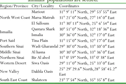
:Twelve sampling localities in 9 provinces from Egypt desert selected in the present study (Table 1).
Table 1: The coordinates of the studied regions and localities of the Egyptian deserts were the sampled
animals’ populations are settled.
Region/Province City/Locality Coordinates
North West Coast
Mariout 31° 9′ 11″ North, 29° 53′ 55″ East Marsa Matruh 31° 21′ 0″ North, 27° 14′ 0″ East El Salloum 31° 30′ 13″ North, 25° 6′ 54″ East
Ismailia Qantara Shark 30° 51Ismalia ′ 0″ North, 32° 18′ 36″ East 30° 36′ 0″ North, 32° 17′ 0″ East Port Said Tina Plain 31° 15′ 0″ North, 32° 17′ 0″ East Southern Sinai Wadi Gharandal 29° 30′ 0″ North, 33° 50′ 0″ East Middle Sinai Al hasna 30° 30′ 0″ North, 33° 36′ 0″ East Northern Sinai Bir Al abed 31° 0' 59" North, 33° 0' 38" East Western Desert Siwa Oasis 29° 11′ 0″ North, 25° 33′ 0″ East
New Valley Dakhla Oasis 25° 29′ 29.6″ North,28° 58′ 45.2″ East
South East Coast Shalateen 23° 7′ 54″ North, 35° 35′ 8″ East
Ethical Approval:
All procedures were in accordance with the ARRIVE guidelines which were in accordance to the European (EU) Directive 2010/63/EU for animal experiments and the National Institutes
of Health guide for the care and use of laboratory animals (NIH Publications No. 8023 revised 1978). In addition, were in agreement with the adopted ethics guidelines of the ministry of higher education and scientific research (50/4/10) national research center (10120507); the funding organizations, and desert research center for the care and use of animals in Egypt.
Blood Samples of Investigated Animals
Population:
A total number of 270 animals were investigated for tick infestations, which were examined/sampled seasonally (4 times/year) during the study period from June 2014 to July 2016. The inspected animals were; 110 camels, 120 sheep, and 40 cattle. EDTA-whole blood samples were collected from jugular veins [41] and used in preparing blood smears for staining
technique and the remaining were stored at -80 ºC until DNA was extracted for molecular studies. Additional 10 ml blood/host/trip was collected on plan tubes for additional serum samples analysis that were separated after centrifugation at 2000 rpm for 10 min.
Ticks
Specimens
and
Taxonomic
Classification by Stereo-Zoom Microscope:
A total of 5223 adult ticks were collected; other developmental stages were excluded. Ticks were captured from the host by forceps and orientated anticlockwise until the capitulum detaches from the host, then they were placed in polyethylene tube, 13 or 25 mm in diameter and 100 mm in height, sealed at one end by a mixture of gypsum and graphite at a ratio of 5:1. The tube was covered with a piece of muslin cloth securely held by a rubber band. The gypsum graphite was moistened to provide adequate humidity for the ticks during transportation to the laboratory [42].
Ticks were brought alive to the laboratory for morphological identification by stereomicroscope and identification keys. The collected ticks were counted and sorted to different genera, species and sex. Tick species were identified morphologically using taxonomic keys of [7, 8, 43]. The tick species were
morphologically examined in details using stereo-zoom microscope, especially the dorsal and ventral surfaces of adult ticks. The adult ticks were photographed by a digital camera fixed on a stereomicroscope.
Detection of Rickettsiae in Stained Smears by
Light Microscope:
All collected samples were examined for Rickettsiae using staining technique. According to Burgdorfer [44], hemolymph was
impressed on slide following scissors amputating the distal portion of the legs, fixed by air dry. Both blood and hemolymph slides were stained with Gimenez stain [45]. Then, prepared slides
were examined under oil emersion lens using an ordinary microscope (Zeiss).
DNA Purification:
Genomic DNA was extracted from camel, cattle, and sheep blood specimens using GF-1 Tissue Blood Combi DNA Extraction Kit (Vivantis) according to the manufacturer’s instructions. Also, total DNA was extracted and purified from the tissues of adult ticks after dissection of each tick into quarters using high salt concentration protocol [46]. DNA concentration
This is an open access journal, and articles are distributed under the terms of the Creative Commons Attribution-Non Commercial-ShareAlike 4.0 License, which allows others to remix, tweak, and build upon the work non-commercially, as long as appropriate credit is given and the new creations are licensed under the identical terms.
© 2018 Journal of Advanced Pharmacy Education & Research | Published by SPER Publication
66
Molecular Confirmation of Ticks’ Taxonomy:
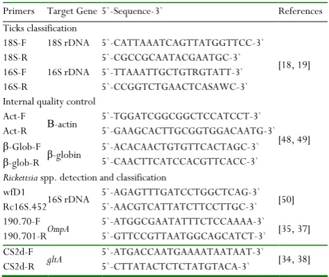
Ten randomly selected adult ticks/species/locality were investigated by molecular techniques to confirm the previous morphological classification of ticks’ into species. Two-locus sequences typing included 18S rDNA and 16S rDNA; the nuclear and mitochondrial genes, respectively, was carried out. The 18S-F and 18S-R (Vivantis) primers pair previously designed were used in 18S rDNA gene amplification [18, 19] with predicted
products size 780 bp(Table 2). Moreover, the 16S rDNA gene primers' set (16S-F and 16S-R, Vivantis) were designed according to previous publications [18, 19], with predicted products
size 455 bp, respectively(Table 2). Each PCR mixture contained 25-50 ng/μl genomic DNA, 10 pM/μl of each primer, 12.5 μl ×2 Dream Taq Green PCR master mix (×2 buffer, 0.4 mM dNTP and 4 mM MgCl2; Thermo Scientific), and 9 μl nuclease free water (Qiagen) to complete the total volume of the reactions. All amplifications were performed in a PTC-100™ thermal cycler (MJ Research Inc., USA) utilizing the following cycling profile; one cycle at 94°C for 5 min of initial dena-turation, then 30 cycles denaturation at 94°C for 1min, annealing at 45°C for 1 min and elongation at 72°C for 1 min, and the final elongation at 72°C for 10 min [18, 19, 47]. A reagent blank was run
as control simultaneously with every PCR.
Table 2: Synthesized oligonucleotides primers used during PCR amplifications and sequencing of markers genes
Primers Target Gene 5`-Sequence-3` References
Ticks classification 18S-F
18S-R
18S rDNA 5`-CATTAAATCAGTTATGGTTCC-3` 5`-CGCCGCAATACGAATGC-3`
[18, 19] 16S-F
16S-R
16S rDNA 5`-TTAAATTGCTGTRGTATT-3` 5`-CCGGTCTGAACTCASAWC-3` Internal quality control
Act-F
Act-R Β-actin
5`-TGGATCGGCGGCTCCATCCT-3` 5`-GAAGCACTTGCGGTGGACAATG-3`
[48, 49] β-Glob-F
β-glob-R β-globin
5`-ACACAACTGTGTTCACTAGC-3` 5`-CAACTTCATCCACGTTCACC-3`
Rickettsia spp. detection and classification wfD1
Rc16S.452 16S rDNA
5`-AGAGTTTGATCCTGGCTCAG-3` 5`-AACGTCATTATCTTCCTTGC-3` [50] 190.70-F
190.701-R OmpA
5`-ATGGCGAATATTTCTCCAAAA-3` 5`-GTTCCGTTAATGGCAGCATCT-3` [35, 37] CS2d-F
CS2d-R gltA
5`-ATGACCAATGAAAATAATAAT-3` 5`-CTTATACTCTCTATGTACA-3` [34, 38]
Classification of Ticks-Borne Rickettsiae by
Multi-Genes Amplification and Sequencing:
Rickettsiae 16S rDNA gene primers' set (wfD1 and Rc16S.452, Vivantis) were designed according to Ogo et al. [50], with
predicted products size 426 bp, respectively(Table 2). The OmpA
gene primers were designed to span the nucleotides positions from 70 to 90 and from 701 to 681 (Table 2), respectively, and the predicted product size ranged from 590-634 bp [35, 37, 51].
Moreover, primers CS2d-F and CSEnd-R could amplify the
full-length of the gltA gene (Table 2), as the predicted product size ranged from 852 to 1265 bp, therefore; CS2d primer was designed to be completely homologous to the corresponding portion of the gene in R. conorii for only SFG [34, 38]. The
amplification reactions were performed in a PTC-100™ thermal cycler (MJ Research Inc., USA) under complete aseptic conditions. Each 25 µl total volumes of each PCR mixture contained 25-50 ng/µl genomic DNA (host or vector), 10 pM/µl of each primer, 12.5 µl of 2x Dream Taq Green PCR master mix (Thermo Scientific) and 9 µl nuclease free water (Qiagen) to complete the total volume of the reaction. All amplifications were performed utilizing the following cycling profile for OmpA primers; one cycle at 94 ºC for 5 min (initial denaturation) followed by 40 cycles consisting of denaturation at 94 ºC for 1 min, annealing at 59 ºC for 1 min and elongation at 72 ºC for 1 min, and the final elongation at 72 ºC for 10 min [12].
While gltA protocol included one cycle at 94 ºC for 5 min (initial denaturation) followed by 40 cycles of denaturation at 94 ºC for 1.5 min, annealing at 52 ºC for 1.5 min and elongation at 72 ºC for 1.5 min, then the final elongation at 72 ºC for 20 min [12]. A
reagent blank was run simultaneously as negative control with each PCR.
Amplified products were electrophoresed in 1% agarose gels in TBE buffer then stained with ethidium bromide (Sigma Aldrich). A 100 bp ladder (Alliance Bio, USA) was used with each gel. Gels photos were analyzed by Lab Image software (BioRad). Sequencing reactions were performed in an MJ Research PTC-225 Peltier Thermal Cycler using an ABI PRISM®BigDye™
Terminator Cycle Sequencing Kits with AmpliTaq®DNA polymerase (Applied Biosystems), following the protocols sup-plied by the manufacturer.
Internal Quality Control of Molecular Assays:
A semi-qualitative internal control to verify the efficiencies of the DNA isolation and the PCR assays were applied [48]. The primers
used derivative from highly conserved regions in both the host species and ticks vectors genomes; β-globin and β-actin genes sequences according to Konnai et al. [49], (Table 2). All
amplifications were performed in a PTC-100™ thermal cycler (MJ Research Inc., USA) utilizing the following cycling profile; one cycle at 94°C for 5 min initial denaturation, then 30 cycles of denaturation at 94°C for 1min, annealing at 60°C for 1.5 min and elongation at 72°C for 2 min, and the final elongation at 72°C for 10 min [48, 49].
Data Analyses by NCBI Blastn:
Journal of Advanced Pharmacy Education & Research | Jan-Mar 2018| Vol 8 | Issue1 67
database. Multiple sequences alignments for evolutionary relationships in between new Egyptian records and Genbank reference isolates were inferred [51, 52].
Statistical Analysis:
The Chi-square was carried out on the data collected in order to test homogeneity in the number of ticks collected according to; a) Infected hosts with Rickettsia in different localities and b) Infected different species of hard ticks with Rickettsia in different localities. This method analyzed using The FREQ Procedure Model of SAS[53] for Windows Evaluation Version. The method
adopted was carried out according to Snedecor and Cochran[54].
Probability values (P-value) < 0.05 were considered of statistical significant and < 0.001 were considered of high statistical significant.
Results
Medical Inspection of Animals’ Population:
The inspected animals were: 110 camels, 120 sheep, and 40 cattle. Ixodid ticks' collection began as a routine method of flocks’ examination to detect the predilection sites of the ticks. It had begun by examining the head of the animal, then the outer and inner sides of ears, neck, lateral and medial aspects of the fore and hind limbs, abdomen, inguinal and anal regions as well as under the tail. The infestation sites examined were 14 on each animal inspected four times per year, each site on each host designated in 16 cm2 area (4x4cm), in all species infestation were
found of significant differences synergistic to the stage of infestation.
The epidemiological records (not shown) included: age, sex, breed, the purpose of rearing animal, production/reproduction records, external infestation, date of ticks’ collection, clinical signs, suspected disease and time and type of treatments. The main clinical signs observed on infested animal hosts during sampling were fever, anorexia, lethargy, anemia, enlargement of superficial lymph nodes and emaciation, other than being apparently healthy in the majority of the inspected population. Older individuals were more susceptible for external infestation.
Morpho-Molecular Identification of Ticks
Species:
Fourteen tick species were identified in the specimens collected from the 9 provinces of Egypt that were investigated during the present survey. The classified 5223 adult ticks were categorized into 1741 males and 3482 females. The classification was by both their taxonomic keys (Chart 1) then confirmed by their amplified and sequenced molecular contents. They were designated to 4 genera; Amblyomma, Hyalomma, Rhipicephalus and Boophilus, as the following: Amblyomma gemma (n=5), A. lepidum (n=6), A. variegatum (n=5), B. annulatus (n=2922), H. albiparmatum
(n=25), H. anatolicum excavatum (n=127), H. dromedarii
(n=1288), H. impeltatum (n=91), H. marginatum marginatum
(n=105), H. m. rufipes (n=53), H. truncatum (n=90), Rh. humeralis
(n=20), Rh. pulchellus (n=22) and Rh. sanguineus(n=307, only
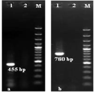
from animal spp. included in the study). The PCR screening utilizing the specific primers of 18S rDNA and 16S rDNA, proved the length of amplified fragments in all tick species of both genes were 780 and 455 bp, respectively (Fig 1: A & B). When the corrected sequences of each tick species were aligned against GenBank records, the identities of the Egyptian sequences confirmed the morphological classification of ticks to their designed species with similarities presents ranged from 97-100 % to records in the GenBank databases.
Chart 1:The infestation density of different ticks’ species on all studied hosts in all destinations included in the present study.
Figure 1.Molecular identification of tick species by PCR amplification of DNA markers detected in 1.5% agarose gels stained with ethidium
bromide. A) lane 1: 100 bp DNA ladder, lane 2: Control negative, lane 3: 780 bp amplicon of 18S rDNA gene. B) lane 1: 100 bp DNA
ladder, lane 2: Control negative, lane 3: 455 bp amplicon of 16S rDNA gene.
Epidemiological Analyses of Infestation
Incidence and Density on Hosts with regards
to Geographical Distribution:
Allam NAT, et al.: Molecular Epidemiological Updates on Spotted Fever Rickettsioses in Animal Species and their Hard Ticks Settling Egyptian Desert
68 Journal of Advanced Pharmacy Education & Research | Jan-Mar 2018| Vol 8 | Issue1
different species of ticks on different hosts was highly significant (P < 0.001). During the period of study, the adult B. annulatus
[Say (1821)] was the highest density abundant among species on cattle; 2922 adult ticks (55.94%), followed by H. dromedarii
[Koch (1844)] on camels; 1288 adult ticks (24.66%), then Rh. sanguineus [Latreille (1806)] on sheep; 307 adult ticks (5.88%), (Chart 2: A, B & C). In comparison, B. annulatus the most common species on cattle (55.94%) was higher than H. dromedarii
species on camels (24.66%), (Chart 2: A, B & C). According to hosts susceptibility for ticks’ infestation; with regards to all ticks species, the infested camels and cattle percentage recorded 33.62% & 59.03%, respectively, were higher than infested sheep recording 7.35%, (Chart 2: A, B & C).
Rickettsiosis Incidence with regards to
Microscopical Detection by Stained Smears
Examination:
Gimenez staining of hemolymph smears was a successful traditional tool for primary detection of Rickettsia infections in ticks but was not successful on examination of ruminants blood smear (Fig 2: A & B). Therefore, prevalence of Rickettsia spp. in animal hosts recorded 0%, while their tick species revealed 9% using Gimenez staining technique. These results triggered the importance of molecular identification of infection in both specimens type by PCR.
Figure 2: Ticks haemolymph smears stained with Gimenez showing:
A) Positive slides containing Rickettsiae (red cocci) inside or around green haemocytes, and B) Negative slide containing green haemocytes,
(Magnification Power = 1000x).
Chart 2:The ticks’ infestation density on camel (a), cattle (b), and sheep (c) investigated during the study with regards to ticks’ species.
Ticks-Borne Rickettsiosis Incidence with
regards
to
Geographical
Distribution
Utilizing Multi-Genes Typing in the Studied
Hosts and their Ticks Species:
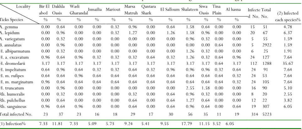
Hosts and ticks’ samples were both screened molecularly by 16S rDNA, OmpA and gltA genes primers sets in two steps molecular detections (additional primers sets of 6 genes were applied but results are not shown in this study) considering the representation of different localities. Samples with amplified fragments of 455, 600 and 1200 bp recorded for amplified 16S rDNA, OmpA and gltA genes were considered Rickettsia positive after alignments of products’ sequences against GenBank database, respectively (Fig. 3: A & B). The localities investigated with positive results for Rickettsia spp. as well as the incidence and number of positive ticks and their host with are statistically analyzed in (Tables 3 & 4). The hard ticks of H. dromedarii species were the highest susceptible vectors to Rickettsia (35.67%) than other species (Table 3). The most prevalent localities with infected hard tick species; vector for SGF Rickettsia, were Shalateen, Dakhla and Siwa Oasis recording 17.83 & 11.78 & 11.15%, respectively, followed by El-Salloum and Marsa Matruh recording 9.55 & 9.24%, respectively (Table 3). The highest
Journal of Advanced Pharmacy Education & Research | Jan-Mar 2018| Vol 8 | Issue1 69
infection percentage was in camels recorded 9.8% in both Shalateen and Wadi Gharandal districts which belong to the Red Sea and Southern Sinai Governorates,respectively. Followed by Marsa Matrouh district which belongs to Marsa Matruh Governorate with infection percentage recorded 7.84% (Table 4). Moreover, the highest infection incidence in sheep was 7.84% in Bir El abed district that belongs to Northern Sinai Governorate (Table 4). Generally, the highest incidence for rickettsiosis was in Bir El abed, Shalateen and Wadi Gharandal recording 14.71, 13.73 & 12.75%, respectively, followed by Al hasna, Marsa Matruh and El Salloum recording 11.76% and 10.78%, respectively (Table 4). Camels were proved the most susceptible to rickettsiosis (64.71%) than other animals’ species studied (even dogs which were not shown in this study) (Table 4). On the other hand,cattle rickettsiosis was documented in 5.88% of studied population; 1.96% of cattle populations in Dakhla Oasis in the Western desert, and Qantara Shark and Tina Plain in the Suez Canal region, which a result that needs further confirmation and explanation, hence, Rickettsia infections are not recorded in cattle previously (Table 4).
The alignment of OmpA and gltA genes sequences against previously recorded isolates in GenBank indicate novel isolates that have less than 95% sequences similarity to the previously identified Rickettsia isolates in Sinai in Egypt; which was molecularly characterized to R. aeschlimannii-like; in all ticks species except A. variegatum, H. albiparmatum, Rh. sanguineus and
Rh. humeralis, while R. africae-like; in all ticks species except Rh. sanguineus. Moreover, the obtained sequences of amplified genes indicated the existence of new species identified R. sibrica mongolitimonae-like. In continence, further molecular investigations to fully characterize these species/isolates was urgent (additional 6 genes are not shown in this study). The similarity percent of identity matrix of Egyptian Rickettsia
sequences was based on multiple alignments. The R. africae-like
and R. aeschlimannii-like isolate from H. anatolicum excavatum, H. dromedarii, H. impeltatum, H. marginatum marginatum, H. m. rufipes
were clustered in a separate clade from other R. africae reference strains with a bootstrap value ranged from 85-99 (trees are not shown in this study), which indicated a novel strain of R.
africae-like within ticks picked from camel from Sinai provinces, Shalateen and Western Desert Oasis. Moreover, R. sibrica mongolitimonae-like is the first detection in Saini and Shalateen provinces in ticks; A. variegatum, H. m. rufipes, Rh. sanguineus and
Rh. pulchellus, respectively, collected from sheep (as far as this publication is released).
Figure 3.Molecular identification of tick-borne Rickettsiae by PCR amplification of the OmpA (A) and gltA (B) genes detected in Rickettsia positive samples of both hosts and/or vectors in 1.5% agarose gels
stained with Ethidium bromide.
Lane M: 100 bp DNA ladder, lane 2: Negative control, (A): lanes 1 & 4 OmpA positives with molecular-sized bands of 600 bp, whereas, (B):
lanes 1& 3 gltA positives with molecular size band of 1200 bp.
Table 3. Rickettsia incidence within hard ticks species in the investigated localities of egyptian desert.
Locality
Ticks Species
Ticks Species
Bir El abed
Dakhla Oasis
Wadi
Gharandal Ismailia Mariout Marsa Matruh
Qantara
Shark El Salloum Shalateen Siwa Oasis
Tina
Plain Al hasna Infecte d No.
Total
No. (2) Infected each species%
% % % % % % % % % % % %
A. gemma 0.00 0.64 0.00 0.00 0.32 0.96 0.00 0.64 1.58 0.64 0.00 0.00 15 51 4.78
A. lepidum 0.00 0.96 0.00 0.00 0.32 1.27 0.00 1.26 1.58 0.96 0.00 0.00 20 67 6.37
A. variegatum 0.00 0.32 0.00 0.00 0.00 0.00 0.00 0.00 0.96 0.32 0.00 0.00 5 55 1.59
B. annulatus 0.00 0.96 0.00 0.00 0.00 0.00 0.00 0.00 0.00 0.00 0.64 0.00 5 2922 1.59
H. albiparmatum 0.00 0.32 0.00 0.00 0.00 0.00 0.00 0.00 1.26 0.32 0.00 0.00 6 25 1.91 H. a. excavatum 0.96 0.64 0.96 0.32 0.32 0.32 0.64 0.32 1.26 0.32 0.64 0.96 24 127 7.64 H. dromedarii 3.17 3.17 3.17 3.17 3.17 3.17 3.17 3.17 3.17 3.17 0.64 3.17 112 1288 35.67
H. impeltatum 0.64 0.96 0.64 0.32 0.32 0.64 0.32 0.96 0.96 0.96 0.32 0.64 24 91 7.64
H. m. rufipes 0.64 0.64 0.96 0.64 0.64 0.64 0.64 0.64 0.64 0.64 0.64 0.32 24 53 7.64
H. m. marginatum 0.96 0.64 0.64 0.64 0.64 0.64 0.64 0.64 0.64 0.64 0.64 0.32 24 105 7.64
H. truncatum 0.00 0.96 0.00 0.00 0.00 0.00 0.00 0.00 2.55 1.58 0.00 0.00 16 90 5.10
Rh. humeralis 0.00 0.32 0.00 0.00 0.00 0.32 0.00 0.64 0.96 0.32 0.00 0.00 8 20 2.55
Rh. pulchellus 0.00 0.64 0.00 0.00 0.00 0.64 0.00 0.64 1.27 0.64 0.00 0.00 12 22 3.82
Rh. sanguineus 0.96 0.64 0.96 0.00 0.00 0.64 0.00 0.64 0.96 0.64 0.00 0.64 19 307 6.05
Total infected No. 23 37 23 16 18 29 17 30 56 35 11 19 314 5223
(1) Infection% 7.33 11.81 7.33 5.09 5.73 9.24 5.41 9.55 17.79 11.15 3.52 6.05
Allam NAT, et al.: Molecular Epidemiological Updates on Spotted Fever Rickettsioses in Animal Species and their Hard Ticks Settling Egyptian Desert
70 Journal of Advanced Pharmacy Education & Research | Jan-Mar 2018| Vol 8 | Issue1
Table 4. Rickettsia incidence within animal species in the investigated localities of egyptian desert
Hosts Species Camel
(n= 120)
Cattle (n= 40)
Sheep (n= 110)
Total (n= 270)
Governorate Locality No. Infected No. Infected No. Infected No.
(1)Infected Hosts
No. % No. % No. % No. %
North West Coast
Mariout 15 4 3.92 0 0 0.00 10 0 0.00 25 4 3.92
Marsa Matruh 14 8 7.84 0 0 0.00 10 3 2.94 24 11 10.78
El Salloum 10 7 6.86 0 0 0.00 10 3 2.94 20 10 9.80
Ismailia Qantara Shark Ismailia 0 7 0 3 0.00 2.94 10 0 2 0 0.00 1.96 5 5 0 0 0.00 0.00 12 15 3 2 1.96 2.94
Port said Tina Plain 0 0 0.00 10 2 1.96 10 0 0.00 20 2 1.96
Southern Sinai Wadi Gharandal 12 10 9.80 0 0 0.00 10 3 2.94 22 13 12.75
Middle Sinai Al hasna 10 6 5.88 0 0 0.00 10 6 5.88 20 12 11.76
Northern Sinai Bir Al abed 12 7 6.86 0 0 0.00 10 8 7.84 22 15 14.71
Western Desert Siwa Oasis 10 6 5.88 0 0 0.00 10 2 1.96 20 8 7.84
New Valley Dakhla Oasis 10 5 4.90 20 2 1.96 10 1 0.98 40 8 7.84
South East Coast Shalateen 20 10 9.80 0 0 0.00 10 4 3.92 30 14 13.73
(2)Infected host%
66 64.71 6 5.88 30 29.41 102
DF =22, Chi-Square (X) = 73.33, Prob. = ** highly Significant, (1) Infection% = infection percentage for each locality regardless of host species, (2) Infected each species % = infected each host percentage with regards to total Infected hosts number within all localities, Total No. = Total numbers of collected infected host within each locality, Infected No.= Number of each infected host regardless to locality, Total infected No.= Total number of infected hosts in each locality.
Discussion
No doubt, ticks’ infestation hampered the growth of the livestock sector and imposed serious constraints on the health and productivity of domesticated animals settling Egyptian deserts [13-15, 41, 42].In Egypt, the ethnic most important and abundant hard
ticks included Hyalomma, Boophilusand Rhipicephalus spp.[6, 7].
Statistically, the distribution of infestation density of tick’s species on hosts was highly significant (P < 0.001). Still, camels had demonstrated a substantial load of hard ticks’ infestation; precisely H. dromedarii species, which is in agreement with previous reports [8, 12, 13, 15, 42]. Other tick species reported to
infest camels included H. impeltatum, H. marginatum, H. excavatum, H. rufipes and H. anatolicum which were also in agreement with previous investigations[5, 8, 12, 13-15, 42]. However,
partially disagreed with published results of Diab et al. [55], El
Kammah et al.[8] and Abdel-Shafy et al.[12, 42] who illustrated that
grazing animal species are parasitized mostly by genus Hyalomma, hence, the most dominant species were H. dromedarii, H. impeltatum, H. excavatum, H. anatolicum, H. truncatum, H. marginatum, H. rufipes, H. turanicum, H. schulzei and H. impressum.
On the other hand, B. annulatus density was recorded higher than
Rh.sanguineus. This is in accordance with the findings of Gabaj et al.[56] and Yassin et al. [15] who found that B. annulatus; despite its
specificity to cattle, was quite common on the coastal belts [57].
This might be attributed to suitability of climatic and other environmental factors to these tick species and their main animal hosts [8, 14, 15, 55-60].
From the topology of Egypt's map, urbanization and international animal mobility either legal from countries at the western and southern borders including Sudan, Somalia, Ethiopia, and Libya and/or illegal in Sinai eastern borders from Gaza strip through tunnels are factors that led to rapid extension of exotic ticks as well as emerging of novel SFG rickettsioses [15, 56]. Camels are the
main animal-structure in the Bedouin desert communities, in
addition to, horses, sheep, goats, and dogs, hence, animal grazing is the main activity practiced (shepherds) by the citizens at eastern, western and southern borders, and Sinai. On the other hand, agriculture is the main practice by the smallholdings farmers at Western Oasis (southwestern border) and Suez Canal region; therefore, cattle were the main animal-structure, in addition, to donkeys, sheep, camels and dogs. Over and above, the burden of infested small mammals and/or migratory birds [61-63] holding and/or spreading the immature stages of ixodid ticks
was obvious yet needed more clarification. Either passage from place to place as rodents or wandering across countries as birds whichconsumed an important role in the distribution of ticks’ species, hence, the present detection of mutant isolates (R. africae-likeand R. aeschlimannii-like), and novel SFG species (R. sibrica mongolitimonae-like) inducing rickettsioses [63, 64]. The
obtained results could be justified by the socio-ecological characteristics of Egyptian society, the hard ticks’ fauna, and structure of the animal population which influenced the zoogeographical range, as well as, the molecular epidemiology, and clinical aspects of SFG rickettsioses in the governorates included in the present investigation [8, 14].Therefore, the present
study not only confirmed the endemic status of the R. africae and
R. aeschlimannii infections, but also, declared a poll of Rickettsia
spp. that had biodiversity form previously detected ones [39].
Nevertheless, emphasize the possibility of future epidemics due to novel species not clearly classified; R. sibrica mongolitimonae -like, in addition to, unclassified Rickettsia spp. Nonetheless, R. sibrica mongolitimonae was previously detected in specimens of travelers coming back from southern Egypt (Shalateen) to France
[65], moreover, in hard ticks collected from a camel in Israel [66-68].
Despite the previous detection of Rickettsia spp., little is known about the epidemiology of tick-borne rickettsioses in Egypt in both animals and humans [12, 31, 32, 68]. The three animal hosts
Journal of Advanced Pharmacy Education & Research | Jan-Mar 2018| Vol 8 | Issue1 71
camels’ is being reservoirs [12-14, 31, 32, 68], illustrated sheep
increased susceptibility [15], yet cattle rickettsioses needs more
justification. In addition, the results highlighted queries about the equines and canines long in contact with grazing herd and/or inside the stables. Moreover, illustrated novel pathological role of Rh. sanguineus in the adaptation of rickettsioses in cattle; which is a hypothesis that needs fundamental studies before being accepted. Until now, the role of other insect species in vector-borne SFG rickettsioses transmission is a strongly suggested hypothesis that needs future investigations taking in consideration equines rickettsioses [12-15, 40, 68]. So far, the fatality
of SFG rickettsiosesin grazing animals and their owners’ remains poorly understood. Unfortunately, the loss of livestock productivity has been associated with stress factors; hot weather, vaccination, dehorning/deworming, heavy infestation, long-distance transportation and animal movement [13, 42, 68].
Additionally, the expanded geography of infections into new governorates; which were not included in previous studies that are characterized by diverse socio-ecological structures was a dangerous alarm [12-16, 42, 68]. Furthermore, co-infections with
more than one Rickettsia species and/or other tick-borne pathogens are the scenarios that have been deserted in numerous tick-borne epidemics[39, 42, 68]. Unquestionably, a phenomenon
that had previously prompted disastrous mutations in all recorded zoogeographical infections, hence, shift the pathogen’s infectivity and virulence, in turn, pathogenicity and fatality [40, 42, 68].
Conclusion
Still, the mitochondrial DNA markers annotation is reliable identification technology of ticks at both intra and inter-species level. A novel pathological role of Rh. sanguineus in the adaptation of rickettsioses in cattle; which is a hypothesis that needs fundamental studies before being accepted. Until now, the role of other insect species in vector-borne SFG rickettsioses transmission is a strongly suggested that needs future investigations.
DNAs of R. aeschlimannii-like, R. africae-like, in addition to, unclassified Rickettsia spp. were detected. Moreover, R. sibirica mongolitimonae-like was detected for the first time within Egyptian livestock’s population. Of priority, the urgent need for comprehensive molecular characterization of obtained novel
Rickettsia species detected in camels, cattle and sheep and their tick vectors which will help in better understanding epidemiology of tick-borne rickettsioses in Egypt.
Acknowledgment
The authors would like to thank the field veterinarians who helped in collecting specimens and follow up the animal population with in investigated localities.
Funding
This study was funded by the research project No. 10120507 offered by National Research Centre, Egypt. In addition to, project No. 50/4/10 offered by Ministry of Higher Education and Scientific Research, Egypt.
References
1. Dick GWA, Lewis EA. A rickettsial disease in east Africa transmitted by ticks (Rhipicephalus simus and
Haemaphysalis leachi). Transact Royal Soc Trop Med Hyg. 1947; 41(3): 295.
2. Parola P, Raoult D. Ticks and tick-borne bacterial diseases in humans: an emerging infectious threat. Clin Infect Dis. 2001; 32: 897-928.
3. Socolovschi C, Mediannikov O, Raoult D, Parola P. The relationship between spotted fever group Rickettsiae and ixodid ticks. Vet. Res. 2009; 40:34. 4. Kernif T, Socolovschi C, Bitam I, Raoult D, Parola P.
Vector-borne rickettsioses in North Africa. Infect Dis North Am. 2012;26: 455-478.
5. Abdel-Shafy S, Allam NAT. Quantitative real-time RT-PCR detection of flaviviruses associated with camel ticks in Egypt. Glob. Vet. 2013;10: 394-402. 6. Hoogstraal H, Kaiser MN. The ticks (Ixodoidea) of
Egypt: A brief review and keys. J Egypt Public Health Assoc. 1958; 33:51–58.
7. Walker AR, Bouattour A, Camicas J-L, Estrada-Pena A, Horak IG, Latif AA, et al. Ticks of Domestic Animals in Africa: a Guide to Identification of Species. Bioscience Reports, Edinburgh, United Kingdom; 2003.
8. El-Kammah KM, Oyoun LM, El Kady GA, Abdel-Shafy S. Investigation of blood parasites in livestock infested with argasid and ixodid ticks in Egypt. J Egypt Soc Parasitol. 2001; 31: 365-371.
9. Mahran OM, Saleh MA. Prevalence of ectoparasites and their effect on some biochemical indices in camels (Camelus dromedarius) at Shalatin City. Assiut Vet Med J. 2004; 50 (100): 164-187.
10. Hamed MI, Zaitoun AA, El-Allawy TAA, Mourad MI. Zootiological studies on tick among dromedary camels in upper Egypt. Assiut Vet Med J. 2010; 56(127): 223-235.
11. El-Seify MA, Mahran OM, El-Aal AMIA. Epidemiological studies on hard ticks and tick borne parasites, in Shalatin city, red sea governorate, Egypt. Assiut Vet Med J. 2011; 57(130): 305-332. 12. Abdel-Shafy S, Allam NAT, Mediannikov O, Parola P,
Raoult D. Molecular detection of spotted fever group rickettsiae associated with ixodid ticks in Egypt. Vector Born Zoonotic Dis. 2012; 12:346–359. 13. Abdullah HHAM, El-Molla A, Salib FA, Allam NAT,
Ghazy AA, Abdel-Shafy S. Morphological and molecular identification of the brown dog tick
Allam NAT, et al.: Molecular Epidemiological Updates on Spotted Fever Rickettsioses in Animal Species and their Hard Ticks Settling Egyptian Desert
72 Journal of Advanced Pharmacy Education & Research | Jan-Mar 2018| Vol 8 | Issue1
dromedarii (Acari: Ixodidae) vectors of Rickettsioses in Egypt. Vet World. 2016; 9(10): 1087-1101.
14. Barghash SM, Hafez AA, Darwish AM, El-Naga TRA. Molecular Detection of Pathogens in Ticks Infesting Camels in Matrouh Governorate, Egypt. J Bacteriol Parasitol. 2016; 7:269.
15. Yassin SS, Abd El BakySMM, KhalilMS, AllamNAT. Incidence of Hard Ticks Infestations in Ruminants Settling Egyptian Deserts regarding Morpho-Molecular Characteristics. Bull NRC. 2016-2017; 1(Bi.1): 32-53.
16. Guglielmone AA, Robbins RG, Apanaskevich DA, Petnery TN, Estrada-Pena A. The Hard Ticks of the World (Acari: Ixodidae). Springer, Dordrecht; 2014: 738.
17. Nava S, Estrada-Pena A, Petney T, Beati L, Labruna BM, Szabo PJM, et al. The taxonomic status of
Rhipicephalus sanguineus (Latreille, 1806). Vet Parasitol.2015; 208: 2-8.
18. Lv J, Wu S, Zhang Y, Zhang T, Feng C, Jia G, et al. Development of a DNA barcoding system for the Ixodida (Acari: Ixodida). MitochondDNA.2014a; 25: 142-149.
19. Lv J, Wu S, Zhang Y, Chen Y, Feng C, Yuan X, et al. Assessment of four DNA fragments (CO1, 16S rDNA, ITS2, 12S rDNA) for species identification of the Ixodida (Acari: Ixodida). Parasit Vector. 2014b; 7: 1-11.
20. Nava S, Mastropaolo M, Venzal JM, Mangold AJ, Guglielmone AA. Mitochondrial DNA analysis of
Rhipicephalus sanguineus sensu lato (Acari: Ixodidae) in the Southern Cone of South America. Vet Parasitol. 2012; 190: 547-555.
21. Lui GH, Chen YZ, Song HQ, Lin RQ, Zhou DH, Zhu XQ. Complete mitochondrial genome sequence data provides evidence that dog tick Rhipicephalus sanguineus
(Acari: Ixodidae) represents a species complex. Int J Biol Sci. 2013; 9: 361-369.
22. Mangold AJ, Bargues MD, Mas-Coma S. Mitochondrial 16S rDNA sequences and phylogenetic relationships of species of Rhipicephaus and other tick genera among Metastriata (Acari: Ixodidae). Parasitol Res. 1998;84: 478-484.
23. Dobson SJ, Barker SC. Phylogeny of the hard ticks (Ixodidae) inferred from 18S rRNA indicates that the genus Aponomma is paraphyletic. Mol Ecol Resour. 1999; 11: 288-295.
24. Navajas M, Fenton B (2000): The application of molecular markers in the study of diversity in acarology: A review. Exp Appl Acarol. 2000;24: 751-774.
25. Parola P, Raoult D. Ticks and tick borne bacterial diseases in human: An emerging infectious threat. Clin Infect Dis. 2001; 32: 897-928.
26. Dantas-Torres F, Chomel BB, Otranto D. Ticks and tick-borne diseases: A one health perspective. Trends Parasitol. 2012; 28: 437-446.
27. Botros BA, Soliman AK, Darwish M, El Said S, Morrill JC, Ksiazek TG. Seroprevalence of murine typhus and
fievre boutonneuse in certain human populations in Egypt. J Trop Med Hyg. 1989; 92: 373-378. 28. Soliman AK, Botros BA, Ksiazek TG. Seroprevalence
of Rickettsia typhi and Rickettsia conorii infection among rodents and dogs in Egypt. J Trop Med Hyg. 1989;92: 345-349.
29. Corwin A, Habib M, Olson J. The prevalence of arboviral, rickettsial, and Hantaan-like viral antibody among schoolchildren in the Nile River Delta of Egypt. Trans R Soc Trop Med Hyg.1992; 86: 677-679. 30. Lange JV, El Dessouky AG, Manor E. Spotted fever
Rickettsiae in ticks from the Northern Sinai Governate, Egypt. Am J Trop Med Hyg.1992; 46: 546-551.
31. Loftis AD, Reeves WK, Szumlas DE, Abbassy MM, Helmy IM, Moriarity JR, et al. Rickettsial agents in Egyptian ticks collected from domestic animals. Exp. Appl. Acarol. 2006a; 40: 67-81.
32. Loftis AD, Reeves WK,Szumlas DE, Abbassy MM, Helmy IM, Moriarity JR, Dasch GA. Population Survey of Egyptian Arthropods for Rickettsial Agents. Ann NY Acad Sci. 2006b; 1078: 364–367.
33. Roux V, Fournier PE, Raoult D. Differentiation of spotted fever group rickettsiae by sequencing and anal-ysis of restriction fragment length polymorphism of PCR-amplified DNA of the gene encoding the protein
rOmpA. J Clin Microbiol.1996; 34: 2058-2065. 34. Roux V, Rydkina E, Eremeeva M, Raoult D. Citrate
synthase gene comparison, a new tool for phylogenetic analysis, and its application for the Rickettsiae. Int J Syst Bacteriol. 1997; 47: 252-261.
35. Fournier PE, Roux V, Raoult D. Phylogenetic analysis of spotted fever group rickettsiae by study of the outer surface protein rOmpA. Int J Syst Bacteriol. 1998; 48: 839-849.
36. Dumler JS, Barbet FA, Bekker PJC, Dasch AG, Palmer HG, Ray CS, et al. Reorganization of genera in the families’ Rickettsiaceae and Anaplasmataceae in the order Rickettsiales: Unification of some species of Ehrlichia
with Anaplasma, Cowdria with Ehrlichia and Ehrlichia
with Neorickettsia, descriptions of six new species combinations and designation of Ehrlichia equi and ‘HGE agent’ as subjective synonyms of Ehrlichia phagocytophila. Int J Syst Evol Microbiol. 2001; 51: 2145-2165.
Journal of Advanced Pharmacy Education & Research | Jan-Mar 2018| Vol 8 | Issue1 73
38. Mediannikov OY, Sidelnikov Y, Ivanov L. Acute tick-borne rickettsiosis caused by Rickettsia heilongjiangensis
in Russian Far East. Emerg Infect Dis. 2004;10: 810-817.
39. Vayssier-Taussat M, Kazimirova M, Hubalek Z, Hornok S, Farkas R, Cosson JF, et al. Emerging horizons for tick-borne pathogens: from the 'one pathogen-one disease' vision to the pathobiome paradigm. Future Microbiol. 2015; 10(12): 2033-43. 40. Yang J, Liu Z, Niu Q, Liu J, Han R, Guan G, et al. A
novel zoonotic Anaplasma species is prevalent in small ruminants: potential public health implications. Parasit Vectors. 2017; 10: 264
41. Engelbrecht H, Lirovec O, Nemeseri L, Rosicky B. Parasitologische Arbeitsmethoden in Medizin und Veterinarmedizin. Akadenic Verlag, Berlin, Thesis; 1965.
42. Abdel-Shafy S, Allam NAT, Mahmoud MS. Molecular Description of Anaplasma Species Biodiversity regarding 16S rDNA, msp4, hsp60, and rpoB Profiles in Ixodid Ticks Infesting Animals from Some Egyptian Provinces. Bull NRC. 2016-2017; 41(Bi.2): 121-136. 43. Estrada-Penã A, Bouattour A, Camicas JL, Walker AR. Ticks of domestic animals in the Mediterranean region, A guide to identification of species. 1st edn.
Bioscience Reports, Edinburgh; 2004: 131.
44. Burgdorfer W. Hemolymph test, a technique for detection of Rickettsiae in ticks. Am J Trop Med Hyg. 1970; 19: 1010-1014.
45. Gimenez, DF. Staining Rickettsiae in yolk sack cultures. Stain Technol. 1964; 39:135–40.
46. Zilberman N, Reikhav S, Hulata G, Ron M. High-throughput genomic DNA extraction protocol from Tilapiaʼs fine tissue. Aquaculture. 2006; 255: 597-599.
47. Chitimia L, Lin R, Cosoroaba I, Wu X, Song H, Yuan Z, Zhu X. Genetic characterization of ticks from Southwestern Romania by sequences of mitochondrial
cox1 and nad5 genes. Exp Appl Acarol. 2010; 52: 305-311.
48. Murase Y, Konnai S, Hidano A, Githaka NW, Ito T, Takano A, et al. Molecular detection of Anaplasma phagocytophilum in cattle and Ixodes persulcatus ticks.Vet Microbiol. 2011; 149: 504–507.
49. Konnai S, Imamura S, Nakajima C, Witola WH, Yamad, S, Simuunz, M, et al. Acquisition and transmission of Theileria parva by vector tick,
Rhipicephalus appendiculatus. Acta Trop. 2006; 99: 34-41.
50. Ogo NI, de Mera IG, Galindo RC, Okubanjo OO, Inuwa HM, Agbede RI, et al. Molecular identification of tick-borne pathogens in Nigerian ticks. Vet Parasitol. 2012; 187: 572-577.
51. Regnery RL, Spruill CL, Plikaytis D. Genotypic identification of rickettsiae and estimation of
intraspecies sequence divergence for portions of two Rickettsial genes. J Bacteriol. 1991; 73: 1576-1589. 52. Tamura K, Nei M, Kumar S. Prospects for inferring
very large phylogenies by using the neighbor-joining method. Proc Natl Acad Sci USA.2004; 101: 11030-11035.
53. SAS. SAS user Guide statistics. SAS Inst., Carry NC; 2009.
54. Snedecor GW, Cochran WG. Statistical method. 8th
ed., Iowa State Univ., Press Ames, Iowa, USA; 1989. 55. Diab FM, El-Kady GA, Shoukry A. Bionomics of ticks collected from Sinai Peninsula: 2-abundance, attachment sites, and density estimators of ticks infesting Arabian camels. J Egypt Soc Parasitol. 2001; 31(2): 479-489.
56. Gabaj MM, Awan MA, Beesley WN. A survey of ticks on farm animals in Libya. Ann Trop Med Parasitol. 1992; 86(5): 543-548.
57. Abbas AA, Abd El Baky SMM, Nadia HA, Abd El-Mohsen A, Awad Elham S. Seasonal Abundance of hard ticks (Ixodidae) in two localities (Giza and Esmaielya governorates) of Egypt. Sci J Fac Sci Minoufiya Univer. 2006; Vol. XX: 65-87.
58. Abd El-Magid S, Abou-Zeid IM. Domestic animals infested by hard ticks in Dakahlia, Egypt. J Soc Parasitol. 1983; 13(1): 135-138.
59. Shoukry A, El-Kady GA, Merdan AI, El-Said S. Distribution and host-relationship of ticks (Ixodoidea) infesting domestic animals and rodents in Sinai Peninsula. J Egypt Soc Parasitol. 1993; 23(2): 459-469.
60. Ahmed BM, EL Hussein AM, El Khiders AO. Some observations on ticks (Acari: Ixodidae) infesting sheep in River Nile Province of Northern Sudan. Onderstepoort J Vet Res. 2005; 72:239-243. 61. Literak I, Kocianova E, Dusbabek F, Martinu J,
Podzemny P, Sychra O. Winter infestation of wild birds by ticks and chiggers (Acari: Ixodidae, Trombiculidae) in the Czech Republic. Parasitol Res. 2007; 101(6): 1709-1711.
62. Ogrzewalska M, Uezu A, Labruna MB. Ticks (Acari: Ixodidae) infesting wild birds in the Atlantic Forest in northeastern Brazil, with notes on rickettsial infection in ticks. Parasitol Res. 2010; 106(4): 809-816. 63. Kiffner C, Vor T, Hagedorn P, Niedrig M, Rühe F.
Factors affecting patterns of tick parasitism on forest rodents in tick-borne encephalitis risk areas, Germany. Parasitol Res. 2011; 108(2): 323-335.
64. Harrus S, Baneth G. Drivers for the emergence and re-emergence of vector-borne protozoal and bacterial diseases. Inter J Parasitol. 2005; 35: 1309-1318. 65. Socolovschi C, Barbarot S, Lefebvre M, Parola P,
Allam NAT, et al.: Molecular Epidemiological Updates on Spotted Fever Rickettsioses in Animal Species and their Hard Ticks Settling Egyptian Desert
74 Journal of Advanced Pharmacy Education & Research | Jan-Mar 2018| Vol 8 | Issue1
66. Harrus S, Perlman-Avrahami A, Mumcuoglu KY, Morick DG, Baneth G. Molecular detection of
Rickettsia massiliae, Rickettsia sibirica mongolitimonae and
Rickettsia conorii israelensis in ticks from Israel. Clin Microbiol Infect. 2011; 17: 176-180.
67. Kleinerman G, Baneth G, Mumcuoglu KY, van Straten M, Dalia Berlin D, Apanaskevich DA, et al. Molecular Detection of Rickettsia africae, Rickettsia aeschlimannii, and Rickettsia sibirica mongolitimonae in Camels and
Hyalomma spp. Ticks from Israel. Vector Born Zoonotic Dis. 2013; 13(12): 851-856.