Original Article
This is an open access journal, and articles are distributed under the terms of the Creative Commons Attribution-Non Commercial-ShareAlike 4.0 License, which allows others to remix, tweak, and build upon the work non-commercially, as long as appropriate credit is given and the new creations are licensed under the identical terms.
428
acy Education & Research | Published by SPER Publication
Journal of Advanced Pharm 7
© 201
Low level laser therapy versus eccentric exercises in the
treatment of shoulder impingement syndrome
Mariam Abd El-Rhman Mohamed Abd-Allah
1, Hatem Mohamed El-Azizi
2, Maha Mostafa
Mohammed
31Assistant Lecturer of Orthopaedic Physical Therapy, Faculty of Physical Therapy, Beni-Suef University. Egypt, 2Prof. of Radiology, Faculty of Medicine, Cairo University,
Egypt, 3Lecturer of Physical Therapy for Musculoskeletal Disorders and their Surgeries, Faculty of Physical Therapy, Cairo University, Egypt
Correspondence: Mariam Abd El-Rhman Mohamed Abd-Allah, Department of Physical Therapy for Musculoskeletal Disorder, Faculty of Physical Therapy, Beni-Suef University. Egypt. E.Mail:[email protected]
ABSTRACT
Background: Subacromial impingement syndrome (SIS) is the most common cause of shoulder pain, accounting for 44-65% of all shoulder pain complaints. Many factors have been reported in the etiology of SIS, as narrowing of the subacromial space and the enlargement of the subacromial structures including bursae and/ or tendons. Physiotherapy should not only focus on decreasing pain, but should be additionally directed to the tendon degeneration. The eccentric exercises were used successfully to treat tendinopathies; the positive clinical effect of the eccentric exercises was correlated to normalization of tendon structure. In addition, low level laser therapy (LLLT) is also evident in SIS, the positive clinical effect of LLLT was also correlated to its beneficial effects in tissue regeneration. Objective: to investigate the effect of the eccentric exercises added to the scapular and the rotator cuff muscles strengthening and flexibility exercises versus LLLT added to the same exercises used in the first group in SIS patients. Subjects: Thirty patients diagnosed as right unilateral SIS for at least 3 months and a maximum of 6 months from the onset, age ranged from 25- 45 years, from both genders. They were divided into 2 groups, group A (11 females, 4 males) received the eccentric exercises in addition to flexibility and strengthening exercises and group B (8 females and 7 males) received LLLT in addition to the same flexibility and strengthening exercises. Both groups received 4 weeks of training. Methods: Patients were evaluated pre and post treatment for pain, and supraspinatus tendon structure (echo-texture) using visual analogue scale and ultrasonography (US) respectively. Results: There was significant improvement in both groups for pain and tendon structure with no significant difference between them. Conclusion: The results proved that both eccentric exercises and LLLT are effective in the treatment of SIS when added to scapular and rotator cuff strengthening and flexibility exercises.
Contribution of paper:
This is the first study that investigated the effect of the eccentric exercises on supraspinatus tendon structure. This is the first study that investigated the effect of LLLT on supraspinatus tendon structure.
Keywords: Subacromial impingement, eccentric exercises, low level laser therapy, scapular and rotator cuff strengthening
Introduction
Shoulder pain is the third most common musculoskeletal complaint encountered in clinical practice after low back
pain and neck pain [1]. Subacromial impingement syndrome (SIS) is most common cause of shoulder pain, accounting for 44-65% of all shoulder pain complaints presented to physicians [2].
Evidence suggests that SIS may result from rotator cuff pathology as the rotator cuff subjected to tendon degeneration which increase the tendon size, causing impingement against the acromion with shoulder movement (intrinsic mechanism) [3].
Access this article online
Website: www.japer.in E-ISSN: 2249-3379
Journal of Advanced Pharmacy Education & Research | Oct-Dec 2017 | Vol 7 | Issue 4 429
Many factors have been reported in the etiology of SIS, as narrowing of the subacromial space and the enlargement of the subacromial structures including bursae and/or tendons. It is thought that the rotator cuff muscles dysfunction results in superior migration of the humeral head causing impingement against the acromion [4]. It has also been reported that the coordination between the shoulder external and internal rotators was affected which might reduce the acromiohumeral distance (AHD) and increase the possibility of impingement to the supraspinatus tendon or subacromial tissues [5].
Evidence suggests that SIS may result from rotator cuff pathology, the rotator cuff subjected to the tendon degeneration which increase the tendon size, causing impingement against the acromion with shoulder movement [6].
Evaluation of SIS is often based on clinical examination, several clinical tests were evaluated, these tests include (Neer sign, Hawkins-Kennedy test, painful arc of abduction, empty and full can of abduction, resisted isometric shoulder abduction and resisted isometric shoulder external rotation). The authors used US as the gold standard and found that the Hawkins-Kennedy test was the most accurate one for diagnosing any degree of SIS. The authors reported that the clinical tests were insufficient in identifying the definite diagnosis but assist in clinical evaluation [7].
Diagnostic Ultrasound (US) has the ability to evaluate the supraspinatus tendon echotexture and the tendon thickness compared to the subacromial space which would help in understanding the mechanism of this pathology and to find the relationship between these structures, the authors found that the supraspinatus tendon in SIS was thicker than the normal ones, supporting an intrinsic mechanism of SIS (tendon degeneration) [8].
Concerning the treatment, there were evidence supporting the use of low-level laser therapy (LLLT) in SIS[9]. There were also systematic reviews that have provided strong evidence for exercise therapy in the management of SIS with no enough evidence about which exercises are the most effective[10,11].
Eccentric exercises are one of these exercises used in practice for tendinopathies, it has been firstly presented
by Stanish et al. . The eccentric exercises were used successfully to treat other tendinopathies, especially of the achilles and patellar tendonsis[13,14]. Tendon degeneration in patients with impingement found to be the same as with patellar and achillestendinopathy[15]. In patients with achillestendinopathy, there was an evidence that eccentric training not only decreases pain and improves function but also assists in tendon regeneration[16]. There were 3 studies that reported favorable clinical results for eccentric exercises in SIS[17,18,19].
The authors correlated the positive clinical effects of the eccentric exercises to the increased in the fibroblastic activity, acceleration of collagen formation, increase in type I collagen, collagen organization and realignment (remodeling)[20,21]. It has been demonstrated that there was a localized decrease in tendon thickness and a normalized tendon structure in patients with chronic achillestendinosis after the treatment with the eccentric training[22]. It was also reported that the eccentric exercises resulted in disappearance of neovessels correlated to pain in patients with achillestendinopathy[23].
Low level laser therapy (LLLT), is also a physical modality that showed biostimulatory effects, it assists in tissue regeneration and pain relief by inducing collagen formation and improves tissue tensile strength which proved the efficacy of LLLT in tissue repair and pain control [24]. The current study is aiming to compare the effect of the eccentric exercises to that of the LLLT in the treatment of SIS on pain and supraspinatus tendon structure (echotexture).
Materials and Methods
This study was conducted in the Orthopaedic Outpatient clinic, Faculty of Physical Therapy, Cairo University, Egypt from 2015 to 2017.
Subjects:
Thirty patients diagnosed as right unilateral SIS were randomly allocated into 2 groups (11 females, 4 males in the group A and 8 females and 7 males in the group B) participated in this study as following:
a- Group A (n=15): received flexibility and strengthening exercises in addition to eccentric exercises.
Mohamed Abd-Allah MA, et al.; Low level laser therapy
430 Journal of Advanced Pharmacy Education & Research | Oct-Dec 2017 | Vol 7 | Issue 4 Inclusion Criteria:
Thirty male and female patients diagnosed as right unilateral SIS were included in the study if they met the following criteria:
1-Positive 3 tests of the following (Neer impingement sign, Hawkins–Kennedy, Jobe supraspinatus test, painful arc between 60 and 120 degrees during active abduction, tenderness on palpation of the supraspinatus tendon insertion on the greater tuberosity).
2-Subacromial impingement with pain (antero-lateral shoulder region) for at least 3 months and a maximum of 6 months.
3-Age range from 25-45 years.
Exclusion Criteria:
Patients were excluded if they had:
1-Full thickness tear of supraspinatus tendon.
2-Hook-shaped acromion.
3-Diabetes mellitus.
4-Rheumatological disease e.g rheumatoid arthritis.
5-Neurological impairment e.g stroke.
6-History of previous upper limb trauma and/ or surgeriese.g shoulder dislocation.
7-Steroid injection at lesion site.
8-Bilateral involvement.
2- Instrumentations:
a- Assessment Instrumentations: 1-Visual Analogue Scale (VAS).
2-Diagnostic Ultrasound (US).
b- Treatment instumentations: 1-Gallium Arsenide (GaAs) LASER.
2-Thera-elastic band.
3-Over head sling.
3- Procedures
The assessment procedure was conducted as following: - Before the treatment.
- Immediately after the treatment (12 sessions).
A- Assessment procedures
1-Pain intensity: was evaluated using VAS, the patient was asked to mark a point that corresponds to his level of pain. The VAS is presented as a 10-cm line,
anchored by verbal descriptors, usually ‘no pain’ and
‘worst imaginable pain’. The distance was measured from end point on the right anchor to the patient mark [25].
2-Diagnostic ultrasound: the tendon structure was assessed using General electric logiqS5 pro with transducer (12-15 MHz, USA), by a single experienced radiologist for more than 20 years in the field of musculoskeletal ultrasoundas following: Tendon echo-texture: The echotexture of the supraspinatus tendon was captured for its homogenecity, with the patient shoulder behind his/her back, according to the following scale: grade 0 (normal tendon, uniform normal striated echo pattern throughout the tendon); grade 1 (area of reduced reflectivity occupying upto 25% of the height of the tendon); grade 2 (25%-50%); grade 3 (50%-75%); grade 4 (more than 75%) [26,27].
B-Treatment Procedures
All patients received 3 sessions per week, for 4 weeks. Both groups received flexibility and strengthening exercises as following:
1-Stretching of posterior shoulder capsule, 3times at the beginning of the treatment session, 30 seconds hold and 10 seconds rest between repetitions[28].
2- Strengthening exercises using thera-elastic band which include serratus anterior strength[29], shoulder scaption, shoulder external rotation, scapular retraction[28]. Three sets of 10 repetitions for each exercise were completed with a 1-minute rest period between the sets. All patients were instructed to do the exercise in the maximal range of motion they could complete with minimal shoulder pain. The level of resistance was adjusted and controlled through changing the level of thera-band. When the patient has the ability to do 3 sets of 10 repetitions with minimal or no pain, they were allowed to exercise with stronger band which is arranged from light to strong resistance as following (Orange, red, and blue)[30].
Group A: Eccentric exercises Group:
Journal of Advanced Pharmacy Education & Research | Oct-Dec 2017 | Vol 7 | Issue 4 431
repetitions with 1-minute rest between sets. The patient was trained to perform the eccentric exercises as long as the pain was tolerable, or not exceeding 5 on a 0–10 pain scale[31], the weight was progressed by 1/2 Kg, as long as the patient was able to do the last set without or with
minimal pain[32].
Group B: LLLT Group:
GaAs LASER (IR) (Phyaction 740, Gymna Unify N.V., Belgium) at a wave-length of 905 nanometer, 3 MHz pulsed mode was used in this study. The LLLT applied for 4 points (3 points anteriorly and 1 point laterally), with a distance about 1 cm between each point measured with a ruler, 150 seconds for each point. The treatment was administered using the direct contact technique. The treatment applied with patient shoulder behind the patient back[33,34].
Results
A total of 36 participants were checked and 30 were included in the final data analysis, four patients were excluded after US evaluation due to full thickness tear of
supraspinatus tendon and two withdrew due to personal issues.
The current study was conducted on thirty patients (19 females and 11 males) suffering from unilateral SIS. They were assigned randomly into two equal studies groups. Group A consisted of 15 patients (11females and 4 males) with mean age, height, weight, and BMI values of 41.46±6.97 years, 160.26±7.5 cm, 76.66±8.33 kg, and 29.8±1.88 kg/m2 respectively. Group B consisted of 15 patients (8 females and 7 males) with mean age, height, weight, and BMI values of 41.46±6.97 years, 162.13±10.52 cm, 79.93±9.03 kg, and 30.38±1.5 kg/m2 respectively. As indicated by the independent t test, there were no significant differences (p>0.05) in the mean values of age, height, weight, and BMI between both tested groups (Table 1).
*SD: standard deviation, P: probability, S: significance, NS: non-significant.
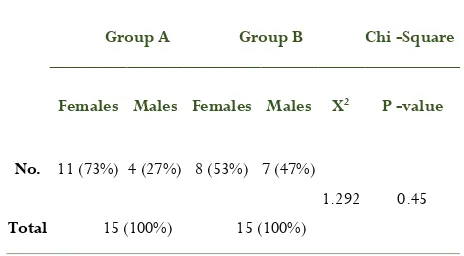
Sex distribution:
The sex distribution of group A revealed that there were 11 females with reported percentage of 73 % and 4 males with reported percentage of 27%. The sex distribution of group B revealed that there were 8 females with reported percentage of 53 % and 7 males with reported percentage of 47 % as shown in table (2). Chi square revealed there was no significant differences between both groups in sex distribution (p>0.05)
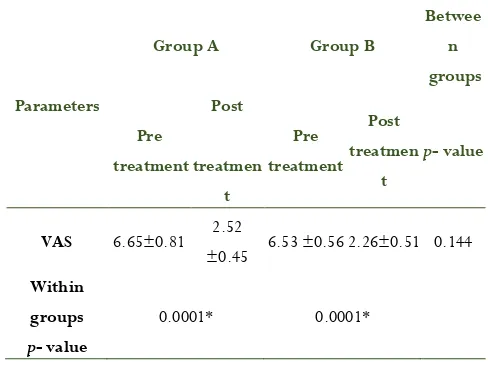
VAS
Statistical analysis using mixed design MANOVA analyzed thirty patients assigned into two equal groups. It revealed that there were significant within subject effect (F = 470.517, p = 0.0001). While there was no significant between subject effect (F= 0.92, p = 0.411) and treatment*time effect (F = 1.086, p = 0.352). Table (3) present descriptive statistic and multiple pairwise comparison tests (Post hoc tests) for the all dependent variables. In the same context regarding within subject effect, the multiple pairwise comparison tests revealed that there were significant decreases (p <0.05) in VAS in the post treatment condition compared with the pretreatment in both groups. Regarding between subject effects multiple pairwise comparisons revealed that there was no significant difference of dependent variables between both groups (p >0.05).
Table 1: Physical characteristics of patients in both groups
Items Group A Group B Comparison
S
Mean ± SD Mean ± SD t-value P-value
Age (yrs) 41.46±6.97 42.46±6.13 -0.417 0.68 NS
Height (cm) 160.26±7.5 162.13±10.52 -0.559 0.58
Weight (kg) 76.66±8.33 79.93±9.03 -1.029 0.312
BMI (kg/m2)
29.8±1.88 30.38±1.5 -0.934 0.358
Table 2: Distribution of sex in both groups
Group A Group B Chi -Square
Females Males Females Males X2 P -value
No. 11 (73%) 4 (27%) 8 (53%) 7 (47%)
1.292 0.45
Mohamed Abd-Allah MA, et al.; Low level laser therapy
432 Journal of Advanced Pharmacy Education & Research | Oct-Dec 2017 | Vol 7 | Issue 4
*Significant level is set at alpha level <0.05.
Tendon Echo-Texture:
Regarding within group's comparison, statistical analysis using Wilcoxon Signed Rank tests revealed that there was a significant reduction in tendon echotexture at post treatment compared to pretreatment at both groups with (p < 0.05). Table (4) present descriptive statistic (median) and comparison tests (within and between groups) for tendon echotexture. Considering the effect of the tested group (first independent variable) on tendon echotexture, "Mann-Whitney U test" revealed that there was no significant difference between both groups at Post treatment (p> 0.05).
*Significant level is set at alpha level <0.05, p-value: probability value
Discussion
This study was conducted to investigate the effect of eccentric exercises versus LLLT on pain and ultra-sonographic represented as (echotexture) of supraspinatus tendon in SIS.
Pain intensity and eccentric exercises:
The results of the current study showed that there was significant improvement of pain for the eccentric exercises group.
The current results are supported by a pilot study[17] in which the authors evaluated 9 patients (with long duration of shoulder pain) after eccentric training in chronic painful SIS, the eccentric training were directed to supraspinatus and deltoid muscles and performed for 3 sets of 15 repetitions, 2 times per day, 7 days per week for 12 weeks. The patients tried different treatments and they were prepared for surgery, their mean VAS before the treatment 71 mm and after 12 weeks of treatment reached 18 mm. Fifty-two weeks’ follow-up after the treatment, the 5 patients were still satisfied with treatment (their mean VAS was 31 mm) and had withdrawn from the waiting list for surgery.
In spite of the difficulty to compare the results of the current study to that of aforementioned study due to difference in the duration of symptoms, inclusion criteria (type 3 shaped acromion) and the dose of the treatment program, a conclusion can be drawn about the
effectiveness of the eccentric exercises in pain reduction,
as the sample used in that study was with chronic, resisted symptoms. It is well known that the more chronic the condition, the more the resisted the symptoms which increase the difficulty of the treatment. On the other hand, the patients were depressed as they were on the list of surgery, but they were persuaded and withdrawn from surgery which indicated that their pain level was effectively decreased.
The current results are also supported by a study[18] in which authors concluded that exercise concentrated on eccentric strength training for patients with SIS can be effective for pain reduction in patients with tendinitis and subacromial compression. Eight of the 10 patients showed significant decrease in the pain intensity (reduction in pain intensity between 17 and 58 mm) which is considered significant clinical changes.
Despite the difference of the aforementioned study[18] from the current study in the inclusion criteria of the sample (older patients with longer duration symptoms), it provided some support to the results of the current study. As it is well accepted that the treatment of old patients
Table 3:.Descriptive statistics of the all dependent variables
Parameters
Group A Group B
Betwee n groups
Pre treatment
Post
treatmen t
Pre treatment
Post treatmen
t
p- value
VAS 6.65±0.81 2.52
±0.45 6.53 ±0.56 2.26±0.51 0.144
Within groups
p- value
0.0001* 0.0001*
Table 4: Tendon echo texture
Tendon echo texture
Pre test Post
test p- value
Median Median
Group A 4 (1) 2(1) 0.001*
Group B 4 (1) 2(1) 0.001*
Journal of Advanced Pharmacy Education & Research | Oct-Dec 2017 | Vol 7 | Issue 4 433
with chronic symptoms is more difficult than that of young with subacute symptoms patients.
Another study[11] came into agreement with the current results in which 102 patients diagnosed as SIS with long duration of symptoms, they were waiting for arthroscopic subacromial decompression. Patients were divided into 2 groups, 55 in each group, one received unspecific exercises, the experimental group received 2 eccentric exercises (supraspinatus, infraspinatus and teres minor), 3 scapular stabilizing exercises (middle and lower trapezius, rhomboids and serratus anterior) and stretching for posterior shoulder capsule. The results showed significantly greater improvement in pain for the eccentric group compared to the control exercise group. In addition, patients in the eccentric exercise group were satisfied and decided to exclude surgical option (63%). That study provided strong support to the current results, as it was applied for larger number of patients.
Supraspinatus tendon echotexture and eccentric exercises:
The results of the current study showed that there was significant improvement of supraspinatus tendon echotexture (homogenous tendon) for the eccentric exercises group.
Up to our knowledge, there was no studies investigated the effect of the eccentric exercises on supraspinatus tendon echotexture, regeneration, in another words. However, a conclusion can be drawn about the effect of eccentric exercises on supraspinatus tendon echotexture from studies on other tendons as achilles and patellar tendinosis, as it was reported that histological examinations of the supraspinatus tendon in patients with SIS shown changes (tendinosis), similar to what have been found in chronic painful achilles and patellar tendinosis[15]. painful eccentric calf muscle training resulted in good clinical results in patients with chronic Achilles tendinosis[34]. The positive effects of eccentric exercises on pain intensity was attributed to its positive effects on tendon structure[35].
The results of the current study were also supported by a study[22] that investigated the effect of eccentric training on tendon structure in patients with Achilles tendinopathy. The patients were examined before the treatment by ultrasonography that showed abnormal
achilles tendon structure (irregular alignment of collagen) in all tendons, and after the treatment, the ultrasonography revealed normalization of tendon structure in 19 of 26 pathological tendons. The positive effect of the eccentric exercises in patients with achillestendinopathy was attributed to normalization of tendon structure (regeneration).
Promising results were also reported in patients with achillestendinopathy after the eccentric exercises, twelve players with achillestendinopathy were included in this study, the concentration of collagen turnover was measured before and after the treatment, the results revealed increase in collagen synthesis in response to the eccentric training load[16].
In spite of the above 2 studies being applied to achillestendinopathy, it has been clarified before that the supraspinatus tendon in patients with SIS shows changes similar to that found in achillestendinopathy[15]. Therefore, the findings can be extended to include the supraspinatus tendon in patient with SIS.
Pain intensity and LLLT:
The results of the current study showed that there was significant decrease of pain intensity for LLLT group.
The current results are supported by a study[9] in which authors evaluated the additive effects of LLLT with exercise on SIS. Patients were assigned into 2 groups, 40 patients on each group. Experimental group received LLLT in addition to exercises and the control group received placebo laser and the same exercises were given to the first group. The results showed significant pain reduction in LLLT group than the control group receiving exercises alone. That study provided strong evidence of LLLT efficacy on pain relief in SIS patients, due to its larger sample size than the current one.
Mohamed Abd-Allah MA, et al.; Low level laser therapy
434 Journal of Advanced Pharmacy Education & Research | Oct-Dec 2017 | Vol 7 | Issue 4
Another study[37] came into agreement with the current results, in which authors examined the effect of LLLT in supraspinatus tendinitis, the results revealed significant pain reduction in LLLT than the control group. Instead, LLLT used in that study was with different wavelength (820nm) and different dose (30 J) per point, it might give us a key about the positive effect of LLLT on supraspinatus tendon while using different parameters. The positive effects of LLLT can be explained by the ability of different parameters of LLLT in achieving improvements on pain intensity.
A study about the effect of LLLT on the pain control[24] agreed with the current results, that study concluded that LLLT was highly effective for pain relief.
In contrast to the current results, there was a study[38] that investigated the effect of LLLT in patients with SIS compared to the control group and found that there was significant improvement in both groups with no significant difference. Those opposing results could be explained by the different application procedures, in their study they applied the LLLT over the tender points rather than the anatomical site, those points were located by the patients and away from the actual lesion site that excepted to cause pain. On the other hand, the biostimulatory effect of LLLT which stimulate collagen synthesis for healing purposes within a low range between 0.4 to 4 J/cm2[39] which is not applied in that study (5 J/cm2).
Supraspinatus tendon echotexture and LLLT:
The current results showed that there was significant improvement of supraspinatus tendon echotexture for LLLT group.
To our knowledge there was no study investigated the effect of LLLT on supraspinatus tendon echotexture. However, a conclusion can be drawn about the healing effect of LLLT on other type of tendinopathies studies.
Come into agreement with the current results, a systematic review concluded that LLLT has an anti-inflammatory effect (at 1-12 joules) in 21 out of 24 controlled laboratory trials, and a biostimulatory effect (at 0.2-4 joules) on collagen production in 31 out of 36 trials[40].
There was a study[41] that investigated the effects of LLLT (Gl-As, 904nm, 3 J) on collagen fibers organization in rats achilles tendon and they demonstrated that LLLT was capable of promoting the collagen fiber organization throughout the tendon, indicating a better tendon repair. Although, the results emphasized the healing effect of LLLT on tendon structure. However, the translation of results to human tendons could be taken with caution due to difference in the response between animals and humans to the same parameters. A positive effect of LLLT on tendon regeneration can be suggested.
Another study[42] supported the current results, in that study LLLT efficacy on tissue repair was investigated, they concluded that the LLLT was a highly effective form of treatment for tissue regeneration, with stronger evidence from animal than human ones. That study reported that the animal studies have observed strong evidence of the LLLT effect than the human ones, as in the human studies there were insufficient prescription of the used parameters[43].
Conclusion
It could be concluded from the current study results that eccentric exercise is as effective as LLLT in relieving pain and improving supraspinatus tendon structure in SIS patients when added to scapular and rotator cuff muscles strengthening and flexibility exercises.
References
1. Green, S. Buchbinder, R. Hetrick, S. (2003).
Physiotherapy interventions for shoulder
pain.Cochrane Data- base Syst Rev. CD004258.
2. Van der Windt DAWM, Koes BW, De Jong BA,
Bouter LM (1995). Shoulder disorders in general practice: Incidence, patient characteristics and management. Ann Rheum Dis. 54(12):959-64.
3. Cook, J. Purdam, C. (2009).Is tendon pathology? A
pathology model to explain the clinical presentation of load-induced tendinopathy.Br J Sports Med. 43,409-416.
4. Michener, LA. McClure, PW. Karduna, AR. (2003).
Journal of Advanced Pharmacy Education & Research | Oct-Dec 2017 | Vol 7 | Issue 4 435
Biomechanics. 18 (5),369–379.
5. Leong, H T. Tsui, S. Ying, M. Vivian, Yee-fong Leung
V Y. Fua, S N. (2012). Ultrasound measurements on acromio-humeral distance and supraspinatus tendon thickness: Test–retest reliability and correlations with shoulder rotational strengths.
Journal of Science and Medicine in Sport. 15, 284–291.
6. Cook, J. Purdam, C. (2009). Is tendon pathology? A
pathology model to explain the clinical presentation of load-induced tendinopathy. Br J Sports Med. 43,409-416.
7. Kelly, S. Brittle, N. Allen, G. (2010). The value of
physical tests for subacromial impingement syndrome: A study of diagnostic accuracy.
ClinRehabil. 24,149-158.
8. Michener, L. Subasi Yesilyaprak, S. Seitz, A.
Timmons, M. Walsworth, M. (2013).Supraspinatus tendon and subacromial space parameters measured on ultrasonographic imaging in subacromial impingement syndrome. Knee Surg Sports Traumatol
Arthrosc. 5(6),1-7.
9. Abrisham, S. Kermani-Algoraishi, M. Ghahramani, R.
et al. (2011). Additive effects of low-level laser therapy with exercise on subacromial syndrome: A randomised, double-blind, controlled trial. Clin
Rheumatol. 30,1341–1346.
10. Hanratty, C. McVeigh, J. Kerr, D. Basford, J. and
Finch, M. (2012). The effectiveness of
physiotherapy in subacromial impingement
syndrome: A systematic review and meta-analysis.
Semin Arthritis and Rheum.42,297–316.
11. Holmgren, T. Bjornsson Hellgren, H. Oberg, B.
Adolfsson, L. Johansson, K. (2012). Effect of specific exercise strategy on need for surgery in patients with subacromial impingement syndrome: randomised controlled study. BMJ.344:e787(1-9).
12. Stanish, W. Rubinovich, R. Curwin, S.
Orthop. 208,65–8.
13. Silbernagel, K. Thomee,´R. Thomee,´P. Karlsson, J.
(2001).Eccentric overload training for patients with chronic Achilles tendon pain a randomized controlled study with reliability testing of the evaluation methods.Scan J Med Sci Sports. 11,197–
206.
14. Jonsson, P. Alfredson, H. (2005). Superior results
with eccentric compared to concentric quadriceps
training in patients with jumper’s knee: a
prospective randomized study. Br J Sports Med. 39,847–50.
15. Khan, K. Cook, J. Bonar, F. Harcourt, P. Astrom, M.
(1999).Histopathology of common tendinopathies: update and implications for clinical management.
Sports Med. 27,393-408.
16. Langberg, H. Ellingsgaard, H. Madsen, T. Jansson, J.
Magnusson, S. Aagaard, P. Kjaer, M. (2007). Eccentric rehabilitation exercise increases peritendinous type I collagen synthesis in humans with achillestendinosis. Scand J Med Sci Sports. 17,61– 66.
17. Jonsson, P. Wahlstro, P. O¨ hberg, L. Alfredson, H.
(2006). Eccentric training in chronic painful impingement syndrome of the shoulder: results of a pilot study. Knee Surg Sports Traumatol Arthrosc. 14,76–81.
18. Bernhardsson, S. Klintberg, I. Wendt, G.
(2011).Evaluation of an exercise concept focusing on eccentric strength training of the rotator cuff for patients with subacromial impingement syndrome.
Clin Rehabil. 25,69–78.
19. Camargo, P. Avila, M. Alburquerque-Sendin, F. Asso,
Mohamed Abd-Allah MA, et al.; Low level laser therapy
436 Journal of Advanced Pharmacy Education & Research | Oct-Dec 2017 | Vol 7 | Issue 4
Bras Fisioter. 16,74–83.
20. Jeffery R, Cronin J, Bressel E.(2005): Eccentric
strengthening: Clinicalapplications to Achilles tendinopathy. New Zealand J Sports Med. 33; 22–30.
21. Peers KH, Lysens RJ. (2005): Patellar tendinopathy in
athletes: currentdiagnostic and therapeutic recommendations. Sports Med. 35: 71-87.
22. Ohberg, L. Lorentzon, R. Alfredson, H. (2004).
Eccentric training in patients with chronic achillestendinosis: normalised tendon structure and decreased thickness at follow up. Br J Sports Med. 38: 8-11.
23. Ohberg L, Alfredson H. (2004). Effects on
neovascularisation behind the good results with
eccentric training in chronic
midportionachillestendinosis? Knee Surg Sports
TraumatolArthrosc. 12; 465-470.
24. Enwemeka, C. Parker, J. Dowdy, D. Harkness, E.
Sanford, L. and Woodruff, L. (2004). The efficacy of low-power lasers in tissue repair and pain control: a meta-analysis study. Photomed Laser Surg. 22,323–329.
25. Williams, G. Gangel, T. Arciero, R.
(1999).Comparison of the single assessment numeric evaluation method and two shoulder rating scales. Am J Sports Med. 27(2),214–221.
26. O'Connor, PJ. Grainger, AJ. Morgan, SR. Smith, KL.
Waterton, JC. Nash, AF. (2004). Ultrasound
assessment of tendons in asymptomatic
volunteers.EurRadiol. 14: 1968-1973.
27. Ingwersen, KG. Hjarbaek, J. Eshoej, H. Larsen,
CM.Vobbe, J. Juul-Kristensen, B.
(2016):Ultrasound assessment for grading structural tendon changes in supraspinatus tendinopathy: an inter-rater reliability study. BMJ. 6;1-8.
28. Bang, MD. and Deyle, GD. (2000): Comparison of
Supervised Exercise With and Without manual physical Therapy for Patients With Shoulder
Impingement Syndrome. Journal of Orthopaedic and
Sports Physical Therapy. 30(3):126-137.
29. Ludewig, PM. Cook, TM. (2000). Alterations in
shoulder kinematics and associated muscle activity in
people with shoulder impingement
syndrome.Physical Therapy. 80: 276–291.
30. Camargo, PR. Haik, MN. Filho, RB. Mattiello-Rosa,
SMG.Salvini, TF. (2008). Bilateral deficits in muscle contraction parameters during shoulder scaption in patients with unilateral subacromial
impingement syndrome.Isokinetics and Exercise
Science. 16: 93–99.
31. Thomee´, R. (1997). A comprehensive treatment
approach for patellofemoral pain syndrome in young women. PhysTher. 77, 1690–703.
32. Maenhout, AG. Mahieu, NN. De Muynck, M. De
Wilde, LF. Cools, AM. (2013). Does adding heavy load eccentric training to rehabilitation of patients with unilateral subacromial impingement result in
better outcome? A randomized, clinical
trial.KneeSurg Sports TraumatolArthrosc. 12 (5):1158-1167.
33. Bjordal, J.M. Couppe, C. Ljunggren, A.E. (2001).
Low level Laser Therapy for Tendinopathy: Evidence of a Dose Response Pattern. Physical
Therapy Reviews. 6, 91-99.
34. Alfredson, H. Pietila¨, T. Jonsson, P. Lo- rentzon, R.
(1998). Heavy load eccentric calf-muscle training for treatment of chronic Achilles tendinosis. Am J
Sports Med. 26,360–366.
35. Mahieu, N. McNair, P. Cools, A. D’Haen, C.
Vandermeulen, K. Witvrouw, E. (2008).Effect of eccentric training on the plantar flexor muscle-tendon tissue properties.MedSci Sports Exerc.40,117–
123.
36. Haslerud, S. Magnussen, LH. Joensen, J.
Journal of Advanced Pharmacy Education & Research | Oct-Dec 2017 | Vol 7 | Issue 4 437
controlled trials. Physiother. Res. Int. 20,108-125.
37. Saunders, L. (1995). The efficacy of low-level laser
therapy in supraspinatus tendinitis.Clinical
Rehabilitation. 9:126-134.
38. Dogan, SK. Saime, AY. Evcik, D. (2010). The
effectiveness of low level laser therapy in subacromial impingement syndrome: a randomized placebo controlled double-blind prospective study.
Clinics.65(10):1019-1022.
39. Reddy, G. Stehno-Bittel, L. and Enwemeka, C.
(1998). Laser Photostimulation of Collagen Production in Healing Rabbit Achilles Tendons.
Lasers SurgMed. 22,281-287.
40. Lopes-Martins, R. Penna, SC. Joensen, J. Iversen,
VV. Bjordal, JM. (2007). Low level laser therapy (LLLT) in Inflammatory and Rheumatic Diseases: A
review of Therapeutic Mechanisms. Current
Rheumatology. 3(2):147-154.
41. Arruda, ERB. Rodrigues, NC. Taciro, C. Parizotto,
NA. (2007). Influences of different low level laser therapy wavelengths in rat tendon regeneration after
tenotomy. Brazilian Journal of Physical
Therapy.11(4):247-252.
42. Fulop, AM. Dhimmer, S. Deluca, JR. Johanson, DD.
Lenz, RV. Patel, KB. Douris, PC. Enwemeka, CS. (2009). A meta-analysis of the efficacy of phototherapy in tissue repair. Photomed Laser Surg. 27:695–702.
43. Huang, YY. Chen, AC. Carrol, JD. Hamblim, MR.