K
RZYSZTOFS
KIBA1, R
OMANR
UTOWSKI1, R
OMANW
IĄCEK1, P
AWEŁR
EICHERT1,
K
RZYSZTOFD
UDEK2Treatment of Subcutaneous Ruptures
of the Achilles Tendon in Own Material
Leczenie podskórnych przerwań ścięgna Achillesa w materiale własnym
1Department of Traumatology and Hand Surgery in Wrocław, Poland
2Institute of Machines Design and Operation, Wrocław University of Technology, Poland
Adv Clin Exp Med 2006, 15, 3, 471–480 ISSN 1230−025X
ORIGINAL PAPERS
Abstract
Background.Traumatic subcutaneous Achilles tendon ruptures are a serious problem in traumatic and orthopedic surgery. They constitute a considerable percentage of injuries not only in persons practicing sports professionally, but also in those who do it for pleasure. The choice of the treatment method, i.e. the use of traditional conventio− nal surgery, conservative treatment, or microsurgical reconstruction of the tendon, largely depends on the level of destruction in tendon continuity as assessed by ultrasound as well as CT and magnetic resonance results.
Objectives.The assessment of surgical results in the treatment of traumatic subcutaneous ruptures of Achilles tendons.
Material and Methods.In 1992–2004, 115 patients underwent treatment, including 83 surgical interventions, at the Department of Traumatology and Hand Surgery. They were 67 males (80.7%) and 16 females (19.3%) and their age range was 20–90 years. The average post−injury time was five days. The patients were operated under supra− dural anesthesia and limb ischemia both in emergency and scheduled procedures. The majority were patients brought from other centers. The procedures were carried out using classical methods (Bunnel’s, Strell’s, Kessler’s methods, Christensen’s and Gebhardt’s reconstruction methods) as well as with the microsurgical recon− struction introduced in the Department of Traumatology and Hand Surgery.
Results. Surgical treatment brings the best results in early ruptures of Achilles tendon. Selection of treatment de− pends on the level and the degree of rupture. At the typical site of rupture, microsurgical reconstruction is the method of choice in the majority of cases. The assessment of mobility of the operated limb showed very good results of this method. In none of the cases were early or late complications observed in the form of repeated ruptu− res, which often occur in patients treated with traditional surgical methods. Tendon inveterate ruptures which require plastic surgery incorporating the plantar muscle, especially the more difficult plastic surgical procedure involving the aponeurosis of the calf muscle, are quite a different group of cases.
Conclusions.The microsurgical reconstruction method deserves the highest recognition. Patients operated on with this method showed no complications and their work−disability period was much shorter than that of patients in whom other methods were applied. Microsurgical reconstruction constituted 50.6% of the total procedures (Adv Clin Exp Med 2006, 15, 3, 471–480).
Key words: Achilles tendon, treatment, microsurgical reconstruction, diagnostic examination, ultrasound, thermo− vision.
Streszczenie
Wprowadzenie. Urazowe podskórne przerwania ścięgna Achillesa są poważnym problemem w chirurgii urazowo− −ortopedycznej. Stanowią znaczny odsetek urazów nie tylko u osób profesjonalnie uprawiających sport, ale rów− nież rekreacyjnie. Wybór metody leczenia – zastosowania tradycyjnych konwencjonalnych form leczenia opera− cyjnego, leczenia zachowawczego czy też mikrochirurgicznej rekonstrukcji ścięgna – jest uwarunkowany pozio− mem i stopniem zniszczenia ścięgna, wykorzystując ultrasonograficzną ocenę jego ciągłości, badanie tomograficz− ne i rezonans magnetyczny.
Cel pracy. Ocena wyników leczenia operacyjnego chorych z urazowymi podskórnymi przerwaniami ścięgna Achillesa.
Materiał i metody. W Klinice Chirurgii Urazowej i Chirurgii Ręki AM we Wrocławiu leczono w latach 1992–2004 łącznie 115 pacjentów, w tym operacyjnie 83 chorych: 67 mężczyzn (80,7%) i 16 kobiet (19,3%) w przedziale wiekowym 20 + 90 lat. Średni czas od urazu do operacji wynosił 5 dni. Pacjentów operowano w znie−
Traumatic subcutaneous ruptures of the Achil− les tendon have been a serious problem in trauma− tic and orthopedic surgery for years. Their preva− lence has increased considerably in persons parti− cipating in sports both professionally or for pleasure, especially in such disciplines as skiing, volleyball, basketball, tennis, football (i.e. soccer) and hand ball. According to some authors [17], tendon rupture occurs in 400–500 persons every year, and in 80% it is the result of practicing sports. Achilles tendon ruptures are most often observed in persons aged 30–50. They involve men more than women and, according to German authors, they prevail (56%) in the dominant left lower limb in right−handed persons [12, 17].
Achilles tendon rupture usually takes place at the so−called typical site located 2–5 cm above the tuber attachment of the calcaneal bone (96%). Pro− ximal segment breaks (2%) as well as breaks in the tuber attachment of the calcaneal bone (2%) con− stitute much smaller percentages [16].
Mechanics explains the pathomechanism of the tendon’s rupture. In accordance with this theo− ry, a sudden uncoordinated contraction of the tri− ceps muscle of the calf with a plantarly flexed foot and the limb straightened at the knee joint very often results in tendon rupture. Sportsmen also show muscular hypertrophy as an effect of conti− nuous practice as well muscles tension, which fa− vor tendon breaks [12]. The degenerative theory also attributes the effect of tendon rupture to some degenerative changes such as collagen fibers frag− mentation or homogenization and mucopolysac− charide basis decay, which appears in people over 35 and is manifested as loss of tendon resilience and distension resistance.
Numerous diseases, such as contagious dis− eases, infections or long corticosteroid therapy in diabetic patients, pyelonephritis, and local admini−
stration of steroids predispose patients to tendon rupture.
On the basis of investigations of tensile strength performed on the tendons of corpses, the authors followed the pathomechanism and asses− sed the degree of tendon strength in relation to age as it definitely decreases after the age of 35. They also estimated the most common level of rupture which, in the experimental studies, is the same as the clinical status [19, 20].
At present, the diagnostics of Achilles tendon injuries is not a problem due to ultrasound exami− nation (USG), computed tomography (CT), ma− gnetic resonance (MR), and thermography, the la− test diagnostic achievement [1–4, 14, 15, 18].
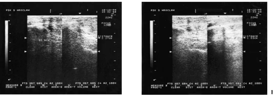
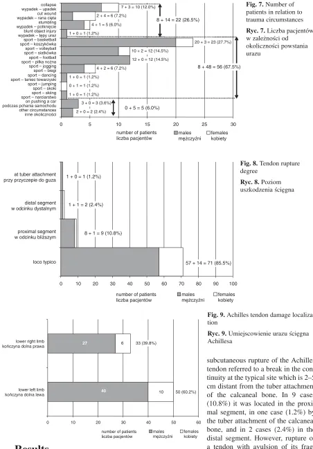
Ultrasound examination plays the most impor− tant role in both the pre− and post−operative diag− nostics of Achilles tendon ruptures as it enables an exact differentiation of partial and total break. It shows the degree of end separation of the ruptured tendon, helps to assess the size of hematoma, as well as the space between the two ends of the ten− don, which greatly influences the decision of the treatment method and enables the evaluation of surgical treatment results (Figs. 1, 2).
If both ends of the tendon approach at the plantar flexure, only conservative treatment (pla− ster cast immobilization) should be considered. Surgical treatment can be performed if the distance between the two tendon ends exceeds 6 mm, which can be seen on ultrasound examination. Ultrasound scan and thermovision examination (Fig. 3) enable the assessment of the healing pro− cess of the Achilles tendon as well as the evalua− tion of surgical and conservative treatment results. They allow a suitable rehabilitation schedule and the patient’s return to professional activity.
There are numerous methods of surgical treat− ment depending on the time which has passed since
Wnioski. Zasługująca na największe uznanie okazała się metoda mikrochirurgicznej rekonstrukcji. U pacjentów operowanych tą nową metodą nie obserwowano powikłań, a powrót do pracy następował znacznie szybciej niż u operowanych pozostałymi metodami. Metoda ta stanowiła 50,6% wszystkich operacji (Adv Clin Exp Med 2006, 15, 3, 471–480).
the injury. Thus there are so−called recent and inve− terate tendon ruptures (three weeks after the injury) [5, 6, 8–10]. Traditional surgical methods are pre−
ferred in countries of central Europe, while in Scandinavian countries, North America, and Great Britain, conservative treatment forms are used.
In 1993, a new method of tendon reconstruc− tion was elaborated and introduced in the Depart−
Fig. 1.a) Post microsurgical reconstruction, condition of the right lower limb Achilles tendon, distance of 1.7 cm from the tuber of the calcaneal bone in a 28−year−old male 9 months after the surgery. Poorly evident post−operative scar. The tendon characteristic for the regular course of bundles, slightly thickened (1.18 cm); correct course of normal line− ar bundles. B.Left lower limb Achilles tendon of the same individual, the tendon thickness at the same level: 1.14 cm
Ryc. 1. A. Stan po rekonstrukcji mikrochirurgicznej ścięgna Achillesa kończyny dolnej prawej w odległości 1,7 cm od przyczepu do guza kości piętowej u 28−letniego mężczyzny – 9 miesięcy od operacji. Blizna pooperacyjna mało widoczna. Ścięgno o regularnym przebiegu wiązek, nieznacznie pogrubiałe – 1,18 cm; przebieg wiązek prawidłowy, linearny. B.Ścięgno Achillesa kończyny dolnej lewej tego samego pacjenta, grubość ścięgna na tym samym poziomie – 1,14 cm
A B
Fig. 2. A.Bunuel’s method, post−suturing condition of the left lower limb Achilles tendon in a 54−year−old female at a distance of 1.8 cm from the tuber of the calcaneal bone. At the suture site the tendon is clearly thickened up to 2.3 cm and the course of bundles remains irregular. B.The right lower limb Achilles tendon is 1.0 cm wide with correct and regular course of bundles
Ryc. 2. A. Stan po zeszyciu ścięgna Achillesa kończyny dolnej lewej u 54−letniej kobiety sposobem Bunuela w odległości 1,8 cm od przyczepu do guza kości piętowej. Ścięgno Achillesa w miejscu zeszycia wyraźnie pogru− białe do 2,3 cm. Przebieg wiązek nieregularny. B.Ścięgno Achillesa kończyny dolnej prawej szerokości 1,0 cm o prawidłowym, regularnym przebiegu wiązek
A B
Fig. 3.Demonstration thermograph of the Achilles tendon area in a 34−year−old patient operated with the microsurgical reconstruction method, 5 months after the injury
ment of Traumatology and Hand Surgery. This method uses microsurgical techniques and has al− ready been described in the journal “Ortopedia Polska” [7, 8]. It combines the advantages of ope− rative methods and, due to the application of mi− crosurgical techniques, it prevents tendon trauma− tization and allows successful convalescence and restoration of sport activity (Fig. 4).
Material and Methods
This article presents a retrospective asses− sment of the results of treatment of traumatic inju− ry to the Achilles tendon. In the years 1992–2004,
115 patients were treated at the Department of Traumatology and Hand Surgery in Wrocław, 83 of whom required surgical intervention. These were 67 males and 16 females (Fig. 5) aged 20–90 (Fig. 6) admitted to the hospital for subcutaneous rupture of the Achilles tendon. Table 1 presents the basic statistics regarding ages and the periods of time between injury and operation.
Some patients (27.8%) were admitted immedi− ately after the injury from the emergency ward, whereas the majority of them (72.2%) underwent scheduled surgical procedures, having been trans− ported from other centers, usually 4 days after the injury. Besides routine biochemical tests, ultra− sound examination was performed in all patients to assess the rupture level and the degree of Achilles tendon damage and to provide a basis for the choice of treatment method.
The patients were operated with supradural anesthesia and were placed on the abdomen with plantar flexure of the foot and with the posterior− medial or sinuous incision of Torklus and Nicol (Fig. 11). After revealing the ruptured tendon, the sheath was always incised and the existing hematoma removed. The appropriate method was selected in relation with the intra−operative condition. Figures 7–10 present the circumstances of the injuries, tendon rupture levels, as well as the meth− ods of treatment.
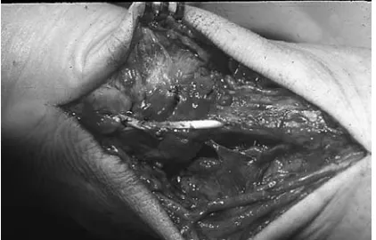
Fig. 4. A.Intra−operative photograph of ruptured Achilles tendon five days after the injury. Hematoma is visible after revealing both ends of the ruptured tendon. In the first operation stage, the tendon is ‘tufty’ delami− nated, with the bunches of different length. B.Groups of tendon bunches adopted with absorbable microsurgi− cal sutures. C.The condition after suturing the tendon sheath with resorbable sutures. The tendon sheath co− vers the reconstructed Achilles tendon
Ryc. 4. A. Zdjęcie śródoperacyjne przerwanego ścię− gna Achillesa 5 dni po urazie. Po odsłonięciu obu koń− ców przerwanego ścięgna widoczny krwiak. Widoczne rozwarstwione „pędzelkowato” ścięgno o różnej dłu− gości pęczkach (1. etap operacji). B. Zaadaptowane wchłanianymi nićmi mikrochirurgicznymi grupy pęcz− ków ścięgna. C. Stan po zeszyciu pochewki ścięgna pojedynczymi resorbowanymi nićmi. Pochewka ścię− gna pokrywa zrekonstruowane ścięgno Achillesa
A B
C
males mężczyźni 80.7%
females kobiety 19.3%
67
16
Fig. 5.The structure of the operated patients according to sex
The 83 patients (87%) treated operatively included 25 emergency cases (30.1%), 47 (56.6%) scheduled procedures, and 11 patients (13.3%) who underwent conservative treatment with foot− crus plaster cast immobilization for about 6 weeks. Sport injuries (67.5%) were the most common cause of Achilles tendon rupture (Fig. 7). They usually took place during basketball (23 persons, 27.7%), volleyball (12 persons, 14.5%), football (soccer, 12 persons, 14.5%), jogging (6 persons, 7.2%), jumping (1 person, 1.2%), skiing (1 person, 1.2%), and dancing (1 person, 1.2%). These most− ly involved young persons aged 20–50 (mean: 38.7 years). Other causes were accidents such as collapse in the street (10, 12%), open wound with glass (6, 7.2%), stumbling (5, 6.0%), blunt object injury (1, 1.2%), pushing a car (3, 3.6%), and other circumstances (2, 2.4%) (Fig. 7).
In 10 cases, patients with inveterate injures (6 months after) were operated on. They had been treated in other medical centers with conservative methods such as Christensen’s and Gebhardt’s method (Fig. 12) and the plastic surgery technique of Strelli (Fig. 13).
In each case, after surgery the limb was immo− bilized in a foot−femur plaster cast with foot plan− tar flexure for a period of 2–3 weeks, correcting the foot to the intermediary position and reducing the foot plantar flexure. In the early postoperative period as well as after 6 and 12 months after surgery, ultrasound and thermovision follow−up examination were performed. This helped to mon− itor both the tendon healing process and the restoration of limb function.
10 20 30 40 50 60 70 80 90
0 5 10 15 20 25 30 35 40
statistics females males total statystyka kobiety mężczyźni razem
mean value: 38.4 41.2 40.6 średnia:
standard deviation: 8.5 13.1 12.4 odchylenie standardowe:
median: 39 38 38 mediana:
minimum: 20 20 20 minimum:
maximum: 52 90 90 maksimum:
age wiek number of patients
liczba pacjentów
Fig. 6.Patients age histogram with normal distribution and basic statistics
Ryc. 6.Histogram wieku pacjen− tów na tle rozkładu normalnego i podstawowe statystyki
Table 1.Statistical values characterizing the patients
Tabela 1.Wartości statystyczne charakteryzujące pacjentów
Females Males Total (Kobiety) (Mężczyźni) (Suma)
Age – years; 38.4 41.2 40.6
mean value (Wiek – lata; wartość średnia)
Standard deviation 8.5 13.1 12.4 (Odchylenie
standardowe)
Median 39 38 38
(Mediana)
Minimum 20 20 20
(Minimum)
Maximum 52 90 90
(Maksimum)
Number of days 11.4 18.0 16.7 between injury and surgery; mean value (Liczba dni dzielących uraz od operacji; wartość średnia)
Standard 28.7 45.2 42.3
deviation (Odchylenie standardowe)
Median 3 4 4
(Mediana)
Minimum 0 0 0
(Minimum)
Maximum 108 224 224
Results
The selection of the surgical method was done individually depending on the results of ultrasound examination and intra−operative condition. In majority of cases (71 patients, 85.5%), traumatic
subcutaneous rupture of the Achilles tendon referred to a break in the con− tinuity at the typical site which is 2–5 cm distant from the tuber attachment of the calcaneal bone. In 9 cases (10.8%) it was located in the proxi− mal segment, in one case (1.2%) by the tuber attachment of the calcaneal bone, and in 2 cases (2.4%) in the distal segment. However, rupture of a tendon with avulsion of its frag− ments in a distal segment at the site of the tuber attachment of the calcaneal bone was not observed. According to other reports, this occurs very rarely.
Achilles tendon rupture is very characteristic.
0 5 10 15 20 25 30
other circumstances inne okoliczności on pushing a car podczas pchania samochodu
males mężczyźni
females kobiety
2 + 0 = 2 (2.4%) 3 + 0 = 3 (3.6%)
0 + 5 = 5 (6.0%)
number of patients liczba pacjentów
sport – narciarstwo
0 10 20 30 40 50 60 70 80 90 100 loco typico
proximal segment w odcinku bliższym distal segment w odcinku dystalnym at tuber attachment przy przyczepie do guza
males mężczyźni
females kobiety 57 + 14 = 71 (85.5%) 1 + 0 = 1 (1.2%)
8 + 1 = 9 (10.8%) 1 + 1 = 2 (2.4%)
number of patients liczba pacjentów
Fig. 8.Tendon rupture degree
Ryc. 8.Poziom uszkodzenia ścięgna
0 10 20 30 40 50 60
lower left limb kończyna dolna lewa lower right limb kończyna dolna prawa
males mężczyźni
females kobiety 50 (60.2%) 33 (39.8%)
27
40 10
6
number of patients liczba pacjentów
Fig. 9.Achilles tendon damage localiza− tion
The tendon fiber bunches usually get torn at vari− ous levels and become tufty. The tendon sheath is damaged and suturing seems indispensable to avoid dermal and tendinous adhesions.
In the majority of cases (56%), tendon recon− struction was done microsurgically, excising
abnormal bunches of fiber and binding them together under a surgical microscope. The surgi− cal microscope was used in adopting single or grouped bunches by applying up to 20 single resorbable sutures. Tendon sheath continuity was always restored, thus preventing the formation of the dermal and tendinous adhesions which appear in more traditional, conventional methods of treatment.
In 6 cases (7.2%) of microsurgical reconstruc− tion of the Achilles tendon, tissue glue was suc− cessfully applied (Fig. 15) [11, 13]. The results of microsurgery proved very good. No early or late complications were observed in the group of patients operated with the microsurgical recon− struction method, including those in whom tissue glue was applied. Six−week limb immobilization provided proper healing of the tendon, and subse− quent rehabilitation and bio−stimulating laser allowed uneventful recovery and further sport practice (Fig. 16).
So−called inveterate tendon ruptures presented
0 5 10 15 20 25 30 35 40 45 50
microsurgery mikrochirurgia method by Bunnel metoda Bunnela conservative treatment leczenie zachowawcze method by Strelli metoda Strelli microsurgery + fibrin glue mikrochirurgia + klej fibrynowy plasty by Christensen plastyka wg Christensena
males mężczyźni
females kobiety 37
6 9 6
6 3 1
2
2
6
5
number of patients liczba pacjentów 4 (4.8%)
6 (7.2%)
8 (9.6%)
11 (13.3%)
12 (14.5%)
42 (50.6%)
Fig. 10.Methods of treating Achilles ten− don rupture
Ryc. 10.Metody leczenia zerwanego ścięgna Achillesa
Fig. 11.Posterior−median access of Torklus
Ryc. 11. Dostęp tylno−przyśrodkowy wg Torklusa
Fig. 12. Intra−operative image, plastic surgery technique of Christensen and Gebhardt with inverted flap of the gas− trocneminus muscle aponeurosis: A.calf lateral image, B.calf posterior image
Ryc. 12.Zdjęcie śródoperacyjne – plastyka według Christensena i Gebhardta z odwróconym płatem z rozcięgna mię− śnia brzuchatego łydki: A.widok z boku, B.widok z tyłu łydki
a greater problem due to the considerable defect in tendon continuity and the inability to mobilize its ends (10 cases). In these cases, the plastic surgical method of Strelli with use of the plantar muscle tendon had to be performed (8 cases), or even the more complicated procedure with the use of the inverted aponeurosis of the gastrocneminus muscle in the Christensen and Gebhardt technique (3 cases). The plastic surgery method of Strelli using the plan− tar muscle tendon, which remained undamaged in all rupture cases, proved efficient both in recent
and inveterate tendon ruptures. Neither early nor late complications were observed.
In one patient operated on with the method of Christensen six months after the injury, early com− plications appeared in the form of abscess and fis− tula. Interstitial tissue staining was performed with disulphine blue dye and necrotic tissues were excised intra−operatively (Fig. 17). A healing effect was achieved; however, the functional qual− ity of the foot was not satisfactory.
Discussion
In traumatic subcutaneous ruptures of the Achilles tendon, the choice between an operative treatment method or the introduction of conserva− tive treatment depends on the degree of tendon rupture, its degree of destruction, as well as the dehiscence of the tendon ends. Ultrasound exami− nation and magnetic resonance prove very useful in such cases. Ultrasound examination also helps to monitor the tendon healing process. Based on their clinical observations, the authors have found thermovision examinations very useful in the
Fig. 13. Achilles tendon suture with the use of the plantar muscle tendon by the Strelli method
Ryc. 13.Szew ścięgna Achillesa z użyciem ścięgna mięśnia podeszwowego sposobem Stelli
Fig. 14.Operated limb immobilization method
Ryc. 14.Sposób unieruchomienia operowanej kończyny
Fig. 15.The use of fibrin glue in microsurgical nerve reconstruction
Ryc. 15.Zastosowanie kleju fibrynowego w mikrochi− rurgicznej rekonstrukcji nerwów
Fig. 16. Lower right limb efficacy after surgery
assessment of both the tendon reparative process− es and post−treatment limb aptitude.
In the majority of cases of traumatic subcuta− neous ruptures of the Achilles tendon, the effects of surgical treatment depend on the time passed since the injury. An emergency procedure as well as treatment administered a few days after the injury, described as recent ruptures, bring the best results. In the majority of inveterate ruptures requiring various plastic procedures, the effects are usually unsatisfactory and the percentage of early and late complications is significant. The new method of Achilles tendon microsurgical reconstruction brings the best effects, as previous− ly proved by survey results. It allows full recon− struction of the tendon with the sheath cover to provide healing without the formation of hypertro−
phied scarring of the connective tissue or dermal and tendinous adhesions. Such conditions allow convalescence and the patients’ return to practic− ing sport.
The authors concluded, that: 1) males pre− vailed (82.8%) among the treated patients, 2) most often (85.5% of cases), the tendon rupture was found at the typical location: 2–5 cm distant from the tuber attachment of the calcaneal bone, 3) the majority of patients (51%) were operated on with the use microsurgical reconstruction, which brings excellent effects in the treatment of recent tendon ruptures, 4) advanced methods allowed the treat− ment of tendon rupture patients and recovery of function of the operated limb, 5) the choice of method largely depends on ultrasound assessment and the intra−operative condition.
Fig. 17. A.Achilles tendon necrotic tissues made visible by use of disulphine blue dye. B.Patient’s appearance after disulphine blue dye injection
Ryc. 17. A. Widoczne wybarwione barwnikiem disulphine bluetkanki martwicze ścięgna Achillesa. B.Wygląd cho− rego po wstrzyknięciu barwnika disulphine blue
A B
References
[1] Burhardt H, Krebs U, Fuchs M, Stankovic P: Die sonographische Beurteilung von Gleitverhalten und Gleitlager operierter Achillessehnen. Unfallchirurg 1991, 94, 589–593.
[2] Dudek K:Thermography as diagnosis system. System Journal of Transdyscyplinary System Science 1998, 3, 2, 68–79.
[3] Grechening W, Clement HG, Fellinger M, Seggl W: Wertigkeit der Sonographie der Achillessehne in der Traumatologie. Radiologe 1997, 37, 322–329.
[4] Khan KM, Forster BB, Robinson J, Cheong Y, Louis I, Maclean L, Taunton JE:Are ultrasound and magnetic resonance imaging of value in assessment of Achilles tendon disorders? A two year prospective study. Br J Sports Med 2003, 37, 2, 149–153.
[5] Kroepfl A, Obrist J: Zur plastische versorgung der verzoegert operierten subcutanen Achillessehnenruptur. Unfallchirurg 1987, 90, 386–390.
[6] Kuś WM, Zawadziński S:Pierwotny szew ścięgna achillesa zmodyfikowany sposobem Blautha. Chir Narz Ruchu Ortop Pol 1980, XLV, 2, 133–135.
[7] Kuś H, Maciejewska M, Rutowski R, Skiba K, Żynda L: Mikrochirurgiczna rekonstrukcja przerwanego ścięgna Achillesa. Chir Narz Ruchu Ortop Pol 1994, LIX, Supl. 3, 515–519.
[8] Kuś H, Rutowski R, Skiba K:Mikrochirurgiczna rekonstrukcja przerwanego ścięgna Achillesa. In: Wybrane zagadnienia z mikrochirurgii. Red. Mackiewicz Z, Szymczyński GA. Eds.: Wydawnictwo Andromed, Bydgoszcz 1998, pp. 99–104.
[9] Lindholm A: A new method of operation in subcutaneous rupture of the Achilles tendon. Acta Chir Scand 1959, 117, 261–270.
[17] Thermann H, Zwipp H:Achillessehnenruptur. Orthopaede 1989, 18, 321–335.
[18] Thermann H, Zwipp H, Milbradt H, Reimer P: Die Ultraschalsonographie in der Diagnostik und Verlaufskontrolle der Achillessehnenruptur. Unfallchirurg 1989, 92, 266–273.
[19] Wiczkowski E, Gabryszewski Z, Skiba K:Wyniki rozciągania ścięgna Achillesa w I obszarze fizjologicznym. Acta Bioeng Biomech 2000, 2, Suppl. 1, 591–598.
[20] Wiczkowski E, Skiba K, Gabryszewski Z:Wyznaczanie sił zrywających ścięgno Achillesa w czasie statycznej próby rozciągania. Acta Bioeng Biomech 1999, 1, Suppl. 1.
Address for correspondence:
Krzysztof Skiba
Department of Traumatology and Hand Surgery Silesian Piasts University of Medicine
Traugutta 57/59 50−417 Wrocław Poland
E−mail: [email protected]
Conflict of interest: None declared.
Received: 3.01.2006 Revised: 10.02.2006 Accepted: 10.02.2006
Praca wpłynęła do Redakcji: 3.01.2006 r. Po recenzji: 10.02.2006 r.