Cite as
Putra-Szczepaniak M, Maj J, Jankowska-Konsur A, Czarnecka A, Hyncewicz-Gwóźdź A. Palmoplantar pustulosis: Factors causing and influencing the course of the disease. Adv Clin Exp Med. 2020;29(1):157–163. doi:10.17219/acem/112613
DOI
10.17219/acem/112613
Copyright
© 2020 by Wroclaw Medical University This is an article distributed under the terms of the Creative Commons Attribution 3.0 Unported (CC BY 3.0) (https://creativecommons.org/licenses/by/3.0/)
Address for correspondence
Anita Hryncewicz-Gwóźdź E-mail: [email protected]
Funding sources
None declared
Conflict of interest
None declared
Received on August 2, 2018 Reviewed on August 22, 2018 Accepted on September 25, 2019
Published online on January 28, 2020
Abstract
Palmoplantar pustulosis (PPP) is a chronic inflammatory disease, most often occurring in middle-aged women. In the course of the condition, painful skin lesions appear on the hands and feet, i.e., areas that are extremely important in everyday life. Therefore, the disease significantly reduces quality of life. The pathogenesis of this disease is poorly understood, although it is known that genetic, immunological and environmental factors play a role in its development. Clinical observations confirm the role of nicotine and contact allergens in the development of the lesions. The skin lesions can also occur as a side effect of certain medications. In some cases, PPP coexists with other diseases, i.e., seronegative arthropathies, as well as celiac and thyroid diseases. There is also a connection between the disease and infectious bacterial foci. Exacerbation of the skin lesions is triggered by stress. Therefore, patients require multidirectional tests, since finding the cause of the disease is essential to administering effective treatment.
Key words: etiopathogenesis, palmoplantar pustulosis, exacerbating factors
Palmoplantar pustulosis: Factors causing
and influencing the course of the disease
Magdalena Putra-Szczepaniak
1,A,C,D, Joanna Maj
1,B,C,E, Alina Jankowska-Konsur
1,B,C,E,
Anna Czarnecka
2,3,C,D, Anita Hyncewicz-Gwóźdź
1,A,C,D,F1 Clinic of Dermatology, Venereology and Allergology, Wroclaw Medical University, Poland 2 Regional Specialist Hospital, Research and Development Centre, Wrocław, Poland 3 Faculty of Physiotherapy, University School of Physical Education, Wrocław, Poland
A – research concept and design; B – collection and/or assembly of data; C – data analysis and interpretation; D – writing the article; E – critical revision of the article; F – final approval of the article
Palmoplantar pustulosis (PPP) is a chronic inflammatory disease, the pathogenesis of which is poorly understood. It is classified either as a variant of psoriasis or as a separate entity.1 The distinct nature of PPP as opposed to psoriasis
is indicated by an absence of psoriasis in the family, a late onset of the disease, a lack of typical psoriatic lesions, and in-creased incidence of contact allergy to metals.2,3 Palmoplantar
pustulosis is more common in women than men, and most often occurs in middle age; it results in impairment of daily functioning and fulfilling social roles. Lesions are found on the palms and soles, usually symmetrically.1 Sterile
pus-tules are formed on an erythematous base and subsequently dry within a few days. Desquamation and linear fissures can be observed. The disease becomes periodically exacerbated with eruptions of new pustules. Hyperkeratosis of the nails is frequently observed, and pustules may form beneath the nail plates (Fig. 1).1 Patients experience pain, itching or burning.
Lesions in the course of PPP develop in areas that are rich in eccrine sweat glands, which probably play a role in the pathogenesis of the disease. The results of recent research indicate that acrosyringium, the intraepidermal part of the excretory sweat gland ducts, is the main loca-tion where pustules form in the course of PPP.4
Biopsies of the skin lesions reveal changes similar to those seen in psoriasis: parakeratosis, a loss of the granular layer and spongiosis. Sterile pustules found in the upper layers of the epidermis are filled with neutrophils and eosino-phils. Scattered, mixed infiltrations consisting of lympho-cytes, neutrophils, eosinophils, and mast cells are found in the upper dermis and perivascularly. T lymphocytes predominate (CD3+ expression).5,6
Contact dermatitis, pityriasis rubra pilaris, dyshidrotic eczema, and tinea should be taken into consideration in the differential diagnosis of PPP.
Etiopathogenesis
of palmoplantar pustulosis
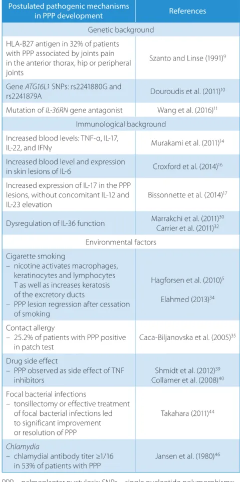
Genetic, immunological and environmental factors play a role in the development of PPP (Table 1).1
Genetic background
The genetic background of PPP is not fully understood. Some authors consider PPP a variant of psoriasis, and stud-ies comparing the prevalence of certain genes in these 2 diseases have been published. Genes coding for HLA-Cw6, WWCC HCR and CDSN5, located in the locus PSORS1 (6p21) on chromosome 6, are considered the main genetic basis of psoriasis.7 On the other hand, studies conducted
on Swedish and British populations have shown that none of the abovementioned alleles are related to the occurrence of PPP. The frequency of occurrence of HLA-Cw6 is simi-lar among patients with PPP (19–20%) and healthy subjects
Fig. 1. Pustulosis localized on the sole
Table 1. Postulated pathogenic mechanisms in PPP development
Postulated pathogenic mechanisms
in PPP development References
Genetic background HLA-B27 antigen in 32% of patients with PPP associated by joints pain in the anterior thorax, hip or peripheral joints
Szanto and Linse (1991)9
Gene ATG16L1 SNPs: rs2241880G and
rs2241879A Douroudis et al. (2011)10
Mutation of IL-36RN gene antagonist Wang et al. (2016)11 Immunological background
Increased blood levels: TNF-α, IL-17,
IL-22, and IFNγ Murakami et al. (2011)14
Increased blood level and expression
in skin lesions of IL-6 Croxford et al. (2014)16
Increased expression of IL-17 in the PPP lesions, without concomitant IL-12 and
IL-23 elevation Bissonnette et al. (2014)
17
Dysregulation of IL-36 function Marrakchi et al. (2011)Carrier et al. (2011)3230
Environmental factors Cigarette smoking
– nicotine activates macrophages, keratinocytes and lymphocytes T as well as increases keratosis of the excretory ducts
– PPP lesion regression after cessation of smoking
Hagforsen et al. (2010)5
Elahmed (2013)34
Contact allergy
– 25.2% of patients with PPP positive
in patch test Caca-Biljanovska et al. (2005)
35
Drug side effect
– PPP observed as side effect of TNF inhibitors
Shmidt et al. (2012)39 Collamer et al. (2008)40 Focal bacterial infections
– tonsillectomy or effective treatment of focal bacterial infections led to significant improvement or resolution of PPP
Takahara (2011)44
Chlamydia
– chlamydial antibody titer ≥1/16
in 53% of patients with PPP Jansen et al. (1980)
46
(15%). Similarly, WWCC HCR occurs in 50% of patients with PPP and 40% of healthy individuals.8,9
Another important genetic factor playing a role in the pathogenesis of psoriasis is the mutation of the ATG16L1
gene located on chromosome 2. ATG16L1 participates in the immunological response, and mutation of this gene leads to decreased production of antimicrobial peptides and enhanced production of pro-inflammatory cytokines interleukin 1 (IL-1) and IL-18, propagating systemic in-flammation. More frequent occurrence of this muta-tion has been identified in inflammatory diseases such as psoriasis and Crohn’s disease. More frequent occur-rence of rs2241880 G and rs2241879A single nucleotide polymorphisms (SNPs) of the ATG16L1 gene has been observed among patients with PPP compared to healthy individuals.10
Due to the possible coexistence of PPP with arthritis and arthralgia in some patients, the genetic base of PPP has been compared with that of psoriatic arthritis. A cor-relation was found between the HLA-B27 antigen, char-acteristic of psoriatic arthritis, and PPP. The HLA-B27 antigen was identified in 32% of patients with PPP associ-ated with arthralgia in the area of the anterior thorax, hip or peripheral joints.9
Recent studies also indicate the role of antagonist receptor IL-36 (IL-36RN) gene mutation in the development of PPP, as well as in various forms of psoriasis: the plaque type, the generalized pustular type and the pustular palmoplan-tar type.11 This mutation is known to be the genetic basis
of deficiency of IL-36 receptor antagonist (DITRA), a rare inherited autosomal recessive disorder characterized by dis-seminated pustular lesions resembling pustular psoriasis or acute generalized exanthematous pustulosis (AGEP).12
Immunological background
Immunological processes in the course of PPP lead to ac-cumulation of large numbers of granulocytes in the area of the sweat gland excretion tract, the acrosyringium.5,13
Cytokines regulating immunological processes play a key role in the pathogenesis of the disorder. As in psoriasis, in PPP, increases in blood levels of pro-inflammatory in-terleukins such as tumor necrosis factor alpha (TNF-α), IL-17, IL-22, and interferon gamma (IFNγ) are observed.14
However, psoriatic and PPP lesions have been found to be different. In psoriatic lesions, there are large amounts of cytokines produced by the Th17 lymphocytes (IL-12, IL-17 and IL-23), while a significant increase in the expres-sion of IL-17 alone has been noted in PPP lein the expres-sions, without concomitant IL-12 and IL-23 elevation. Therefore, isolated increases in IL-17 levels in PPP indicate that neutrophils, not Th-17 lymphocytes, are the main source of this re-sponse.15–17 A high level of IL-17 found locally
in the epi-dermis stimulates keratinocytes to produce IL-6, which activates neutrophils and monocytes and exerts a chemo-tactic effect, attracting granulocytes to the epidermis, which
leads to the formation of pustules. Interleukin 6 probably plays a crucial role in the formation of lesions in PPP, and increased levels of this cytokine have been found within the skin lesions as well as in the blood of patients with this disease.16 Interleukin 6 is a pro-inflammatory cytokine and
belongs to the gp130 cytokine family.18,19 It activates
recep-tor kinase JAK1/JAK2 and Tyk2, and regulates the STAT1/ STAT3 SHP2-MAPK signaling duct.19–21 Interleukin 6
stimulates synthesis of acute phase proteins, differentia-tion of B cells into mature plasma cells, differentiadifferentia-tion and activation of T cells, and activation of Th17 lymphocytes and other non-immunological cells such as keratinocytes and fibroblasts.19,22,23 Moreover, IL-6 stimulates production
of chemokines, e.g., IL-8 and monocyte chemotactic pro-tein 1 (MCP-1), via macrophages and adhesion molecules in the vascular endothelium. This leads to increased mi-gration of granulocytes and the formation of pustules.23–25
Thus, IL-6 may be an attractive new target in the treatment of PPP.20 Interleukin 36, produced by activated
keratino-cytes, is another cytokine that plays a role in the patho-genesis of psoriasis and PPP. This cytokine directly affects immune system cells and stimulates production of IL-1, IL-6, IL-23, TNF-α, and IFNγ,26–28 which reciprocatively
induces keratinocytes to produce IL-6, IL-8 and antibac-terial peptides S 100 A7 and A15. Peptides S 100 A7 and A15, like other pro-inflammatory cytokines, enhance granulocyte migration.29–32 Recent genetics research
in-dicates that IL-36 has a crucial role in the pathogenesis of PPP. As mentioned above, the IL-36RN gene mutation encoding the IL-36Ra protein, the antagonist of the IL-36 receptor, has been demonstrated in patients with localized and generalized forms of pustulosis, occurring in families and sporadically. As a result of this mutation, the function of IL-36 is dysregulated, resulting in increased production of IL-1, IL-6 and IL-8, and the development of an inflam-matory reaction.30,31,33
Relationship between palmoplantar
pustulosis and smoking
Palmoplantar pustulosis is very common in smokers. There are many reports of lesion regression in patients with PPP after they quit smoking.34 Smoking causes oxidative
stress and leads to an accumulation of inflammatory cells in the epidermis. Newly created free radicals stimulate cel-lular signaling ducts, as in psoriasis. Consequently, protein kinases are activated, which is also triggered by bacterial antigens. Moreover, nicotine triggers macrophages and keratinocytes to release cytokines, and activates T lym-phocytes, which sustains the chronic inflammatory pro-cess.5,13 Recent studies indicate that acrosyringium plays
is regulated by nicotinic Ach transferase, and can affect the functioning of the sweat glands. It has been shown that the level of Ach is lower in the lower layers of the ac-rosyringium, because the large quantities of esterase pres-ent there decompose the neurotransmitter. In the absence of Ach, nicotine binds to the nAch receptors. It is believed that nicotine Ach receptors, activated by nicotine and not Ach, may play a role in the pathogenesis of PPP and lead to the accumulation of neutrophils and eosinophils, and to the formation of pustules. Nicotine also affects kera-tinocytes around the sweat glands and causes increased keratosis of the excretory ducts.5,13
Impact of contact allergens
on skin lesions in palmoplantar pustulosis
Based on clinical observations, contact hypersensitivity plays a role in the background of PPP. Clinical studies have shown that contact allergies to one or more substances occurred in 25.2% patients diagnosed with PPP, whereas in a control group comprising people diagnosed with pso-riasis vulgaris, only in 11% of patients had contact allergies. Significantly more women than men suffering from PPP presented positive patch tests. The following allergens were the most common: nickel, rubber additives, Peru balsam, chromium, mercury, and various fragrances. Therefore, performing patch tests in PPP patients who do not re-spond to treatment can be helpful in further medical care. In some centers, patch test are performed routinely in the course of diagnosing PPP, and avoidance of contact allergens is an important element of therapy.35,36
Palmoplantar pustulosis
as a side effect of drugs
Palmoplantar pustulosis primarily affects adults and the elderly. Many patients with PPP are on long-term medi-cation due to the coexistence of other chronic diseases, and skin lesions may appear as a side effect of these drugs.37
Hagforsen et al. documented that in a group of patients with PPP, 30% were treated with ß-blockers, angiotensin-convert-ing-enzyme (ACE) inhibitors or calcium channel blockers, 13% with anti-diabetic drugs, 30% with hormonal therapy, and 15% received antidepressants.38 Recently, widely used
biological agents bring huge therapeutic benefits to many groups of patients. Although TNF inhibitors (adalimumab, infliximab and etanercept) have high efficacy in the treat-ment of psoriasis, their side effects include formation of pso-riatic lesions. During TNF inhibitor therapy, exacerbation of psoriasis and the development of inverse, pustular and erythrodermic forms of the disease have been observed.39
Some reports consider palmoplantar pustular psoriasis the most common type of psoriasis induced by TNF inhibi-tors.39,40 The mechanism of the formation of psoriatic lesions
as a side effect of TNF inhibitors is not fully understood. It has been suggested that blocking the crucial functions
of pro-inflammatory cytokines, such as TNF, triggers alter-native paths of T lymphocyte action that result in the occur-rence of psoriatic lesions in predisposed patients.41
Relationship between palmoplantar
pustulosis and bacterial infections
Clinical observations indicate an association between some inflammatory skin diseases and focal bacterial infec-tions.42 Many case reports have described PPP associated
with tonsillitis, chronic sinusitis or odontogenic infec-tion. Tonsillectomy or effective treatment of focal bacte-rial infections led to significant improvement or resolution of PPP.43,44 In a group of 116 patients with PPP, 109 showed
improvement after tonsillectomies.44 The association
be-tween PPP and tonsillitis has also been proved by the results of immunological and molecular tests. Increased expres-sion of class II activation markers, such as CD25 and skin homing receptors (CLA and CCR6), on T cells in the pala-tine tonsils and in peripheral blood in patients with pustulo-sis has been observed. Moreover, there are ligands for these receptors, such as E-selectin and ligand 20, in psoriatic skin lesions. Excessive stimulation of T lymphocyte migration to the skin by bacteria colonizing the palatine tonsils prob-ably plays a very important role in the pathogenesis of PPP as a focal disease.44 Furthermore, increased expression
of the inducible co-stimulatory molecule (ICOS) receptor on tonsillar tissue T cells in patients with PPP also confirms the relationship between PPP and infectious foci. The ICOS receptor, like CD28, belongs to the co-stimulatory recep-tor family and is not present on resting lymphocytes. Its expression indicates stimulation of the immunological cells and plays a role in triggering T lymphocyte response.42
Other microbiological factors than tonsillar and dental infections are also regarded as cause or aggravating factors in PPP. Erythematous-infiltrative skin lesions with erup-tions resembling PPP are observed in some patients with Reiter’s syndrome and classified as a spondyloarthropathy reactive arthritis. The classic triad of Reiter’s syndrome symptoms consists of urethritis, arthritis and conjunctivitis. Some patients also suffer from dermal and mucosal lesions, such as balanitis circinata located on the penis glans, kera-toderma blennorrhagicum located on the hands and feet, aphthous ulcers within the oral mucosa, and nail disorders. Skin lesions are mainly observed coexisting with Chlamydia trachomatis (Ch. trachomatis) infections.45 It has been
pos-tulated that Ch. trachomatis infections may be a poten-tial cause of isolated pustulosis. This theory is supported by the observation of high antibody titers (equal to or higher than 1/64) against Chlamydia more frequently in patients with PPP (38%) than in psoriatic patients (13%), patients with eczema or urticaria (12%) and a control group of healthy individuals (3%). Chlamydia antibody titers greater than or equal to 1/16 were observed in 53% of patients with PPP.46
Anecdotal case reports also indicate Helicobacter pylori
Stress as a possible factor aggravating
the course of palmoplantar pustulosis
Psychological factors, particularly stress, can play a sig-nificant role in the pathogenesis of certain dermatological diseases, including PPP. About 90% of patients with PPP report exacerbations of the disease in association with stress. Psychological tests have confirmed these clinical observations. A study based on the Eysenck Personal-ity Questionnaire (EPQ), which measures 3 personalPersonal-ity factors (extraversion–introversion, neuroticism and psy-choticism), showed that fear, anxiety and psychosomatic disorders occur in 43% of patients with PPP, compared to 19% of people in the control group.48 Patients with PPP
have also been subjected to the Inventory of Situations and Responses to Anxiety (ISRA). Fear and activation of the au-tonomic nervous system, manifested by tachycardia, dry mouth and sweating, was observed in 85% of patients with PPP, compared to 19% in the control group.48
Relationship between palmoplantar
pustulosis and gluten intolerance
It is suspected that gluten intolerance might play a role in the pathogenesis of PPP, but the results of published studies are inconclusive. In a population of Swedish pa-tients with PPP, the presence of IgA anti-gliadin antibodies was demonstrated in 18% and of anti-transglutaminase tissue in 7–10% of the subjects who did not report gas-trointestinal symptoms. However, the duodenal mucosa biopsy in a group of patients with anti-gliadin antibodies demonstrated intestinal villi atrophy. In most patients, adherence to a gluten-free diet resulted in regression of the skin lesions.38,49 Different results were obtained
in patients from Germany; neither anti-gliadin antibodies nor anti-transglutaminase tissue were detected in patients with PPP. The discrepancies may be related to many fac-tors, such as ethnic differences between the populations surveyed.50
Coexistence of palmoplantar
pustulosis with other diseases
Seronegative arthropathies coexisting
with palmoplantar pustulosis
Palmoplantar pustulosis is one of the symptoms of a group of syndromes classified as seronegative spon-dyloarthropathies. These disorders include synovitis/acne/ pustulosis/hyperostosis/osteitis (SAPHO) syndrome, So-nozaki syndrome and Reiter’s syndrome.
The SAPHO syndrome was described in 1987 as the co-existence of synovitis, acne, PPP, hyperostosis, and oste-itis.51 Skin lesions can precede, occur simultaneously with
or follow osteoarticular symptoms. The timespan between lesion formation and osteoarticular symptoms does not ex-ceed 2 years.52 The etiopathogenesis of SAPHO syndrome
is unknown. The adverse effect of retinoids among patients treated for severe acne was considered as a possible cause, but this theory was not confirmed, since a significant number of patients with SAPHO syndrome had not been treated with retinoids.53 Infectious agents have also been
suspected as part of the etiology of SAPHO syndrome. In some patients, Corynebacterium has been isolated from the sternoclavicular joints. However, the role of this patho-gen is questionable.53,54
Pustulotic arthro-osteitis (PAO) was first described by Sonozaki in 1979 as symmetrical erythematous and pustular lesions on the hands and feet accompanied by sternoclavicular joint pain. Recently, it has been sug-gested that Sonozaki syndrome should be classified as a spectrum of SAPHO syndrome. However, SAPHO syndrome and other spondylopathies, like psoriatic arthri-tis and Reiter’s syndrome, are related to the HLA B27 an-tigen, while in Sonozaki syndrome, the prevalence of this antigen is low.53
Thyroid diseases in patients
with palmoplantar pustulosis
Thyroid diseases have been observed more frequently in women with PPP than in a healthy population. Thyroid dysfunction coexists in 25% of studied women suffering from PPP. These disorders include hypothyroidism, hy-perthyroidism, struma nodosa, and any thyroid surgery. Due to the fact that PPP coexists with thyroid diseases, it is advisable to gather more data concerning PPP patients and conduct screening of their thyroid functions.55
Summary
ORCID iDs
Magdalena Putra-Szczepaniak https://orcid.org/0000-0001-6340-7164 Joanna Maj https://orcid.org/0000-0001-8300-8208
Alina Jankowska-Konsur https://orcid.org/0000-0003-4944-5388 Anna Czarnecka http://orcid.org/0000-0002-6621-9537
Anita Hyncewicz-Gwóźdź https://orcid.org/0000-0002-1601-471X
References
1. Yamamoto T. Extra-palmoplantar lesions associated with palmoplan-tar pustulosis. J Eur Acad Dermatol Venereol. 2009;23(11):1227–1232. 2. Ammoury A, El Sayed F, Dhaybi R, Bazex J. Palmoplantar pustulosis
should not be considered as a variant of psoriasis. J Eur Acad
Derma-tol Venereol. 2008;22(3):392–393.
3. de Waal AC, van de Kerkhof PC. Pustulosis palmoplantaris is a dis-ease distinct from psoriasis. J Dermatolog Treat. 2011;22(2):102–105. 4. Murakami M, Ohtake T, Horibe Y, et al. Acrosyringium is the main site
of the vesicle/pustule formation in palmoplantar pustulosis. J Invest
Dermatol. 2010;130(8):2010–2016.
5. Hagforsen E, Michaëlsson G, Stridsberg M. Normal and PPP-affected palmoplantar sweat gland express neuroendocrine markers chro-mogranins and synaptophysin differently. Arch Dermatol Res. 2010; 302(9):685–693.
6. Eriksson MO, Hagforsen E, Lundin IP, Michaëlsson G. Palmoplantar pustulosis: A clinical and immunohistological study. Br J Dermatol. 1998;138(3):390–398.
7. Allen MH, Ameen H, Veal C, et al. The major psoriasis susceptibility locus PSORS1 is not a risk factor for late-onset psoriasis. J Invest
Der-matol. 2005;124(1):103–106.
8. Asumalahti K, Ameen M, Suomela S, et al. Genetic analysis of PSORS1 distinguishes guttate psoriasis and palmoplantar pustulosis. J Invest
Dermatol. 2003;120(4):627–632.
9. Szanto E, Linse U. Arthropathy associated with palmoplantar pustu-losis. Clin Rheumatol. 1991;10(2):130–135.
10. Douroudis K, Kingo K, Traks T, et al. ATG16L1 gene polymorphisms are associated with palmoplantar pustulosis. Hum Immunol. 2011;72(7): 613–615.
11. Wang TS, Chiu HY, Hong JB, Chan CC, Lin SJ, Tsai TF. Correlation of IL36RN mutation with different clinical features of pustular psoria-sis in Chinese patients. Arch Dermatol Res. 2016;308(1):55–63. 12. Nakai N, Sugiura K, Akiyama M, Katoh N. Acute generalized
exan-thematous pustulosis caused by dihydrocodeine phosphate in a patient with psoriasis vulgaris and a heterozygous IL36RN mutation.
JAMA Dermatol. 2015;151(3):311–315.
13. Hagforsen E, Hedstrand H, Nyberg F, Michaëlsson G. Novel findings of Langerhans cells and interleukin-17 expression in relation to the acrosyringium and pustule in palmoplantar pustulosis. Br J Dermatol. 2010;163(3):572–579.
14. Murakami M, Hagforsen E, Morhenn V, Ishida-Yamamoto A, Iizuka H. Patients with palmoplantar pustulosis have increased IL-17 and IL-22 levels both in the lesion and serum. Exp Dermatol. 2011;20(10):845–847. 15. Kagami S, Rizzo HL, Lee JJ, Koguchi Y, Blauvelt A. Circulating Th17,
Th22, and Th1 cells are increased in psoriasis. J Invest Dermatol. 2010; 130(5):1373–1383.
16. Croxford AL, Karbach S, Kurschus FC, et al. IL-6 regulates neutrophil microabscess formation in IL-17A-driven psoriasiform lesions. J Invest
Dermatol. 2014;134(3):728–735.
17. Bissonnette R, Nigen S, Langley RG, et al. Increased expression of IL-17A and limited involvement of IL-23 in patients with palmoplantar (PP) pustular psoriasis or PP pustulosis: Results from a randomised controlled trial. J Eur Acad Dermatol Venereol. 2014;28(10):1298–1305. 18. Neurath MF, Finotto S. IL-6 signaling in autoimmunity, chronic inflamma-tion and inflammainflamma-tion-associated cancer. Cytokine Growth Factor Rev. 2011;22(2):83–89.
19. Rincon M. Interleukin-6: From an inflammatory marker to a target for inflammatory diseases. Trends Immunol. 2012;33(11):571–577. 20. Jones SA, Scheller J, Rose-John S. Therapeutic strategies for the clinical
blockade of IL-6/gp130 signaling. J Clin Invest. 2011;121(9):3375–3383. 21. Miyoshi K, Takaishi M, Nakajima K, et al. Stat3 as a therapeutic target for the treatment of psoriasis: A clinical feasibility study with STA-21, a Stat3 inhibitor. J Invest Dermatol. 2011;131(1):108–117.
22. Ataie-Kachoie P, Pourgholami MH, Morris DL. Inhibition of the IL-6 signaling pathway: A strategy to combat chronic inflammatory dis-eases and cancer. Cytokine Growth Factor Rev. 2013;24(2):163–173. 23. Ishihara K, Hirano T. IL-6 in autoimmune disease and chronic
inflam-matory proliferative disease. Cytokine Growth Factor Rev. 2002;13(4–5): 357–368.
24. Romano M, Sironi M, Toniatti, et al. Role of IL-6 and its soluble recep-tor in induction of chemokines and leukocyte recruitment. Immuni-ty. 1997;6(3):315–325.
25. Bartoccioni E, Scuderi F, Marino M, Provenzano C. IL-6, monocyte infiltration and parenchymal cells. Trends Immunol. 2003;24(6): 299–300.
26. Blumberg H, Dinh H, Dean C Jr, et al. IL-1RL2 and its ligands con-tribute to the cytokine network in psoriasis. J Immunol. 2010;185(7): 4354–4362.
27. Tortola L, Rosenwald E, Abel B, et al. Psoriasiform dermatitis is driv-en by IL-36-mediated DC-keratinocyte crosstalk. J Clin Invest. 2012; 122(11):3965–3976.
28. Johnston A, Xing X, Guzman AM, et al. IL-1F5, -F6, -F8, and -F9: A novel IL-1 family signaling system that is active in psoriasis and promotes keratinocyte antimicrobial peptide expression. J Immunol. 2011;186(4):2613–2622.
29. Skov L, Beurskens FJ, Zachariae CO, et al. IL-8 as antibody therapeutic target in inflammatory diseases: Reduction of clinical activity in pal-moplantar pustulosis. J Immunol. 2008;181(1):669–679.
30. Marrakchi S, Guigue P, Renshaw BR, et al. Interleukin-36-receptor antagonist deficiency and generalized pustular psoriasis. N Engl
J Med. 2011;365(7):620–628.
31. Tripodi D, Conti F, Rosati et al. IL-36 a new member of the IL-1 family cytokines. J Biol Regul Homeost Agents. 2012;26(1):7–14.
32. Carrier Y, Ma HL, Ramon HE, et al. Inter-regulation of Th17 cytokines and the IL-36 cytokines in vitro and in vivo: Implications in psoriasis pathogenesis. J Invest Dermatol. 2011;131(12):2428–2437.
33. Capon F. IL36RN mutations in generalized pustular psoriasis: Just the tip of the iceberg? J Invest Dermatol. 2013;133(11):2503–2504. 34. Elahmed HH. Rapid improvement of palmoplantar psoriasis after
cessation of smoking. Sultan Qaboos Univ Med J. 2013;13(1):188–189. 35. Caca-Biljanovska N, V’lckova-Laskoska M, Balabanova-Stefanova M,
Grivceva-Panovska V. Frequency of delayed-type hypersensitivity to contact allergens in palmoplantar psoriasis. Prilozi. 2005;26(2): 131–141.
36. Ito T, Mori T, Fujiyama T, Tokura Y. Dramatic exacerbation of palmo-plantar pustulosis following strongly positive nickel patch testing.
Int J Dermatol. 2014;53(5):e327–e329.
37. Bordel-Gómez MT, Sánchez-Estella J, Martínez-González O, Cardeño-so-Alvarez ME. Palmoplantar psoriasis: A paradoxical adverse reac-tion induced by adalimumab. J Eur Acad Dermatol Venereol. 2009; 23(4):444–445.
38. Hagforsen E, Michaëlsson K, Lundgren E, et al. Women with palmo-plantar pustulosis have disturbed calcium homeostasis and a high prevalence of diabetes mellitus and psychiatric disorders: A case-control study. Acta Derm Venereol. 2005;85(3):225–232.
39. Shmidt E, Wetter DA, Ferguson SB, Pittelkow MR. Psoriasis and pal-moplantar pustulosis associated with tumor necrosis factor-α inhib-itors: The Mayo Clinic experience, 1998 to 2010. J Am Acad Dermatol. 2012;67(5):e179–e185.
40. Collamer AN, Guerrero KT, Henning JS, Battafarano DF. Psoriatic skin lesions induced by tumor necrosis factor antagonist therapy: A lit-erature review and potential mechanisms of action. Arthritis Rheum. 2008;59(7):996–1001.
41. Bordel-Gómez MT, Sánchez-Estella J, Martínez-González O, Cardeño-so-Alvarez ME. Palmoplantar psoriasis: A paradoxical adverse reac-tion induced by adalimumab. J Eur Acad Dermatol Venereol. 2009; 23(4):444–445.
42. Kobayashi S. Tonsil-related skin diseases and possible involvement of T cell co-stimulation in chronic focal infection. Adv
Otorhinolar-yngol. 2011;72:83–85.
43. Tsuboi H, Katsuoka K. Pustulosis palmaris et plantaris with promi-nent hyperkeratosis of the soles. J Dermatol. 2006;33(12):892–895. 44. Takahara M. Clinical outcome of tonsillectomy for palmoplantar
45. Quint KD, van der Helm-van Mil AH, Bergman W, Lavrijsen AP. Muco-cutaneous abnormalities in Chlamydia trachomatis-induced reactive arthritis [in Dutch]. Ned Tijdschr Geneeskd. 2010;154:A1614
46. Jansen CT, Hollmén, Pajarre R, Terho P. Antichlamydial antibodies in chronic palmoplantar pustulosis. Acta Derm Venereol. 1980;60(3): 263–266.
47. Martin Hübner A, Tenbaum SP. Complete remission of palmoplan-tar psoriasis through Helicobacter pylori eradication: A case report.
Clin Exp Dermatol. 2008;33(3):339–340.
48. Sáez-Rodríguez M, Noda-Cabrera A, Alvarez-Tejera S, et al. The role of psychological factors in palmoplantar pustulosis. J Eur Acad
Der-matol Venereol. 2002;16(4):325–327.
49. Michaëlsson G, Kristjánsson G, Pihl Lundin I, Hagforsen E. Palmoplan-tar pustulosis and gluten sensitivity: A study of serum antibodies against gliadin and tissue transglutaminase, the duodenal mucosa and effects of gluten-free diet. Br J Dermatol. 2007;156(4):659–666. 50. Weisenseel P, Kuznetsov AV, Ruzicka T, Prinz JC. Palmoplantar
pustu-losis is not inevitably associated with antigliadin antibodies. Br
J Der-matol. 2007;156(6):1399–1400.
51. Károlyi Z, Harhai I, Erós N. Dermatologic aspects of SAPHO-syndrome.
Orv Hetil. 2001;142(33):1801–1804.
52. Chamot AM, Benhamou CL, Kahn MF, Beraneck L, Kaplan G, Prost A. Acne-pustulosis-hyperostosis-osteitis syndrome: Results of a nation-al survey (85 cases). Rev Rhum Mal Osteoartic. 1987;54(3):187–196. 53. Stanford CW, Kollipara R, Melookaran AM, Hall JC. Palmoplantar
pus-tular psoriasis following initiation of a beta-blocker: Disease control with low-dose methotrexate. Cutis. 2014;94(3):153–155.
54. Matzaroglou Ch, Velissaris D, Karageorgos A, Marangos M, Panagi-otopoulos E, Karanikolas M. SAPHO Syndrome diagnosis and treat-ment: Report of five cases and review of the literature. Open Orthop J. 2009;3:100–106.