Liaqat Hussain
1, A–C, Muhammad S. H. Akash
2, A, D–F, Sabah Naseem
1, B,
Kanwal Rehman
3, D–F, Kwaja Z. Ahmed
1, AAnti-Ulcerogenic Effects of Salmalia Malabarica
in Gastric Ulceration – Pilot Study
1 Government College, University Faisalabad, Pakistan
2 College of Pharmaceutical Sciences, Institute of Pharmacology, Toxicology and Biochemical Pharmaceutics, Zhejiang University, Hangzhou, China
3 Department of Toxicology, School of Medicine and Public Health,Zhejiang University, Hangzhou, China
A – research concept and design; B – collection and/or assembly of data; C – data analysis and interpretation;
D – writing the article; E – critical revision of the article; F – final approval of article; G – other
Abstract
Background. According to an estimation of the WHO, almost 80% of people globally are treated by traditional medicine.
Objectives. We evaluated the anti-ulcerogenic potential of Salmalia malabarica extract in rats using aspirin-, alco-hol- and pylorus ligation-induced ulcer models.
Material and Methods. Two different doses (200 and 400 mg/kg body weight) of Salmalia malabarica extract was administered intraperitoneally (i.p.) to all 3 ulcer-induced models for 5 consecutive days. The anti-ulcerogenic potential in rats treated with 2 doses of Salmalia malabarica extract and omeprazole (20 mg/kg, i.p.) was deter-mined and compared to the control groups.
Results. Salmalia malabarica extract showed a significant decrease in ulcer index as compared to the control group in a dose-dependent manner. Salmalia malabarica extract also showed protection of 66.22% and 74.54% in asprin-, 73.79% and 78.14% in alcohol- and 68.94% and 78.84% in pylorus ligation-induced ulcers. However, omeprazole showed protection of 84.73%, 85.5% and 86.12% in aspirin-, alcohol- and pylorus ligation-induced ulcers, respec-tively. Furthermore, Salmalia malabarica extract significantly decreased the volume of gastric juice, free and total acidity, whereas it increased gastric pH when directly compared to the control group.
Conclusions. Conclusively, Salmalia malabarica possesses anti-ulcerogenic, antisecretory, and cytoprotective potential and can be used as a supplement for the treatment of gastric ulcers in a dose dependent manner (Adv Clin Exp Med 2015, 24, 4, 595–605).
Key words:Salmalia malabarica, ulcer index, cytoprotective mechanism, peptic ulcer.
ORIGINAL PAPERS
Adv Clin Exp Med 2015, 24, 4, 595–605
DOI: 10.17219/acem/28115 © Copyright by Wroclaw Medical University ISSN 1899–5276
Peptic ulcer disease is a term used for both stomach and duodenal ulcer. In clinical practice, peptic ulcer disease is most commonly known as a gastrointestinal disease which is interlinked with the acute and chronic inflammation of the gas-tric and duodenal epithelium [1]. According to the pathophysiology, peptic ulcer disease appears as a result of an imbalance between offensive and defensive factors. The major offensive factors that potentiate a peptic ulcer are acid, pepsin, and He-licobacter pylori, whereas the defensive factors which antagonize the harmful effects of the offen-sive factors are mucin, prostaglandin, bicarbonate,
nitric oxide and growth factors [2]. Other factors that may influence the potentiation for peptic ulcer are improper digestion, metabolism and elimina-tion of food. Mental and physical stress might al-so participate in the development of peptic ulcers. Chronic users of non-steroidal anti-inflammatory drugs (NSAID’S) also represent the pathogenesis of peptic ulcer disease. Alcohol consumption is al-so a major risk factor and contributes in the forma-tion of gastric ulcers [3, 4].
tract [5], which ruins the balance between the de-fensive and ofde-fensive agents. Therapy for pep-tic ulcer disease has been broadly classified into 2 major types. The first type of these drugs causes a decrease in acid or pepsin secretion such as H2-blockers, M1-blockers and proton pump in-hibitors. The second class of these drugs causes an increase in mucosal defensive factors [6]. Ninety eight percent of all gastrointestinal ulcers are most likely to be found in the duodenum and stomach at an approximate ratio of 4 : 1 [7]. There are remark-able findings that have suggested boiling water, ab-solute ethanol, 25% sodium chloride, 0.2 N sodium hydroxide and 0.6 N hydrochloric acid are necro-tizing agents participating in ulcerogenesis [8].
The traditional systems for the treatment of different diseases via medicinal plants have gained strong acceptance. Today, natural products are the foremost symbol of safety for the treatment of vari-ous diseases as compared to synthetic drugs [9–16]. Different parts of Salmalia malabarica [Malvaceae
(Bombacaceae)] have been proven traditionally and scientifically for the treatment of several ailments. The gum of Salmalia malabarica has been tradition-ally used mainly in the treatment of haemoptysis of pulmonary tuberculosis, influenza and menor-rhagia. It is also known for various other proper-ties such as astringent, antiangiogenic, expectorant, analgesic, emetic, stimulant, anti-inflammatory, an-ti-hypertensive and antioxidant properties [17]. It has also been used in bladder disorders, calculus, leucorrhoea, tuberculosis and to cure conjunctivi-tis [18]. Salmalia malabarica has been proven ef-fective in the treatment of acne and skin eruption problems [19]. Antibacterial and antifungal charac-teristics are also reported to be found in Salmalia malabarica. The methanol extract of Salmalia mala-barica has shown a potent antibacterial activity [20]. Moreover, hypoglycemic and hypolipidemic effects were also studied in an n-hexane extract of sepal of
Salmalia malabarica [21]. However, other effects of
Salmalia malabarica still need to be elucidated. The purpose of our present study was to in-vestigate the anti-ulcerogenic and ulcer healing potential of a methanol extract of Salmalia mal-abarica using three experimental models i.e. aspi-rin-, alcohol- and pylorus ligation-induced gastric ulceration using albino Wistar rats.
Material and Methods
Plant Material
The fresh bark of Salmalia malabarica was col-lected in March 2013 from nearby areas of Layyah from the Punjab province of Pakistan. The bark
was cut into small pieces and dried. The plants, i.e. bark, was grounded to a fine powder by Wiley mill (standard model 4). The plant was authenticated by the Botany Department, University of the Pun-jab, Lahore, Pakistan and the voucher specimen no. of Salmalia malabarica, LAH-3112013 ML, was preserved.
Chemicals
Omeprazole (OMZ) was purchased from Getz Pharma Pvt. Ltd., Karachi, Pakistan. Aspirin was purchased from Highnoon Laboratories Pvt. Ltd., Lahore, Pakistan. Methanol (95%) was obtained from Sigma-Aldrich (St. Louis, MO, USA). All other chemicals used in this study were analytical grade and used without further modification.
Animals
Healthy adult male albino Wistar rats (130– –180 g) were used for the anti-ulcerogenic study. The animals were purchased from the animal house of the University of Agriculture, Faisalabad, Pakistan. The animals were acclimatized with stan-dard environmental conditions of a 12 h light: dark cycle, temperature of 25 ± 2° and relative humidity of 65 ± 10%. They were provided with a standard rodent pellet diet. Access to food and water was provided ad libitum. The food was withdrawn 18– 24 h and water was withheld 2 h before the start of the experimental procedure.
Ethical Considerations
The experimental animals were used under the guidelines of the Institute of Laboratory Ani-mals Resource, Commission on Life Sciences, Na-tional Research Council and approved by the Ethi-cal Committee of Government College University Faisalabad, Faisalabad, Pakistan.
Preparation of Crude Extract
of Salmalia Malabarica
Phytochemical Analysis and
Determination of LD50 in Rats
Salmalia malabarica were subjected to the prelim-inary screening of phytochemical compounds using various test as described previously with some modi-fications [22, 23]. Briefly, alkaloids were detected using the Mayer’s test. Tannins were detected using a gela-tin test. Flavonoids were detected using an alkaline re-agent test. Proteins and amino acids were detected us-ing a xanthoproteic test and ninhydrin test.
We investigated the acute toxicity of Salma-lia malabarica extract in order to select the suitable dose for an investigation of the anti-ulcerogenic po-tential of Salmalia malabarica on Wistar rats. We administered a single i.p. dose of methanol extract of Salmalia malabarica with the dose range of 0.1– –20 g/kg. Following the treatment period, the ani-mals were observed for up to 72 h and all the ob-servational changes and death were recorded dur-ing the whole observation period. The % of death in each group was calculated and LD50 was de-termined as described previously [24], with some modifications.
Treatment Schedule
The standard dose of plant extract Salma-lia malabarica was diluted at a dose of 200 mg/kg (SM-200) and 400 mg/kg body weight of the rats (SM-400). The treatment therapy was continued for 5 consecutive days in all groups of experimen-tal animals. The extracts of Salmalia malabarica
were administered intraperitoneally according to the weight of the animal. Omeprazole (OMZ) was used as a standard anti-ulcer drug. OMZ was given at a dose of 20 mg/kg PO in 5 mL of normal saline.
Evaluation of Anti-Ulcerogenic
Potential of Extract of Salmalia
Malabarica
Aspirin-Induced Ulcer
Healthy albino Wistar rats were divided into 5 groups (5 × 7). Group I was treated as placebo, group II served as a negative control treated with aspirin, group III was treated with the standard drug OMZ, group IV with the 200 mg/kg body weight of extract of Salmalia malabarica (SM-200) and group V with 400 mg/kg body weight of ex-tract of Salmalia malabarica (SM-400). The ani-mals were fasted for 24 h and water was withdrawn 2 h prior to the surgical procedures. After 45 min of the administration of OMZ and extracts of SM-200 and SM-400, aspirin was administered at a dose of
20 mg/mL of body weight. The animals were killed by cervical dislocation after 4 h and the stomach was cut along the greater curvature following the procedure as described previously [25]. The ulcers were measured with the help of a microscope.
Alcohol-Induced Ulcer
Healthy albino Wistar rats were divided into 5 groups with 7 mice in each group. Group I was treated as placebo, group II served as a negative control treated with ethanol, group III with the standard drug OMZ, group IV with SM-200 and group V with SM-400. The animals were fasted for 24 h and water was withheld 2 h before the surgical procedures. Absolute ethanol (96%) was given PO at a dose of 1 mg/200 gm/kg body weight of rats after 45 min of the administration of the methanol SM extracts and OMZ treatment [26]. The animals were euthanized by cervical dislocation after 1 h of ethanol administration. The stomach was taken out and incised along the greater curvature and pinned on a soft board. The ulcer was scored microscopi-cally for the measurement of gastric lesions.
Pylorus Ligation-Induced Ulcer
Healthy albino Wistar rats were divided into 4 equal groups each containing 7 animals. Group I was considered as positive control which re-ceived normal saline. Group II was treated with the standard dose of OMZ. Group III was treated with 200 mg/kg of extract of Salmalia malabarica
(SM-200) and group IV was treated 400 mg/kg of extract of Salmalia malabarica (SM-400). On the 5th
Measurement of Ulcer Index and
Percantage of Protection from Ulcer
We examined the opened stomach of each rat in order to calculate the ulcer index (UI) by giv-ing a scorgiv-ing number [2]. The ulcer score was es-timated by viewing the ulcers with a magnifying glass. The UI was calculated as follows where “X” was the ratio of total mucosal area to that with ul-cerated area. The percentage of protection was cal-culated using the following formulaas described previously [2].
Gastric Secretion Study
The centrifuged gastric volume was measured from measuring the cylinder after transferring the supernatant liquid from centrifuge tubes [27]. The supernatant fluid was taken from the centrifuged tubes. 1 mL of gastric juice was taken in a 100 mL conical flask and diluted with 10 mL of distilled water. Two–three drops of Topfer’s reagent was poured into the conical flask and was titrated against 0.01 N NaOH until the color of the solu-tion turned to yellowish orange and all traces of red color disappeared. The volume of NaOH was not-ed to measure the pH of the gastric acid, volume of the gastric secretion and free acidity following the method as described previously [28]. For the determination of total acidity, 2–3 drops of phe-nolphthalein indicator was poured into the conical flask and again titrated against NaOH, a clear red tinge reappeared. The volume of alkali added was noted and the total volume used determined the total acidity. Total acidity was calculated following the method as described previously [29].
Histopathology of Ulcer Models
The rats were made unconscious by overdose of chloroform. The rats were euthanized by cer-vical dislocation with the help of a presterilized sharp blade. Tissues from the gastric walls were separated and fixed in 10% buffered formalin over-night and processed in a paraffin tissue process-ing machine. Sections of the stomach were made at a thickness of 5 μ and stained with haematoxy-lin and eosin (H & E) for histological evaluation. Histochemical alterations were observed in these tissues and images were taken using Olympus col-or video camera with Histolab software (Biocom).
Statistical Analysis
The results were expressed as mean ± SEM. The data of all groups was analyzed using a one-way anal-ysis of variance (ANOVA), Student-Neuman-Keuls
multiple comparison tests or Student’s t-test us-ing Graph Pad Prism 5 (Graph Pad, Software Inc., USA). The criteria for statistically significant differ-ence between the treated group and that of the con-trol group were adjusted at p < 0.05.
Results
Phytochemical Analysis and
Determination of LD50 in Rats
Preliminary phytochemical analysis of Salma-lia malabarica revealed the presence of a rich amount of phytochemicals such as alkaloids, tan-nins, flavonoids, proteins and amino acids. The ex-tract of Salmalia malabarica was found to be non- -toxic when administered i.p. to Wistar rats with dose range of 0.1–20 g/kg and the LD50 value was found to be safe at the highest dose of Salmalia malabarica.
Anti-Ulcer Effect
of Salmalia Malabarica
Effects of Salmalia Malabarica
on Aspirin-Induced Ulcer
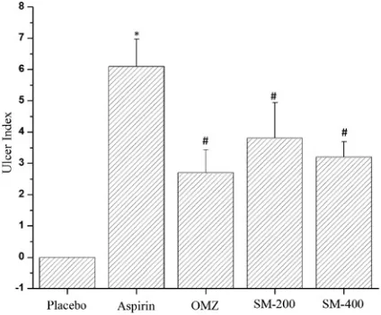
In the aspirin-induced ulcer model, the pla-cebo group did not show any kind of ulcer index. The rat group treated with aspirin alone showed an elevated value of the mean ulcer index at its signif-icant level of 6.1 ± 0.872 (Fig. 1). The OMZ-treat-ed group showOMZ-treat-ed the lesser values of the ulcer in-dex (2.7 ± 0.734) when directly compared with that
of the aspirin-treated group. The other 2 groups, treated with SM-200 and SM-400, showed values of the ulcer index of 3.8 ± 1.140 and 3.2 ± 0.489, respectively, as shown in Fig. 1A. The percentage of protection of the OMZ-treated groups was high-est, at 84.73%, whereas the rat groups treated with SM-200 and SM-400 maintained the % protection of 66.22% to 74.54% (Table 1). The mean ulcer index obtained from the rat groups treated with SM-200 and SM-400 also showed a significant dif-ference between the values of the ulcer index of the aspirin-treated group.
Effects of Salmalia Malabarica
on Alcohol-Induced Ulcer
In alcohol-induced ulcer, the rat group treat-ed with alcohol alone showtreat-ed the highest ulcer in-dex, of 5.8 ± 0.492, whereas the rat group treated with OMZ showed the least value of ulcer index, at 2 ± 0.694, when directly compared to that of the alcohol-treated rat group (Fig. 2). The OMZ- -treated groups also showed the highest percent-age of protection against alcohol-induced ulcer (Table 1). The effect of Salmalia malabarica ex-tract at the dose of SM-200 showed the least signif-icant effect against ulcer protection (3.7 ± 0.832 vs. 5.8 ± 0.492 when compared to the alcohol-treated group, and 3.7 ± 0.832 vs. 2.6 ± 0.694 when com-pared to the OMZ-treated group), with a percent-age of protection of 78.79%, whereas the Salmalia malabarica extract at the dose of SM-400 showed a highly significant effect in gastric protection against ulceration caused by alcohol (2.9 ± 1.092 vs. 5.8 ± 0.492) when directly compared to that of the OMZ-treated and/or SM-200-treated groups, respectively (Fig. 1B). The percentage of protec-tion against ulcer in the SM-400-treated group was significantly higher than that in the SM-200-treat-ed group (Table 1).
Effects of Salmalia Malabarica
on Pylorus Ligation-Induced Ulcer
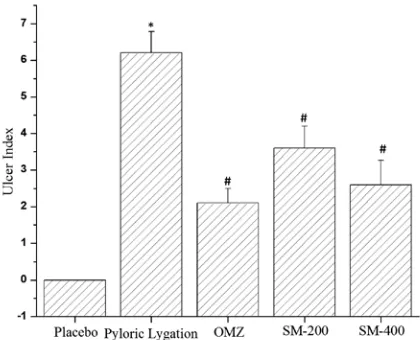
In pyloric ligation-induced ulcer, group I served as a control group that only received normal saline, and showed a significant mean ulcer index with the highest value of 6.2 ± 0.583. Group II was treated with OMZ, which showed the lowest value of mean ulcer index (2.1 ± 0.400 vs. 6.2 ± 0.583) when compared to the mean ulcer index value of the control group (Fig. 1C), with a significant val-ue of %protection at the level of 66.12% (Table 1). Group III and group IV were treated with Salmalia malabarica extracts of SM at 2 different doses. The SM-400 showed the lowest value of mean ulcer in-dex when directly compared to that of the control
Table 1. Effect of methanol extract of Salmalia Malabarica in 3 experimental induced ulcers (n = 5)
Sr.# Treatments Percent protection
dose administered aspirin-induced
ulcer (%) alcohol-induced ulcer (%) pyloric ligation- -induced ulcer (%)
1 Control (placebo) – – – –
2 Control (normal saline) 1 mL/kg (i.p.) – – – 3 Omeprazole 20 mg/kg (i.p.) 84.73 85.5 86.12 4 SM-200 200 mg/kg (i.p.) 66.22 63.79 66.94 5 SM-400 400 mg/kg (i.p.) 74.54 78.14 79.84 OMZ – omeprazole, SM – Salmalia malabarica, i.p. – intraperitoneal.
group (2.6 ± 0.663 vs. 6.2 ± 0.583). However, the group treated with SM-200 extract of Salmalia malabarica showed a mean ulcer index value of 3.6 ± 0.600. A non-significant difference was al-so observed between the mean ulcer values of the rat group treated with SM-400 extract of Salma-lia malabarica and the OMZ-treated rat group (Fig. 3). We also measured the percentage of pro-tection against ulcer and found that the rat group treated with SM-400 extract of Salmalia malabar-ica showed maximum percentage of protection against ulcer when directly compared to the group treated with SM-200 extract of Salmalia malabar-ica (Table 1).
Effect of Salmalia Malabarica
on Gastric Secretion
At the end of the treatment, we also measured the effects of Salmalia malabarica on the secretion of gastric contents in pyloric ligation-induced ul-cer. We measured the volume of gastric juice, the free acidity, total acidity and pH of the gastric se-cretion in all rat groups of pyloric ligation-induced ulcer. The control group, treated with normal sa-line, showed a significantly high volume of gas-tric juice (6.86 ± 0.272) when directly compared to that of the other 3 groups (Fig. 4A). Similarly, the rat group treated with SM-400 extract of Salma-lia malabarica showed the lowest value of gastric juice when directly compared to that of the control group (2.88 ± 0.287 vs. 6.86 ± 0.272). The OMZ- -treated groups also showed a decreased secretion
of gastric juice as compared to both the control group (2.16 ± 0.333 vs. 6.86 ± 0.272) and the rat group treated with SM-200 extract of Salmalia malabarica (2.16 ± 0.333 vs. 3.52 ± 0.655).
In pyloric ligation-induced ulcer, we mea-sured free acidity in all rat groups. Due to the oc-currence of ulcer, the control groups showed a sig-nificantly higher value of free acidity when directly compared to the OMZ-treated group and the rat groups treated with extract of Salmalia malabar-ica SM-200 and SM-400 (Fig. 4B). The value of free acidity in the rat group treated with extract of
Salmalia malabarica SM-400 showed a non-signif-icant difference when directly compared to that of the OMZ-treated group (Fig. 4B). We also mea-sured the total acidity in all rat groups. The control group followed the same pattern for total acidity as it followed in the case of free acidity (Fig. 2C). However, the value of total acidity was signifi-cantly very high as compared to the OMZ-treated group and/or rat groups treated with the extracts of Salmalia malabarica (Fig. 4C).
We also measured the pH of the gastric con-tents of the pyloric ligation-induced ulcer model, where the control group revealed the lowest value of pH when directly compared to the OMZ-treated and/or extracts of Salmalia malabarica rat groups (Fig. 4D). The extract of Salmalia malabarica with a dose of SM-400 increased the value of pH signifi-cantly very highly (p < 0.001) as compared to the ex-tract of Salmalia malabarica with a dose of SM-200.
Histopathology of Ulcer Models
At the end of the treatment period, we also as-sessed the histopathological alterations in ulcer- -induced models treated with extract of Salmalia malabarica (Fig. 5). The specimens of the gastric walls from each rat were fixed in 10% buffered for-malin overnight and processed in a paraffin tis-sue processing machine. Sections of the stomach were made at a thickness of 5 μ and stained with haematoxylin and eosin (H & E) for histological evaluation. In the pyloric ligation-induced ulcer model, the rat groups exhibited damaged mucosal epithelium, proliferated fibroblasts, inflammato-ry exudates and the infiltration of leucocytes, due to which necrosis occurred (Fig. 5A). Extract of
Salmalia malabarica showed a significant protec-tion against these histopathological changes in py-loric ligation-induced ulcer with apparent epitheli-alizations and regeneration of mucosa (Fig. 5A-D).
Discussion
According to the World Health Organization (WHO), 80% of the world’s population relies on traditional medicines for the treatment of different
diseases and their primary health care needs [30]. Methanol extract of Salmalia malabarica has also shown pronounced characteristics of antibacteri-al and antifungantibacteri-al activities. The present study elu-cidated the effect of methanol extract of Salmalia malabarica at 2 doses, i.e. SM-200 and SM-400, on ulcer induction by aspirin-, alcohol- and pylorus ligation-induced ulcers.
Preliminary phytochemical analysis of Salma-lia malabarica showed rich custody of phytochem-icals such as tannins, fatty acids, amino acids, fla-vonoids and alkaloids. Several studies have proved that the phytoconstituents such as saponins, tan-nins, terpenoids and flavonoids have been report-ed as gastroprotective agents [31–34]. As Salmalia malabarica showed a rich amount of these phyt-oconstituents, we focused on the anti-ulcerogen-ic potential using three ulcer-induced experimen-tal models in rats.
Initially, an aspirin-induced ulcer model was used for the study of antiulcer activity. This meth-od is a well-substantiated ulcer mmeth-odel because it has been used by various researchers [5, 31, 35–40]. Aspirin is used for the induction of ulcer. It is an over the counter NSAID used as an analgesic and anti-inflammatory drug. Aspirin mostly damag-es gastrointdamag-estinal mucosa, causing supprdamag-ession of the biosynthesis of prostaglandins and altering mu-cosal permeability [41]. NSAIDs are weakly acid-ic in nature and remain in a unionized lipophilacid-ic form in the acidic environment of the stomach and exert topical irritant effects on the linings of GIT. NSAIDs can move across the lipid membranes of epithelial cells and cause cell injury by trapping intracellularly in an ionized form. Aspirin causes an increase in the pH of the stomach and the sup-ply of HCO3 ions is also stopped from the blood stream, this may contribute to the accumulation
Fig. 4. Measurement of gastric contents in pyloric ligation-induced ulcer model. Effects of methanol extract of
of acid in GIT [20]. Hence NSAIDs correspond to the initial stages of destruction of the muco-sal cell membrane, parietal cells and endothelial cells [42]. Prostaglandins play a crucial role in the maintenance of gastroduodenal mucosal integrity and repairing of GIT [43]. NSAIDs also cause dis-turbance in prostaglandin synthesis and thus can damage mucosal defense. Arachidonic acid orig-inates from membrane phospholipids by the en-zyme phospholipase A2. The metabolism of ara-chidonic acid to prostaglandins is catalyzed by the cyclooxygenase (COX) pathway [44]. COX-1 and COX-2 are 2 isoforms having similarity in their amino acid sequences but have different roles in the pathophysiological cascade of events [45]. As-pirin is a potent COX blocker which also decreases gastroduodenal bicarbonate secretion [46]. Ulcers induced by NSAIDs resulted in the production of excess amounts of acid leading to pyloric obstruc-tion and mucosal necrosis. Although various fac-tors are involved for the induction of ulcerogen-esis, the most important is an imbalance between offensive and defensive factors. Methanol extract of Salmalia malabarica significantly decreased the ulcer index in aspirinduced ulcer and in-creased the percentage of protection up to 74.54%, as shown in Fig. 1 and Table 1. Methanol extract of
Salmalia malabarica showed its anti-ulcerogenic
effects in a dose-dependent manner. A high dose of extract of Salmalia malabarica showed signif-icant ulcer healing effects when compared to the low dose of Salmalia malabarica extract (Fig. 1). We also found that a high dose of Salmalia mal-abarica extract showed a non-significant differ-ence in ulcer healing effect when compared to the standard dose of OMZ. The methanol extract of
Salmalia malabarica showed a significant potential of protection against ulceration. This effect may be due to the presence of phytoconstituents, especial-ly tannins, which help prevent the corrosion of the gastric wall from excessive acid and promotes the healing effect. Tannins are powerful astringent compounds that are phenolic in nature and have an ability to bind with the proteins and leads to denaturation of protons [20]. Tannins act by their action on protein precipitation and vasoconstric-tion, and also form a protective layer that protects the mucosa from ulcerogens [34].
Alcohol causes necrotic damage and enhances the process of lipid peroxidation in response to re-active oxygen species (ROS) generation. These ox-ygen free radicals lead to gastric injury visible as red streaks of lesions [47]. ROS plays an important role in acute and chronic ulceration, and superoxide anion and hydroperoxy free radicals are produced in the metabolism of alcohol [48]. Alcohol induces
gastric lesions by augmentation of aggressive tors while weakening the mucosal protective fac-tors [49]. Alcohol also acts through the destruction of cells and the epithelium layer by increasing lipid peroxidation linked with the generation of oxygen free radicals [50]. The alcohol-induced ulcer mod-el has long been used to investigate the ulcer heal-ing effects of various medicinal plants [1, 2, 25]. Keeping in mind the destructive role of alcohol in ulcerogenesis, we also used the alcohol-induced ul-cer model to investigate the cytoprotective effects of Salmalia malabarica extract at 2 dosage levels, SM-200 mg/kg and SM-400 mg/kg. The extract showed significant potential to protect peptic ulcer induced by ethanol. Salmalia malabarica extract showed a % protection against ulcer from 63.79% to 78.14% in a dose dependent manner (Table 1).
Salmalia malabarica extract also significantly de-creased the value of the mean ulcer index when directly compared to that of the alcohol-induced ulcer group (Fig. 2). The high dose of Salmalia malabarica extract (SM-400) also showed the same effects against ulcer as observed in the OMZ-treat-ed group. The phytoconstituents of Salmalia mala-barica also contain flavonoids that are efficient free radical scavengers, and also reduce the secretion of histamine from mast cells [51] which, in turn, may protect the gastric mucosa from ulcerogenesis.
Various factors are known to be involved in the generation of ROS, which play an important role in the pathogenesis of ulcerative mucosal le-sions. Flavonoids are usually attributed with their antioxidant activity, which might be involved in gastroprotection during ulcerogenesis. In our pre-liminary phytochemical analysis, we found that
Salmalia malabarica also contains flavonoids. Li-gation-induced ulcer of the pyloric end of the du-odenum is an important method for the mea-surement of mean ulcer index in ulcerogenesis. It causes the accumulation of gastric juice in the stomach and this may result in an increase in total acid output, which is the root cause of ulcer [49]. Pyloric ligation-induced ulcers are suggested to be caused by autodigestion of mucosa by the gastric juice that leads to the loss of integrity of the muco-sal barrier by excessive acid-pepsin secretion [35]. The major cause of gastric ulceration in the liga-tion of the pyloric end model may be the stress-inducing secretion of HCl in excess amounts from the parietal cells, which ultimately leads to gastric ulcer. Extract of Salmalia malabarica in the pylor-ic ligation-induced model reduced the mean ulcer index in a dose dependent manner (Fig. 3) produc-ing a percentage of protection index of 68.94% and 79.84% at doses of 200 mg/kg and 400 mg/kg body weight, respectively. We also noticed that Salmalia malabarica extract showed a significant reduction
of ulcer index in pyloric ligation-induced ulcer as compared to aspirin- and/or alcohol-induced ul-cer models (Fig. 1) whereas, a non-significant dif-ference was observed in the % protection of extract of Salmalia malabarica in all 3 ulcer-induced rat models (Table 1).
We also measured the effects of Salmalia mal-abarica extract on gastric contents in a pyloric liga-tion-induced ulcer model. In this model, Salmalia malabarica extract showed a significant reduction in the volume of gastric juice, free acidity, total acidity and pH when directly compared to the con-trol rat group treated with normal saline (Fig. 4). The results in this model clearly suggest the cyto-protective ability of Salmalia malabarica showing its effects on the offensive and defensive factors. The enhanced synthesis of cytoprotective prosta-glandins might protect the epithelial cells from the ulcerogens and increase the healing ability of mu-cosa. Thereby, we also investigated the histopath-ological alterations in the pyloric ligation-induced ulcer model. The gastric mucosa of rats in the py-loric ligation-induced ulcer model revealed that pyloric ligation resulted in hemorrhagic necrosis of the gastric mucosa in rats. However, pretreat-ment of these rats with extract of Salmalia mala-barica reduced the pyloric ligation-induced haem-orrhagic necrosis of the rat stomach which was similar to that of the OMZ-treated rats. Our in-vestigations are in validation with the anti-ulcero-genic activity of the extract of Salmalia malabari-ca observed under the studies on pharmacological evaluation. Further work is also required to eluci-date the actual mechanism involved in the anti-ul-cerogenic activity of Salmalia malabarica.
In our present study, the dose was selected keeping in mind the method of allometric dose translation which is based upon normalization of body surface area [52].
Acknowledgments. The authors would like to acknowledge the Government College University for the financial sup-port to conduct this study.
References
[1] Devaraj V, Asad M, Prasad S: Effect of leaves and fruits of Moringa oleifera on gastric and duodenal ulcers. Pharm Biol 2007, 45, 332–338.
[2] Choudhary MK, Bodakhe SH, Gupta SK: Assessment of the antiulcer potential of Moringa oleifera root-bark extract in rats. J Acupunct Meridian Stud 2013, 6, 214–220.
[3] Khan MSA, Hussain SA, Jais AMM, Zakaria ZA, Khan M: Anti-ulcer activity of Ficus religiosa stem bark etha-nolic extract in rats. J Med Plants Res 2011, 5, 354–359.
[4] Avinash K, Abha D, Ganesh NS: Peptic ulcer: a review with emphasis on plants from Cucurbetaceae family with antiulcer potential. IJRAP 2011, 2, 1714–1716.
[5] Pimple B, Kadam P, Patil M: Ulcer healing properties of different extracts of Origanum majorana in streptozoto-cin-nicotinamide induced diabetic rats. Asian Pac J Trop Dis 2012, 2, 312–318.
[6] Goel R, Sairam K: Anti-ulcer drugs from indigenous sources with emphasis on Musa sapientum, Tamrabhasma,
Asparagus racemosus and Zingiber officinale. Indian J Pharmacol 2002, 34, 100–110.
[7] Singh S, Khajuria A, Taneja SC, Khajuria RK, Sing J, Johr RK, Qazi GN: The gastric ulcer protective effect of boswellic acids, a leukotriene inhibitor from Boswellia serrata, in rats. Phytomedicine 2008, 15, 408–415.
[8] Wallace JL: Prostaglandins, NSAIDs, and gastric mucosal protection: why doesn’t the stomach digest itself? Physiol Rev 2008, 88, 1547–1565.
[9] Gill NS, Garg M, Bansal R, Sood S, Muthuraman A, Bali M, Sharma PD: Evaluation of Antioxidant and Antiulcer potential of Cucumis sativum L. seed extract in rats. Asian J Clin Nutr 2009, 1, 131–138.
[10] Akash MSH, Rehman K, Rasool F, Sethi A, Abrar MA, Irshad A, Abid A, Murtaza G: Alternate therapy of type 2 diabetes mellitus (T2DM) with Nigella (Ranunculaceae). J Med Plants Res 2011, 5, 6885–6889.
[11] Rehman K, Akash MSH, Azhar S, Khan SA, Abid R, Waseem A, Murtaza G, Sherazi TA: A Biochemical and Histopathologic Study Showing Protection and Treatment of Gentamicin-Induced Nephrotoxicity in Rabbits Using Vitamin C. Afr J Tradit Complement Altern Med 2012, 9, 360–365.
[12] Ibrahim M, Farooq T, Hussain N, Hussain A, Gulzar T, Hussain I, Akash MSH, Rehmani FS: Acetyl and butyryl cholinesterase inhibitory sesquiterpene lactones from Amberboa ramose. Chem Cent J 2013, 7, 116.
[13] Akash MSH, Rehman K, Chen S: Effects of coffee on type 2 diabetes mellitus. Nutrition 2014, 30, 755–763.
[14] Parveen A, Akash MSH, Rehman K, Mahmood Q, Qadir MI: Analgesic, anti-inflammatory and anti-pyretic activities of Caesalpinia decapetala. Bioimpacts 2014, 4, 43–48.
[15] Akash MSH, Rehman K, Chen S: Spice plant Allium cepa: Dietary supplement for treatment of type 2 diabetes mellitus. Nutrition 2014, DOI: 10.1016/j.nut.2014.02.011.
[16] Hussain L, Ikram J, Rehman K, Tariq M, Ibrahim M, Akash MSH: Hepatoprotective Effects of Malva sylvestris
L. against Paracetamol-induced Hepatotoxicity. Turk J Biol 2014, DOI: 10.3906/biy-1312-32.
[17] Rani P, Khullar N: Antimicrobial evaluation of some medicinal plants for their anti-enteric potential against multi-drug resistant Salmonella typhi. Phytother Res 2004, 18, 670–673.
[18] Jain DL, Baheti AM, Jain SR, Khandelwal KR: Use of medicinal plants among tribes in Satpuda region of Dhule and Jalgaon districts of Maharashtra – An Ethnobotanical survey. Indian J Trad Knowledge 2010, 91, 152–157.
[19] Kumar A, Baboota S, Agarwal S, Ali J, Ahuja A: Treatment of acne with special emphasis on herbal remedies. Expert Rev Dermatol 2008, 3, 111–122.
[20] Prabhu K, Karar P, Hemalatha S, Ponnudurai K: Antiulcer activity of Ethanolic leaf extracts of three Viburnum Linn. Species – A comparative evaluation. JRAP 2011, 2, 787–792.
[21] De D, Chatterjee K, Ali K, Mandal S, Barik B, Ghosh D: Antidiabetic and antioxidative effects of hydro-meth-anolic extract of sepals of Salmalia malabarica in streptozotocin induced diabetic rats. J Appl Biomed 2010, 8, 23–33.
[22] Harbone JP: Phytochemical methods, a guide to modern technique of plant analysis. Chapman and Hall, London 1973, 1–271.
[23] Kokate CK, Purohit AP, Gokhale SB: Pathway to screen phytochemical nature of natural drugs. A text book of pharmacognosy. Pune: Nirali Prakashan 2006, 34th ed., 593–597.
[24] Paget E: The LD50 test LD50 and possible alternatives. Acta Pharmacolgica Toxicol 1981, 52, 1–14.
[25] Sairam K, Rao CV, Babu MD, Kumar KV, Agrawal VK, Goel RK: Antiulcerogenic effect of methanolic extract of Emblica officinalis: an experimental study. J Ethnopharmacol 2002, 82, 1–9.
[26] Gurbuz I, Yesilada E, Ito S: An anti-ulcerogenic flavonol diglucoside from Equisetum palustre L. J Ethnopharmacol 2009, 121, 360–365.
[27] Kakub G, Gulfraz M: Cytoprotective effects of Bergenia ciliata Sternb, extract on gastric ulcer in rats. Phytother Res 2007, 21, 1217–1220.
[28] Patil PH, Patil JY, Mahale JN, Patel JB, Surana SJ: Evaluation of antiulcer activity of the terpenoid fraction from the leaves of Thespesia populnea (L) (Malvaceae) in albino rats. Res J Pharm Biol Chem Sci 2010, 1, 495–513.
[29] Sivaraman D, Muralidharan P: Cytoprotective effect of Morinda tinctoria Roxb. against surgical and chemical factor induced gastric and duodenal ulcers in rats. Ulcers 2011, 2011, 2–9.
[31] Lewis DA, Fields WN, Shaw GP: A natural flavanoid present in unripe banana pulp (Musa sapientum L. var. paradisiacal) protects the gastric mucosa from aspirin-induced erosions. J Ethnopharmacol 1999, 65, 283–288.
[32] Tsuda T: The role of anthocyanins as an antioxidant under oxidative stress in rats. Biofactors 2000, 13, 133–139.
[33] Al-Rehaily AJ, Al-Howiriny TA, Al-Sohaibani MO, Rafatullah S: Gastroprotective effects of ‘Amla’ Emblica officinalis on in vivo test models in rats. Phytomedicine 2002, 9, 515–522.
[34] Arumugam S, Selvaraj SV, Velayutham S, Natesan SK, Palaniswamy K: Evaluation of anti-ulcer activity of
Samanea saman (Jacq) merr bark on ethanol and stress induced gastric lesions in albino rats. Indian J Pharmacol 2011, 43, 586–590.
[35] Sairam K, Rao CV, Babu MD, Goel RK: Prophylactic and curative effects of Bacopa monniera in gastric ulcer models. Phytomed 2001, 8, 423–430.
[36] Brzozowska I, Targosz A, Sliwowski Z, Kwiecień S, Drozdowicz D, Pajdo R, Konturek PC, Brzozowski T, Pawlik M, Konturek SJ, Pawlik WW, Hahn EG: Healing of chronic gastric ulcers in diabetic rats treated with native aspirin, nitric oxide (NO)-derivative of aspirin and cyclooxygenase (COX)-2inhibitor. J Physiol Pharmacol 2004, 55, 773–790.
[37] Malairajan P, Gopalakrishnan G, Narasimhan S, Veni K, Kavimani S: Anti-ulcer activity of crude alcoholic extract of Toona ciliata Roemer (heart wood). J Ethnopharmacol 2007, 110, 348–351.
[38] Mabrouk MA, Nnawodu FI, Tanko Y, Dawud F, Mohammed A: Effect of aqueous garlic (Ag) on aspirin induced gastric mucosal lesion in albino Wistar rats. Curr Res J Biol Sci 2009, 1, 15–19.
[39] Sivaraman D, Muralidharan P: Anti-ulcerogenic evaluation of root extract of Ficus hispida Linn. in aspirin ulcer-ated rats. Afr J Pharmacy Pharmacol 2010, 4, 79–82.
[40] Nair V, Arjuman A, Gopalkrishnan HN, Dorababu P, Mirshad PV, Bhargavan D, Chatterji D: Evaluation of the anti-ulcer activity of NR-ANX-C (a polyherbal formulation) in aspirin and pyloric ligature induced gastric ulcer in albino rats. Indian J Med Res 2010, 132, 218–223.
[41] Jainu M, Mohan KV, Devi CSS: Gastroprotective effect of Cissus quadrangularis extract in rats with experimen-tally induced ulcer. Indian J Med Res 2006, 123, 799–806.
[42] Vinothapooshan G, Sundar K: Anti-ulcer activity of Mimosa pudica leaves against gastric ulcer in rats. Res J Pharm Biol Chem Sci 2010, 1, 606–614.
[43] Goulart YCF, Sela VR, Obici S, Martins JVC, Otobone F, Cortez DA, Audi EA: Evaluation of gastric anti-ulcer activity in a hydro-ethanolic extract from Kielmeyera coriacea. Braz Arch Biol Technol 2005, 48, 211–216.
[44] Wolfe MM, Lichtenstein DR, Singh G: Gastrointestinal toxicity of nonsteroidal anti-inflammatory drugs. N Eng J Med 1999, 340, 1888–1899.
[45] Maricic N, Ehrlich K, Gretzer B, Schuligoi R, Respondek M, Peskar BM: Selective cyclo-oxygenase-2 inhibitors aggravate ischaemia-reperfusion injury in the rat stomach. Br J Pharmacol 2009, 128, 1659–1666.
[46] Thamotharan G, Sekar G, Ganesh T, Sen S, Chakraborty R, Kumar S: Antiulcerogenic effects of Lantana camara Linn. leaves on in vivo test models in rats. Asian J Pharm Clin Res 2010, 3, 57–60.
[47] Ubaka M, Ukwe V, Okoye C, Adibe O: Investigation into the anti-ulcer activity of the aqueous leaf extract of
Aspilia africana CD Adams. Asian J Med Sci 2010, 2, 40–43.
[48] Zaman R, Rehman A: Anti-helicobacter pylori and protective effects of aqueous Fumaria vaillantii L extract in pylorus-ligated, indomethacin-and toxic-induced ulcers in rats. Afr J Pharm Pharmacol 2010, 4, 256–262.
[49] Patra KC, Pareta SKN, Harwansh R, Kumar M, Meena KP: Evaluation of Shivaksharpachan churna for its Gastroprotective Activity. Pharmacologyonline 2011, 2, 731–737.
[50] Devi MR, Subramanian NS, Anbazhagan S, Telrandhe UB: Anti gastric ulcer activity of Ficus nervosa bark in Wistar albino rats. J Chem Pharm Res 2012, 4, 1288–1295.
[51] Borrelli F, Izzo AA: The plant kingdom as a source of anti-ulcer remedies. Phytother Res 2000, 14, 581–591.
[52] Reagan-Shaw S, Nihal M, Ahmad N: Dose translation from animal to human studies revisited. FASEB J 2008, 22, 659–661.
Address for correspondence:
Faculty of Pharmaceutical Sciences Government College University FaisalabadPakistan
E-mail: sajidakash@gmail.com
Conflict of interest: None declared