www.fm.viamedica.pl
Address for correspondence: I. Komarnitki, Department of Descriptive and Clinical Anatomy, Warsaw University Medical School, ul. Chałubińskiego 5, 02–004 Warszawa, Poland, tel/fax: +48 22 629 52 83, e-mail: anatomy@onet.eu
Clinical anatomy of the auriculotemporal
nerve in the area of the infratemporal fossa
I. Komarnitki
1, A. Andrzejczak-Sobocińska
1, J. Tomczyk
2, K. Deszczyńska
1, B. Ciszek
11Department of Descriptive and Clinical Anatomy, Warsaw University Medical School, Warsaw, Poland
2Department of Anthropology, Institute of Ecology and Bioethics, Cardinal Stefan Wyszynski University,
Warsaw, Poland
[Received 17 May 2012; Accepted 2 July 2012]
The auriculotemporal nerve is a sensory branch extending from the posterior section of the mandibular nerve trunk. Its nerve roots form a short trunk, which gives off a number of branches, innervating: the temporomandibular joint, the temporal re-gion, structures of the external ear: auricle, and external acoustic meatus, and the parotid gland. It also conducts excretory fibres to the buccal and labial glands. Ana-tomical relationships between the auriculotemporal nerve and the muscles of masti-cation, temporomandibular joint, and surrounding vessels in the area of the infratem-poral fossa create favourable conditions for entrapment syndromes. Entrapment of the auriculotemporal nerve plays a role in the pathogenesis of temporomandibular joint pain syndromes, headaches, as well as pain symptoms or paraesthesias within the external acoustic meatus and auricle. The current study was performed on 16 spe-cimens containing the infratemporal fossa. Some variations in the nerve roots of the auriculotemporal nerve were found and described as one-, two-, three-, four-, and five-root variants. The topography of the auriculotemporal nerve and its close rela-tionship to the structures of the temporomandibular joint were described. Individu-ally, the variable topography of the nerve course may play a role in the symptomato-logy of headaches and localisation of pain in the face regions and masticatory sys-tem. (Folia Morphol 2012; 71, 3: 187–193)
Key words: auriculotemporal nerve, auriculotemporal neuralgia, facial pain syndrome, auriculotemporal nerve entrapment
INTRODUCTION
Pain syndromes of the face are a serious diagnostic and therapeutic problem. Among the many different causes of pain symptoms are neuropathies arising as a result of masticatory system pathology [7, 10, 13, 17, 19, 23, 26, 27, 29, 34]. Symptoms usually occur as a result of compression of the nerve or nerve branch by neighbouring structures. The nerve may be pressed by normal anatomical structures or by pathologically changed ones, for example, hypertrophic lateral ptery-goid muscle or dislocated structures of the temporo-mandibular joint (TMJ) [17, 23, 28]. Auriculotemporal
also been reported [24, 29]. While the mechanism of ATN entrapment has still not been clearly explained, the symptoms are related to the area of its innervation. ATN usually starts with two roots from the posterior margin of the mandibular nerve below its exit through the foramen ovale. Variations of one-, two-, three-, and four-roots of ATN have been described [3, 9, 31]. In the case of the two-root variation, the roots surround the middle meningeal artery (MMA), which goes toward the foramen spinosum. The roots of the nerve run be-tween the lateral pterygoid muscle and posterior parts of tensor veli palatini. They fuse and form a short trunk, which extends laterally from the sphenoid spine and sphenomandibular ligament, and medially to the TMJ. The roots, ATN trunk, mandibular nerve, lingual nerve, inferior alveolar nerve, and maxillary artery are surround-ed by a strong connective tissue sheath. Then the nerve trunk gives off numerous branches, which include: branches communicating with the facial nerve, articu-lar branches, branches to the external acoustic meatus, anterior auricular nerve, superficial temporal branch, parotid branches, vascular branches, branches commu-nicating with the otic ganglion and the mandibular nerve. Most descriptions are based on a two-root vari-ant of ATN [1, 3–5, 8, 18, 20, 28, 30, 32, 36].
Due to the numerous disagreements about the mechanism of ATN entrapment relative to the small number of studies on the anatomy of this nerve, we decided to perform an anatomical study to evalu-ate the course and variability of ATN at the infrevalu-atem- infratem-poral fossa and to find potential correlations be-tween the ATN course and the possibility of entrap-ment syndrome developentrap-ment.
MATERIAL AND METHODS
This study was carried out on 16 specimens with the infratemporal fossa fixed in 4% formaldehyde solu-tion. The specimens were dissected using classical mi-cro anatomical instruments and an operating mimi-cro- micro-scope. Every step of dissection was documented. The dissected nerves: auriculotemporal, mandibular, lingual, and inferior alveolar, were described and measured.
The number of roots was determined by the order of their point of origin from the mandibular nerve trunk or the inferior alveolar nerve. The reference point was the foramen ovale. The thickness of the roots was measured at each point of origin from the mandibu-lar nerve or inferior alveomandibu-lar nerve. Length measure-ments were made in two ways: morphological and simplified length. Morphological length was measured from the point of root origin to the point of its con-nection with other roots. The simplified length was
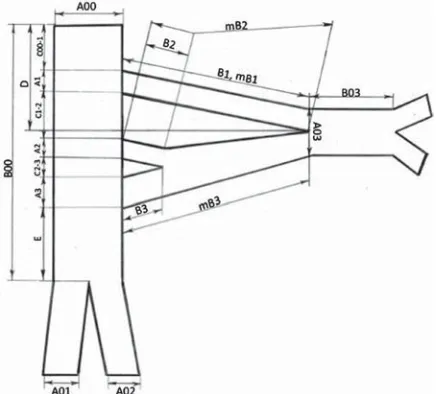
equal to the sum of the morphological length of the root and all the additional inter-radicular connections until the point of formation of the ATN trunk. The thick-ness of the ATN trunk was measured at the point of its origin formed by the combination of the roots. The length of the trunk was equal to the distance from the point of origin to the point of its first branch origin. The distance between the foramen ovale and the ini-tial point of the ATN trunk was also measured (Fig. 1). The relationships of ATN to MMA and lateral pterygoid were also described. Results of the study were then analysed statistically using the Spearman statistical analysis method.
RESULTS
The course of the MMA was variable. Most fre-quently, the artery went between the first and sec-ond root of the ATN (in 7 cases out of 16) (Table 2). In one case an anatomical variant was seen in which the nerve pierced the lateral pterygoid muscle (Fig. 4). In this five-root variation long roots pene-trated the lateral pterygoid in its upper and medial parts. Then nerve roots emerged from the lower edge near the insertion of the muscle to fuse in the short for the second root, 1.14 mm for the third root, 1.19 mm
for the fourth root, and 0.98 mm for the fifth root. Typically, the first roots were the thickest and the fifth roots were the thinnest. The average simplified length of roots was, respectively, as follows: 14.76 mm for the first root, 11.95 mm for the second root, 12.22 mm for the third root, 18.35 mm for the fourth root, and 19.83 mm for the fifth root. The average morphological root lengths were: 14.41 mm for the first root, 7.95 mm for the second root, 7.74 mm for the third root, 4.58 mm for the fourth root, and 3.54 mm for the fifth root. Average ATN trunk thickness ranged from 2.02 mm to 5.62 (mean 3.18 ± 0.84) mm. The average ATN trunk length ranged from 0 to 20.7 (mean 6.32 ± 5.91) mm (Table 1).
Figure 2. Variations and topographical relationships of auriculotem-poral nerve and middle meningeal artery; 1-a, b, c, d, e—one-root variants; 2-a, b, c, d, e—two-root variants; 3-a—three-root vari-ant; 4-a—four-root variant; 5-a, b, c, d — five-root variants.
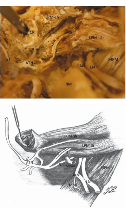
Figure 3. Variation of rightauriculotemporal nervewith 5 roots. Late-ral view. The ramus of mandible and middle part of lateLate-ral pterygoid was removed; CP — condyloid process; LPM — lateral pterygoid muscle; MN — mandibular nerve; IAN — inferior alveolar nerve; LN — lingual nerve; ATN — auriculotemporal nerve (trunk); MMA — middle meningeal artery; 1, 2, 3, 4, 5 — particular roots of ATN.
Table 1. Measurements of auriculotemporal nerve (ATN)
Min. Max. Mean ± SD
D 3.77 20.47 11.52 ± 4.33 E –21.90 7.94 –2.97 ± 8.27 B00 3.57 24.16 12.06 ± 5.56 B03 0.00 20.70 6.32 ± 5.91 A03 2.02 5.62 3.18 ± 0.84 COO-1 0.00 5.12 2.47 ± 1.87 C1-2 0.00 6.28 2.59 ± 2.45 C2-3 0.00 8.55 1.87 ± 2.74 C3-4 0.00 7.41 2.56 ± 3.03 C4-5 0.00 7.00 3.53 ± 3.14 A1 1.30 8.08 2.67 ± 1.65 A2 0.55 3.46 1.70 ± 1.02 A3 0.52 2.06 1.14 ± 0.57 A4 0.70 1.81 1.19 ± 0.51 A5 0.54 1.72 0.98 ± 0.56 B1 1.40 38.00 14.76 ± 8.11 B2 2.45 32.00 11.95 ± 7.75 B3 6.55 24.50 12.22 ± 6.53 B4 3.55 50.16 18.35 ± 18.28 B5 10.80 37.99 19.83 ± 12.29 mB1 1.40 38.00 14.41 ± 8.34 mB2 1.00 28.00 7.95 ± 7.84 mB3 2.00 20.50 7.74 ± 6.98 mB4 1.07 16.67 4.58 ± 6.78 mB5 1.50 6.05 3.54 ± 1.96
trunk on the medial surface of the TMJ capsule. The upper or first root passed between the heads of the lateral pterygoid muscle, the second and third roots passed through the inferior head of lateral pterygoid, and the fourth root was very short and — just from the point of origin from the mandibular nerve trunk — was connected to the fifth root, which ran under the lower head of the muscle (Fig. 4).
DISCUSSION
Our knowledge of ATN anatomy is based on dif-ferent anatomical textbooks and atlases [1, 5, 6, 8, 18, 32, 36, 37]. In the literature the ATN is usually discussed in chapters about trigeminal nerve branch-es and infratemporal fossa anatomy. However, thbranch-ese sources always replicate the fixed bifurcated pattern of ANT that is pierced by the MMA. Meanwhile, some authors have pointed out the high variability of this nerve [3, 9]. On the basis of previous research re-sults, the number of ATN roots is highly variable.
In some studies, one can find a description of nerve variants having from one to four roots [3]. Generally the one- and two-root variants are men-tioned as the most common variants, but there are some discrepancies in these research results. Baumel et al. [3] consider the two-root variant as the most common (62/85 cases). In our own research, we only found a small number of other variants: three-root (12/85), one-root (10/85), and four-root (1/85).
On the other hand, Gülekon et al. [9] observed that the one-root variant is the most common (16/ /32 cases), relative to two-root (12/32), three-root (3/32), and four-root variants (1/32). In one study a two-root variation is described in which the lower root is divided into three smaller roots [31]. Despite so many articles describing the high variability of the ATN in the infratemporal part, many studies limit themselves to describing the nerve in its segment located outside the infratemporal fossa [3, 12, 14, 28]. Most papers, which are the main source of anatomical knowledge about the ATN, list only the two-root variant [1, 5, 6, 8, 15, 18, 32, 36, 37].
There is no clear criterion for determining the precise architecture of ATN roots. The structures that one group classify as the nerve roots [9], others clas-sify as connecting branches [15]. In our study, all the structures that originated from the mandibular nerve or inferior alveolar nerve and directly or indi-rectly participated in the formation of the ATN trunk were considered as nerve roots. In two cases of one-root variations, there was an additional branch that ran vertically from the mandibular nerve to the
infe-Table 2. Relationships between the auriculotemporal
nerve and the middle meningeal artery
Variation Location of middle meningeal artery
Laterally Medially 1–2 2–3 3–4 4–5
1 root 1 4
2 roots 1 1 3 3 roots 1
4 roots 1
5 roots 3 1
[9]. According to Baumel et al. [3], the average length of all roots is equal to 15 mm, and individual lengths vary from 5 to 23 mm. However, these authors do not specify exactly how the measurements of roots were taken. It can be assumed that they were measured from the point of origin on the mandibular nerve trunk or the inferior alveolar nerve trunk to the point of ori-gin of the ATN: a value that in our measurements was called the “simplified length”. The morphological length of the root changes according to the number of roots, being highest for the first root and succes-sively smaller for each of the next.
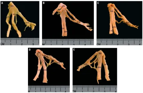
The average length of the nerve trunk according to the literature is 6 mm [3]. It is comparable to our results. Furthermore, in our analysis, a statistically sig-nificant positive correlation between total root thick-ness and the number of roots (r = 0.62, p < 0.01) (Fig. 6) was found. This means that the greater the number of roots in a particular variation, the great-er the sum of the thicknesses. So, if we compare a one-root variation with a multi-root variation of the nerve, the sum of the thicknesses of their roots will not be equal but will increase with the number of roots. This is logical and can be explained by the presence of the nerve sheath. The value of the mea-Figure 5. The dissectedauriculotemporal nerve(ATN) exposed of photographs; A. One-root variant of ATN, left side; B. Two-root variant of ATN, left side; C. Three-root variant of ATN, left side; D. Four-root variant of ATN, left side; E. Five-root variant of ATN, right side; MN — mandibular nerve; IAN — inferior alveolar nerve; LN — lingual nerve; auriculotemporal nerve (trunk); 1, 2, 3, 4, 5 — particular roots of ATN.
rior alveolar nerve (Figs. 2, 5), which can be treated as one root of a divided mandibular nerve trunk. Similar variations have been reported before [2].
Some authors point out that ATN roots do not al-ways have their origin from the mandibular nerve but branch off much lower — from the inferior alveolar nerve [3, 9]. We observed such a situation in three cases of five-root variant and in one case each of four-root and two-four-root variants — in 5/16 cases in total (Fig. 2). It can be concluded that multi-root variations predispose to nerve roots exiting very low from the trunk. Such low exits of the ATN nerve roots are likely to be the cause of atypical symptoms of anaesthesia of the external ear during inferior alveolar nerve ana-esthesia in dental procedures [25].
According to Gülekon et al. [9], the thickness of the ATN roots varies from 0.65 mm to 2.54 mm for the first root, from 1.96 mm to 0.49 mm for the second root, from 0.66 mm to 1.17 mm for the third root, and is equal to 0.67 mm for the one case of four-root variant. These results are similar to our data (Table 1).
The length of the nerve roots is also similar to re-sults obtained by other authors. Typically these are 18.5 mm for the first root, 18.7 mm for the second, 17.48 mm for the third, and 29.66 mm for the fourth
A B C
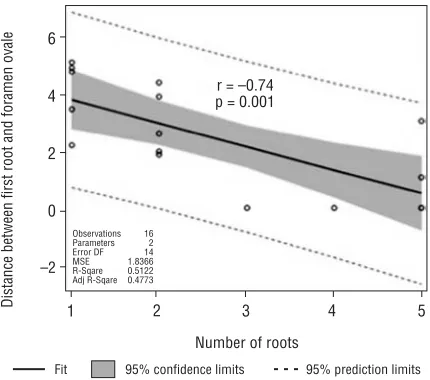
surements is a sum of the value of nerve fibre bun-dle thickness; in particular, the root and nerve sheath double thickness for each of the roots. Therefore, in the observed multi-root variation the total thickness of the roots increases. We also observed a statisti-cally significant negative correlation between the number of roots and the distance from the point of origin of the first root to the foramen ovale (r = –0.74, p < 0.001) (Fig. 7). Already at the stage of preparation, it was observed that a high point of origin of the first nerve root is often associated with the presence of more nerve roots in the nerve. This
was confirmed by the results of statistical analysis. In conclusion, it appears that the smaller the dis-tance between the foramen ovale and the point of origin of the first nerve root, the greater the ber of its roots. And vice versa: the greater the num-ber of roots, the higher the first root is located. In addition, a few more correlations were found: a positive correlation between the length of the nerve trunk and the number of roots (r = 0.49, p < 0.056) and a negative correlation between the distance from the foramen ovale to the ATN trunk and the number of its roots (r = –0.73, p < 0.001).
Comparing the relationship of the ATN to the MMA, our results are very similar to results obtained by other authors in previous studies. According to Gülekon et al. [9], in the one-root variation, the artery ran laterally to the nerve in 8/16 cases, medially in 7/16 cases, while in one case the authors did not identify the location of the vessel. In the two-root variation the vessel proceed-ed laterally in 2/12 cases, and between the first and second root in 10/12 cases. In the three-root variation (1/3 of cases), the artery ran medially in one third of the cases and between the first and second root in the re-maining two-thirds. In the four-root variation, the ar-tery typically ran between the first and second root. Other authors observed similar nerve architecture. In two-root variations, they described the artery between the first and second root in 52/62 cases, medially to the nerve in 6/62 cases, and laterally to the nerve in 4/62 cases. In one-root variations, the artery was located laterally to the nerve in 8/10 cases and medially in 2/10 cases. In three-root variants, the artery was seen to be placed between the first and second roots in 6/12 cases, and between the second and third roots in the other 6/10 cases [3]. In our study, in all variants in which the MMA was surrounded by the roots of ATN, the roots lying on the artery were located lateral to it. This posi-tion was observed in all such cases (Fig. 2, Table 2). However, in one study, the authors found a two-root variation in which the upper root was placed medially to the artery, while the lower one was placed laterally. The relative topography of the ATN and MMA is impor-tant during any sub-temporal approach to the middle cranial fossa or infratemporal fossa. In such cases the MMA is a reference structure to the ATN. Frequently MMA have to be dissected free, coagulated, and divid-ed without ATN injury.
Topographical relations in the area of infratempo-ral fossa promote ATN entrapment. Nearly all the space of the infratemporal fossa is filled by the muscles be-longing to the functional group of mastication, as well as with important blood vessels and nerves [1, 5, 8, 21]. Figure 6. Correlation of summarised root thickness and number
of auriculotemporal nerve roots.
Figure 7. Correlation of distance between first root and foramen ovale and number of auriculotemporal nerve roots.
Summarised
root thickness
10.0
7.5
5.0
2.5
0.0
1
Fit Plot for SumaGrubosciK???
2 3 4 5
r = 0.62 p < 0.01 Observations
Parameters Error DF MSE R−Sqare Adj R−Sqare
16 2 14 3.7424 0.3856 0.3417
Fit 95% confidence limits 95% prediction limits Number of roots
Distance
between
first root and
foramen
ovale
6
4
2
0
–2
1
Fit Plot for Coo–1???
2 3 4 5
r = –0.74 p = 0.001
Observations Parameters Error DF MSE R−Sqare Adj R−Sqare
16 2 14 1.8366 0.5122 0.4773
Number of roots
Overloading and hypertrophy of masticatory mus-cles can occur as a result of dysfunction within the sto-matognathic system [7]. Hypertrophic masticatory muscles, especially the lateral pterygoid, reduce the free space at the infratemporal fossa, creating favourable conditions for the compression of the ATN. Previous studies have described an anatomical variation in which mandibular nerve branches pass through the lateral pterygoid muscle [7, 11, 22, 30]. Penetration of the lat-eral pterygoid muscle by ATN is quite rare [22, 30]. This kind of variation appeared only in one case in our study. It is also worth noting that in all cases, the ATN lying at the TMJ level (at this level the roots are ty-pically already united in the trunk) ran directly adja-cent to the medial surface joint capsule. Similar re-sults were observed by other authors [28].
Such topographical relationships probably play a role in nerve entrapment. Dislocations of the TMJ structures, caused by inflammation or injury, may exert pressure on the nerve, producing symptoms of entrapment [19, 23, 26]. Based on previous data, we can point to two main factors that determine the presence of ATN entrapment in its course within the infratemporal fossa. The first is anatomical variation, while the second is the presence of various types of dysfunction within the masticatory system that ini-tiates a series of morphological changes and ultimate-ly leads to entrapment. In such situations, even small functional or structural changes within the stomato-gnathic system can lead to pain syndromes.
REFERENCES
1. Aleksandrowicz R, Ciszek B (2007) Anatomia kliniczna głowy i szyi. PZWL, Warszawa.
2. Anil A, Peker T, Turgut HB, Gülekon IN, Liman F (2003) Variations in the anatomy of the inferior alveolar nerve. Br J Oral Max Surg, 41: 236–239.
3. Baumel J, Vanderheiden J, McElenney J (2005) The auriculotem-poral nerve of man. Am J Anat, 130: 431–440.
4. Becser Andersen N, Bovim G, Sjaastad O (2000) The frontotem-poral peripherial nerves. Topographic variations of the supraor-bital, supratrochlear and auriculotemporal nerves and their pos-sible clinical significance. Surg Radiol Anatom, 23: 97–104. 5. Bochenek A, Reicher M (2007) Nerw żuchwowy (V3). In: Łasiński W
ed. Anatomia człowieka. Vol. V. PZWL, Warszawa, pp. 198–207. 6. Bourgery JM, Jacob NH (2008) The atlas of anatomy and surgery.
Vol. 3. TASCHEN, Hong Kong.
7. Dupas PH (2009) Dysfunkcja czaszkowo-żuchwowa. Powstanie dysfunkcji czaszkowo-żuchwowej. PZWL, Warszawa, pp. 3–19. 8. Gray H (1995) The spheno-palatine ganglion and its branches. In:
Pickering Pick T, Howden R eds. Gray’s anatomy. Barnes Noble, New York, pp. 713–717.
9. Gülekon N, Anil A, Poyraz A, Peker T, Basri Turgut H, Karakose M (2005) Variation in the anatomy of the auriculotemporal nerve. Clin Anatom, 18:15–22.
10. Hargitai A, Bertrand PM (2004) Characteristics and demograph-ics of an orofacial pain population: review of 255 consecutive cases. Clin Update, 26: 37–39.
11. Isberg A, Isaacson G, Williams W, Loughner B (1987) Lingual numbness and speech articulation deviation associated with tem-poromandibular joint disc displacement. Oral Surg Oral Med Oral Pathol, 64: 9–14.
12. Janis JE, Hatef DA, Ducic I, Ahmad J, Wong CH, Osborn T (2010) Anatomy of the auticulotemporal nerve: variation in its rela-tionship to the superficial temporal artery and implications for the treatment of migraine headaches. Plast Recon Surg, 125: 1422–1428.
13. Karolakowska W, Durko A (1991) Leczenie zaburzeń czynnoś-ciowych narządu żucia u chorych z bólami głowy. Neur Neuro-chir Pol, 25: 634–639.
14. Keersmaekers K, De Boever JA, Van Den Berghe L (1996) Otal-gia in patients with temporomandibular joint disorders. J Pros-thet Dent, 75: 72–76.
15. Kim SY, Hu KS, Chung IH, Le EW, Kim HJ (2004) Topografic anato-my of the lingual nerve and variations in communication pattern of the mandibular nerve branches. Surg Radiol Anat, 26: 128–135. 16. Kleinrok M (1987) Przewlekłe bóle głowy a zaburzenia
czyn-nościowe narządu żucia. Pol Tyg Lek, 62: 298–303.
17. Kleinrok M, Fałęcka A (1990) Objawy oczne u chorych z zaburze-niami czynnościowymi układu ruchowego narządu żucia. Prot Stom 60: 260–267.
18. Köpf-Maier P (2002) Atlas of human anatomy. Vol. 2. PZWL, Warszawa.
19. Kowalik S, Mazurkiewicz J, Wojcieszyn M (1971) Otoneurolo-giczne objawy po urazach stawu skroniowo-żuchwowego. Czas Stomat, 24: 391–395.
20. Kwak H, Park H, Youn K, Hu KS, Koh KS, Han SH, Kim HJ (2004) Branching patterns of the facial nerve and its communication with the auriculotemporal nerve, Surg Radiol Anat, 26: 494–500. 21. Łasiński W (1993) Anatomia głowy dla stomatologów. PZWL,
Warszawa.
22. Loughner BA, Larkin LH, Mahan PE (1990) Nerve entrapment in the lateral pterygoid muscle. Oral Surg Oral Med Oral Pathol, 69: 299. 23. McGrath CJR, Egbert MA, Tong DC, Myall WT (1996) Unusual
presentations of injuries associated with the mandibular condyle in children. Br J Oral Max Surg, 34: 311–314.
24. Myrayama R, Stuginski-Barbosa J, Moraes N, Speciali J (2009) Toothache referred from auriculotemporal neuralgia: case re-port. Int Endod J, 42: 845–851.
25. Ngeow W, Chai W (2009) Numbness of the ear following infe-rior alveolar nerve block: the forgotten complication. Br Dent J, 207: 19–21.
26. Ramirez ALM, Sandoval OGP, Ballesteros LE (2005) Theories on otic symptoms in temporomandibular disorders: past and present. Int J Morphol 23: 141–156.
27. Schames J, Schames M, Boyd JP, King EL, Ulansey S (2002) Trigeminal pharyngioplasty: treatment of the forgotten acces-sory muscles of mastication which are associated with orofacial pain and ear symptomology. J Pain Manag, 12: 102–112. 28. Schmidt BL, Pogrel MA, Necoechea M, Kearns G (1998) The
dis-tribution of the auriculotemporal nerve around the temporo-mandibular joint. Oral Surg Oral Med Oral Pathol, 86: 165–168. 29. Schrenker H (2006) Mioartropatie uwarunkowane protetycznie — przykłady. In: Frątczak B ed. Ograniczenia i kompromisy w protetyce. RAABE, Warszawa, pp. 127–161.
30. Shimokawa T, Akita K, Sato T, Ru F, Yi SQ, Tanaka S (2004) Pe-netration of muscles by branches of the mandibular nerve: a possible cause of neuropathy. Clin Anatom, 17: 2–5. 31. Simmi S, Gayatri R, Rajesh S, Venkat RV (2009) Unusual
organi-zation of auriculotemporal nerve and its clinical implications. J Oral Maxil Surg, 67: 448–450.
32. Snell RS (1978) Atlas of clinical anatomy. Little, Brown and Com-pany, Boston.
33. Speciali G, Godoi Concalves DA (2005) Auriculotemporal neu-ralgia. Curr Pain Headache Rep, 9: 277–280.
34. Trescot AM (2000) Headache management in an interventional pain practice. Pain Physician, 3: 197–200.
35. Wanyura H, Gołębiowski M, Pączek L, Stopa Z, Chmielewski W, Samolczyk-Wanyura D (2004) Wstępna kliniczno-radiologiczna oraz artroskopowa ocena zaburzeń czynnościowych i zmian morfologicznych występujących w chorobie stawów skronio-wo-żuchwowych. Czas Stomat, 57: 804–819.
36. Weber JC (2000) Sekcja zwłok. Podręcznik Shearera. PZWL, Warszawa.