Bone material properties in osteogenesis imperfecta Nick Bishop MB ChB MRCP MD FRCPCH
University of Sheffield and Sheffield Children’s NHS Foundation Trust Academic Unit of Child Health,
Sheffield Children’s Hospital Western Bank
Sheffield S10 2TH United Kingdom
Relevant disclosures
Consultancy for Amgen, Mereo, UCB Grant income from Amgen
Abstract
Osteogenesis imperfecta entrains changes at every level in bone tissue, from the disorganisation of the collagen molecules and mineral platelets within and between collagen fibrils, to the macro-architecture of the whole skeleton. Investigations using an array of sophisticated instruments at multiple scale levels have now determined many aspects of the effect of the disease on the material properties of bone tissue.
The brittle nature of bone in osteogenesis imperfecta reflects both increased bone mineralisation density – the quantity of mineral in relation to the quantity of matrix within a specific bone volume – and altered matrix-matrix and matrix mineral interactions. Contributions to fracture resistance at multiple scale lengths are discussed, comparing normal and brittle bone.
Integrating the available information provides both a better understanding of the effect of current approaches to treatment – largely improved architecture and possibly some macro-scale toughening - and indicates potential opportunities for alternative strategies that can influence fracture resistance at longer length scales.
Introduction and background
Multiple genetic mechanisms give rise to bone fragility (Table 1). Osteogenesis imperfecta (OI) is in danger of becoming a catch-all term for early-onset bone fragility. The key feature that discriminates OI from other early-onset bone fragility conditions is the hyper-mineralisation of the bone material itself – hence the alternative name “brittle bone disease” – although increased mineralisation density is not the only contributor to brittleness. Mutations in the type I collagen synthesis and processing pathways, along with defects in accessory proteins such as PEDF (type VI OI) and those associated with reduced type I collagen production (type V OI) all share the bone material hyper-mineralisation phenotype.
This review of bone material properties in OI describes the multiscale
abnormalities from a molecular level up and also assesses their contribution to the brittle phenotype in both animal model systems and, where available, in human tissue.
Normal bone tissue; molecular to fibrillar scale
In the healthy skeleton, mineral accounts for 65-70% of bone mass, water around 10% with the remainder being matrix proteins, principally type 1 collagen, and a small amount of citrate (approximately 2%). (1) Each type 1 collagen molecule comprises two type 1 collagen 1 and one type 1 collagen 2 α α chains. The internal core of each trimer is hydrophobic.
Each individual heterotrimeric collagen molecule (also called tropocollagen) is approximately 1.5nm wide and 300nm long. (2) The individual collagen
molecules self-assemble(3) into a structure that is clearly recognisable by electron microscopy (EM), initially held together by non-covalent interactions. (4) The mature structure is formed of overlapping and cross-linked type I collagen molecules in a quasi-hexagonal array of groups of five collagen
documented, the lighter bands corresponding to the “gap space” of 36 nm. These groups of 5 collagen molecules might reasonably regarded as “microfibrils”. Collagen molecules are joined to one another by two types of crosslink,
enzymatic and non-enzymatic. Enzymatic crosslinks are the result of the activity of lysyl oxidase and lysyl hydroxylase. (6) Initially such crosslinks are divalent. With increasing maturity some become trivalent; (6) these include the
pyridinoline and pyrrole crosslinks that when cleaved during collagen
degradation can be measured as biomarkers reflecting bone resorption. The non-enzymatic crosslinks are the result of apparently spontaneous reactions between sugars such as pentosidine and exposed amino acid residues. (7) The number of non-enzymatic crosslinks increases with age. (7) Karim also observed variation in the accumulation of non-enzymatic crosslinks between cancellous and cortical bone. (8) Enzymatic crosslinks are usually regarded as contributing positively to bone strength; non-enzymatic crosslinks, by contrast, are widely held to
adversely affect bone material properties. (9)
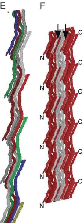
The collagen molecules within each microfibril group coil gradually around each other, with specific kinks that create pockets and potential binding sites for other matrix proteins (5) as well as interactions with cells through integrin-binding. (10) [INSERT Figure 1 here; use ORGEL doi_10.1073_pnas.0502718103 - figure 3E]
The non-collagenous proteins within bone have been suggested to have a range of functions, from the formation of collagen fibrils and initiation of mineral platelet formation, to mineral maturation and collagen cross-linking. (11) In addition, osteopontin and osteocalcin are proposed to have roles in energy dissipation at the microfibrillar level through the formation of dilatational voids (12) and, additionally for osteopontin, through the forming, loss and reforming of sacrificial bonds with divalent cations such as calcium. (13, 14)
The individual microfibril groups are separated from their near neighbours by so-called “gap channels”. (15) Water molecules both within and between the microfibrils contribute to the overall stability of the whole structure through the formation of multiple hydrogen bonds. (16) Individual fibrils are 80-100nm in diameter; fibril length is indeterminate. (17)
Molecular changes in Osteogenesis Imperfecta
The vast majority of OI (more than 85% of cases) is caused by mutations in one of the two genes encoding the type I collagen molecules. (18) In broad terms, for “classical OI” as described by Sillence, (19) null alleles (i.e. absent or
non-functional gene product) in the COL1A1 gene, resulting in reduced amounts of normal 1 chain give rise to a milder phenotype; missense mutations (point α mutation giving rise to altered amino acid sequence) in either COL1A1 or COL1A2 give rise to more severe phenotypes, including lethal disease. No clear skeletal phenotype has been identified arising from a heterozygous COL1A2 null allele. Both collagen genes code for a repetitive, staggered (Gly-X-Y)n amino acid
create fibrils; the severity of the bone disease that results seems likely to be a consequence both of the alterations in the 3-dimensional fibrillar structure and also the specific siting of the mutation. In particular, two “major ligand binding regions” in tropocollagen are identified as sites of interaction with other matrix proteins that, when disrupted as a result of mutation in the type I collagen genes, often have a severe or lethal OI phenotype. (20)
Fibrillar changes in OI
Much of the exploration of fibrillar mechanics in OI has been undertaken using the oim mouse model, which carries a mutation in the cola-2 gene. (21) In oim-/- mice, homozygous for the mutation, the altered 2 chain of the type 1 collagen α heterotrimer is unable to associate with the 1 chains and the type 1 collagen α molecule is thus a homotrimer of 1 chains. The mice have a moderately severe α OI phenotype; they are smaller at birth and grow less well than wild type
littermates, have low bone mass, fracture with minimal trauma, and develop bone deformities. At a tissue level, cortical thinning and reductions in trabecular number are seen on light microscopy, along with a lack of lamellar architecture and increased osteocyte density. (21)
At an ultrastructural level, collagen content is reduced by 20% (22) and a reduction in the size of the D-space within oim-/- collagen fibrils of
approximately 1% vs wild type across a range of D-space sizes from 60-70nm has been observed using atomic force microscopy. (23) The individual
tropocollagen molecules are more prone to kinking (24) and there is more water associated with the tropocollagen and between the microfibrils (25). The bone matrix compressive elastic modulus of oim-/- tibia by nanoindentation was reduced by 20% compared to wild type; in addition, resistance to plastic deformation was 8% higher, implying reduced toughness. (26) The overall picture that emerges from the mouse model reports is of a disorganised bone matrix, more loosely woven, less capable of responding to normal mechanical inputs, and less able to absorb and dissipate energy that might lead to fracture. Mineralisation of bone matrix
Collagen in sites outside the skeleton and teeth should not mineralise. This implies either the presence of one or more inhibitors of mineralisation in non-skeletal sites, or the presence of factors that promote mineralization in bone, or both. In remodeling sites, mineralisation of osteoid is linked both to local mineral concentrations and the incorporation of osteoblasts into the matrix (27)– clearly, osteoblasts are not present in non-mineralising tissues. During endochondral ossification occurring with growth or fracture repair, matrix vesicles create mineral crystals within themselves that then seed into the local environment as they grow and rupture the vesicle walls. (28) The incorporation of mineral into tissue in endochondral ossification begins with the calcification of the
cartilaginous matrix laid down by hypertrophic chondrocytes, with longitudinal septae more heavily calcified than transverse septae. The invasion of endothelial cells precedes the ingress of both osteoclasts, which remove the transverse septae, and osteoblasts, which deposit bone onto the remaining longitudinal calcified cartilage cores to form the bony trabeculae of the primary spongiosa. (29)
variety of inter-linked mechanisms have been identified that regulate the site and degree of mineralisation activity, including the presence of mineralisation inhibitors such as pyrophosphate (32) and osteopontin (33), and the potential mineral nucleation initiator bone sialoprotein which may act in concert with alkaline phosphatase. (34) Multiple other factors may also have a role, recently reviewed in Staines et al. (35) In both endochondral ossification and matrix mineralisation, however, it seems likely that an initial phase of amorphous mineral deposition precedes the formation of more highly organised mineral. (36, 37)
Bone mineralisation in OI
In most cases of OI, mineralisation processes seem to be in place and normally functioning. Two exceptions, however, may be the types V and VI OI initially identified as having distinct bone histological appearances (type V mesh-like under polarised light; type VI osteomalacic) at the level of light microscopy. (38, 39) Type V infants initially display altered metaphyseal modelling suggestive of a delay in endochondral ossification, (40) with a subsequent increase in
metaphyseal density, but no alteration in mineralisation rate or extent of remodelling sites in the trabecular bone of transiliac bone biopsies. (38) By contrast, although there is a clear mineralisation defect at remodelling sites in type VI patients, (39) there is no apparent defect in endochondral ossification. Despite these appearances at a light microscopic level, primary osteoblasts from type V OI patients demonstrate increased mineralisation in culture,(41) and bone tissue from type VI patients have been shown to have hyper-mineralised bone tissue by back scattered electron imaging. (42) These findings suggest that the final degree of mineralisation at small length scales is not directly dependent on the rate of mineralisation observed at larger scale lengths. The genetic
origins of these forms – an activating mutation in IFITM5/BRIL for type V, (43, 44) and loss of function in SERPINF1 (encoding pigment epithelium derived factor, PEDF) for type VI (45, 46) – suggest no direct connection with collagen synthesis or processing. However, reduced type 1 collagen production has been reported recently in primary cultured osteoblasts from type V patients, (41) and PEDF binds to the secreted collagen heterotrimer at two distinct sites. (47) IFITM5/BRIL may have a role in PEDF regulation; osteoblast PEDF production was reduced and typical type VI histology seen in a patient with a novel
inactivating mutation in IFITM5/BRIL. (48) Mineral platelets
There is general agreement that the mineral platelet’s long axis is aligned with the long axis of the collagen fibrils (49) and that some staggering of the platelets occurs. (50) There is, however, a lack of consensus concerning the exact spatial relationships of the collagen molecules with the mineral platelets that contribute to the material properties of bone.
This may reflect the variety of techniques and preparation methods used, the hydration state of the samples (51) and the difficulty in creating model systems that allow accurate recapitulation of the microfibrillar-mineral platelet
The mineral platelets are composed of hydroxyapatite; estimates of their size vary, (52) in part as a function of their maturity but also again reflecting the methodologies used to provide the estimates and the conditions in which such estimates are made. A recent study using ion-milled cryogenic femoral bone found consistent platelet sizes of 5nm thick, 70nm wide and >200nm long. (53) The platelets may be arranged in stacks, (1) so that several are placed side by side in a particular gap channel, or extending between fibrils across a series of gap channels. (15) Whilst their long axes align with that of the adjacent collagen fibril, the planes of stacks around and between fibrils may not be similar; there is some evidence that they vary significantly across local areas. (54)
In addition to the highly ordered mineral platelet arrays/stacks, there is a disorganised hydrogen-phosphate phase, with both the ordered and disordered mineral elements being strongly associated with both citrate and water. (1) A recent paper suggests that citrate acts to maintain the ordered platelet parallel arrays and also holds non-platelet hydrogen-phosphate in a relatively immobile, highly hydrated phase between the platelets, thus maintaining a degree of disorder and preventing crystals increasing in size or thickness (which would result in increased bone fragility). (1) This arrangement could be conceptualised as a series of stacked multilayer sandwiches (see Figure 2c). Water may also play an important role in the interactions between matrix and mineral. (55) Layered water provides multiple hydrogen bonds at interfaces between mineral platelets, and also allows interactions between platelets and fibrils. (56) Molecular
dynamics simulations indicate that water can significantly alter the interactions between platelets and fibrils during loading. (57)
Mineral platelets in OI
Investigations of mineral platelet size and orientation, and the effects of mineral on intrinsic material properties in OI have been undertaken in both human tissue and in mouse models. There is general agreement that mineral platelet size is reduced; (58) that there are more, thinner, platelets; (59) that the composition of the platelets is altered in terms of the ratio of phosphate to carbonate; (23) and that although the alignment of the platelets is generally concordant with the fibrils, there is less overall homogeneity of alignment of platelets within the tissue, (58) likely reflecting matrix disorganisation. Overall, tissue mineralisation density is increased in OI, more so in more severely affected individuals, (60) and even more so specifically in those with c-propeptide cleavage site mutations, (61) BMP-1 mutations (62) and in OI type VI. (42) The roles of water and citrate have not been studied in OI bone; simulation studies of mineralised OI bone have not been undertaken as yet.
The overall increase in tissue mineralisation density is likely to be a major contributor to the brittle nature of OI bone tissue. It is possible that the observed changes listed above reflect increased size of fibrillar gap channels, or possibly the orientation of stacks of platelets across the gap channels, in a manner similar to that suggested by Alexander (15) (see also Figure 2).
Energy dissipation in bone and fracture toughening mechanisms
Intrinsic material properties that contribute to increased fracture resistance include those that promote plasticity and toughness i.e. ductility, energy absorption and dissipation. (64, 65) This requires cooperative deformation of mineral and matrix and is accomplished through multiple mechanisms including inter and intrafibrillary crosslink breakages, (6) shearing between mineral platelets, (66) sliding of mineral platelets relative to the fibrils, fibril deformation and platelet deformation. (67) Fritsch and colleagues have proposed layered water-induced ductile sliding of minerals followed by rupture of collagen crosslinks based on a continuum micromechanics model, upscaled for elastic properties and then applied to a multi-scale representation of bone materials. (68) A multiscale model summarising toughening mechanisms has been proposed by Ritchie. (65)
Toughness is a measure of energy dissipation and cannot be easily estimated in an anisotropic material. Both strength and toughness are influenced by
inhomogeneity (30) and interface properties, (64) which can be highly localised. Fracture resistance extends beyond the intrinsic material properties of bone to encompass all levels of scale up to the whole bone. (17) Whilst stiffness can be considered as an averaged property across a material, strength and toughness cannot. Strength is affected by the “weakest link” problem. (63) In addition, scale matters in strength testing – small defects can result in large decreases in
strength; as scale reduces, the number and size of defects possible decreases and sample strength increases as a consequence.
At scales beyond those at which material properties are assessed, extrinsic biomechanical factors act to shield an existing crack from forces that would extend that crack – usually on a scale of 10-1000 microns, i.e. osteonal level. (69) Fibre orientation can affect crack propagation, and the successive alteration in fibre orientation within the concentric lamellae of an osteon is thought to reflect such an adaptation in bone. (70, 71) Osteonal borders, defined by cement lines, act as barriers to crack propagation, deflecting cracks into more tortuous paths. (72) Cement lines are also found around remodeled bone packets. Mechanisms have to be able to operate in high strain situations i.e. real life rather than the laboratory where the strain rates are often very low. Experimental evidence suggests that the crack-shielding mechanisms are more effective at preventing crack propagation when they are dealing with the lower “every day” strains, (73) rather than the higher strains more often associated with a fall or moderate degree of trauma likely to result in fracture in real life. With higher strains, cracks are seen to be straighter, crossing osteons rather than deflecting around them, with less energy dissipation. (74)
Accumulated microdamage may contribute to increased fracture risk (75) if appropriate remodelling of damaged areas is not undertaken.
Intrinsic mechanisms of fracture resistance are also affected by strain rate; higher strain rates are associated with increased material stiffness and reduced ductility (post-yield plastic deformation). Thus at abnormally high strain rates, bone behaves as if it is more “brittle”. (74)
Many studies have reported the occurrence of microcracks “ahead” of a
Fracture resistance is difficult to measure in vivo. A novel approach using
microindentation has been developed that provides information on the ability of bone to resist a localised force – effectively, the resistance of separation of mineralised fibrils - at an intermediate scale. (80) The output for the in vivo, hand-held device (Osteoprobe™) is given as “Bone Material Strength” and is reported to be reduced independently of bone mineral density in patients with fragility fractures. (81) Multiple outputs are provided by the ex vivo benchtop device (BioDent™); “indentation distance” and “indentation distance increase” indicate the extent to which the probe penetrates initially and further after 10 cycles of indentation into a bone sample and are thought to reflect fracture resistance. (82)
Fracture resistance in OI bone
When a long OI bone breaks, the fracture line tends to be transverse, suggesting that some of the mechanisms that normally promote energy dissipation are abrogated. At lower scale lengths in OI, multiple factors likely interact to reduce the ability of the disorganised matrix to effectively absorb or dissipate fracture-causing energy. In the oim mouse, these include fewer enzymatic and more non-enzymatic crosslinks with associated increased mineralisation density and consequent reduced material elasticity and toughness, (26, 83) as well as
smaller, more densely packed mineral platelets with disordered orientation. (26) These features likely impact on the ability of bone to dissipate energy through sacrificial bond breakage, sliding of platelets relative to fibrils and shearing between platelets.
At longer scale lengths, oim-/- mice demonstrate reduced stable crack extension, crack-initiation toughness and crack-growth toughness with increasing severity of OI and amounts of woven bone; in addition, increased cortical vascular
porosity in oim reduces stable crack growth. (83, 84) Although excessive woven bone is not a clear feature in human OI bone biopsies, increased cortical porosity is, and likely also contributes to increased fracture risk.(85-88) Figure 2
summarises the factors contributing to bone fragility at the different scale lengths.
In the oim-/- mouse, use of microindentation showed an increase in initial indentation distance and total indentation distance in one study, (23) but no relationship of microindentation outcomes was found with stress intensity fracture toughness in another. (89) Microindentation has not been applied in vivo in OI as yet.
Effects of current interventions in OI
The therapeutic options currently employed or under investigation in the treatment of OI in humans either reduce bone remodelling (bisphosphonates, (90, 91) denosumab (92, 93)) or increase bone formation (PTH (94),
anti-sclerostin antibodies (95)). None of these interventions have been found to affect tissue material properties in OI bone.
However, improved femoral geometry and biomechanical strength in the brtl OI mouse was offset by reduced predicted (not measured) elastic modulus
following 12 weeks of alendronate treatment. (96)
intrinsic material properties were shown; the most recent work found reduced toughness (post-yield plastic deformation) in alendronate-treated bone, in association with an increase also in the phosphate to carbonate ratio of mineral crystals reflecting slower bone remodelling and increased mineral crystal maturity.
Thus for the most commonly used intervention, bisphosphonates, there is
evidence for improved extrinsic biomechanical properties consequent on macro-scale architectural change (increased bone width, increased cortical thickness, reduced cortical porosity, retention of new trabeculae in growing bone(86)) which in mild OI may result in reduced fracture risk. (90, 100) There is no evidence, however, that bisphosphonates alter bone material properties in such a way as to further reduce fracture risk. It follows that there is a limit to what current treatment can achieve regarding fracture risk reduction in terms of off-setting increased bone mass and improved macro-architecture against the poor material properties that characterise OI bone.
Anti-TGF antibody treatment of two moderate-severe mouse models of OI β (crtap-/- and Col1a2tm1.1Mcbrmice; not oim-/-) has been reported to restore bone
architecture and reduce hyperosteocytosis, but did not affect bone material properties. (101)
In terms of future therapeutic interventions, new approaches to improving the intrinsic material properties of bone would appear attractive but may not be practicable given the underlying issue of matrix disorganisation. Increasing the proportion of normal collagen within the matrix would require implementation of cellular or genetic approaches. Murine (oim-/-) studies suggest that
mesenchymal stem cells (MSCs) can engraft and produce normal collagen ameliorating the OI phenotype and reducing bone brittleness, (102-104) and human chorionic cells transplanted into newborn oim-/- mice also improved the clinical phenotype. (105) Previous human studies of bone marrow
transplantation shon little mesenchymal lineage engraftment and failed to deliver clear benefit (106, 107) but the recent report of fetal stem cell infusion both before and after delivery in a child thought to have type IV OI was
encouraging. (108)
Ex vivo manipulation of cells ex vivo and then their reintroduction has been widely discussed following the recent successful treatment of a child with leukaemia. In OI, an approach of this type could substantially impact on tissue phenotype. (109) An alternative approach using siRNAs to knock down mutant alleles in MSCs ex vivo has been shown to reduce mutant collagen production by 42% in fibroblasts from the Brtl mouse model of OI. (110)
Summary
The characteristic material feature of bone in OI is its brittleness, and this helps differentiates OI from other disorders associated with early onset bone fragility. The brittleness is contributed to both by increased mineralisation density due to smaller more densely packed mineral platelets and increased numbers of non-enzymatic crosslinks. The bone matrix is looser, allowing more space between collagen molecules and fibrils for other matrix proteins as well as the mineral platelets. At longer scale lengths, the contribution of abnormal architecture to fragility is substantial – increased cortical porosity, thinner cortices and
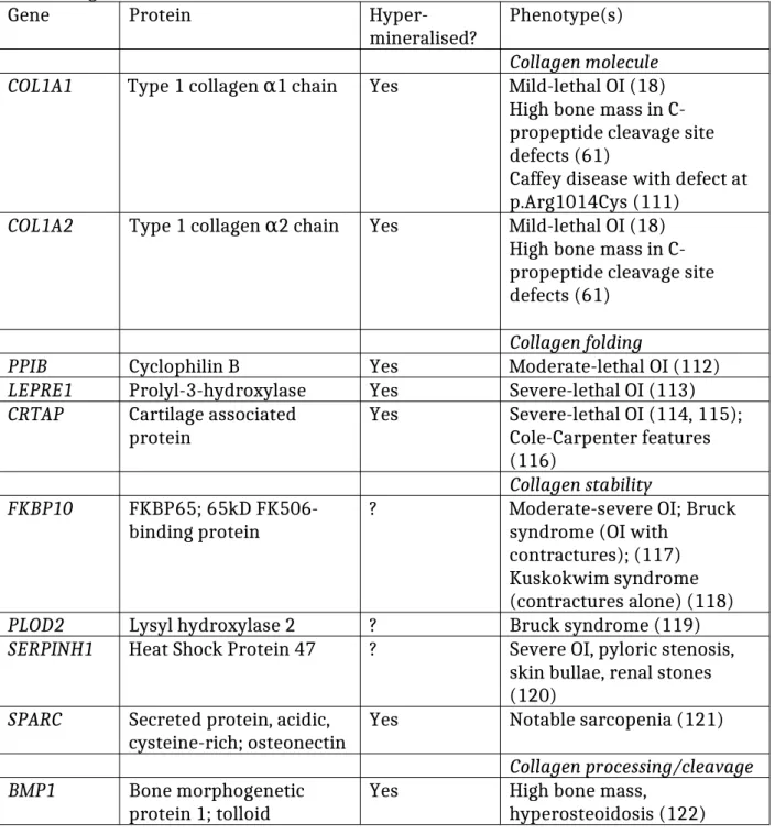
Table 1. Early onset bone fragility syndromes Collagen related
Gene Protein
Hyper-mineralised? Phenotype(s) Collagen molecule COL1A1 Type 1 collagen 1 chainα Yes Mild-lethal OI (18)
High bone mass in C-propeptide cleavage site defects (61)
Caffey disease with defect at p.Arg1014Cys (111)
COL1A2 Type 1 collagen 2 chainα Yes Mild-lethal OI (18) High bone mass in C-propeptide cleavage site defects (61)
Collagen folding
PPIB Cyclophilin B Yes Moderate-lethal OI (112)
LEPRE1 Prolyl-3-hydroxylase Yes Severe-lethal OI (113) CRTAP Cartilage associated
protein Yes Severe-lethal OI (114, 115); Cole-Carpenter features (116)
Collagen stability FKBP10 FKBP65; 65kD
FK506-binding protein ? Moderate-severe OI; Bruck syndrome (OI with contractures); (117) Kuskokwim syndrome (contractures alone) (118) PLOD2 Lysyl hydroxylase 2 ? Bruck syndrome (119) SERPINH1 Heat Shock Protein 47 ? Severe OI, pyloric stenosis,
skin bullae, renal stones (120)
SPARC Secreted protein, acidic,
cysteine-rich; osteonectin Yes Notable sarcopenia (121) Collagen processing/cleavage BMP1 Bone morphogenetic
Osteoblast lineage/function
Gene Protein
Hyper-mineralised? phenotype
Wnt-signalling pathway LRP5/6 Lipoprotein
receptor-related protein 5/6 No Homozygous – osteoporosis pseudoglioma syndrome; (123) Heterozygous – osteoporosis and/or vitreoretinopathy (124) WNT1 Wingless-type MMTV
integration site family, member 1
No Homozygous – severe OI; some have brain malformation; autism, learning difficulties in some. (125)
Heterozygous – early onset osteoporosis, normal growth (126)
Osteocyte dysfunction
PLS3 Plastin 3 ? X-linked early onset severe
osteoporosis without other OI features (126)
Mineralisation regulation SERPINF1 Pigment epithelium
derived factor Yes Slowly progressively worsening OI; osteoid mineralization defect (no endochondral defect) (45)
IFITM5/
BRIL Interferon-induced transmembrane protein 5, or, bone-restricted
IFITM5-like
Yes Severe OI; metaphyseal dysplasia and sclerosis, hypertrophic callus, interosseous membrane calcification. (43, 44, 127) Osteoblast lineage
SP7/OSX Specificity Protein 7;
Osterix ? Typical OI features (128)
ER-related P4HB Prolyl 4-hydroxylase;
protein disulfide isomerase
? Cole-Carpenter syndrome;
craniosynostosis, ocular
proptosis, hydrocephalus (129) TMEM38B Trimeric Intracellular
Cation Channel Type B; TRIC-B
? Severe osteopenia and limb fractures without vertebral fractures (130)
CREB3L1 Old Astrocyte Specifically
Induced Substrate - OASIS ? Severe OI; cardiac failure (131) NBAS Neuroblastoma Amplified
Sequence
? Early onset osteoporosis, recurrent acute liver failure, developmental delay (132) SEC24D Component of COPII
proptosis, hydrocephalus (133) Linker enzyme deficiency
Figure 1 – from Orgel - Use only parts E and F
Figure 2
See previous TIFF file labelled as figure 3 Legend
a. Macro scale –OI bone is narrower, cortices are thinner, there are fewer trabeculae; bone mass is reduced.
b. Sub-macro scale – increased cortical porosity in OI bone; a larger number of vascular channels as well as pores that coalesce and form larger voids. c. Fibrillar level – fibrils are less consistent in size and shape in OI. The
“weave” is looser, allowing more water and mineral between and within fibrils. Mineral platelets are smaller, thinner and more closely packed. Results in hypermineralisation and increased brittleness.
References
1. Davies E, Muller KH, Wong WC, Pickard CJ, Reid DG, Skepper JN, et al. Citrate bridges between mineral platelets in bone. Proc Natl Acad Sci U S A. 2014 Apr 8;111(14):E1354-63. PubMed PMID: 24706850. Pubmed Central PMCID: 3986129.
2. Streeter I, de Leeuw NH. A molecular dynamics study of the interprotein interactions in collagen fibrils. Soft matter. 2011 Apr 7;7(7):3373-82. PubMed PMID: 23526918. Pubmed Central PMCID: 3605786.
3. Orgel JP, Antipova O, Sagi I, Bitler A, Qiu D, Wang R, et al. Collagen fibril surface displays a constellation of sites capable of promoting fibril assembly, stability, and hemostasis. Connective tissue research. 2011 Feb;52(1):18-24. PubMed PMID: 21117898. Pubmed Central PMCID: 3244825.
4. Bailey AJ, Paul RG, Knott L. Mechanisms of maturation and ageing of collagen. Mechanisms of ageing and development. 1998 Dec 1;106(1-2):1-56. PubMed PMID: 9883973.
5. Orgel JP, Irving TC, Miller A, Wess TJ. Microfibrillar structure of type I collagen in situ. Proc Natl Acad Sci U S A. 2006 Jun 13;103(24):9001-5. PubMed PMID: 16751282. Pubmed Central PMCID: 1473175.
6. Saito M, Marumo K. Effects of Collagen Crosslinking on Bone Material Properties in Health and Disease. Calcif Tissue Int. 2015 Sep;97(3):242-61. PubMed PMID: 25791570.
7. Karim L, Vashishth D. Heterogeneous glycation of cancellous bone and its association with bone quality and fragility. PLoS One. 2012;7(4):e35047.
PubMed PMID: 22514706. Pubmed Central PMCID: 3325937.
8. Karim L, Tang SY, Sroga GE, Vashishth D. Differences in non-enzymatic glycation and collagen cross-links between human cortical and cancellous bone. Osteoporos Int. 2013 Sep;24(9):2441-7. PubMed PMID: 23471564. Pubmed Central PMCID: 4550204.
9. Poundarik AA, Wu PC, Evis Z, Sroga GE, Ural A, Rubin M, et al. A direct role of collagen glycation in bone fracture. Journal of the mechanical behavior of biomedical materials. 2015 Dec;52:120-30. PubMed PMID: 26530231. Pubmed Central PMCID: 4651854.
10. Sweeney SM, Orgel JP, Fertala A, McAuliffe JD, Turner KR, Di Lullo GA, et al. Candidate cell and matrix interaction domains on the collagen fibril, the predominant protein of vertebrates. J Biol Chem. 2008 Jul 25;283(30):21187-97. PubMed PMID: 18487200. Pubmed Central PMCID: 2475701.
11. Orgel JP, San Antonio JD, Antipova O. Molecular and structural mapping of collagen fibril interactions. Connective tissue research. 2011 Feb;52(1):2-17. PubMed PMID: 21182410.
12. Poundarik AA, Diab T, Sroga GE, Ural A, Boskey AL, Gundberg CM, et al. Dilatational band formation in bone. Proc Natl Acad Sci U S A. 2012 Nov 20;109(47):19178-83. PubMed PMID: 23129653. Pubmed Central PMCID: 3511118.
13. Fantner GE, Adams J, Turner P, Thurner PJ, Fisher LW, Hansma PK. Nanoscale ion mediated networks in bone: osteopontin can repeatedly dissipate large amounts of energy. Nano letters. 2007 Aug;7(8):2491-8. PubMed PMID: 17645366.
18586839. Pubmed Central PMCID: 2527241.
15. Alexander B, Daulton TL, Genin GM, Lipner J, Pasteris JD, Wopenka B, et al. The nanometre-scale physiology of bone: steric modelling and scanning
transmission electron microscopy of collagen-mineral structure. Journal of the Royal Society, Interface / the Royal Society. 2012 Aug 7;9(73):1774-86. PubMed PMID: 22345156. Pubmed Central PMCID: 3385760.
16. Gautieri A, Pate MI, Vesentini S, Redaelli A, Buehler MJ. Hydration and distance dependence of intermolecular shearing between collagen molecules in a model microfibril. Journal of biomechanics. 2012 Aug 9;45(12):2079-83. PubMed PMID: 22762892.
17. Zimmermann EA, Busse B, Ritchie RO. The fracture mechanics of human bone: influence of disease and treatment. BoneKEy reports. 2015;4:743. PubMed PMID: 26380080. Pubmed Central PMCID: 4562496.
18. Forlino A, Marini JC. Osteogenesis imperfecta. Lancet. 2015 Nov 2. PubMed PMID: 26542481.
19. Sillence DO, Senn A, Danks DM. Genetic heterogeneity in osteogenesis imperfecta. Journal of medical genetics. 1979 Apr;16(2):101-16. PubMed PMID: 458828.
20. Marini JC, Forlino A, Cabral WA, Barnes AM, San Antonio JD, Milgrom S, et al. Consortium for osteogenesis imperfecta mutations in the helical domain of type I collagen: regions rich in lethal mutations align with collagen binding sites for integrins and proteoglycans. Human mutation. 2007 Mar;28(3):209-21. PubMed PMID: 17078022. Pubmed Central PMCID: 4144349.
21. Chipman SD, Sweet HO, McBride DJ, Jr., Davisson MT, Marks SC, Jr., Shuldiner AR, et al. Defective pro alpha 2(I) collagen synthesis in a recessive mutation in mice: a model of human osteogenesis imperfecta. Proc Natl Acad Sci U S A. 1993 Mar 1;90(5):1701-5. PubMed PMID: 8446583.
22. Camacho NP, Hou L, Toledano TR, Ilg WA, Brayton CF, Raggio CL, et al. The material basis for reduced mechanical properties in oim mice bones. J Bone Miner Res. 1999 Feb;14(2):264-72. PubMed PMID: 9933481.
23. Bart ZR, Hammond MA, Wallace JM. Multi-scale analysis of bone chemistry, morphology and mechanics in the oim model of osteogenesis
imperfecta. Connective tissue research. 2014 Aug;55 Suppl 1:4-8. PubMed PMID: 25158170.
24. Chang SW, Shefelbine SJ, Buehler MJ. Structural and mechanical differences between collagen homo- and heterotrimers: relevance for the molecular origin of brittle bone disease. Biophysical journal. 2012 Feb
8;102(3):640-8. PubMed PMID: 22325288. Pubmed Central PMCID: 3274792. 25. Andriotis OG, Chang SW, Vanleene M, Howarth PH, Davies DE, Shefelbine SJ, et al. Structure-mechanics relationships of collagen fibrils in the osteogenesis imperfecta mouse model. Journal of the Royal Society, Interface / the Royal Society. 2015 Oct 6;12(111):20150701. PubMed PMID: 26468064. Pubmed Central PMCID: 4614505.
26. Vanleene M, Porter A, Guillot PV, Boyde A, Oyen M, Shefelbine S. Ultra-structural defects cause low bone matrix stiffness despite high mineralization in osteogenesis imperfecta mice. Bone. 2012 Jun;50(6):1317-23. PubMed PMID: 22449447. Pubmed Central PMCID: 3407875.
27. Prideaux M, Loveridge N, Pitsillides AA, Farquharson C. Extracellular matrix mineralization promotes E11/gp38 glycoprotein expression and drives osteocytic differentiation. PLoS One. 2012;7(5):e36786. PubMed PMID:
28. Anderson HC. Matrix vesicle calcification: review and update. Bone and Mineral Research. 1985;3:109-50.
29. Amizuka N, Hasegawa T, Oda K, Luiz de Freitas PH, Hoshi K, Li M, et al. Histology of epiphyseal cartilage calcification and endochondral ossification. Frontiers in bioscience. 2012;4:2085-100. PubMed PMID: 22202021.
30. Currey JD. The many adaptations of bone. Journal of biomechanics. 2003 Oct;36(10):1487-95. PubMed PMID: 14499297.
31. Kerschnitzki M, Wagermaier W, Liu Y, Roschger P, Duda GN, Fratzl P. Poorly ordered bone as an endogenous scaffold for the deposition of highly oriented lamellar tissue in rapidly growing ovine bone. Cells, tissues, organs. 2011;194(2-4):119-23. PubMed PMID: 21597267.
32. Millan JL. The role of phosphatases in the initiation of skeletal mineralization. Calcif Tissue Int. 2013 Oct;93(4):299-306. PubMed PMID: 23183786. Pubmed Central PMCID: 3594124.
33. Boskey AL, Christensen B, Taleb H, Sorensen ES. Post-translational modification of osteopontin: effects on in vitro hydroxyapatite formation and growth. Biochem Biophys Res Commun. 2012 Mar 9;419(2):333-8. PubMed PMID: 22342723. Pubmed Central PMCID: 3299831.
34. Wang J, Zhou HY, Salih E, Xu L, Wunderlich L, Gu X, et al. Site-specific in vivo calcification and osteogenesis stimulated by bone sialoprotein. Calcif Tissue Int. 2006 Sep;79(3):179-89. PubMed PMID: 16969594.
35. Staines KA, MacRae VE, Farquharson C. The importance of the SIBLING family of proteins on skeletal mineralisation and bone remodelling. The Journal of endocrinology. 2012 Sep;214(3):241-55. PubMed PMID: 22700194.
36. Boonrungsiman S, Gentleman E, Carzaniga R, Evans ND, McComb DW, Porter AE, et al. The role of intracellular calcium phosphate in osteoblast-mediated bone apatite formation. Proc Natl Acad Sci U S A. 2012 Aug 28;109(35):14170-5. PubMed PMID: 22879397. Pubmed Central PMCID: 3435222.
37. Weiner S. Biomineralization: a structural perspective. Journal of structural biology. 2008 Sep;163(3):229-34. PubMed PMID: 18359639.
38. Glorieux FH, Rauch F, Plotkin H, Ward L, Travers R, Roughley P, et al. Type V osteogenesis imperfecta: a new form of brittle bone disease. J Bone Miner Res. 2000 Sep;15(9):1650-8. PubMed PMID: 10976985.
39. Glorieux FH, Ward LM, Rauch F, Lalic L, Roughley PJ, Travers R. Osteogenesis imperfecta type VI: a form of brittle bone disease with a
mineralization defect. J Bone Miner Res. 2002 Jan;17(1):30-8. PubMed PMID: 11771667.
40. Arundel P, Offiah A, Bishop NJ. Evolution of the radiographic appearance of the metaphyses over the first year of life in type V osteogenesis imperfecta: clues to pathogenesis. J Bone Miner Res. 2011 Apr;26(4):894-8. PubMed PMID: 20872883.
41. Reich A, Bae AS, Barnes AM, Cabral WA, Hinek A, Stimec J, et al. Type V OI primary osteoblasts display increased mineralization despite decreased COL1A1 expression. J Clin Endocrinol Metab. 2015 Feb;100(2):E325-32. PubMed PMID: 25387264. Pubmed Central PMCID: 4318905.
42. Fratzl-Zelman N, Schmidt I, Roschger P, Roschger A, Glorieux FH, Klaushofer K, et al. Unique micro- and nano-scale mineralization pattern of human osteogenesis imperfecta type VI bone. Bone. 2015 Apr;73:233-41. PubMed PMID: 25554599.
mutation in the 5'-UTR of IFITM5 causes osteogenesis imperfecta type V. Am J Hum Genet. 2012 Aug 10;91(2):343-8. PubMed PMID: 22863190. Pubmed Central PMCID: 3415533.
44. Semler O, Garbes L, Keupp K, Swan D, Zimmermann K, Becker J, et al. A mutation in the 5'-UTR of IFITM5 creates an in-frame start codon and causes autosomal-dominant osteogenesis imperfecta type V with hyperplastic callus. Am J Hum Genet. 2012 Aug 10;91(2):349-57. PubMed PMID: 22863195. Pubmed Central PMCID: 3415541.
45. Becker J, Semler O, Gilissen C, Li Y, Bolz HJ, Giunta C, et al. Exome sequencing identifies truncating mutations in human SERPINF1 in autosomal-recessive osteogenesis imperfecta. Am J Hum Genet. 2011 Mar 11;88(3):362-71. PubMed PMID: 21353196. Pubmed Central PMCID: 3059418.
46. Homan EP, Rauch F, Grafe I, Lietman C, Doll JA, Dawson B, et al. Mutations in SERPINF1 cause osteogenesis imperfecta type VI. J Bone Miner Res. 2011 Dec;26(12):2798-803. PubMed PMID: 21826736. Pubmed Central PMCID: 3214246.
47. Sekiya A, Okano-Kosugi H, Yamazaki CM, Koide T. Pigment epithelium-derived factor (PEDF) shares binding sites in collagen with heparin/heparan sulfate proteoglycans. J Biol Chem. 2011 Jul 29;286(30):26364-74. PubMed PMID: 21652703. Pubmed Central PMCID: 3143599.
48. Farber CR, Reich A, Barnes AM, Becerra P, Rauch F, Cabral WA, et al. A novel IFITM5 mutation in severe atypical osteogenesis imperfecta type VI impairs osteoblast production of pigment epithelium-derived factor. J Bone Miner Res. 2014 Jun;29(6):1402-11. PubMed PMID: 24519609. Pubmed Central PMCID: 4352343.
49. Landis WJ, Moradian-Oldak J, Weiner S. Topographic imaging of mineral and collagen in the calcifying turkey tendon. Connective tissue research.
1991;25(3-4):181-96. PubMed PMID: 1647935.
50. Jager I, Fratzl P. Mineralized collagen fibrils: a mechanical model with a staggered arrangement of mineral particles. Biophysical journal. 2000
Oct;79(4):1737-46. PubMed PMID: 11023882. Pubmed Central PMCID: 1301068. 51. Tomoaia G, Pasca RD. On the Collagen Mineralization. A Review. Clujul medical. 2015;88(1):15-22. PubMed PMID: 26528042. Pubmed Central PMCID: 4508610.
52. Hassenkam T, Fantner GE, Cutroni JA, Weaver JC, Morse DE, Hansma PK. High-resolution AFM imaging of intact and fractured trabecular bone. Bone. 2004 Jul;35(1):4-10. PubMed PMID: 15207735.
53. McNally EA, Schwarcz HP, Botton GA, Arsenault AL. A model for the ultrastructure of bone based on electron microscopy of ion-milled sections. PLoS One. 2012;7(1):e29258. PubMed PMID: 22272230. Pubmed Central PMCID: 3260135.
54. Falgayrac G, Facq S, Leroy G, Cortet B, Penel G. New method for Raman investigation of the orientation of collagen fibrils and crystallites in the Haversian system of bone. Applied spectroscopy. 2010 Jul;64(7):775-80. PubMed PMID: 20615291.
55. Wilson EE, Awonusi A, Morris MD, Kohn DH, Tecklenburg MM, Beck LW. Highly ordered interstitial water observed in bone by nuclear magnetic
resonance. J Bone Miner Res. 2005 Apr;20(4):625-34. PubMed PMID: 15765182. 56. Wilson EE, Awonusi A, Morris MD, Kohn DH, Tecklenburg MM, Beck LW. Three structural roles for water in bone observed by solid-state NMR.
Pubmed Central PMCID: 1440753.
57. Bhowmik R, Katti KS, Katti DR. Mechanics of molecular collagen is influenced by hydroxyapatite in natural bone. Journal of Materials Science. 2007;42(21):8795-803.
58. Fratzl P, Paris O, Klaushofer K, Landis WJ. Bone mineralization in an osteogenesis imperfecta mouse model studied by small-angle x-ray scattering. J Clin Invest. 1996 Jan 15;97(2):396-402. PubMed PMID: 8567960. Pubmed Central PMCID: 507030.
59. Fratzl-Zelman N, Schmidt I, Roschger P, Glorieux FH, Klaushofer K, Fratzl P, et al. Mineral particle size in children with osteogenesis imperfecta type I is not increased independently of specific collagen mutations. Bone. 2014 Mar;60:122-8. PubMed PMID: 24296239.
60. Boyde A, Travers R, Glorieux FH, Jones SJ. The mineralization density of iliac crest bone from children with osteogenesis imperfecta. Calcif Tissue Int. 1999 Mar;64(3):185-90. PubMed PMID: 10024373.
61. Lindahl K, Barnes AM, Fratzl-Zelman N, Whyte MP, Hefferan TE,
Makareeva E, et al. COL1 C-propeptide cleavage site mutations cause high bone mass osteogenesis imperfecta. Human mutation. 2011 Jun;32(6):598-609. PubMed PMID: 21344539. Pubmed Central PMCID: 3103631.
62. Asharani PV, Keupp K, Semler O, Wang W, Li Y, Thiele H, et al. Attenuated BMP1 function compromises osteogenesis, leading to bone fragility in humans and zebrafish. Am J Hum Genet. 2012 Apr 6;90(4):661-74. PubMed PMID: 22482805. Pubmed Central PMCID: 3322236.
63. Wagermaier W, Klaushofer K, Fratzl P. Fragility of Bone Material Controlled by Internal Interfaces. Calcif Tissue Int. 2015 Sep;97(3):201-12. PubMed PMID: 25772807. Pubmed Central PMCID: 4525333.
64. Tai K, Dao M, Suresh S, Palazoglu A, Ortiz C. Nanoscale heterogeneity promotes energy dissipation in bone. Nature materials. 2007 Jun;6(6):454-62. PubMed PMID: 17515917.
65. Ritchie RO. The conflicts between strength and toughness. Nature materials. 2011 Nov;10(11):817-22. PubMed PMID: 22020005.
66. Gupta HS, Seto J, Wagermaier W, Zaslansky P, Boesecke P, Fratzl P. Cooperative deformation of mineral and collagen in bone at the nanoscale. Proc Natl Acad Sci U S A. 2006 Nov 21;103(47):17741-6. PubMed PMID: 17095608. Pubmed Central PMCID: 1635545.
67. Thurner PJ, Katsamenis OL. The role of nanoscale toughening mechanisms in osteoporosis. Curr Osteoporos Rep. 2014 Sep;12(3):351-6. PubMed PMID: 24969723.
68. Fritsch A, Hellmich C, Dormieux L. Ductile sliding between mineral crystals followed by rupture of collagen crosslinks: experimentally supported micromechanical explanation of bone strength. Journal of theoretical biology. 2009 Sep 21;260(2):230-52. PubMed PMID: 19497330.
69. Carriero A, Zimmermann EA, Shefelbine SJ, Ritchie RO. A methodology for the investigation of toughness and crack propagation in mouse bone. Journal of the mechanical behavior of biomedical materials. 2014 Nov;39:38-47. PubMed PMID: 25084121.
70. Weiner S, Traub W, Wagner HD. Lamellar bone: structure-function relations. Journal of structural biology. 1999 Jun 30;126(3):241-55. PubMed PMID: 10475685.
16341218.
72. O'Brien FJ, Taylor D, Clive Lee T. The effect of bone microstructure on the initiation and growth of microcracks. Journal of orthopaedic research : official publication of the Orthopaedic Research Society. 2005 Mar;23(2):475-80. PubMed PMID: 15734265.
73. Hazenberg JG, Taylor D, Clive Lee T. Mechanisms of short crack growth at constant stress in bone. Biomaterials. 2006 Mar;27(9):2114-22. PubMed PMID: 16243392.
74. Zimmermann EA, Gludovatz B, Schaible E, Busse B, Ritchie RO. Fracture resistance of human cortical bone across multiple length-scales at physiological strain rates. Biomaterials. 2014 Jul;35(21):5472-81. PubMed PMID: 24731707. 75. Seref-Ferlengez Z, Kennedy OD, Schaffler MB. Bone microdamage,
remodeling and bone fragility: how much damage is too much damage? BoneKEy reports. 2015;4:644. PubMed PMID: 25848533. Pubmed Central PMCID:
4371415.
76. Gupta HS, Zioupos P. Fracture of bone tissue: The 'hows' and the 'whys'. Medical engineering & physics. 2008 Dec;30(10):1209-26. PubMed PMID: 18977164.
77. Nalla RK, Kinney JH, Ritchie RO. Mechanistic fracture criteria for the failure of human cortical bone. Nature materials. 2003 Mar;2(3):164-8. PubMed PMID: 12612673.
78. Nalla RK, Kruzic JJ, Ritchie RO. On the origin of the toughness of
mineralized tissue: microcracking or crack bridging? Bone. 2004 May;34(5):790-8. PubMed PMID: 15121010.
79. Voide R, Schneider P, Stauber M, Wyss P, Stampanoni M, Sennhauser U, et al. Time-lapsed assessment of microcrack initiation and propagation in murine cortical bone at submicrometer resolution. Bone. 2009 Aug;45(2):164-73. PubMed PMID: 19410668.
80. Diez-Perez A, Guerri R, Nogues X, Caceres E, Pena MJ, Mellibovsky L, et al. Microindentation for in vivo measurement of bone tissue mechanical properties in humans. J Bone Miner Res. 2010 Aug;25(8):1877-85. PubMed PMID:
20200991. Pubmed Central PMCID: 3153354.
81. Malgo F, Hamdy NA, Papapoulos SE, Appelman-Dijkstra NM. Bone material strength as measured by microindentation in vivo is decreased in patients with fragility fractures independently of bone mineral density. J Clin Endocrinol Metab. 2015 May;100(5):2039-45. PubMed PMID: 25768670. 82. Granke M, Makowski AJ, Uppuganti S, Does MD, Nyman JS. Identifying Novel Clinical Surrogates to Assess Human Bone Fracture Toughness. J Bone Miner Res. 2015 Jul;30(7):1290-300. PubMed PMID: 25639628. Pubmed Central PMCID: 4478129.
83. Carriero A, Zimmermann EA, Paluszny A, Tang SY, Bale H, Busse B, et al. How tough is brittle bone? Investigating osteogenesis imperfecta in mouse bone. J Bone Miner Res. 2014 Jun;29(6):1392-401. PubMed PMID: 24420672. Pubmed Central PMCID: 4477967.
84. Carriero A, Doube M, Vogt M, Busse B, Zustin J, Levchuk A, et al. Altered lacunar and vascular porosity in osteogenesis imperfecta mouse bone as revealed by synchrotron tomography contributes to bone fragility. Bone. 2014 Apr;61:116-24. PubMed PMID: 24373921.
85. Rauch F, Travers R, Glorieux FH. Pamidronate in children with
86. Rauch F, Travers R, Plotkin H, Glorieux FH. The effects of intravenous pamidronate on the bone tissue of children and adolescents with osteogenesis imperfecta. J Clin Invest. 2002 Nov;110(9):1293-9. PubMed PMID: 12417568. 87. Albert C, Jameson J, Smith P, Harris G. Reduced diaphyseal strength associated with high intracortical vascular porosity within long bones of children with osteogenesis imperfecta. Bone. 2014 Sep;66:121-30. PubMed PMID:
24928496. Pubmed Central PMCID: 4467578.
88. Katti KS, Gu C, Katti DR. Anisotropic properties of human cortical bone with osteogenesis imperfecta. Biomechanics and modeling in mechanobiology. 2015 Sep 23. PubMed PMID: 26399513.
89. Carriero A, Bruse JL, Oldknow KJ, Millan JL, Farquharson C, Shefelbine SJ. Reference point indentation is not indicative of whole mouse bone measures of stress intensity fracture toughness. Bone. 2014 Dec;69:174-9. PubMed PMID: 25280470. Pubmed Central PMCID: 4228060.
90. Bishop N, Adami S, Ahmed SF, Anton J, Arundel P, Burren CP, et al. Risedronate in children with osteogenesis imperfecta: a randomised, double-blind, placebo-controlled trial. Lancet. 2013 Oct 26;382(9902):1424-32. PubMed PMID: 23927913. Epub 2013/08/10. eng.
91. Glorieux FH, Bishop NJ, Plotkin H, Chabot G, Lanoue G, Travers R. Cyclic administration of pamidronate in children with severe osteogenesis imperfecta. N Engl J Med. 1998 Oct 1;339(14):947-52. PubMed PMID: 9753709.
92. Hoyer-Kuhn H, Netzer C, Koerber F, Schoenau E, Semler O. Two years' experience with denosumab for children with osteogenesis imperfecta type VI. Orphanet journal of rare diseases. 2014;9:145. PubMed PMID: 25257953. Pubmed Central PMCID: 4180531.
93. Hoyer-Kuhn H, Semler O, Schoenau E. Effect of denosumab on the growing skeleton in osteogenesis imperfecta. J Clin Endocrinol Metab. 2014
Nov;99(11):3954-5. PubMed PMID: 25148238.
94. Orwoll ES, Shapiro J, Veith S, Wang Y, Lapidus J, Vanek C, et al. Evaluation of teriparatide treatment in adults with osteogenesis imperfecta. J Clin Invest. 2014 Feb 3;124(2):491-8. PubMed PMID: 24463451. Pubmed Central PMCID: 3904621. Epub 2014/01/28. eng.
95. Grafe I, Alexander S, Yang T, Lietman C, Homan EP, Munivez E, et al. Sclerostin Antibody Treatment Improves the Bone Phenotype of Crtap Mice, a Model of Recessive Osteogenesis Imperfecta. J Bone Miner Res. 2015 Dec 30. PubMed PMID: 26716893.
96. Uveges TE, Kozloff KM, Ty JM, Ledgard F, Raggio CL, Gronowicz G, et al. Alendronate treatment of the brtl osteogenesis imperfecta mouse improves femoral geometry and load response before fracture but decreases predicted material properties and has detrimental effects on osteoblasts and bone
formation. J Bone Miner Res. 2009 May;24(5):849-59. PubMed PMID: 19113917. Pubmed Central PMCID: 2672204.
97. Weber M, Roschger P, Fratzl-Zelman N, Schoberl T, Rauch F, Glorieux FH, et al. Pamidronate does not adversely affect bone intrinsic material properties in children with osteogenesis imperfecta. Bone. 2006 Sep;39(3):616-22. PubMed PMID: 16644299.
98. Saito M, Mori S, Mashiba T, Komatsubara S, Marumo K. Collagen maturity, glycation induced-pentosidine, and mineralization are increased following 3-year treatment with incadronate in dogs. Osteoporos Int. 2008 Sep;19(9):1343-54. PubMed PMID: 18373056.
Alendronate treatment alters bone tissues at multiple structural levels in healthy canine cortical bone. Bone. 2015 Dec;81:352-63. PubMed PMID: 26253333. 100. Sakkers R, Kok D, Engelbert R, van Dongen A, Jansen M, Pruijs H, et al. Skeletal effects and functional outcome with olpadronate in children with osteogenesis imperfecta: a 2-year randomised placebo-controlled study. Lancet. 2004 May 1;363(9419):1427-31. PubMed PMID: 15121405.
101. Grafe I, Yang T, Alexander S, Homan EP, Lietman C, Jiang MM, et al.
Excessive transforming growth factor-beta signaling is a common mechanism in osteogenesis imperfecta. Nat Med. 2014 Jun;20(6):670-5. PubMed PMID:
24793237. Pubmed Central PMCID: 4048326. Epub 2014/05/06. eng.
102. Panaroni C, Gioia R, Lupi A, Besio R, Goldstein SA, Kreider J, et al. In utero transplantation of adult bone marrow decreases perinatal lethality and rescues the bone phenotype in the knockin murine model for classical, dominant
osteogenesis imperfecta. Blood. 2009 Jul 9;114(2):459-68. PubMed PMID: 19414862. Pubmed Central PMCID: 2714216.
103. Guillot PV, Abass O, Bassett JH, Shefelbine SJ, Bou-Gharios G, Chan J, et al. Intrauterine transplantation of human fetal mesenchymal stem cells from first-trimester blood repairs bone and reduces fractures in osteogenesis imperfecta mice. Blood. 2008 Feb 1;111(3):1717-25. PubMed PMID: 17967940.
104. Vanleene M, Saldanha Z, Cloyd KL, Jell G, Bou-Gharios G, Bassett JH, et al. Transplantation of human fetal blood stem cells in the osteogenesis imperfecta mouse leads to improvement in multiscale tissue properties. Blood. 2011 Jan 20;117(3):1053-60. PubMed PMID: 21088133.
105. Jones GN, Moschidou D, Abdulrazzak H, Kalirai BS, Vanleene M, Osatis S, et al. Potential of human fetal chorionic stem cells for the treatment of osteogenesis imperfecta. Stem cells and development. 2014 Feb 1;23(3):262-76. PubMed PMID: 24028330. Pubmed Central PMCID: 3904514.
106. Horwitz EM, Prockop DJ, Fitzpatrick LA, Koo WW, Gordon PL, Neel M, et al. Transplantability and therapeutic effects of bone marrow-derived
mesenchymal cells in children with osteogenesis imperfecta. Nat Med. 1999 Mar;5(3):309-13. PubMed PMID: 10086387.
107. Horwitz EM, Prockop DJ, Gordon PL, Koo WW, Fitzpatrick LA, Neel MD, et al. Clinical responses to bone marrow transplantation in children with severe osteogenesis imperfecta. Blood. 2001 Mar 1;97(5):1227-31. PubMed PMID: 11222364.
108. Gotherstrom C, Westgren M, Shaw SW, Astrom E, Biswas A, Byers PH, et al. Pre- and postnatal transplantation of fetal mesenchymal stem cells in
osteogenesis imperfecta: a two-center experience. Stem cells translational medicine. 2014 Feb;3(2):255-64. PubMed PMID: 24342908. Pubmed Central PMCID: 3925052.
109. Chamberlain JR, Schwarze U, Wang PR, Hirata RK, Hankenson KD, Pace JM, et al. Gene targeting in stem cells from individuals with osteogenesis imperfecta. Science. 2004 Feb 20;303(5661):1198-201. PubMed PMID: 14976317.
110. Rousseau J, Gioia R, Layrolle P, Lieubeau B, Heymann D, Rossi A, et al. Allele-specific Col1a1 silencing reduces mutant collagen in fibroblasts from Brtl mouse, a model for classical osteogenesis imperfecta. European journal of human genetics : EJHG. 2014 May;22(5):667-74. PubMed PMID: 24022296. Pubmed Central PMCID: 3992561.
expands the spectrum of collagen-related disorders. J Clin Invest. 2005 May;115(5):1250-7. PubMed PMID: 15864348. Pubmed Central PMCID: 1087158.
112. van Dijk FS, Nesbitt IM, Zwikstra EH, Nikkels PG, Piersma SR, Fratantoni SA, et al. PPIB mutations cause severe osteogenesis imperfecta. Am J Hum Genet. 2009 Oct;85(4):521-7. PubMed PMID: 19781681. Pubmed Central PMCID: 2756556.
113. Cabral WA, Chang W, Barnes AM, Weis M, Scott MA, Leikin S, et al. Prolyl 3-hydroxylase 1 deficiency causes a recessive metabolic bone disorder
resembling lethal/severe osteogenesis imperfecta. Nat Genet. 2007 Mar;39(3):359-65. PubMed PMID: 17277775.
114. Barnes AM, Chang W, Morello R, Cabral WA, Weis M, Eyre DR, et al. Deficiency of cartilage-associated protein in recessive lethal osteogenesis imperfecta. N Engl J Med. 2006 Dec 28;355(26):2757-64. PubMed PMID: 17192541.
115. Morello R, Bertin TK, Chen Y, Hicks J, Tonachini L, Monticone M, et al. CRTAP is required for prolyl 3- hydroxylation and mutations cause recessive osteogenesis imperfecta. Cell. 2006 Oct 20;127(2):291-304. PubMed PMID: 17055431.
116. Balasubramanian M, Pollitt RC, Chandler KE, Mughal MZ, Parker MJ, Dalton A, et al. CRTAP mutation in a patient with Cole-Carpenter syndrome. American journal of medical genetics Part A. 2015 Mar;167A(3):587-91. PubMed PMID: 25604815.
117. Schwarze U, Cundy T, Pyott SM, Christiansen HE, Hegde MR, Bank RA, et al. Mutations in FKBP10, which result in Bruck syndrome and recessive forms of osteogenesis imperfecta, inhibit the hydroxylation of telopeptide lysines in bone collagen. Hum Mol Genet. 2013 Jan 1;22(1):1-17. PubMed PMID: 22949511. Pubmed Central PMCID: 3606010. Epub 2012/09/06. eng.
118. Barnes AM, Duncan G, Weis M, Paton W, Cabral WA, Mertz EL, et al. Kuskokwim syndrome, a recessive congenital contracture disorder, extends the phenotype of FKBP10 mutations. Human mutation. 2013 Sep;34(9):1279-88. PubMed PMID: 23712425. Pubmed Central PMCID: 3770534.
119. Ha-Vinh R, Alanay Y, Bank RA, Campos-Xavier AB, Zankl A, Superti-Furga A, et al. Phenotypic and molecular characterization of Bruck syndrome
(osteogenesis imperfecta with contractures of the large joints) caused by a recessive mutation in PLOD2. American journal of medical genetics Part A. 2004 Dec 1;131A(2):115-20. PubMed PMID: 15523624.
120. Christiansen HE, Schwarze U, Pyott SM, AlSwaid A, Al Balwi M, Alrasheed S, et al. Homozygosity for a missense mutation in SERPINH1, which encodes the collagen chaperone protein HSP47, results in severe recessive osteogenesis imperfecta. Am J Hum Genet. 2010 Mar 12;86(3):389-98. PubMed PMID: 20188343. Pubmed Central PMCID: 2833387.
121. Mendoza-Londono R, Fahiminiya S, Majewski J, Care4Rare Canada C, Tetreault M, Nadaf J, et al. Recessive osteogenesis imperfecta caused by missense mutations in SPARC. Am J Hum Genet. 2015 Jun 4;96(6):979-85. PubMed PMID: 26027498. Pubmed Central PMCID: 4457955.
122. Hoyer-Kuhn H, Semler O, Schoenau E, Roschger P, Klaushofer K, Rauch F. Hyperosteoidosis and hypermineralization in the same bone: bone tissue
analyses in a boy with a homozygous BMP1 mutation. Calcif Tissue Int. 2013 Dec;93(6):565-70. PubMed PMID: 24091809.
LDL receptor-related protein 5 (LRP5) affects bone accrual and eye
development. Cell. 2001 Nov 16;107(4):513-23. PubMed PMID: 11719191. 124. Hartikka H, Makitie O, Mannikko M, Doria AS, Daneman A, Cole WG, et al. Heterozygous mutations in the LDL receptor-related protein 5 (LRP5) gene are associated with primary osteoporosis in children. J Bone Miner Res. 2005 May;20(5):783-9. PubMed PMID: 15824851.
125. Fahiminiya S, Majewski J, Mort J, Moffatt P, Glorieux FH, Rauch F. Mutations in WNT1 are a cause of osteogenesis imperfecta. Journal of medical genetics. 2013 May;50(5):345-8. PubMed PMID: 23434763.
126. Keupp K, Beleggia F, Kayserili H, Barnes AM, Steiner M, Semler O, et al. Mutations in WNT1 cause different forms of bone fragility. Am J Hum Genet. 2013 Apr 4;92(4):565-74. PubMed PMID: 23499309. Pubmed Central PMCID: 3617378.
127. Balasubramanian M, Parker MJ, Dalton A, Giunta C, Lindert U, Peres LC, et al. Genotype-phenotype study in type V osteogenesis imperfecta. Clin
Dysmorphol. 2013 Jul;22(3):93-101. PubMed PMID: 23612438. Epub 2013/04/25. eng.
128. Lapunzina P, Aglan M, Temtamy S, Caparros-Martin JA, Valencia M, Leton R, et al. Identification of a frameshift mutation in Osterix in a patient with
recessive osteogenesis imperfecta. Am J Hum Genet. 2010 Jul 9;87(1):110-4. PubMed PMID: 20579626. Pubmed Central PMCID: 2896769.
129. Rauch F, Fahiminiya S, Majewski J, Carrot-Zhang J, Boudko S, Glorieux F, et al. Cole-Carpenter syndrome is caused by a heterozygous missense mutation in P4HB. Am J Hum Genet. 2015 Mar 5;96(3):425-31. PubMed PMID: 25683117. Pubmed Central PMCID: 4375435.
130. Shaheen R, Alazami AM, Alshammari MJ, Faqeih E, Alhashmi N, Mousa N, et al. Study of autosomal recessive osteogenesis imperfecta in Arabia reveals a novel locus defined by TMEM38B mutation. Journal of medical genetics. 2012 Oct;49(10):630-5. PubMed PMID: 23054245.
131. Symoens S, Malfait F, D'Hondt S, Callewaert B, Dheedene A, Steyaert W, et al. Deficiency for the ER-stress transducer OASIS causes severe recessive
osteogenesis imperfecta in humans. Orphanet journal of rare diseases. 2013;8:154. PubMed PMID: 24079343. Pubmed Central PMCID: 3850743. 132. Capo-Chichi JM, Mehawej C, Delague V, Caillaud C, Khneisser I, Hamdan FF, et al. Neuroblastoma Amplified Sequence (NBAS) mutation in recurrent acute liver failure: Confirmatory report in a sibship with very early onset, osteoporosis and developmental delay. European journal of medical genetics. 2015
Dec;58(12):637-41. PubMed PMID: 26578240.