Identification of an adaptor for the
Drosophila
Ciass iA phosphoinositide 3-kinase and the
study of its function
In vivo
and
In vitro
David Weinkove
A thesis submitted to the University of London for the Degree of
Doctor of Philosophy
May 1999
Ludw ig Institute for Cancer Research
91 R iding H ouse Street
London W IP 8BT
Departm ent o f Biochem istry and M olecular B iology
U niversity C ollege London
Gower Street
London WCIE 6BT
MRC Laboratory for M olecular Cell B iology
U niversity C ollege London
Gower Street
ProQuest Number: 10010362
All rights reserved
INFORMATION TO ALL USERS
The quality of this reproduction is dependent upon the quality of the copy submitted.
In the unlikely event that the author did not send a complete manuscript and there are missing pages, these will be noted. Also, if material had to be removed,
a note will indicate the deletion.
uest.
ProQuest 10010362
Published by ProQuest LLC(2016). Copyright of the Dissertation is held by the Author.
All rights reserved.
This work is protected against unauthorized copying under Title 17, United States Code. Microform Edition © ProQuest LLC.
ProQuest LLC
789 East Eisenhower Parkway P.O. Box 1346
Abstract
Class Ia phosphoinositide 3-kinases (PI3Ks) regulate several cellular
processes in response to receptor tyrosine kinase (RTK) activation. This
response is mediated by the SH2 domain-containing adaptors for Class Ia
PI3Ks, which, by binding to phosphotyrosines on activated RTKs or their
substrates, bring Class Ia PI3Ks close to their lipid substrates at the membrane.
This thesis describes the identification of p60, the adaptor for the Drosophila
Class Ia PI3K, D p i 10. p60 was isolated by affinity purification w ith a
phosphopeptide derived from a human RTK, and the corresponding cDNA
was cloned using peptide sequence data. Like the mammalian Class Ia PI3K
adaptors, p60 possesses tw o SH2 domains, which enable receptor binding, and
an inter-SH2 domain, which facilitates D p i 10 binding. Biochemical analyses
show ed that the D p ll0 /p 6 0 complex is w idely expressed and possesses lipid
and protein kinase activity. After determining the genomic structure of the p60
region and characterising candidate mutations, tw o p60 mutants were
generated. Zygotic p60 mutants, like D p l l O mutants, show ed a lethal
phenotype resulting from defective larval development. Clones of pSO or
D p l l O mutant cells survived and proliferated during imaginai disc
development, but were reduced in both size and number. To address its in vivo
function further, p60 w as ectopically expressed in eye and w ing imaginai
discs. Previously, it w as show n that ectopically-expressed D pllO promotes
imaginai disc growth. In contrast, p60 ectopic expression resulted in small
w ings and eyes. Expressed together, D pllO and p60 had little effect on growth,
even though the proteins were present at higher levels w hen coexpressed than
w hen expressed alone. Together, these results demonstrate a specific function
for D p ll0 /p 6 0 in imaginai disc development. M odels for the regulation of
D pllO by p60 and the control of organ growth by D p llO /p 6 0 are proposed,
Table of Contents
Title P a g e ...1
Abstract...2
Table of Contents... 3
List of Figures and T a b les... 8
Chapter 1: Introduction... 10
1.1 O verview ...12
1.2 The three classes of phosphoinositide 3-kinases... 12
1.3 PI3Ks may mediate many of the functions of receptor tyrosine kinases...15
1.3.1 Signalling downstream of RTKs via proteins that recognise and bind phosphotyrosine... 15
1.3.2 Class Ia PI3Ks associate with activated RTKs through SH2 domain-containing adaptors...16
1.3.2.1 The adaptors for Class Ia PI3Ks... 17
1.3.2.2 The SH2 domains of the adaptors for Class Ia PI3KS... 19
1.3.3 The activation of Class Ia PI3Ks... 19
1.3.4 RTK specificity in the activation of Class Ia PI3Ks... 22
1.3.5 Other means of activation of Class Ia PI3Ks...23
1.3.6 Downregulation of Class Ia PI3K activity... 24
1.4 Downstream effectors of Class Ia PBKs... 25
1.4.1 The protein serine/threonine kinases, PKB and P D K l...26
1.4.2 Guanine nucleotide exchange factors... 28
1.4.3 PLCy... 29
1.4.4 Other downstream effectors... 30
1.5 The cellular functions of Class Ia PI3Ks... 30
1.5.1 Methods of investigation of Class Ia PI3K fu nction ... 31
1.5.2 Stimulation of proliferation by Class Ia PBKs ...34
1.5.3 Protection from apoptosis by Class Ia PBKs...35
1.5.4 Effects of Class Ia PBKs on glucose and lipid m etabolism ... 36
1.5.5. Regulation of protein synthesis by Class Ia PBKs... 37
1.5.7 Cytoskeletal rearrangements mediated by Class Ia
PI3KS...39
1.5.8 Class Ia PI3Ks and cell transformation... 40
1.5.9 Other roles of Class Ia PI3Ks... 41
1.6 The role of Class Ia PI3Ks in the w hole organism ...42
1.6.1 Genetic analysis of Class Ia PI3K function in C. elegans... 43
1.6.2 Analysis of Class Ia PI3Ks in m ic e ...46
1.7 Using Drosophila to study Class Ia PI3Ks...46
1.7.1 The Drosophila Class Ia PI3K, D pllO , plays a role in growth control...47
1.8 Imaginai disc grow th... 47
1.8.1 Imaginai disc d evelop m en t... 47
1.8.2 The regulation of growth during developm ent...51
1.8.2.1 Intrinsic and extrinsic controls of imaginai disc grow th... 51
1.8.2.2. The involvement of proliferation, apoptosis and protein synthesis in imaginai disc d evelop m en t... 52
1.8.2.3 The coordination of growth and patterning during imaginai disc developm ent... 55
Chapter 2: Experimental procedures... 57
2.1 Culture of cells and f lie s ... 58
2.1.1 Cell culture...58
2.1.2 Fly cu ltu re...58
2.2 Biochemical techniques... 58
2.2.1 Preparation of ly sa te s...58
2.2.2 Affinity purification...59
2.2.3 Immunoprécipitation...59
2.2.4 SDS polyacrylamide gel electrophoresis... 59
2.2.4.1 Staining SDS PAGE g e ls ... 60
2.2.5 Western b lotting... 60
2.2.6 Peptide sequencing...61
2.2.7 Making antisera... 61
2.2.8 Protein kinase a ssa y s...61
2.2.9 Lipid kinase a ssa y s ... 61
2.3 Molecular biology techniques...62
2.3.1 Transforming £. coli with plasmid D N A ... ...62
2.3.2 Preparation of genomic D N A ... 63
2.3.3 Preparation of mRNA and first strand c D N A ...63
2.3.4. Radioactive probe lab ellin g... 63
2.3.5 Southern blotting... 63
2.3.6 Northern b lottin g... 64
2.3.7 Library screening... 64
2.3.8 PCR: standard, degenerate, long ra n g e ... 65
2.3.9 D N A sequencing... 65
2.3.10 Subcloning and plasmid construction... 65
2.4 Molecular G enetics... 66
2.4.1 Mapping P element insertion sites and deletion breakpoints... 66
2.5 G enetics... 66
2.5.1 Making transgenic f lie s ...66
2.5.2 Lines used in experim ents...67
2.6 Phenotypic analysis... 69
2.6.1 Light microscopy and CCD cam era...69
2.6.2 Preparation of eye section s...69
2.6.3 Mounting w in g s ...70
2.6.4 Scanning electron m icroscopy... 70
Chapter 3: Identification and characterisation of p60, an adaptor for the Drosophila Class Ia PI3K... 71
3.1 Introduction...72
3.2 Identification of an adaptor for D p llO ... 73
3.2.1 Affinity purification of p60 using pYXXM phosphopeptides... 73
3.2.2 Cloning of the p60 cDNA through the use of degenerate PC R ... 75
3.3 Analysis of the p60 amino acid sequence... 77
3.3.1 H om ology with mammalian adaptors...77
3.3.2 The SH2 domains of p 6 0 ... 77
3.3.3 The inter-SH2 domain of p 6 0 ... 80
3.4 Biochemical analysis of D p ll0 /p 6 0 ...82
3.4.1 Generation of p60-specific an tisera...82
3.4.2 Affinity purification of p60 and D pllO from different stages of the Drosophila life c y c le ...84
3.4.3 Immunoprécipitation of p60 and D p llO ... 86
3.5 D iscu ssion ... 88
Chapter 4: Understanding p60 function using g en etics...90
4.1 Introduction...91
4.2 Characterisation of the p60 region...92
4.2.1 Localisation of the p60 g e n e ...92
4.2.2 Determination of the exon/intron structure of the p60 g e n e ...94
4.2.3 Characterisation of other genes in the p60 re g io n ... 97
4.3 Characterisation of mutations and other chromosomal aberrations in the p60 region ...98
4.3.1 Large deficiencies in the p60 r e g io n ... 100
4.3.2 Mutations in the p60 region ...100
4.3.3 Small deletions in the p60 region: mbm is not p 6 0...103
4.4 Construction and analysis of genomic rescue constructs for genes in the p60 region and the generation of p60 m u ta n ts...106
4.4.1 Analysis of P[gR10], a genom ic rescue construct for dU2AF^8 and d S T I l...106
4.4.2 Construction of a genomic rescue construct for pSO and the generation of p60 m u tan ts...106
4.5 Analysis of the p60 mutant phenotype and comparison with the D p l l O mutant p henotype...107
4.5.1 Phenotypic analysis of p60~ larvae... 107
4.5.2 Comparison of the p60~ and D p llO '- larvae... 108
4.6 Analysis of p60~ and D p llO '- c e lls... 110
4.6.1 Generation of clones of p60' cells in the e y e ... 110
4.6.2 Analysis of p60' cells in tangential sections of the e y e ... 112
4.6.3 Comparison of p60 and D p l l O cellular phenotypes... 115
4.7 D iscu ssion ... 115
Chapter 5: Investigation of the in vivo function and regulation of p60 and D pllO using ectopic exp ression ...117
5.1 Introduction... 118
5.2 Analysis of the effects of ectopic p60 and Ap60 expression...119
5.2.1 Generation of constructs to ectopically express HAp60 and HAAp60 in Drosophila...119
5.2.2 The effect of HAp60 and HAAp60 ectopic expression in the eye and w ing d isc...121
5.3. Investigation of the relationship between D pllO and p 6 0 ...124
5.3.1 Ectopic expression of m ycD pllO and HAp60 in the e y e ...124
5.3.2 Ectopic expression of D pllO and p60 in the w in g ...126
5.4. The biochemical properties of ectopically expressed p60 and D p llO ...127
5.4.1 Developm ent of the hsG A L 4/U A S system as a protein expression s y ste m ... 127
5.4.2 The expression levels of HAp60 and m yc D pllO w hen both proteins are ectopically coexpressed... 131
5.4.3 The association of recombinant D pllO and p 6 0 ...131
5.5 D iscu ssion ...133
Chapter 6: D iscu ssio n ... 134
6.1 O verview ... 135
6.2 The role of p60 as the adaptor for D p llO ... 135
6.2.1 p60 as a signal transducer... 135
6.2.2 p60 as a stabiliser of D p llO ...137
6.2.3 p60 as an inhibitor of D p llO ...138
6.3. The cellular functions of D pllO and p 60... 143
6.3.1 Cellular processes implicated in causing changes in cell s iz e ... 144
6.3.2 Cellular processes implicated in causing changes in cell num ber...145
6.3.3 Potential upstream regulators and downstream effectors of D p ll0 /p 6 0 ...146
6.4. The function of D pllO and p60 in the w hole organism ...148
6.4.1 The control of imaginai disc size and the involvem ent of D p l l 0 / p 6 0 ... 150
6.4.2 Implications of the new m odel for the function of Class Ia PI3Ks...153
Abbreviations... 156
A cknow ledgem ents... 158
List of Figures and Tables
Figure 1.1. The three classes of PI3Ks and their roles in lipid m etabolism ... 13
Figure 1.2. The role of the adaptor in Class I a PI3K activation... 18
Figure 1.3. SH2 dom ain structure... 20
Figure 1.4. A diagram depicting the dow nstream targets of the 3' phosphoinositides and the some potential signalling pathw ays regulated by these targets... 27
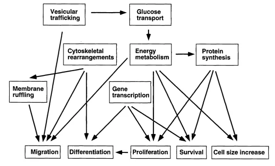
Figure 1.5. A diagram illustrating some of the possible relationships am ong the various cellular processes regulated by Class I a PI3Ks... 33
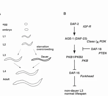
Figure 1.6. The C. elegans life cycle and the pathw ay involving the C. elegans Class Ia PI3K...45
Figure 1.7. Drosophila possess one of each class of P13K and the Drosophila Class Ia P13K prom otes grow th in the wing imaginai d is c ...48
Figure 1.8. Basic features of wing and eye imaginai disc developm ent... 50
Figure 1.9. Various perturbations to cell division, cell survival and protein synthesis have no effect on the size of the resulting o rg an ... 54
Figure 3.1. Affinity purification w ith pYXXM phosphopeptides implicates p60 as the adaptor for D p llO ... 74
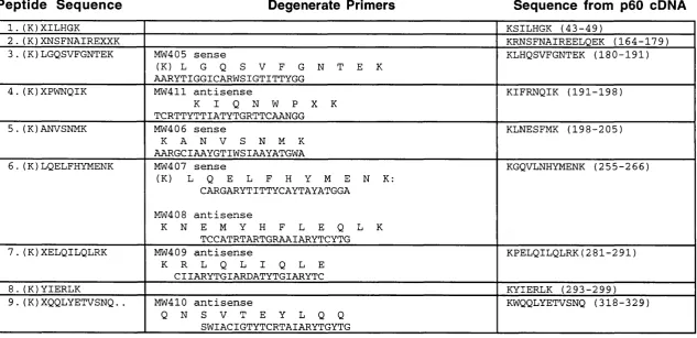
Table 3.1. Peptides and prim ers used to isolated the p60 cD N A... 76
Figure 3.2, Am ino acid sequence comparison of p60 w ith the m am m alian a d a p to rs ... 78
Figure 3.3. Com parison of the dom ain structure of p60 w ith m am m alian adaptors for Class Ia PI3Ks... 79
Table 3.2. BLAST analysis of the inter-SH2 dom ain of p 6 0 ... 81
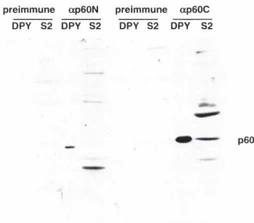
Figure 3.4. Testing ap60 antisera... 83
Figure 3.5. Characterisation of p60 at various stages of the Drosophila life cycle... 85
Figure 3.6. The D p ll0 /p 6 0 complex can be purified by imm unoprécipitation or w ith the DPY phosphopeptide and possesses protein and lipid kinase a c tiv ity ... 87
Figure 4.1. P I clones and STSs in the p60 region... 93
Figure 4.2. Southern blots of genomic clones of the al contig digested w ith EcoRI dem onstrate the localisation and direction of transcription of p 6 0... 95
Figure 4.3. The p60 genomic re g io n ... 96
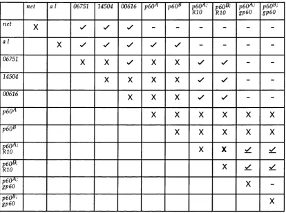
Table 4.1. Results of complem entation analysis of Drosophila lines w ith m utations or other aberrations in the p60 region... 99
Figure 4.4. Southern blot of DNA from P elem ent insertions dem onstrates that P[l(2)kl4504], P[l(2)06751] and P[l(2)k00616] are inserted near the p60 g e n e... 102
Figure 4.5. An example of the Southern blots used to determ ine the breakpoints of
the small p60 d eletions...104
Figure 4.6. p60 purified from mbm^ flies binds to DpllO, is phosphorylated, and is the same size as p60 purified from wild type {Oregon R) flies...105
Figure 4.7. The zygotic phenotypes oip60 m utants, DpllO m utants and DpllO p60 double m u ta n ts ...109
Figure 4.8. The FRT/FLP system ...I l l Figure 4.9. DpllOr and p60" om matidia and cells in the adult eye are reduced in size ... 113
Figure 4.10. Tangential sections of p60' and DpllO' clones in the eye...114
Figure 5.1. Ectopic expression strategies...120
Figure 5.2. Ectopic expression of HAp60 and HAAp60 in the eye or the w ing imaginai disc reduces the size of the adult o rg an s...123
Figure 5.3. The combination of HAp60 and m ycD pllO ectopic expression in the eye imaginai disc...125
Figure 5.4. The combination of HAp60 and m ycD pllO ectopic expression in the wing imaginai disc... 128
Figure 5.5. The effects of ectopically expressed HAp60 and m ycD pllO on levels of recom binant and endogenous proteins...130
Figure 5.6. The biochemical association betw een D pllO and p60 (both recombinant and endogenous)...132
Figure 6.1. Possible m odes of action for ectopically expressed HAp60 and m y cD p llO ...139
Figure 6.2. Two possible m odels describing the interactions betw een D pllO and p 6 0 ... 140
Figure 6.3. Possible pathw ay upstream and dow nstream of D p ll0 /p 6 0 ...149
Chapter 1 : Introduction
1.1 O verview ... 12
1.2 The three classes of phosphoinositide 3-kinases...12
1.3 PI3Ks may mediate many of the functions of receptor tyrosine kinases...15
1.3.1 Signalling downstream of RTKs via proteins that recognise and bind phosphotyrosine... 15
1.3.2 Class Ia PI3Ks associate with activated RTKs through SH2 domain-containing adaptors...16
1.3.2.1 The adaptors for Class Ia PBKs... 17
1.3.2.2 The SH2 domains of the adaptors for Class Ia PBKs... 19
1.3.3 The activation of Class Ia PBKs... 19
1.3.4 RTK specificity in the activation of Class Ia PBKs... 22
1.3.5 Other means of activation of Class Ia PBKs... 23
1.3.6 Downregulation of Class Ia PBK activity... 24
1.4 Downstream effectors of Class Ia PBKs... 25
1.4.1 The protein serine/threonine kinases, PKB and P D K l... 26
1.4.2 Guanine nucleotide exchange factors... 28
1.4.3 PLCy...29
1.4.4 Other downstream effectors...30
1.5 The cellular functions of Class Ia PBKs... 30
1.5.1 Methods of investigation of Class Ia PBK function ... 31
1.5.2 Stimulation of proliferation by Class Ia PBKs...34
1.5.3 Protection from apoptosis by Class Ia PBKs... 35
1.5.4 Effects of Class Ia PBKs on glucose and lipid m etabolism ...36
1.5.5. Regulation of protein synthesis by Class Ia PBKs... 37
1.5.6 Regulation of membrane trafficking by Class Ia P B K s ... 38
1.5.7 Cytoskeletal rearrangements mediated by Class Ia P B K s ... 39
1.5.8 Class Ia PBKs and cell transformation... 40
1.5.9 Other roles of Class Ia PBKs... 41
1.6 The role of Class Ia PBKs in the w hole organism ... 42
1.6.1 Genetic analysis of Class Ia PI3K function in C.
elegans...43
1.6.2 Analysis of Class Ia PI3Ks in m ic e ...46
1.7 U sing Drosophila to study Class Ia PI3Ks... 46
1.7.1 The Drosophila Class Ia PI3K, D pllO , plays a role in growth con trol... 47
1.8 Imaginai disc grow th...47
1.8.1 Imaginai disc d ev elo p m en t... 47
1.8.2 The regulation of growth during developm ent...51
1.8.2.1 Intrinsic and extrinsic controls of imaginai disc grow th...51
1.8.2.2. The involvem ent of proliferation, apoptosis and protein synthesis in imaginai disc d ev elo p m en t...52
Chapter 1 : Introduction
1.1 Overview
Communication between cells is essential for the developm ent and
viability of multicellular organisms. Intercellular communication is often
achieved by one cell producing extracellular ligands that activate receptors on
the surface of another cell. Activation of these receptors generates intracellular
signals that m odify the behaviour of the recipient cell. Thus, intracellular
signalling is vital to the developm ent and function of an organism and its
disruption is a key factor in the pathogenesis of many diseases, including
cancer.
The experiments described in this thesis investigate the function of
Class Ia phosphoinositide 3-kinases (PISKs). These lipid kinases have been
show n to respond to stimulated receptors and regulate several cellular
processes including proliferation, protection from apoptosis, glucose
metabolism, protein synthesis, membrane trafficking and cytoskeletal
rearrangements. Many of the assigned functions of Class Ia PI3Ks have been
inferred from the function of their upstream activators and downstream
effectors, which w ill be described in Sections 1.3 and 1.4 before Class Ia PI3K
function is addressed directly in Section 1.5. Most of the studies of Class Ia
PI3Ks have not dealt with the consequences of Class Ia PI3K activation during
the developm ent and function of multicellular organisms. H owever, recent
studies in C. elegans, Drosophila and mice provide interesting insights into Class
Ia PI3K function and w ill be reviewed in Sections 1.6 and 1.7. Finally, in this
thesis. Class Ia PI3K function is investigated in the developing Drosophila
imaginai disc. Therefore, the developm ent of imaginai discs and the cellular
processes that might be regulated by Class Ia PI3Ks during imaginai disc
developm ent w ill also be discussed (Section 1.8).
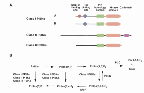
1.2 The three classes of phosphoinositide 3-kinases
PI3Ks are specific lipid kinases that phosphorylate the 3' hydroxyl of the
inositol ring of phosphoinositides (Stephens et al. 1993). All PI3Ks identified to
date, w ith the exception of the PI3Ks in Dictyostelium (Zhou et al. 1995), have
been grouped into three separate classes on the basis of their amino acid
sequence and in vitro lipid substrate specificity (Figure 1.1, Vanhaesebroeck et
al. 1997a). In vitro, Class I PBKs are able to produce phosphatidylinositol
3-phosphate (PtdIns(3)P) from phosphatidylinositol (Ptdlns), PtdIns(3,4)P2 from
adaptor Ras binding binding
site site
PIK homology
domain domainkinase
C2 domain
Class I PISKs
A
B
Class II PISKs
CO
Class III PISKs
B
FLO P td ln s P tdlns(4)P Ptdlns(4,5)P2
C la ss I PISKs C la ss II PISKs C la ss III PISKs
i i
C la ss 1 PISKs C lass 1 PISKs C la ss II PISKs
▼ ▼ ▼
PTEN
P tdlns(3)P Ptdlns(3,4)P2
SHIP
Ptdlns(3,4,5)P3
lns(1,4,5)P3
+
DAG
PtdIns(4)P, and Ptdlns(3,4/5)P3 from PtdIns(4,5)P2. In contrast. Class II PISKs
are only able to produce PtdIns(3)P and PtdIns(3,4)P2 (MacDougall et al. 1995;
Domin et al. 1997; Arcaro et al. 1998) and Class III PISKs are only able to
produce PtdIns(3)P (Schu et al. 1993; Volinia et al. 1995).
PISKs have been further classified on the basis of their binding partners
and their proposed method of activation. Class Ia PISKs have been found in
complex w ith src hom ology 2 (SH2) domain-containing adaptors, allowing
them to be responsive to receptor tyrosine kinase (RTK) stimulation (Section
1.3). Class Ib PISKs, on the other hand, have been found in association w ith a
101 kD protein and have been show n to be stimulated by G-protein coupled
receptors (Stoyanov et al. 1995; Stephens et al. 1997) and Class III PISKs have
been show n to bind to a serine/threonine kinase (Panaretou et al. 1997). In
contrast, no binding partners have yet been identified for Class II PISKs, and
their method of activation has yet to be established, though there have been
some suggestions that Class II PISKs and Class I PISKs can be activated by the
same stimuli (Figure 1.1, Turner et al. 1998; Arcaro et al. 1999). Interestingly, all
Class II PISKs possess a common carboxy-terminal (C-terminal) domain that is
termed a C2 domain because of its hom ology with a family of domains that
includes the 2nd conserved domain of the protein kinase C (PKÇ) family. The
C2 domain family also includes the Ca^+ binding domain of synaptotagmin, a
protein that mediates secretion at synaptic junctions. H owever, many C2
domains, including those of the Class II PISKs, lack the critical aspartate
residues necessary to bind Ca^+ (Rizo and Sudhof 1998), and no function has
yet been ascribed to the C2 domains of Class II PISKs.
PISKs from each of the above classes have been identified in several
different species. Furthermore, in many species, more than one member of
each class is present. Thus, so far it appears that mammals possess three Class
Ia PISKs (p i 10a, p and 8), one Class Ib PISK (pllO'^, three Class II PISKs
(PISKC2a, p [possibly not in mice] and y) and one Class III PISK (Wymann and
Pirola 1998). With the possible exception of the PISKs discovered in the slime
m ould Dictyostelium (Zhou et al. 1995), which are hard to classify by the
scheme described above. Class I and II PISKs have only been found in
metazoan organisms. The completed genom e sequence of Saccharomyces
cerevisiae reveals that the Class III PISK, VpsS4p, is the only PISK in budding
yeast (M ewes et al. 1997). In contrast, the completed genom e sequence of
Caenorhabditis elegans reveals one Class Ia PISK, one Class II PISK and one
Class III PISK (Ruvkun and Robert 1998, Section 1.6). Similarly, only one PISK
from each class has been identified in Drosophila (Section 1.7, Figure 1.7).
The kinase domains of all PISKs are related in sequence to the kinase
domains of all serine/ threonine (S/T) protein kinases (Carpenter and Cantley
1998), and all PISKs tested so far display S /T protein kinase activity in vitro.
Comparison of the amino acid sequences of the PISKs with the sequences of
other kinases has allowed the PISK family to be placed in a larger kinase
superfamily (Hunter 1995). This family also includes the PI 4-kinase family as
w ell as a family of high molecular w eight protein kinases that includes the
ataxia telangiectasia gene product (ATM), the DNA-dependent protein kinase
(DNA-PK) and the targets of [the immunosuppressant] rapamycin (TORs, also
called FRAP for FKBP12-rapamycin associated protein and RAFT for
rapamycin and FKBP12 target).
1.3 PISKs may mediate many of the functions of receptor tyrosine
kinases
RTKs have been show n to stimulate many cellular processes upon
extracellular stimulation including differentiation, mitogenesis, changes in
metabolic activity and cytoskeletal rearrangement (van der Geer et al. 1994).
Early experiments demonstrated that the intracellular levels of PtdIns(3,4,5)P3
increase rapidly w hen cells are treated with ligands that stimulate RTKs or with
other classes of ligand that stimulate receptors coupled to intracellular tyrosine
kinases or G-proteins (Auger et al. 1989; Stephens et al. 1993). In contrast,
increases in PtdIns(3,4)P2 levels occur more slow ly and to a lesser extent than the
increases in PtdIns(3,4,5)P3 levels follow ing receptor stimulation. Interestingly,
the levels of PtdIns(3)P remain unchanged in vivo after receptor stimulation.
Taken together, these discoveries suggested that PtdIns(3,4,5)P3 and
PtdIns(3,4)P2 act as second messengers for RTKs and because PI3Ks produce
these lipids, they were implicated as the regulators of several of the cellular
functions initiated by extracellular ligands. H owever, the fact that PtdIns(3)P is
produced very efficiently by all PI3Ks in vitro but is not increased by RTK
activation in vivo remains an enigma. (Lists of stimuli that increase
PtdIns(3,4,5)P3 can be found in Stephens et al. 1993).
1.3.1 Signalling downstream of RTKs via proteins that recognise and
bind phosphotyrosine
U pon the binding of extracellular ligands, RTKs (which are constitutive
dimers or which dimerise upon stimulation) transphosphorylate on specific
exposed tyrosine residues. In addition, certain RTKs also phosphorylate
or phosphotyrosine-binding (PTB) domains, w hich recognise
phosphotyrosines in particular amino acid sequence contexts, can then
associate with the tyrosine-phosphorylated RTKs and RTK substrates (Pawson
and Scott 1997). As w ell as SH2 or PTB domains, these downstream proteins
also contain domains with enzymatic activity or contain domains that facilitate
further protein: protein interactions. Signals are therefore transduced to the
cellular machinery through the activities of enzym es, such as kinases,
phosphatases and phospholipases, that are recruited to RTKs directly or
indirectly, through interactions w ith other proteins.
Several signalling mechanisms triggered by activated RTKs have been
w ell characterised (Pawson 1995). For example, the protein Grb2 binds to
activated RTKs through its SH2 domain. The N-terminal src hom ology 3 (SH3)
domain of Grb2 binds to proline-rich sequences in the guanine nucleotide
exchange factor (GEF) Sos, which in turn catalyses the exchange of guanine
nucleotide on inactive GDP-bound Ras to generate active GTP-bound Ras
(Buday and Downward 1993; Reif et al. 1994). The generation of Ras-GTP at the
plasma membrane causes the translocation and activation of the S /T kinase,
Raf, which in turn triggers a cascade of S /T kinases and the phosphorylation
of multiple targets proteins, including transcription factors, phosphatases and
metabolic enzym es (Marshall 1994).
Together, the numerous pathways triggered by tyrosine
phosphorylation of RTKs and their substrates have been proposed to mediate
all RTK functions (Pawson and Scott 1997). This proposal has been w ell
supported both by experiments in cultured cells overexpressing RTK mutants
lacking various tyrosine motifs and, more recently, by genetic studies. For
example, a m ouse with a mutant Met RTK in w hich two tyrosines had been
converted to phenylalanine had the same phenotype as a null mutation in the
Met RTK (Maina et al. 1996).
1.3.2 Class I
aPISK
sassociate with activated RTKs through SH2 domain-
containing adaptors
Class Ia PI3Ks have been show n to bind to tyrosine phosphorylated
RTKs and their substrates through the SH2-domain containing adaptor
proteins with which all Class Ia PI3Ks associate (Figure 1.2A). The association
of Class Ia PI3Ks w ith RTKs has been proposed to transduce a signal by
allowing Class Ia PBKs to reach the plasma membrane, where their
phosphoinositide substrates are located. H owever, studies from several
laboratories have suggested that the mechanism of activation of Class Ia PBKs
is more complicated than this simple m odel (Section 1.3.3, Section 6.2).
1.3.2.1 The adaptors for Class I
aPISK
sIn mammals, three genes have been identified that encode adaptors for
the mammalian Class I a PISKs: p85a, p8SP and p55y (or p55^^*^) (Figure 1.2B,
Escobedo et al. 1991; Otsu et al. 1991; Skolnik et al. 1991; Pons et al. 1995; Dey et
al. 1998). Four additional adaptors are encoded by splice variants of the p85a
transcript: p55a, p50a, p85aAS53 and p55aAS53 (Antonetti et al. 1996; Fruman
et al. 1996; Inukai et al. 1996). All of these adaptors contain tw o SH2 domains
separated by a large domain, called the inter-SH2 domain, which has been
show n to be responsible for the association of the adaptors w ith the Class Ia
PI3Ks (Dhand et al. 1994a). The p85a and p85p adaptors also possess long
N-terminal extensions that contain an SH3 domain and a breakpoint cluster
region hom ology (BH) domain. In contrast, the p55y, p55a and p55ocAS53
adaptors have short N-terminal extensions (Figure 1.2B). In addition, p85a and
p85p have a proline-rich, SH3 domain-binding stretch of amino acids (termed
PI) that is situated between the SH3 and BH domains. Furthermore, all
mammalian isoforms have a second proline-rich sequence (P2) located
approximately 20 residues N-terminal of the N-terminal SH2 domain.
The existence of the BH and SH3 domains and the proline-rich
sequences has raised the possibility that the adaptors for Class Ia PI3Ks may
receive signals from, or transduce signals to, molecules besides RTKs and
Class Ia PI3Ks. The BH domains have structural hom ology with GTPase
activating proteins (GAPs) but have not been show n to possess any GAP
activity; thus, their function is unclear. The SH3 domains of p85a and p85p
recognise proline-rich peptide motifs and have been show n to bind a number
of proteins in vitro (Gout et al. 1993; Harrison-Findik et al. 1995; Soltoff and
Cantley 1996). The situation in vivo may be different, however, because the
SH3 domains of p85a have also been show n to bind to the proline-rich
sequences found in the adaptors themselves and, together w ith the BH
domain, have been suggested to mediate the dimérisation of the p85 adaptors
w hen they are in complex with Class Ia PI3Ks (Layton et al. 1998).
In spite of the structural diversity described above, there has been no
experimental demonstration of selectivity of binding between different
adaptors and the mammalian Class I a PI3Ks, p i 10a, p or Ô (Vanhaesebroeck et
al. 1997b). H owever, there have been reports that the different adaptors can
localise to different compartments within the cell (Shepherd et al. 1997), which
PDGF
insulin
insuiin receptor PDGF
receptor
adaptor C lass U PI3K
IRS-1
B
p85u
p85uAS53
p55a
p55oAS53
p50a
p85p
pSSPIK/y
SH3
1
SH2 lnter-SH2 SH2PI P2
8 extra amino acids
V
1
8 extra amino acids
V
LL
^
'
1
Figure 1.2. The role of the adaptor in Class Ia PI3Ks activation. (A) The SH2
domains of the adaptor allow the Class Ia PI3K to be translocated to the membrane by activated RTKs such as the PDGF receptor or by phosphorylat ed receptor substrate molecules such as IRS-1. (B) Seven adaptors are encod ed by three genes. There are five splice variants of p85a.
1.3.2.2 The SH2 domains of the adaptors for Class I
aPISK
sThe binding specificity of distinct SH2 domains lies in the ability of
these domains to recognise phosphotyrosine in the context of particular amino
acid sequences C-terminal to the phosphotyrosine. The preferred
phosphotyrosine binding motifs of several different SH2 domains have been
established using purified SH2 domains to screen a library of phosphotyrosine
peptides (Songyang et al. 1993). This approach predicted that the SH2 domains
of p85a w ould bind to phosphotyrosine in the context pYXXM where X can be
any amino acid. Consistent with this prediction, peptides containing pYXXM
motifs have been utilised to purify the adaptors in complex w ith Class Ia
PISKs (Otsu et al. 1991; Fry et al. 1992, Chapter 3). Structural studies of many
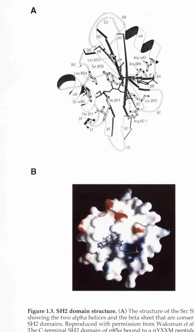
SH2 domains using X-ray crystallography and nuclear magnetic resonance
have revealed that SH2 domains share a common fold consisting of a central
beta sheet flanked by two alpha helices (Figure 1.3A, Waksman et al. 1993). All
SH2 domains contain a "pocket" that can bind phosphotyrosine. Binding
specificity is achieved by other, more variable regions of the dom ain that form
binding surfaces or pockets for amino acids that lie C-terminal to the
phosphotyrosine in the bound peptide. Structural studies of the N- and
C-terminal SH2 domains of p85a in complex with pYXXM peptides have
revealed that, in addition to the phosphotyrosine-binding pocket, both
domains form a hydrophobic pocket that binds to the methionine residue
situated three amino acids C-terminal of the phosphotyrosine in the bound
peptide (Figure 1.3B, Breeze et al. 1996; Nolte et al. 1996).
1.3.3 The activation of Class I
aPI3K
sPurified Class Ia PI3Ks in complex w ith their adaptors have been
show n to be constitutively active in vitro (Hiles et al. 1992; Vanhaesebroeck et
al. 1997b). These studies suggest that Class Ia PI3Ks are constitutively active in
the cell and that the regulated production of 3' phosphoinositides is achieved
in vivo by bringing the active enzym es to their substrates in the membrane.
This m odel is supported by studies show ing that downstream pathways are
activated by overexpressed Class Ia PI3Ks targeted to the membrane by the
addition of amino acid sequences that become farnesylated or myristylated or
by the addition of transmembrane sequence of the protein, CD2 (Klippel et al.
1996; Leevers et al. 1996; Brennan et al. 1997). However, other studies have
suggested that translocation to the membrane is not sufficient for full
activation of Class Ia PI3Ks. For example, it has been show n that, although
overexpressed membrane-targeted p i 10a activates downstream signalling
Arg pB 5
B
Figure 1.3. SH2 domain structure. (A) The structure of the Src SH2 dom ain showing the tw o alpha helices and the beta sheet that are conserved in all SH2 domains. Reproduced w ith perm ission from W aksm an et al. 1993. (B)
The C-terminal SH2 dom ain of p85a bound to a pYXXM peptide. Repro duced with perm ission from Breeze et al. 1996.
p85a to the N-terminus of p i 10a (Hu et al. 1995). Thus, the adaptors might
upregulate Class Ia PI3K activity by a mechanism in addition to membrane
localisation. Consistent with this notion, another study show ed that the simple
coexpression of the inter-SH2 domain of p85a with p i 10a resulted in a
dramatic increase in PI3K activity (Frevert and Kahn 1997). Furthermore,
several reports have demonstrated that the addition of pYXXM
phosphopeptides to Class Ia PI3K/adaptor complexes increases lipid kinase
activity 2-3 fold in vitro (Carpenter et al. 1993a; Rordorf Nikolic et al. 1995). In
addition, pYXXM phosphopeptides can be used to activate Class Ia PI3Ks in
vivo (Kotani et al. 1994; Derossi et al. 1998).
Recently, Yu et al. (1998b) show ed that p i 10a overexpressed in a
mammalian cell line w as unstable at 37 °C but could be stabilised by reducing
the temperature to 30 °C or by adding various extensions to the N-terminus.
Therefore, these authors argued that the apparent activation of Class Ia PI3Ks
by the addition of N-terminal extensions that had been reported by other
investigators w as actually a consequence of the increased stability of these
fusion proteins compared w ith that of the untagged p i 10a (Yu et al. 1998b).
Furthermore, it was demonstrated that the lipid kinase activity of p i 10a
stabilised by an N-terminal tag and synthesised at 30 °C w as inhibited by the
adaptor, p85a. Experiments in which p i 10a and p85a were overexpressed in
Sf9 insect cells were also used to show that p85a could inhibit p llO a , and that
this inhibition was relieved by the addition of pYXXM phosphopeptides (Yu et
al. 1998b). Thus, the observed activation of Class Ia PI3Ks by the addition of
phosphopeptides may, in reality, be a relief of inhibition by the adaptor.
Consistent w ith this hypothesis, other experiments have show n that the
adaptors can inhibit lipid kinase activity w hen in complex w ith Class Ia PI3Ks.
Expression of a mutated version of p85a, which w as truncated from the
C-terminal end of the inter-SH2 domain through chromosomal rearrangement,
w as show n to cause an increase in PI3K activity in vivo, possibly because
translocation of the Class Ia PI3K to the membrane was increased (Jimenez et
al. 1998). Therefore, the C-terminal SH2 domain a n d /o r the C-terminal region
of the inter-SFI2 domain may inhibit the activity of associated Class Ia PBKs
by altering their intracellular location. H owever, in vitro experiments have
show n that the N-terminal SH2 domain and the inter-SH2 domain are
necessary and sufficient for the adaptor to inhibit Class Ia PBK kinase activity
(Yu et al. 1998a). Thus, it is possible that the adaptors can inhibit Class Ia PBKs
by two distinct mechanisms, firstly, by directly inhibiting kinase activity by
association and, secondly, by restricting the access of the associated Class Ia
regulation of Class Ia PI3K function in the light of the results described in this
thesis w ill be discussed in Section 6.2.
1.3.4 RTK specificity in the activation of Class I
aPISK
sReceptor tyrosine kinases form a large family of molecules that have
specific and diverse functions in the control of cellular processes and the
developm ent of multicellular organisms (van der Geer et al. 1994). The
remarkable phosphotyrosine-binding specificity of the adaptors for Class Ia
PI3Ks has led to the prediction that all RTKs with YXXM motifs that become
tyrosine-phosphorylated upon stimulation w ill activate Class Ia PBKs (Section
1.3.4).
However, this m odel is not completely reliable for a number of reasons.
Firstly, pYXXM motifs are recognised by the SH2 domains of other signalling
molecules including phospholipase Cy (PLCy, Bourette et al. 1997, Chapter 3)
and the multidomain protein. Nek (Nishimura et al. 1993). Secondly, Class Ia
PBK /adaptor complexes have been show n to associate with the pYVXV motif
of the Met receptor, albeit with a lower affinity than that for pYXXM motifs
(Ponzetto et al. 1993). Thirdly, several RTKs that lack YXXM motifs, such as the
epidermal growth factor receptor (EOF receptor or ErbBl) have also been
show n activate Class Ia PBKs upon stimulation (Stephens et al. 1993). The
translocation of Class Ia PBKs to the membrane upon activation of these RTKs
is achieved by the adaptors binding to pYXXM motifs on receptor substrate
molecules that are phosphorylated by the activated RTKs. In particular,
activated insulin and insulin-like growth factor (IGF) receptors have been
show n to tyrosine phosphorylate insulin receptor substrates (1RS 1-4), thereby,
inducing the association of the substrate m olecules with Class Ia PBKs
(Backer et al. 1992; Yenush and White 1997). Other RTKs can phosphorylate the
related substrate molecule, G abl, w hich also contains YXXM motifs
(Holgado-Madruga et al. 1996; Ingham et al. 1998). In addition, cytokine receptors
without intrinsic tyrosine kinase activity have been show n to activate Class Ia
PBKs through the activation of intracellular protein tyrosine kinases that
phosphorylate YXXM motifs on other proteins (a comprehensive list of
cytokines that activate PBKs can be found in W ymann and Pirola 1998).
Finally, some RTKs such as the fibroblast growth factor (FGF) receptor are
thought to stimulate Class Ia PBKs solely through the activation of Ras (van
Weering et al. 1998, Section 1.3.5). Thus, although the presence of a
phosphorylated YXXM motif on a receptor is a good indicator that it will
activate Class Ia PBKs, it cannot be concluded that the motif w ill not also
activate other proteins or that an RTK without a pYXXM w ill not activate Class
Ia PISKs.
1.3.5 Other means of activation of Ciass I
aPISK
sAs w ell as being activated by their association w ith pYXXM motifs.
Class Ia PISKs are also activated by other molecules. These molecules include
tyrosine phosphorylated IkB, the inhibitor of the transcription factor NFkB
(Beraud et al. 1999), and Ca^+-bound calmodulin, which binds to the SH2
domains of p85a (Joyal et al. 1997). H owever, the only m olecule that has been
show n to be able to activate Class Ia PISKs independently of the adaptor is the
small GTPase Ras (Section l.S .l). Specifically, it has been show n that PISK
activity can be co-purified with Ras, and overexpression of constitutively
active Ras increases the levels of S' phosphorylated inositol lipids in vivo
(Sjolander et al. 1991; Rodriguez-Viciana et al. 1994). In addition to Raf and Ral
CDS (guanine nucleotide dissociation stimulator), active GTP-bound Ras is
able to associate with and activate the Class Ia PISK, p i 10a
(Rodriguez-Viciana et al. 1994). Mutant forms of Ras that bind preferentially to one of these
three effectors have been generated by the introduction of specific point
mutations, which were first identified in a yeast two-hybrid screen for
modifiers of Ras binding (White et al. 1995; Rodriguez-Viciana et al. 1997).
Overexpression of constitutively active Ras w ith one of these effector
mutations (V12 Ras C40) in COS cells increases Class Ia PISK activity (though
to a lesser extent than V I2 Ras without the effector domain mutation) but does
not activate Raf or Ral CDS (Rodriguez-Viciana et al. 1997). These studies
provide a useful tool to activate Class Ia PISKs in cells as w ell as further
evidence that Class Ia PISKs are direct effectors of Ras. In addition, the region
of p i 10a that interacts with Ras has been mapped (Figure 1.1). Consistent with
the proposal that the association of Ras w ith this region of p i 10a leads to
activation of the kinase, a form of p i 10a with a point mutation in the
Ras-binding region possesses increased lipid kinase activity in vitro
(Rodriguez-Viciana et al. 1996). This discovery again provides a reagent that can be used to
mimic Class Ia PI3K activation.
The ability of Ras-GTP to activate Class Ia PISKs in at least some
experimental situations suggests that there is another mechanism in addition
to direct association w ith the adaptors for Class Ia PISKs by which RTKs can
increase S' phosphoinositide production. H owever, the level of Ras-induced
activation of Class Ia PISKs by the FGF receptor, w hich activates Ras but does
not possess YXXM motifs, has been found to be significantly less than the level
adaptors via their YXXM motifs (van Weering et al. 1998). Interestingly,
reactive free radical species generated by nitric oxide donors have been
reported to induce the association of pllO ô and pllO p but not p llO a with Ras,
providing a mechanism for Class Ia PI3K activation that is independent of
tyrosine kinase activation (Deora et al. 1998).
Together, these studies can be used to argue that Ras functions
upstream of p i 10a. However, other studies have show n that overexpressed,
activated p i 10a can increase the amount of Ras-GTP in NIH 3T3 cells (Hu et al.
1995). Furthermore, the cellular effects of activated p i 10a expression can be
inhibited by the coexpression of dominant negative Ras and mimicked by the
expression of activated Ras (Hu et al. 1995). These results are more consistent
with Ras acting downstream of p i 10a. Another apparently conflicting report
has show n that dominant negative Ras has no effect on Class Ia PI3K
signalling in 3T3 LI adipocytes (Gnudi et al. 1997). Therefore, the interaction
between Ras and Class Ia PI3Ks is still controversial and may be cell type
specific. Genetic studies are required to demonstrate the importance of the
interaction in normal signalling processes.
1.3.6 Downregulation of Class I
aPI3K activity
In theory, there are several w ays in w hich the downregulation of Class
Ia PI3K activity might be achieved. The possibilities include 1) the spatial
restriction of the enzym e away from its lipid substrates in the membrane; 2)
the direct attenuation of its catalytic activity or, 3) the depletion of the 3'
phosphorylated lipid products. As discussed in Section 1.3.3, the adaptors may
utilise the first two mechanisms to inhibit Class Ia PI3Ks. The lipid kinase
activity of p i 10a is also attenuated after it phosphorylates its associated p85a
on serine 608 (Carpenter et al. 1993b; Dhand et al. 1994b). Interestingly, p i 106
does not phosphorylate associated adaptors but does phosphorylate itself,
which also leads to a reduction in lipid kinase activity (Vanhaesebroeck et al.
1999a).
There is good evidence for the third mechanism of downregulation of
Class Ia PI3K activity, namely, the degradation of the 3' phosphoinositides.
The cellular level of PtdIns(3,4,5)P3 rises rapidly upon extracellular stimulation
but is followed by a sharp decline (Auger et al. 1989; Stephens et al. 1993). The
peak in the level of PtdIns(3,4,5)P3 is consistently followed by a later peak in
the level of PtdIns(3,4)P2, suggesting that the increase in PtdIns(3,4)P2 arises
from the dephosphorylation of PtdIns(3,4,5)P3 by a 5' phosphatase rather than
from the phosphorylation of PtdIns(4)P by PI3Ks. If PtdIns(3,4,5)P3 rather than
PtdIns(3,4)P2 is the important signalling product of Class Ia PI3K, such a 5'
phosphatase w ould be expected to downregulate the effects of Class Ia PI3Ks.
The SH2 domain-containing 5' inositol phosphatase (SHIP) is one of a family of
5' inositol phosphatases and has been show n to dephosphorylate
Ptdlns(3,4/5)P3 to generate PtdIns(3,4)P2 (Woscholski and Parker 1997).
Importantly, the overexpression of SHIP inhibits the activation of the
downstream targets of Class Ia PI3Ks (Aman et al. 1998; Vollenweider et al.
1999).
The protein PTEN has been identified as a tumour suppressor both
because cells from many human cancers have somatic loss of function
mutations in PTEN, and because PTEN is mutated in the germline of patients
with the cancer-related syndromes, Cowden disease, Lhermitte-Duclos disease
and Bannayan-Zonana syndrome (Maehama and Dixon 1999). Interestingly,
PTEN possesses 3' lipid phosphatase activity and dephosphorylates
PtdIns(3,4,5)P3 to form PtdIns(4,5)P2 (Maehama and Dixon 1998; Stambolic et
al. 1998). PtdIns(3,4)P2 and PtdIns(3)P are also dephosphorylated by PTEN but
at a significantly lower rate (Maehama and Dixon 1998). Studies in which SHIP
or PTEN have been inhibited, mutated or overexpressed suggest that these
lipid phosphatases antagonise PI3K activity in vivo (Aman et al. 1998;
Stambolic et al. 1998), making them excellent reagents for the study of Class Ia
PI3K function (Section 1.5).
1.4 Downstream effectors of Class I
aPI3K
sAlthough the association of Class Ia PI3Ks with RTKs and the sudden
increase in PtdIns(3,4,5)P3 upon RTK stimulation implicated Class Ia PI3Ks
and their lipid products in the control of various cellular processes, a
mechanism by which these lipids act as second messengers w as also required.
In other words, how do lipids w ith a particular chemical structure cause
changes in cell behaviour? There have been suggestions that the 3'
phosphoinositides alter cell behaviour through biophysical effects on
membrane structure or by interfering with actin polymerisation. However, the
existence of specific proteins that associate w ith the 3' phosphorylated inositol
lipids is also an attractive model.
The search for proteins that transduce the signal from the 3’
phosphorylated inositol lipids to the cellular machinery took tw o different
routes. Eirstly, RTK activated proteins that were implicated in regulating the
same cellular processes as Class Ia PI3Ks were examined. Secondly, searches
were performed for proteins that specifically bound the 3' phosphorylated
benefited enormously from the use of synthetic 3' phosphoinositides.
Importantly, these lipids were synthesised and made available in both the
naturally-occurring and non-naturally-occurring chiral forms, thus providing
an excellent control for the non-specific binding of charged lipids to proteins
(Gaffney and Reese 1997). Together, research based on these two approaches
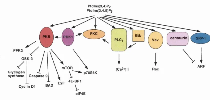
led to the discovery that certain proteins containing pleckstrin hom ology (PH)
domains could bind specifically to PtdIns(3,4,5)P3 and PtdIns(3,4)P2 (Figure
1.4, Lemmon et al. 1996). Interestingly, only a subset of PH domains have been
show n to bind these lipids in preference to other phosphoinositides.
Furthermore, other classes of domain may also bind specifically to the 3'
phosphorylated inositol lipids (Section 1.4.6). (An extensive list of
PtdIns(3,4,5)P3-binding proteins can be found in Shepherd et a l 1998).
Many of the m ethods used to investigate Class Ia PI3K function
(Section 1.5.1) have also been used to establish that particular proteins are
downstream targets of Class Ia PI3Ks. Once it has been established that a
protein is directly activated (or inhibited) by Class Ia PI3Ks, investigation of
the function of that protein can provide further evidence for the functional
studies of Class Ia PI3Ks. Therefore, the best characterised downstream
effectors w ill be discussed before addressing Class Ia PI3K function (Section
1.5).
1.4.1 The protein serine/threonine kinases, PKB and PDK1
The S /T kinase, protein kinase B (PKB, also known as Akt [the cellular
form of the oncogene v-Akt] and RAC-PK [Related to A and C protein kinase])
has been show n to act downstream of Class Ia PI3Ks. The initial experiments
that implicated PKB as a target of Class Ia PI3Ks demonstrated that PKB was
activated by the RTK ligand platelet derived growth factor (PDGF) and that
this activation was dependent not only on Class Ia PI3K activity, but also on
the association of Class Ia PI3Ks with the PDGF receptor (Burgering and
Coffer 1995; Franke et al. 1995; Kohn et al. 1995). The PH dom ain of PKB can
bind selectively to PtdIns(3,4)P2 and PtdIns(3,4,5)Pa, and there have been
suggestions that lipid binding can relieve the inhibition of PKB activity by its
PH domain (James et a l 1996; Franke et a l 1997; Freeh et al. 1997; Klippel et a l
1997). H owever, PKB is also activated by Class Ia PI3Ks through the action of
3' phosphoinositide-dependent kinase 1 (PDKl), which can also bind to
PtdIns(3,4,5)Ps through its PH domain (Alessi et al. 1997a; Stephens et al. 1998).
PDKl phosphorylates PKB on serine 308, one of the tw o sites on PKB that
must be phosphorylated for its complete activation (Alessi et al. 1997b; Stokoe
et al. 1997). An additional role of PtdIns(3,4,5)P3 and PtdIns(3,4)P2 in the
Ptdlns(3,4)P2
Ptdlns(3,4,5)P3
PKC
Btk
PKB
PDKl
centaurin
GRP-1PLC
yVav
PFK2
GSK-3
ARF
mTOR
Rac
Glycogen
synthase
C aspase
p70S6K
4E-BP1
E2F
BAD
Cyclln D1
elF4E
activation of PKB may be to colocalise PDKl and PKB. It has been suggested
that another protein kinase, termed PDK2, phosphorylates the second site on
PKB, threonine 473, to result in the full activation of PKB. PDK2 has yet to be
identified, but integrin linked kinase, which can phosphorylate threonine 473
on PKB in vitro, is a proposed candidate (Coffer et a l 1998; Delcommerme et al.
1998).
There are several potential targets of PKB, many of which are proteins
that were thought to act downstream of Class Ia PI3Ks but for w hich there
w as no established mechanism for their direct activation by Class Ia PI3Ks
(Figure 1.4). These proteins include glycogen synthase kinase-3 (GSK-3),
phosphofructokinase-2 (PFK-2) and mTOR, all of which w ill be discussed later
in the context of the cellular functions that they are thought to regulate
(Section 1.5). PKB has been implicated in the control of several cellular
processes, including protection from apoptosis, insulin-induced changes in
metabolism, progression through the cell cycle, protein synthesis and glucose
uptake (Coffer et al. 1998, Section 1.5). It is so generally accepted that PKB is
activated by Class Ia PI3Ks that PKB activation is often used as a measure of
Class Ia PI3K activity. H owever, it is important to note that PKB can be
activated independently of Class Ia PI3Ks (Yano et al. 1998). In addition, PDKl
has been show n to phosphorylate several substrates other than PKB, some of
which might be non 3'-phosphoinositide dependent PDKl targets (Alessi et al.
1998; Cheng et al. 1998; Chou et al. 1998; Le Good et al. 1998; Pullen et al. 1998).
1.4.2 Guanine nucleotide exchange factors
Guanine nucleotide exchange factors (GEFs) catalyse the activation of
small GTPases by inducing the exchange of bound GDP for GTP. Several GEFs
possess PH domains, including all GEFs for the Rho family GTPases. The PH
domain of the Rac GEF, Vav, has been show n to bind to PtdIns(3,4,5)P3 and
this interaction increases the GEF activity of Vav towards Rac (Han et al. 1998).
Consistent with this observation, Rac has been show n to be activated by Class
Ia PI3Ks in vivo (Hawkins et al. 1995). Interestingly, the Ras GEF, Sos, also has
a PH domain that binds selectively to PtdIns(3,4,5)P3 (Rameh et al. 1997),
suggesting that Ras family GTPases might also be activated via the stimulation
of GEFs by PtdIns(3,4,5)P3.
Recently, another family of PH domain-containing GEFs comprised of
GRP-1, ARNO and cytohesin-1 has also been show n to bind PtdIns(3,4,5)P3.
These proteins have hom ology with the yeast protein Sec7, and are all GEFs
for ADP-ribosylation factors (ARFs), which are GTPases involved in the
regulation of various vesicular trafficking pathways and the activation of
specific phospholipase D isoforms (Moss and Vaughan 1998). ARNO was
identified through its hom ology with the yeast ARF GEF, G eal (Chardin et al.
1996; Peyroche et al. 1996), whereas GRPl w as identified in a screen of an
expression library for PtdIns(3,4,5)P3-binding proteins (Klarlund et al. 1997).
Independently of studies of ARNO and GRPl, cytohesin-1 w as found in a
yeast two-hybrid screen for proteins that associate w ith the cytoplasmic
domain of an integrin found in T cells. Cytohesin-1 has been proposed to
promote cellular adhesion to extracellular substrates by stimulating the
exocytosis of integrins (Kolanus et al. 1996).
The PH domains of ARNO and cytohesin-1 bind to PtdIns(3,4,5)P3
specifically and are membrane-localised upon stimulation (Nagel et at. 1998;
Venkateswarlu et al. 1998b). Strikingly, w hen fused to green fluorescent
protein (GFP), the ARNO-PH domain can be visualised translocating from the
cytoplasm to the plasma membrane within seconds of stimulating live
adipocytes with insulin (Venkateswarlu et al. 1998b). A similar effect w as seen
with a GRPl PH domain GFP fusion protein in PC12 cells stimulated w ith
NGF or EGF (Venkateswarlu et al. 1998a). Therefore, it has been postulated
that the function of PtdIns(3,4,5)P3 binding in ARF GEF activation is to localise
these proteins to the appropriate cellular environment rather than to stimulate
their GEF activity directly (Moss and Vaughan 1998).
1.4.3 PLCy
The phospholipase Cs are a family of enzym es that hydrolyse
PtdIns(4,5)P2 to generate the second messengers inositol (1,4,5) trisphosphate
and diacylgycerol, molecules which in turn induce intracellular Ca^+ release
and protein kinase C (PKC) activation (Lee and Rhee 1995). Thus, the PECs
share a substrate with Class Ia PBKs. In addition to their catalytic domains,
PLCy isoforms have tw o SH2 domains and a PH domain. The N-terminal SH2
domain of PLCy2 has been show n to bind the same phosphotyrosine on the
macrophage colony stimulating factor (M-CSF) receptor as the adaptors for
Class Ia PI3Ks. Thus, as w ell as sharing a substrate w ith Class Ia PI3Ks, PLCy
isoforms can be recruited to the same RTKs (Bourette et al. 1997, see also
Section 3.2.2). Furthermore, the PH domain and both SH2 domains of PLCyl
have been show n to bind PtdIns(3,4,5)P3 and mediate the translocation of
PLCyl to the plasma membrane (Bae et al. 1998; Falasca et al. 1998). Consistent
with the idea that these multiple interactions between Class Ia PI3K and PLCy
isoforms have physiological significance, overexpression of active Class Ia
PI3K leads to an increase in PLC activity in COS cells (Bae et al. 1998).
of Class IA PI3Ks. Tyrosine phosphorylation of PLCy isoforms by Tec family
kinases may be another mechanism by which Class Ia PI3Ks activate PLCy
isoforms (Section 1.4.4). H owever, various reports have show n that tyrosine
phosphorylation does not always correlate with PLCy activation (Rhee and Bae
1997; Gratacap et al. 1998). It is also possible that the recruitment of PLCy by
PtdIns(3,4,5)Ps to the same environment as Class Ia PI3Ks leads to the
depletion of the substrate for Class Ia PI3Ks, resulting in an effective
downregulation of PI3K activity.
1.4.4 Other downstream effectors
Other proteins that can bind to PtdIns(3,4,5)P3 in vitro, and are therefore
potential targets of PtdIns(3,4,5)Ps in vivo, are certain PKC isoforms (Nakanishi
et ah 1993; Toker et ah 1994; Le Good et ah 1998), centaurin-a, w hich has a
PtdIns(3,4,5)P3-binding PH domain and a domain with hom ology to ARF
GAPs (Hammonds-Odie et ah 1996), and the Tec family of intracellular
tyrosine kinases (August et ah 1997; Scharenberg et ah 1998). In the search for
PH domains that selectively bind to PtdIns(3,4,5,)P3, the PH domain from one
of these kinases, Bruton's tyrosine kinase (Btk) was identified (Salim et ah 1996;
Rameh et ah 1997). Mutations in human Btk are found in patients with
X-linked agammaglobulinaemia, a disease arising from a deficiency in B cells
(Mattsson et ah 1996). Importantly, a number of the point mutations that are
associated w ith this disease map to the PH domain of Btk and disrupt the
binding of PtdIns(3,4,5,)P3 to Btk (Salim et ah 1996).
Interestingly, a newly-identified domain called the FYVE domain has
show n to bind specifically to PtdIns(3)P (Burd and Emr 1998; Gaullier et ah
1998; Patki et ah 1998). Proteins containing these domains are potential
downstream effectors of Class III PI3Ks. It is possible that there are as yet
unidentified FYVE domains that bind to PtdIns(3,4,5)P3. Finally, som e SH2
domains have been show n to bind PtdIns(3,4,5)P3, including the SH2 domain
of p85a, the SH2 domain of Src and the SH2 domains of PLCy (Rameh et ah
1995; Bae et ah 1998; Rameh et ah 1998). Clearly, these interactions create
further possibilities for the downregulation of Class Ia PI3K activity and the
cross-regulation of signalling pathways.
1.5 The cellular functions of Class I
aPI3K
sA s described above, early ideas about Class Ia PI3K function were based
on the correlation of the regulation of cellular processes by RTKs with the
appearance of the lipid products of PI3Ks and the recruitment of Class Ia PI3Ks