Original Article
Title: Standardized in vivo evaluation of biocompatibility and bioresorption of a new synthetic membrane for guided bone regeneration
Lívia da Costa Pereira DDS¹
¹Dentistry School, Postgraduate Program in Dentistry, Universidade Federal Fluminense, Niterói, Rio de Janeiro, Brazil. - [email protected]
Carlos Fernando de Almeida Barros Mourão DDS; MSc; PhD5
5Oral Surgery Department, Dentistry School, Postgraduate Program in Dentistry,
Universidade Federal Fluminense, Niterói, Rio de Janeiro, Brazil.
Adriana Terezinha Neves Novellino Alves DDS; PhD²
²Oral Diagnosis Department, Dentistry School, Universidade Federal Fluminense, Niterói, Rio de Janeiro, Brazil.– [email protected]
Rodrigo Figueiredo de Brito Resende DDS; PhD³
³Oral Surgery Department, Universidade Federal Fluminense, Niterói, Rio de Janeiro, Brazil. [email protected]
Marcelo José Pinheiro Guedes de Uzeda⁴
⁴Oral Surgery Department, Universidade Federal Fluminense, Niterói, Rio de
Janeiro, Brazil. - [email protected] Rafael Seabra Louro¹ DDS; PhD6
6Oral Surgery Department, Dentistry School, Universidade Federal Fluminense,
Niterói, Rio de Janeiro, Brazil. - [email protected]
Mônica Diuana Calasans-Maia7
7Oral Surgery Department, Dentistry School, Universidade Federal Fluminense,
Niterói, Rio de Janeiro, Brazil. – [email protected]
*Correspondence author: Mônica Diuana Calasans-Maia DDS; PhD. Oral
Surgery Department, Universidade Federal Fluminense, Niterói, Rio de Janeiro,
Brazil. Rua Mario Santos Braga, 28/4º andar. [email protected]
ABSTRACT
This study aimed to evaluate the physico-chemical properties, biocompatibility
and bioabsorption of 3 different new membranes for bone guided regeneration
(PLGA associated with hydroxyapatite and -TCP) with three thicknesses (200,
500 and 700 µm) implanted in mice subcutaneously.
Scanning electron microscopy (SEM), X-ray diffraction (XRD), Fourier transform
infrared spectroscopy (FTIR) and the quantification of Carbon, Hidrogen and
Nitrogen were used to characterize the physico-chemical properties. One
hundred Balb-C mice were divided into 5 experimental groups: Group 1 - Sham
(without implantation); Group 2 - 200 μm; Group 3 - 500 μm; Group 4 - 700 μm;
and Group 5 - Pratix®. Each group was subdivided into four experimental periods
(7, 30, 60 and 90 days). Samples were collected and processed for histological
and histomorphometrical evaluation.
The membranes showed no moderate or severe tissue reactions in the
experimental periods studied. The 500 μm membrane did not show tissue
reaction for any experimental periods studied. The 200 μm membrane
membranes began to exhibit fragmentation after 30- day, while the 500 and 700
µm membranes started the fragmentation at 90-day.
All membranes studied were biocompatible, and the 500µm membrane showed
the best results being a promissory membrane for bone guided regeneration.
1. INTRODUCTION
The bone is a specialized connective tissue capable of regenerating
completely. Therefore, regenerative procedures are used for gaining bone tissue,
allowing professionals the possibility of rehabilitating a patient both aesthetically
and restoring function. However, bone tissue’s growth pattern requires a longer
time for its formation compared to non-mineralized connective tissue, since the
fibroblasts proliferate and produce matrices of collagen more quickly than the
osteoblasts that form the bone tissue[1].
Guided Bone Regeneration treatment (GBR), in which the regeneration of
defects is predictably achieved by the application of occlusive membranes, was
developed based on the Guided Tissue Regeneration (GTR) concept. GBR
involves mechanically excluding the population of non-osteogenic cells from the
surrounding soft tissues and allowing the population of osteogenic cells
originating from native bone to inhabit the defect, thus preventing the growth of
unwanted soft tissue in the bone defect[2]. Experimental studies have provided
significant evidence that bone regeneration is significantly increased when the
invasion of soft tissues into bone defects is mechanically prevented[3,4].
In the last 40 years, different membranes have been developed to act as
physical barriers in the treatment of bone defects. Today, resorbable collagen
and aliphatic polyester membranes are the most commonly used materials to
eliminate the need for a second surgery for membrane removal[5,6]. However,
collagen undergoes rapid degradation due to the action of inflammatory cells in
the region, which resultd in poor mechanical resistance leading to collapse[7].
Among the polymers used, the most applicable in medicine are those that,
the organism. The aliphatic polyesters derived from lactic acid and glycolic acid
undergo hydrolytic degradation. However, despite the excellent mechanical
properties of the PLA, its biocompatibility, biodegradability, hydrophobicity,
brittleness, acidic degradation products and high cost of PLA restrict its
application[8]. The PLA polymer shows a slower hydrolysis rate compared to the
PGA polymer in the human body, so for a suitable degradation of the polymer,
the PLA polymer has been combined with the PGA polymer as a copolymer,
improving its characteristics [9]. The insertion of calcium phosphate
(hydroxyapatite and -TCP) prevents collapses by improving mechanical
properties[10].
The aim of this study was to evaluate the physico-chemical properties,
biocompatibility and bioabsorption of 3 different thicknesses of PLGA membranes
associated with calcium phosphate (PLGA + hydroxyapatite + β-TCP) implanted
in mice subcutaneously, following ISO 10993-6: 2016. This study used mice as
an experimental model due to their rapid prolificity, easy handling, known biology
and genome and not very expensive maintenance, as well as similarity of the
results with humans.
2. MATERIALS AND METHODS Control membrane
A commercially available PLGA dental membrane (Pratix®, Baumer, São
Paulo, SP, Brazil) was purchased for comparison of the biocompatibility and
bioabsorption rates in the subcutaneous tissue of rats.
Physico-chemical characterization of experimental membranes
The crystalline mineral phases present in the membranes, their crystallinity
examined by Rietveld XRD Quantification (XRD). The XRD patterns were
obtained with a Empyrean - Panalytical diffractometer operating at 45 kV and
40mA, with CuKα radiation (Cu - 1,540598 Å), with temperature of 25ºC and
relative humidity of the air about 55%. The data were collected in the 2θ range of
10°–70° with a step of 0.02° point per second.
The contents of the hydroxyapatite and β-tricalcium phosphate phases in
the samples were evaluated by the relative intensities of specific peaks of βTCP
and HA XRD patterns in the sample as described by Balmain et al. [11].
Vibrational modes of phosphate and hydroxyl groups in samples were analyzed
by Fourier transform infrared spectroscopy. The spectra were obtained in a
Thermo Scientific, Nicolet iS50, operating in transmission mode from 650 to 4000
cm−1. Scans number: 8, Resolution: 4 cm-1.
The microstructure of the membrane was investigated by using a
scanning electron microscope (FEG - ZEISS® - mod. SUPRA 55VP) of the
surface and the transversal section with 300, 1000, 3000, 10000 and 20000 X.
The carbon, hydrogen and nitrogen contents were quantified in duplicate with the
organic elemental analyzer (CHN SO PE 2400 series II, PerkinElmer – Table 1).
Tabela 1 - CHN SO PE 2400 series II, PerkinElmer
Animals Caracterization and Experimentals Group
This study was carried out in compliance with the guidelines of the 3Rs
Program (Reduction, Refinement and Replacement), whose objective is to
discomfort and avoid euthanasia at the end of experimentation (NC3Rs 2010),
and reported according to the ARRIVE guidelines regarding relevant items¹¹. The
Ethical Committee of Universidade Federal Fluminense approved the study and
the protocol no. is CEUA/UFF: 869. One hundred Balb-C mice, male and female,
weighing approximately 30g, were provided by the Laboratory Animals Center at
Fluminense Federal University (Niterói, Rio de Janeiro, Brazil). The animals were
divided into 5 experimental groups: Group 1—Sham (without membrane
implantation); Group 2—PLGA membrane + HA + β-TCP (200 μm); Group 3—
PLGA membrane + HA + β-TCP (500 μm); Group 4—PLGA membrane + HA +
β-TCP (700 μm); and Group 5—Pratix® membrane implantation. The materials
were supplied by FGM Materiais Odontológicos LTDA. All experimental groups
were subdivided into 4 experimental periods (7, 30, 60 and 90 days) with 5
animals in each group/experimental period. Before and after the study, all animals
were kept in isolators with a maximum of 5 animals in each and fed with pelleted
feed and water ad libitum.
Surgical Procedure and Production of Samples
After a 24-hour fast, all animals were submitted to general anestesia by
the intraperitoneal route, following Fluminense Federal University protocol, with
a 0.6 ml injection of anesthetic solution prepared with 1.0 ml of 10% Ketamine
(Dopalen®-100mg / ml), 0.5 ml of 2% xylazine (Anasedan® 20mg / ml) and 8.5
ml saline sterile (KabiPac®).
Three minutes later, degermation and trichotomy were performed with
chlorhexidine degermant and chlorhexidine alcoholic 2% solutions (Riohex
Scrub®, Rioquimica; São José do Rio Preto, São Paulo, Brazil). An approximately
followed by divulsion of the muscular fascia skin with the aid of scalpel and
blunt-tipped scissors, exposing the subcutaneous tissue for insertion of the membrane
(1 cm) into the subcutaneous region. This was followed by a 5-0 nylon suture
(Ethicon®, Johnson & Johnson, Brazil) and antisepsis with gauze and alcoholic
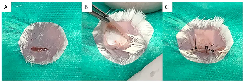
chlorhexidine solution (Figure 1).
Figure 1 - Sequence of surgical procedures performed. (A). Trichotomy, skin antisepsis of the back region of the animals and incision of approximately 1cm in
length on the back region of the animals, according to ISO 10993-6 / 2016; (B).
After divulsion of the cutaneous tissue, a fragment of 1cm membrane was
implanted and tested according to the experimental groups and (C). Replacement of cutaneous tissue over the implanted membrane and suture with 5-0 mononylon
(Ethicon®, Johnson & Johnson, Brazil).
In the postoperative period, the animals were kept in the Animal
Experimentation Laboratory (AEL/UFF) and divided in isolaters based on their
experimental groups, where they received food and water at will. On the day of
surgery and on each of the following two days, 5 mg/kg of Meloxicam (Eurofarma
Laboratórios LTDA, São Paulo, SP, Brazil) was administered subcutaneously
every 24 hours.
The mice were euthanized after the respective experimental periods with
tissue (± 5 mm with safety margins) were collected, fixed, decalcified, dehydrated,
clarified and included in paraffin to obtain 5 μm thickness. The slices were stained
with Hematoxylin and Eosin (HE) and observed using a light field microscope at
40X magnification (Nikon Eclipse E400, Tokyo, Japan). These images were
captured by a high resolution digital camera (Sony® HD DSC HX9V 16.2 Mega
Pixels) using a 10x acroplan objective lens at the Laboratory of Applied
Biotechnology – UFF (LABA) for descriptive and semiquantitative histological
evaluation for the presence of inflammatory infiltrate, vascular neoformation,
extension and type of necrosis, presence of fatty infiltrate and fibrosis and
membrane bioabsorption.
3. RESULTS
3.1. Structure and characteristics of the PLGA membrane
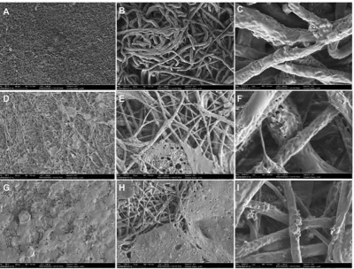
The synthetic PLGA membrane is shown in Figure 2. The membrane
exhibited a micro-fibrous aspect to favor guide bone regeneration. The difference
in the morphology between the different membranes thickness is clearly visible
on the 20.000X magnification observed by SEM.
The micro-fibrous surface exhibited a highly porous structure with
interlaced non-woven fibres. The diameters of the fibres ranged from 0.2 to 2 μm
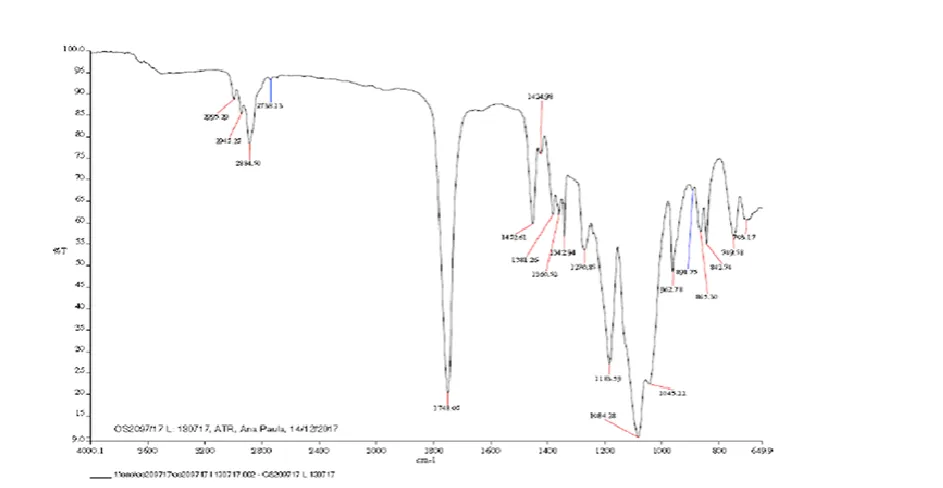
(Figure 2). The XRD evaluation showed two crystallines mineral phases present
in the membranes (HA and -TCP), and the proportion of the hydroxyapatite
(54.2%) and β-tricalcium phosphate phases (45.8%) (Figure 3). The carbon,
hydrogen and nitrogen contents were quantified according the Table 1. It was
observed the mean value for Carbon (39.4%), Hydrogen (4.5%) and Nitrogen
(0.1%). The FTIR showed that samples showed predominant absorptions of poly
Figure 3 - X-ray diffraction pattern (XRD) of membrane sample. Observe the peak representing hydroxyapatite and -TCP. The quantitative analysis showed
the presence of two phases: 45.8% -TCP and 54.2% HA.
3.2. In vivo implantation
Overall, the animals tolerated well the anesthesia, pre-surgery and
post-surgery, without complications or setbacks. Biological effects after the
implantation of the different experimental membranes were evaluated according
to the criteria established by ISO 10993-6: 2016 / Part 6 / Annex E and the
descriptive analysis of the tissue response to the membranes was evaluated as
a function of tissue disposition in the different membranes: bioabsorption,
presence of inflammatory cells, vascular neoformation and presence of fibrosis.
The degree of inflammation was evaluated and quantified manually according to
the number and distribution of inflammatory cells present at the membrane-tissue
interface, that is, polymorphonuclear cells, lymphocytes, plasma cells,
macrophages and giant cells. In addition, the degree of degeneration (debris)
was determined by morphological alterations necrosis extension.
For the 7-day experimental period, small vascular neoformation with
minimal capillary proliferation (presence of 1 to 3 bulbs per examined field) was
observed in Group Sham. Also found were a cicatricial process with an absence
of multinucleated giant cells and macrophages and an abundance of lymphocytes
and plasmocytes, in addition to the moderate presence of polymorphonuclear
cells. In Group 2 (200μm), a moderate infiltration of lymphocytes, macrophages
and multinucleated giant cells was observed, along with a slight presence of
polymorphonuclear cells. Moderate vascular neoformation with capillary
proliferation was also present (presence of a wide range of capillaries per
examined field).
In Group 3 (500 μm), a moderate infiltration of lymphocytes and
cells were observed, as were mild vascular neoformation with proliferation of
capillaries. Group 4 (700 μm) presented integral membranes with a moderate
presence of lymphocytes, plasma cells and macrophages and a discrete
polymorphonuclear infiltration. Local neovascularization with small capillary
proliferation was also observed in this group. The control Group (Pratix ®) did not
visualize the membrane in any cuts performed; therefore, the cellular response
tissue of the periphery membrane was evaluated. There was a moderate
presence of lymphocytes, macrophages and polymorphonuclear infiltration. Also
observed in this group was moderate local neovascularization with small capillary
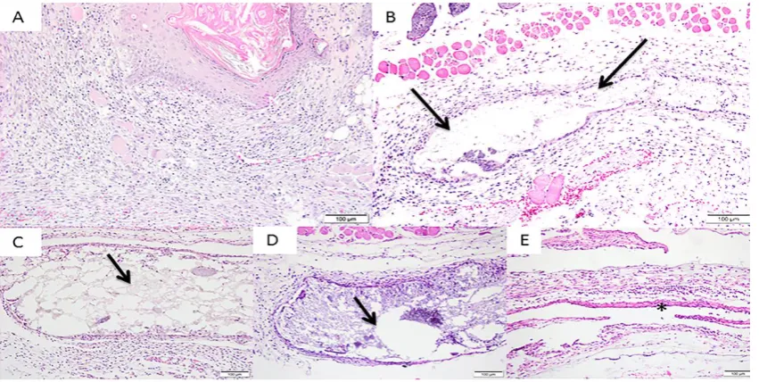
proliferation (Figure 5).
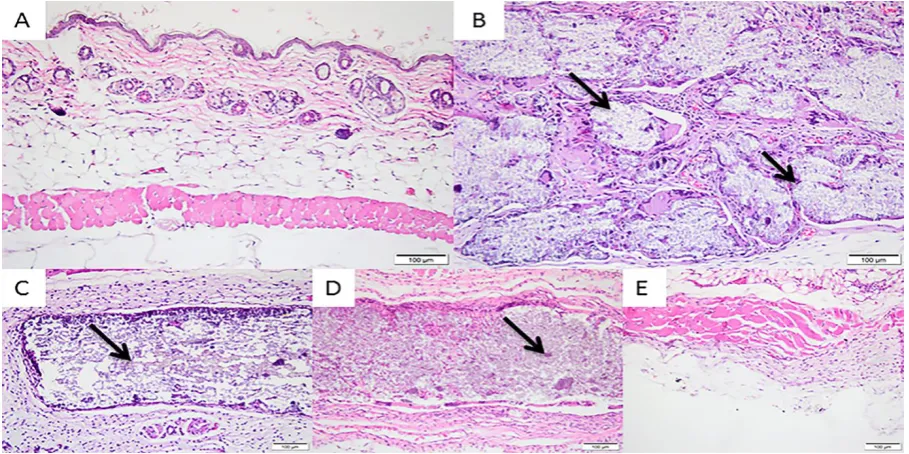
Figure 5 - Photomicrographs of the experimental groups after 7 days of implantation. Group 1 (Sham) – (A); Group 2 (200 μm) - (B); Group 3 (500 μm)
- (C); Group 4 (700 μm) – (D); and Group 5 (Pratix®) – (E). Observe the presence of the fragment-free membrane in all groups. Membranes; * Pratix®
In the 30-day experimental period, a discrete presence of lymphocytes and
plasma cells and a moderate presence of macrophages were observed in Group
Sham. Regarding tissue response, a discrete presence of vascular neoformation
with few vascular proliferations characterized this sample. In Group 2 (200 μm),
membranes began to exhibit fragmentation with tissue invasion. There was a
moderate presence of multinucleated giant cells and macrophages, a discrete
presence of lymphocytes with an absence of polymorphonuclear cells and mild
local neovascularization.
The membranes of the 500 μm and 700 μm Groups showed no changes
in their integrity after 30 days. The discrete presence of lymphocytes, plasma
cells, macrophages and a few multinucleated giant cells, with a moderate tissue
response of local neovascularization, was observed in Group 3. Group 4 had a
discrete presence of lymphocytes and plasma cells, a moderate presence of
multinucleated giant cells and few polymorphonuclear cells. Regarding tissue
response, there was moderate vascular neoformation with proliferation of more
than 7 shoots per field. The Pratix® membranes used as a control group did not
adhere to the subcutaneous tissue, so during the removal of the samples they
were detached from the tissue and the tissue adjacent to the implantation site of
this membrane was observed, revealing a discrete lymphoplasmocytic infiltration
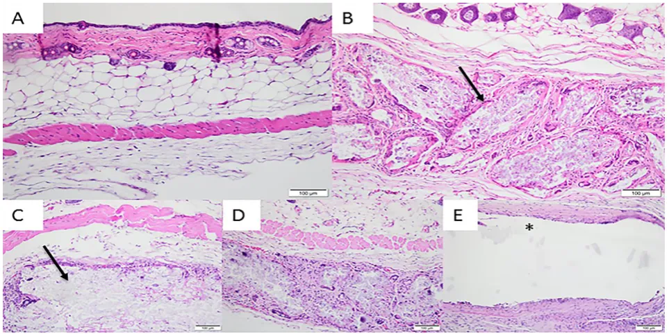
Figure 6 - Photomicrographs of the experimental groups after 30 days of implantation. Group 1 (Sham) – (A); Group 2 (200 μm) – (B); Group 3 (500 μm)
– (C); Group 4 (700 μm) – (D); and Group 5 (Pratix®) – (E). Observe the presence in the 200 μm membrane of fragmented and non-fragmented aspects in the 500
μm and 700 μm groups. Membranes; * Pratix® membrane site;
Magnification: 10 X and 20X, coloring: Hematoxylin and Eosin.
After 60 days, a discrete presence of lymphocytes, plasma cells and
macrophages was observed in the Sham Group. There were no multinucleated
giant cells or polymorphonuclear cells with a slight presence of vascular
neoformation shoots. In Group 2, there was a moderate presence of lymphocytes,
macrophages and multinucleated giant cells, a discrete presence of plasma cells
and an absence of polymorphonuclear cells. Groups 3 and 4 presented a discrete
/ moderate presence of lymphocytes, macrophages, multinucleated giant cells
and plasma cells. However, while Group 3 presented mild vascular neoformation,
macroscopically detached from the tissue, without connection to the
subcutaneous tissue. The presence of multinucleated and polymorphonuclear
giant cells was not observed and there was only a moderate presence of
lymphocytes and macrophages (Figure 7).
Figure 7 - Photomicrographs of the experimental groups after 60 days of implantation. Group 1 (Sham) – (A); Group 2 (200 μm) – (B); Group 3 (500 μm) – (C); Group 4 (700 μm) – (D); and Group 5 (Pratix®) – (E). Membranes; * Pratix® membrane site; Magnification: 10 X and 20X, coloring: Hematoxylin and
Eosin.
After the 90-day period, Group 1 showed discrete lymphocyte and
macrophages infiltration, and an absence of multinucleated giant cells and
polymorphonuclear cells. The 200 μm group presented a moderate presence of
macrophages, lymphocytes and multinucleated giant cells and a discrete
presence of plasma cells. In the 500 μm and 700 μm groups, there was no
multinucleated giant cells and macrophages. The control group had a discrete
presence of polymorphonuclear cells and a moderate presence of macrophages,
multinucleated giant cells, lymphocytes and plasma cells. All presented moderate
vascular neoformation, with the exception of Group 1, which had discrete
neoformation (Figure 8).
Figure 8 - Photomicrographs of the experimental groups after 90 days of implantation. Group 1 (Sham) – (A); Group 2 (200 μm) – (B); Group 3 (500 μm)
– (C); Group 4 (700 μm) – (D); and Group 5 (Pratix®) – (E). Membranes; * Pratix® membrane site; Magnification: 10 X and 20X, coloring: Hematoxylin and
Eosin.
No areas of necrosis, fibrosis or fatty infiltration were present for all
experimental periods.
According to ISO, at the end of the evaluation test samples may present
scores ranging from 0.0 to 2.9 (absence of tissue reaction), 3.0 to 8.9 (discrete
tissue reaction). Thus, after evaluation of each cell type and tissue within the
conditions of this study, following ISO 10993-6/2016, it was observed that the test
sample of 200 μm showed a slight tissue reaction (4) compared to the control
sample after 7 days of implantation, whereas the test samples of 500 μm (2,6)
and 700 μm (0) showed no tissue reactions after 7 days of deployment. After 30
days, the tested membrane of 700 μm showed a slight tissue reaction (4.4) when
compared to the control sample, whereas the 200 μm (1,2) and 500 μm (1,8)
samples did not present a tissue reaction. After 60 days, the 200 μm membrane
presented a slight tissue reaction (7) when compared to the control group. There
was an absence of tissue reaction for the membranes of 500 μm (2,6) and 700
μm (3,2). After 90 days of implantation, the membrane of 700 μm showed tissue
reaction (3.8), while the membrane of 200 μm obtained a score of 0.6 and the
membrane of 500 μm obtained a score of 0, characterizing an absence of tissue
reaction.
4. DISCUSSION
For membranes to be used effectively in the GTR process, they must have
biocompatibility characteristics, occlusive properties, capacity to maintain space,
tissue integration and have easy clinical manageability [12,13]. The desirable
characteristics of the membrane barriers used for guide bone regeneration
therapy include biocompatibility to allow integration into host tissues without
creating an inflammatory response, a degradation profile accompanying tissue
neoformation, mechanical and physical properties sufficient to allow membrane
installation and sufficient sustained force to not collapse and to perform the
Studies demonstrate that membranes containing PLGA + HA and -TCP
can contribute mainly due to their mechanical properties, thus improving
structural integrity and flexibility [15,16]. In addition, the release of calcium and
phosphorus ions during the degradation of HA and -TCP may be involved in
bone metabolism and promote the formation of new bone [17]. Sanaei-Rad et al.
[18] demonstrated that HA improved osteogenic properties, strength and
structural stability. Seyedjafari et al. [19] evaluated the incorporation of
nanohydroxyapatite on the surface of an electrospun poly (l-lactide)-PLLA
associated with human cord blood-derived unrestricted somatic stem cells, in
vitro under osteogenic induction and in vivo after subcutaneous implantation, by
demonstrating adequate mechanical properties and improving the osteogenic
differentiation of somatic stem cells.
The membranes tested did not present moderate or severe tissue
reactions in the experimental periods studied, not differing from the patterns
presented by the control group. Therefore, they are biocompatible. The present
results demonstrate that all membranes were intact after 7 days of implantation,
but after 30 days the 200 μm membrane was partially fragmented and lower
mechanical stability was observed, while the membranes of 500 μm and 700 μm
started the process of fragmentation only after 90 days of implantation. The 200
μm membrane showed a slight tissue reaction after 7 and 60 days, while the
membrane of 700 μm showed a slight tissue reaction after 30 and 90 days. The
500 μm membrane did not show tissue reaction in any experimental periods
studied. To play its role as a barrier, absorbable membranes should remain for at
Therefore, we verified that all thicknesses of membranes are
biocompatible to subcutaneous connective tissue of mice; however, the
membrane of 200 μm presented a faster absorption when compared to those of
another thickness and to the control group. The membrane of 500 μm only started
fragmentation after 90 days, an ideal time to exclude mechanically
non-osteogenic and non tissue response cell populations, thus making it a good
thickness for this type of membrane.
Particles of thermally deproteinized inorganic bone (hydroxyapatite) have
been demonstrated to induce a chronic inflammatory reaction with the presence
of giant cells and fibrosis of the particles in the subcutaneous tissue of rats²¹.
Therefore, it is reasonable to suggest that the recruitment of giant cells may be
related to the persistence of remnants of the mineral phase on the membrane.
5. CONCLUSION
With the limitations of this preclinical study, it is possible to conclude that
the tested membranes are well tolerated by the tissues, are not completely
resorbed at 90 days. Absorbable membranes derived from PLGA + HA + β-TCP
could be used as barriers to promote tissue regeneration in surgical techniques
of guided tissue regeneration, but further in vivo (bone repair model) and clinical
studies should be conducted to determine the clinical efficacy of these
membranes.
AUTHOR CONTRIBUTIONS
Conceptualization - Lívia da Costa Pereira, Mônica Diuana Calasans-Maiaand Rafael Seabra Louro.
Histomorphological analysis - Adriana Terezinha Neves Novellino Alves.
Writing - original draft preparation – Lívia da Costa Pereira.
Supervision – Rafael Seabra Louro and Monica Diuana Calasans-Maia. Writing - review and editing - Carlos Fernando de Almeida Barros Mourão.
FUNDING
This research received no external funding.
ACKNOWLEDGMENTS
This work was supported by the FGM Materiais Odontológicos LTDA (Joinville,
Santa Catarina, Brazil) for the supply of materials.
CONFLICTS OF INTEREST
The authors declare no conflict of interest.
REFERENCES
1. Melcher AH. On the repair potential of periodontal tissues. J Periodontol
1976,47,256-260. DOI: 10.1902/jop.1976.47.5.256
2. Arunjaroensuk S; Panmekiate S; Pimkhaokham A. The Stability of Augmented
Bone Between Two Different Membranes Used for Guided Bone Regeneration
Simultaneous with Dental Implant Placement in the Esthetic Zone. Int J Oral
Maxillofac Implants 2017,33,206–216.DOI: 10.11607/jomi.5492
3. Benic GI; Hammerle CH. Horizontal bone augmentation by means of guided
bone regeneration. Periodontol 2000 2014,66,13-40. DOI: 10.1111/prd.12039
4. Schwarz F; Herten M; Ferrari D; Wieland M; Schmitz L; Engelhardt E; Becker
J. Guided bone regeneration at dehiscence-type defects using biphasic
collagen-coated natural bone mineral (BioOss Collagen®): an immunohistochemical study
in dogs. Int J Oral Maxillofac Surg 2007,36,1198-1206.
5 Guda T; Walker JA; Singleton BM; Hernandez JW; Son JS; Kim SG; Oh DS;
Appleford MR; Ong JL; Wenke JC. Guided bone regeneration in long-bone
defects with a structural hydroxyapatite graft and collagen membrane. Tissue
Eng Part A 2013,19,1879-1888. DOI: 10.1089/ten.TEA.2012.0057
6. Peng SW; Li CW; Chiu IM; Wang GJ. Nerve guidance conduit with a hybrid
structure of a PLGA microfibrous bundle wrapped in a micro/nanostructured
membrane. Int J Nanomedicine 2017,11,421-432. DOI: 10.2147/IJN.S122017
7. Shao HJ; Chen CS; Lee IC; Wang JH; Young TH. Designing a
three-dimensional expanded polytetrafluoroethylene-poly(lactic-co-glycolic acid)
scaffold for tissue engineering. Artif Organs 2009,33,309-317. DOI:
10.1111/j.1525-1594.2009.00721.x
8. Mao D; Li Q; Bai N; Dong H; Li D. Porous stable poly(lactic acid)/ethyl
cellulose/hydroxyapatite composite scaffolds prepared by a combined method for
bone regeneration. Carbohydr Polym 2018,15,104-111. DOI:
10.1016/j.carbpol.2017.10.031
9. Lee SW; Kim SG. Membranes for the Guided Bone Regeneration. Maxillofac
10. Fu L; Wang Z; Dong S; Cai Y; Ni Y; Zhang T; Wang L; Zhou Y. Bilayer
Poly(Lactic-co-glycolic acid)/Nano-Hydroxyapatite Membrane with Barrier
Function and Osteogenesis Promotion for Guided Bone Regeneration. Materials
(Basel) 2017,3,10(3). DOI: 10.3390/ma10030257
11. Balmain N; Legros R; Bonel G. X-ray diffraction of calcined bone tissue:
a reliable method for the determination of bone Ca/Pmolar ratio.
Calcif Tissue Int 1982,34,s93-98.
12. Jin Q; Wang XM; Wang XF; Li XD; Ma JF. The efficacy of
collagen-hydroxyapatite composite membrane on bone regeneration. Hua Xi Kou Qiang
Yi Xue Za Zhi 2011,29,21-26.
13. Buser D; Dula K; Hirt HP; Schenk RK. Lateral ridge augmentation using
autografts and barrier membranes: a clinical study with 40 partially edentulous
patients. J Oral Maxillofac Surg 1996,54,420-432.
14. Hwang KS; Choi JW; Kim JH; Chung HY; Jin S; Shim JH; Yun WS; Jeong
CM; Huh JB. Comparative Efficacies of Collagen-Based 3D Printed
PCL/PLGA/beta-TCP Composite Block Bone Grafts and Biphasic Calcium
Phosphate Bone Substitute for Bone Regeneration. Materials (Basel) 2017,10(4).
DOI: 10.3390/ma10040421
15. Chen G; Xia Y; Lu X; Zhou X; Zhang F; Gu N. Effects of surface
functionalization of PLGA membranes for guided bone regeneration on
proliferation and behavior of osteoblasts. J Biomed Mater Res A 2013,101,44-53.
16. Knecht S; Erggelet C; Endres M; Sittinger M; Kaps C; Stussi E. Mechanical
testing of fixation techniques for scaffold-based tissue-engineered grafts. J
Biomed Mater Res B Appl Biomater 2007,83,50-57.
17. Deng XL; Sui G; Zhao ML; Chen GQ; Yang XP. Poly(L-lactic
acid)/hydroxyapatite hybrid nanofibrous scaffolds prepared by electrospinning. J
Biomater Sci Polym Ed 2007,18,117-130.
18. Sanaei-Rad P; Jafarzadeh Kashi TS; Seyedjafari E; Soleimani M.
Enhancement of stem cell differentiation to osteogenic lineage on
hydroxyapatite-coated hybrid PLGA/gelatin nanofiber scaffolds. Biologicals 2016,44,511-516.
DOI: 10.1016/j.biologicals.2016.09.002
19. Seyedjafari E; Soleimani M; Ghaemi N; Shabani I.
Nanohydroxyapatite-coated electrospun poly(l-lactide) nanofibers enhance osteogenic differentiation
of stem cells and induce ectopic bone formation. Biomacromolecules
2010,8,3118-3125. DOI: 10.1021/bm1009238
20. Minabe M. A critical review of the biologic rationale for guided tissue
regeneration. J Periodontol 1991,62,171-179. DOI: 10.1902/jop.1991.62.3.171
21. Oliveira RC; Menezes R; Cestari TM; Taga EM; Taga R; Buzalaf MAR;
Granjeiro JM. Tissue response to a membrane of demineralized bovine cortical
bone implanted in the subcutaneous tissue of rats. Braz Dent J 2004,15,3-8. DOI: