CXCL14 expression during
chick embryonic development
CHRISTOPHER T. GORDON*, CHRISTINE WADE, INIGO BRINAS and PETER G. FARLIE
Craniofacial Development Laboratory, Murdoch Children’s Research Institute, Royal Children’s Hospital,Parkville, Australia
ABSTRACT Chemokines are small secreted signalling molecules best known for their roles as chemoattractants for cells of the immune system. CXCL12 and its receptor CXCR4 comprise one chemokine signalling pathway with essential functions in non-immune cell types during embryonic development. CXCL14, a chemokine-encoding gene related to CXCL12, is developmentally regulated in zebrafish and Xenopus embryos, but its role during embryogenesis remains unknown. Here we describe the embryonic expression pattern of CXCL14 in an amniote, the chick. Although expression in some regions is conserved with that of fish and frog, chick CXCL14 displays a complex pattern of expression in several novel sites. We analyse the expression pattern in the branchial arches, trige-minal placode and ganglion, inner ear, dorsal midline of the brain, somites, trunk neural tube and limb bud. Expression in several domains raises the possibility that CXCL14 may be involved in some of the same developmental events during which CXCL12-CXCR4 signalling is known to play a role.
KEY WORDS:
chemokine, trigeminal, mesencephalon, somite, otic vesicle.
Chemokines are a large family of secreted proteins that have his-torically been investigated in the context of immune cell trafficking. In this paradigm, chemokines act as chemoattractants, recruiting immune cells that express G-protein coupled chemokine recep-tors. However, some chemokines and their receptors have roles outside the haematopoietic system. The most extensively studied chemokine system during embryonic development consists of the CXCL12 ligand and CXCR4 receptor. Initial investigation of Cxcl12 and Cxcr4 knockout mice revealed that in addition to requirements in haematopoietic cells, these genes had essential roles in cardiac, cerebellar and vascular development (Ma et al., 1998; Nagasawa et
al., 1996; Tachibana et al., 1998; Zou et al., 1998). Subsequently, it
was demonstrated that the cxcl12-cxcr4 pathway was required for migration of primordial germ cells in zebrafish and mice (Doitsidou
et al., 2002; Knaut et al., 2003; Molyneaux et al., 2003), for
migra-tion of the lateral line primordium in zebrafish (Dambly-Chaudiere
et al., 2007; David et al., 2002; Haas and Gilmour, 2006; Valentin et al., 2007), and for coordinating migration of germ layers during
zebrafish gastrulation (Nair and Schilling, 2008). In the mouse cerebral cortex and cerebellum, positioning of Cajal-Retzius cells and granule cells, respectively, is regulated by Cxcl12 and Cxcr4 (Borrell and Marin, 2006; Ma et al., 1998; Paredes et al., 2006; Reiss et al., 2002; Zhu et al., 2002). Roles for Cxcl12 and Cxcr4
www.intjdevbiol.com
*Address correspondence to: Christopher T. Gordon. INSERM U781, Tour Lavoisier – Deuxième Etage, Hôpital Necker-Enfants Malades, 149 Rue de Sèvres, 75743
Paris Cedex 15, France. E-mail: [email protected]
Supplementary Material (four figures) for this paper is available at: http://dx.doi.org/10.1387/ijdb.103258cg
Accepted: 11 January 2011. Final, author-corrected PDF published online: 15 June 2011.
ISSN: Online 1696-3547, Print 0214-6282 © 2011 UBC Press
Printed in Spain
Abbreviations used in this paper: AER, apical ectodermal ridge; HH, Hamburger-Hamilton.
during migration of muscle progenitors (Vasyutina et al., 2005) and cranial neural crest migration (Olesnicky Killian et al., 2009; Theveneau et al., 2010) have also been described. A common theme in most of these examples is expression of Cxcl12 in a tissue through which Cxcr4-expressing cells migrate, while in some cases Cxcl12 is required to enable Cxcr4-positive cells to maintain a fixed position relative to the ligand (reviewed in Raz and Mahabaleshwar, 2009).
Initial characterisation of CXCL14 indicated that although it was widely expressed in normal tissues (as assessed by Northern blotting), its expression was reduced in tumour cell lines and tumour-derived tissue (Cao et al., 2000; Frederick et al., 2000; Hromas et al., 1999; Sleeman et al., 2000). Also, forced expression in mice has demonstrated that Cxcl14 suppresses tumour growth (Izukuri et al., 2010; Ozawa et al., 2006; Tessema et al., 2010). On the other hand, CXCL14 can act as an autocrine stimulator of fibroblast growth and migration (Augsten et al., 2009). Although the identity of its receptor is unknown, CXCL14 has been shown to function as a chemoattractant for some immune cell types (Kurth et
et al., 2005; Starnes et al., 2006). Cxcl14 knockout mice have
defects in feeding behaviour (Tanegashima et al., 2010) and in glucose metabolism (Nara et al., 2007), but have a normal immu-ne system (Meuter et al., 2007). Both groups that have knocked out Cxcl14 reported that a lower than expected number of null mice were recovered in the perinatal period (Meuter et al., 2007; Nara et al., 2007; Tanegashima et al., 2010). Although no major phenotypic abnormalities were noted, this finding may indicate a subtle developmental defect that initiates during embryonic stages. Embryonic functions for Cxcl14 remain unexplored. In zebrafish embryos, cxcl14 is expressed transiently in discrete domains of the brain and otic vesicle, and in cells of the anterior and posterior lateral line (Long et al., 2000). In Xenopus, CXCL14 is expressed in specific regions of cranial ectoderm, and in the dorsal retina and head mesenchyme (Park et al., 2009). Park et al., (2009) noted that the only region of potential overlap between Xenopus
and zebrafish CXCL14 expression was within the otic vesicle. In this report, we examine the embr-yonic CXCL14 expression pattern in an amniote, the chick, and compare it to that reported for ze-brafish and Xenopus. Our results indicate some similarities in expression amongst these species, but several sites of expression not reported in the anamniotes. Some of the developmental events during which chick CXCL14 expression is observed raise the intriguing possibility that this chemokine may function in parallel to the CXCL12-CXCR4 ligand-receptor system.
Results and Discussion
Branchial arches
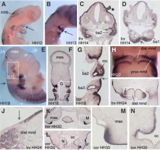
We examined CXCL14 expression in chick by whole-mount and section in situ hybridisation at several stages during embryogenesis. During craniofacial development, CXCL14 is regionally expressed in both branchial arch epithelium and cranial neural crest-derived mesenchyme (Fig. 1). At early stages of first branchial arch expansion,
CXCL14 is expressed within mandibular epithelium
(Fig.1 A,B,D); this domain increases between Hamburger-Hamilton (HH) stages 12 and 13 (Fig. 1 A,B). In Xenopus, CXCL14 is expressed specifically in the cement gland, a thickened layer of ectoderm immediately ventral to the prospective oral opening (Park et al., 2009). This epithelial expression on the ventral side of the mouth in both species may indi-cate homologous regulation of expression. In the second branchial arch, CXCL14 is widely expres-sed in mesenchymal but not epithelial cells, while dorsal ectoderm at the same axial level strongly expresses CXCL14 (Fig. 1C). At HH18, expression is greatly reduced in cranial surface ectoderm relative to that posterior to the otic vesicle (Fig. 1E). This difference is also evident at the junction of surface ectoderm covering the second and third branchial arches (Fig. 1 F,G). Between HH18-24,
CXCL14 expression in mandibular mesenchyme is
more extensive proximally than distally (Fig. 1 H,I
Fig. 1. Expression of CXCL14 during development of the branchial arches. (A) HH12. (B)
HH13. (C) HH14, transverse section through the second branchial arch. (D) HH14, transverse section through the first branchial arch. Arrows in (A,B,D) indicate expression in first branchial arch epithelium. (E-G) HH18. White dashed lines in (E) indicate the approximate plane of sectioning through the mesencephalon and diencephalon in Fig. 2 A,B, respectively. The boxed region in (E) is shown at higher magnification in Supplementary Fig. 1C. (F) Coronal section. (G) Higher magnification image of the region boxed in (F). Black arrows in (E,G) indicate the boundary of surface ectoderm expression between the 2nd and 3rd branchial
arches. (H) HH22. (I,J) HH24, transverse section. (J) Higher magnification image of the box in (I). The arrow in (J) indicates a mesenchymal-epithelial boundary of expression in the distal mandible. (K,L,M,N) HH30, coronal sections. (M,N) Higher magnification images of the boxed regions in (K,L), respectively. Asterisks indicate staining artifacts. mhb, midbrain-hindbrain boundary; ba1, 1st branchial arch; ba2, 2nd branchial arch; ba3, 3rd branchial arch;
mes, mesencephalon; di, diencephalon; mc, mesodermal core; dist mnd, distal mandible; prox mnd, proximal mandible; max, maxilla; oc, oral cavity; trv, transverse; cor, coronal.
and data not shown). Interestingly, the domain of mesenchymal expression along the proximodistal axis of the mandible ceases at a point where expression in the distal mandibular epithelium begins (Fig. 1 I,J). Note also there are scattered positive cells within the mesodermal core (Fig. 1 F,G); a similar pattern has been observed for some genes expressed within the neural crest cells that invade this tissue (Grenier et al., 2009). In first branchial arch derivatives at HH30, a stage subsequent to the initiation of skeletal differentia-tion, CXCL14 continues to be expressed in limited domains of the upper and lower jaws, including a maxillary mesenchyme domain at the junction of expressing and non-expressing epithelia (Fig. 1 K,M), and a thin layer of mesenchyme immediately subjacent to lateral oral epithelium (Fig. 1 L,N).
Trigeminal placode and ganglion
In surface ectoderm between HH7-9, CXCL14 is expressed
G
B
C
D
E
F
H
I
J
K
A
M N
in caudal regions of the embryo and as far anterior as the region overlying the caudal hindbrain, but is absent from cranial surface ectoderm during the same period (Supplementary Fig. 3A and data not shown). By HH12 however, expression is observed in scattered cells in a bilateral swathe adjacent to the midbrain (Fig. 1A and Supplementary Fig. 1A); this distribution is reminiscent of expression of PAX3, a marker of the ophthalmic trigeminal placode (Stark et al., 1997). Transverse sections through this region at HH13 indicate that the positive cells are indeed within the surface ectoderm, rather than within underlying mesenchyme (Supplemen-tary Fig. 1B). Between HH17-22 CXCL14 is expressed in cells of both the ophthalmic and maxillomandibular lobes of the trigeminal placode and ganglion (Supplementary Fig. 1 C-I). At HH17, expres-sion is seen in streams of cells ingressing from the placode into the ganglion (Supplementary Fig. 1D). By HH21, portions of the trigeminal ganglion retain a connection to the surface ectoderm, via CXCL14-expressing cells, while small clumps of cells in the surface ectoderm continue to express CXCL14 without an obvious connection to the ganglion (Supplementary Fig. 1E). By comparing the distribution of ectodermal cells overlying the trigeminal ganglion
Xenopus CXCL14 (Long et al., 2000; Park et al., 2009), however in Xenopus, the transcription factor PAX3 is necessary and sufficient
for CXCL14 expression in the hatching gland, a specialisation of the cranial ectoderm (Park et al., 2009). In the future it will be of interest to test whether chick CXCL14 is also downstream of PAX3 in the ophthalmic trigeminal placodal ectoderm. The lack of PAX3 expression in the maxillomandibular lobe suggests that CXCL14 would be downstream of other transcription factors in that portion of the trigeminal. In addition to the trigeminal, we observed expres-sion of CXCL14 in scattered cells of other cranial ganglia (data not shown). CXCL14 is also expressed in the anterior half of Rathke’s pouch, a placodal epithelium that develops as an outpocketing of the oral cavity (Supplementary Fig. 1J).
Otic vesicle and hindbrain
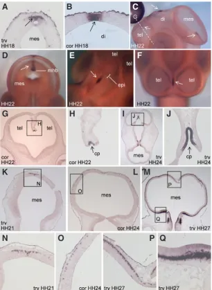
At HH13, CXCL14 is expressed in the dorsal surface ectoderm surrounding the invaginating otic vesicle, but is excluded from the thickened epithelium of the vesicle itself (Supplementary Fig. 2A and 3B). However by HH22 two patches of expression can be discerned at the anterior and posterior limits of the otic vesicle
Fig. 2. Expression of CXCL14 in the dorsal mesencepha-lon, diencephalon and telencephalon. (A,B) HH18. (A) Transverse section through the caudal mesencephalon; (B) coronal section through the diencephalon, at the positions indicated in Fig. 1E. (C-H) HH22. White dashed line in (C) indicates the approximate plane of sectioning through the telencephalon in (G). In (D), the rostral mesencephalon has been removed to allow visualisation of CXCL14 expression in the dorsal midline of the caudal mesencephalon. Arrows in (A-F) indicate expression in the dorsal midline of the cephalic vesicles. (G,H) Coronal section; (H) is a magnified view of the box in (G). (I,J)HH24, transverse section; (J) is a magnified view of the box in (I). (K-Q) expression of CXCL14 in migratory cells of the dorsal mesencephalon. (K,N) HH21, transverse section. (N) is a magnified view of the box in (K).(L,O) HH24, coronal section. (O) Magnified view of the box in (L). (M, P, Q) HH27, transverse section. (P,Q) Magnified images of the boxed regions in (M). tel, telencephalon; di, diencephalon; mes, mesencephalon; mhb, midbrain-hindbrain boundary; epi, epiphysis; cp, choroid plexus; trv, transverse; cor, coronal. at HH18 (Supplementary Fig. 1C) and HH22 (Supple-mentary Fig. 1F), it can be seen that loose clusters of
CXCL14-expressing cells resolve into discrete clumps.
The latter typically overlie the ophthalmic branch of the trigeminal ganglion, in some cases protruding prominently from the surface (Supplementary Fig. 1G), and may correspond to ectopic ganglia described previously within the chick surface ectoderm in a similar region (Kuratani and Hirano, 1990). Cells expressing
CXCL14 in the trigeminal ganglion are mainly localised
in the distal portion of the ganglion (Supplementary Fig. 1I), which is typical of trigeminal placode-derived sensory neurons, in contrast to neural crest-derived neurons that reside more proximally (D’Amico-Martel and Noden, 1983). In zebrafish, cxcr4b is required for correct positioning of sensory neurons within the developing trigeminal ganglion (Knaut et al., 2005), and based on our expression analysis, chick CXCL14 may also be involved in assembly of this ganglion. Trigeminal expression was not reported for zebrafish or
G
O
B
C
D
E
F
H
I
J
K
L
P Q
A
M
(Supplementary Fig. 2B, and similarly at HH24 in Supplementary Fig. 2C). By HH26 a third major domain is distinct (Supplementary Fig. 2E). These three areas most likely correspond to the anterior, posterior and lateral cristae, based on comparison with published markers of these prosensory domains, such as BMP4 and SER1 (Adam et al., 1998; Cole et al., 2000; Wu and Oh, 1996). Prosensory patches give rise to the mechanotransducing hair cells and their support cells. CXCL14 may additionally be expressed within the saccular macula, another sensory patch of the vestibule (Supple-mentary Fig. 2G). Notably, zebrafish cxcl14 is also expressed specifically in sensory patches of the inner ear (Long et al., 2000), suggesting conservation of transcriptional regulation between fish and birds, in this organ.
Between HH22-26, we observed prominent expression of
CXCL14 in the hindbrain (Supplementary Fig. 2 B,D-F), with the
strongest expression occurring in a region adjacent to the otic vesi-cle. In zebrafish, cxcl14 is also expressed in specific populations of cells within the hindbrain (Long et al., 2000).
Telencephalon, diencephalon and mesencephalon
CXCL14 expression is successively activated in a caudal to
rostral fashion in the brain. At HH12-13, the sole site of CXCL14 expression in the central nervous system is a patch in the caudal-most region of the dorsal mesencephalon (Fig. 1A and Supplemen-tary Fig. 1 A,B). Although expression in the dorsal midline of the forebrain is not evident at these stages, by HH18 a small domain of expression is observed in the dorsal diencephalon (Fig. 1E and 2B), with expression in the mesencephalon continuing at this stage (Fig. 1E and 2A). By HH22 CXCL14 is expressed in the dorsal telencephalon (Fig. 2 C,F-H), with maintenance of expression in the mesencephalon and diencephalon at the same stage (Fig. 2 C-E). Central nervous system expression for Xenopus CXCL14 was not reported (Park et al., 2009), however Long et al., (2000) described zebrafish cxcl14 expression in the presumptive epiphyseal region of the diencephalon, the midbrain-hindbrain boundary, and the cerebellum. The expression domain for chick CXCL14 in the dorsal diencephalon lies immediately posterior to the epiphysis (Fig. 2E); therefore this site plausibly represents regulatory conservation between chick and zebrafish. Expression at the midbrain-hindbrain boundary also appears similar between the two species, however we did not observe cerebellar expression for chick CXCL14. Indeed, the expression at the midbrain-hindbrain boundary is confined to the midbrain side of this boundary in chick (Fig. 2D). The CXCL14 expression domain in the dorsal telencephalon may correspond to a portion of the prospective choroid plexus (Fig. 2F-J). This
CXCL14-expressing region expands between HH22 (Fig. 2H) and
HH24 (Fig. 2J).
Although at HH18 CXCL14 expression in the dorsal mesencepha-lon is confined to a small patch (Fig. 2A), we noticed that between HH21-27, individual CXCL14-expressing cells are progressively distributed more ventrally (Fig. 2 K-M; magnified images of the ventral-most cells in the respective sections are shown in Fig. 2 N,O,Q). This pattern is suggestive of expression in a tangentially migrating population. Note that the positive cells are confined to a very superficial layer throughout most of this putative migratory route (Fig. 2 O,P), but are located in a deeper layer in the most ventral region of the mesencephalon (Fig. 2Q). By HH30, CXCL14-positive cells along this entire putative migratory route also appeared to have shifted to a deeper layer within the mesencephalon (data not
shown). The expression pattern of CXCL14 in the mesencephalon is intriguing in light of known roles of Cxcl12 and Cxcr4 in neuro-nal migration. For example, in the mouse telencephalon Cxcr4 is expressed in Cajal-Retzius cells, a population that migrates tangentially within the marginal zone, while Cxcl12 is expressed in the meningeal layer underlying the marginal zone. Studies in targeted mice suggest that Cxcl12, secreted by the meninges, is required for correct positioning of Cxcr4-positive Cajal-Retzius cells. In the case of chick CXCL14, although it may seem counterintui-tive for a secreted chemokine ligand to be expressed in migrating cells, it has been reported that a chemotactic response of myeloid dendritic cells to activin A depends on the production of CXCL14 and CXCL12 from the migrating cells themselves (Salogni et al., 2009). The identity of the CXCL14-expressing cells in the mesen-cephalon awaits further investigation. The possibility that Cxcl14 has developmental or regulatory functions in the mammalian brain is supported by the recent finding that Cxcl14 knockout mice have defects in feeding behaviour when placed in a novel environment (Tanegashima et al., 2010).
Somitogenesis
At HH9-10, we observed prominent CXCL14 expression in somites and surface ectoderm, but not in endoderm, lateral plate mesoderm or neuroepithelium (Supplementary Fig. 3 A,D). During early somitogenesis, the intensity of expression within individual somites varies according to the axial level, such that expression in the oldest somites (ie, the most anterior) is lower than that in younger somites (Supplementary Fig. 3 A-C). At HH13, there is a striking rostrocaudal polarity of expression within individual somites, with expression restricted to the posterior half (Supplementary Fig. 3C). By HH18, a dorsoventral restriction within the somite is apparent, with exclusion from the dorsal and ventral lips of the dermomyoto-me (Suppledermomyoto-mentary Fig. 3E). Several other genes with polarised rostrocaudal somitic expression have been reported, including some members of the NOTCH signalling pathway (Rodrigues et al., 2006). Chick CXCL14 may therefore be regulated by this pathway. Notably, in zebrafish cxcl12a displays restriction to the posterior por-tion of early somites, similar to chick CXCL14, while the duplicated receptor genes cxcr4a and cxcr4b are expressed in the anterior portion (Chong et al., 2001; Chong et al., 2007). In contrast, chick
CXCR4 expression appears restricted to the posterior half of early
somites (Yusuf et al., 2005). Although we have observed complex somitic expression for chick CXCL14, expression of the zebrafish and Xenopus orthologues of CXCL14 was not reported in somites (Long et al., 2000; Park et al., 2009). An intriguing possibility is that different vertebrates utilise different chemokine signalling compo-nents during anteroposterior somitic patterning.
Spinal cord
studies will be required to identify the cell types expressing chick
CXCL14 in the neural tube.
Limbs
Although CXCL14 expression is widespread in the dorsal and ventral surface ectoderm of the limb bud, it is excluded from the apical ectodermal ridge (AER) throughout limb bud development (Supplementary Fig. 4A,B). The AER is a specialised epithelium that provides growth and patterning signals to the underlying mes-oderm (reviewed in Fernandez-Teran and Ros, 2008); one possi-bility is that CXCL14 acts to restrict these AER functions. Within the mesenchyme of the autopod, two major bands of expression were observed; one within the anterior third of the autopod, and a thinner band immediately subjacent to the anterior-most epithelium (Supplementary Fig. 4C,D). Anterior mesenchymal patches persist through to stages at which cartilage elements have appeared, but expression was not observed in chondrocytes (Supplementary Fig. 4E,F). Note that expression is widespread in limb and head surface ectoderm at least as late as HH30 (Fig. 1L and Supplementary Fig. 4E), consistent with reports that CXCL14 is strongly expressed in the epidermis of mouse and human skin (Meuter and Moser, 2008).
Conclusions
Our analysis of embryonic chick CXCL14 expression leads to a number of testable hypotheses. Based on comparison with pu-blished expression patterns, CXCL14 is potentially downstream of several key developmental regulators. For example, CXCL14 may be regulated by PAX3 in the ophthalmic trigeminal placode, and by NOTCH signalling within early somites. These possibilities could be tested by electroporation of overexpression, dominant negati-ve, or knockdown constructs for putative upstream regulators of
CXCL14, followed by analysis of changes in expression of CXCL14,
by in situ hybridisation. Our data also highlight the possibility that CXCL14 participates in some of the embryonic events during which CXCL12-CXCR4 signalling is known to play a role. An important task will be to examine in detail the extent of overlap, temporally and spatially, between expression of CXCL14 and CXCL12 or CXCR4, in the same species. Such analysis may then raise the possibility of functional redundancy between developmental chemokine pathways, thereby requiring parallel loss of function approaches. The major puzzle confronting all studies of CXCL14 is the identity of its receptor; in the future, comparison with our expression data may serve to highlight the likelihood that a given candidate receptor is physiologically relevant. Previous work has indicated that CXCL14 can act as a tumour suppressor, or a growth factor, depending on the context. It is possible that CXCL14 may also regulate tissue growth during embryonic development. Given that several of the
CXCL14 expression sites are within tissues that have traditionally
been amenable to electroporation in the chick embryo, including the neural tube, placodal ectoderm, epithelial somites and inner ear,
CXCL14 misexpression experiments utilising in ovo electroporation
may therefore be fruitful in the study of the embryonic functions of this chemokine in the future.
Materials and Methods
The primers 5’-GAGGACGGGAACACAAGACAG (forward) and 5’-GA-GAAATCATCTTCTGCAGAGC (reverse) were used to amplify a 430 bp
fragment of CXCL14 (spanning the coding sequence of all four exons of the gene), from an embryonic chick cDNA library. The fragment was cloned into pCRII-TOPO (Invitrogen), and the presence of CXCL14 was confirmed by sequencing. Following digestion with XbaI or BamHI, digoxigenin (Roche)-labelled antisense or sense probes were generated using SP6 or T7 primers, respectively. Chicken eggs were obtained from Research Poultry Farm, Research, Victoria. Embryos were processed for whole-mount and section in situ hybridisation essentially as described (Acloque et al., 2008). Probe hybridisation was visualised by incubation with an a-digoxigenin antibody
conjugated to alkaline phosphatase (Roche) followed by addition of the colorimetric substrates NBT and BCIP (Roche). No specific signal was detected in whole embryos or sections hybridised with the sense probe, at all embryonic stages tested (data not shown).
References
ACLOQUE, H., WILKINSON, D.G. and NIETO, M.A. (2008). In situ hybridization analysis of chick embryos in whole-mount and tissue sections. Methods Cell
Biol 87: 169-185.
ADAM, J., MYAT, A., LE ROUX, I., EDDISON, M., HENRIQUE, D., ISH-HOROWICZ, D. and LEWIS, J. (1998). Cell fate choices and the expression of Notch, Delta and Serrate homologues in the chick inner ear: parallels with Drosophila sense-organ development. Development 125: 4645-4654.
AUGSTEN, M., HAGGLOF, C., OLSSON, E., STOLZ, C., TSAGOZIS, P., LEVCHENKO, T., FREDERICK, M.J., BORG, A., MICKE, P., EGEVAD, L. et al., (2009). CXCL14 is an autocrine growth factor for fibroblasts and acts as a multi-modal stimulator of prostate tumor growth. Proc Natl Acad Sci USA 106: 3414-3419.
BORRELL, V. and MARIN, O. (2006). Meninges control tangential migration of hem-derived Cajal-Retzius cells via CXCL12/CXCR4 signaling. Nat Neurosci 9: 1284-1293.
CAO, X., ZHANG, W., WAN, T., HE, L., CHEN, T., YUAN, Z., MA, S., YU, Y. and CHEN, G. (2000). Molecular cloning and characterization of a novel CXC che-mokine macrophage inflammatory protein-2 gamma chemoattractant for human neutrophils and dendritic cells. J Immunol 165: 2588-2595.
CHONG, S.W., EMELYANOV, A., GONG, Z. and KORZH, V. (2001). Expression pattern of two zebrafish genes, cxcr4a and cxcr4b. Mech Dev 109: 347-354. CHONG, S.W., NGUYET, L.M., JIANG, Y.J. and KORZH, V. (2007). The chemokine
Sdf-1 and its receptor Cxcr4 are required for formation of muscle in zebrafish.
BMC Dev Biol 7: 54.
COLE, L.K., LE ROUX, I., NUNES, F., LAUFER, E., LEWIS, J. and WU, D.K. (2000). Sensory organ generation in the chicken inner ear: contributions of bone mor-phogenetic protein 4, serrate1, and lunatic fringe. J Comp Neurol 424: 509-520. D’AMICO-MARTEL, A. and NODEN, D.M. (1983). Contributions of placodal and
neural crest cells to avian cranial peripheral ganglia. Am J Anat 166: 445-468. DAMBLY-CHAUDIERE, C., CUBEDO, N. and GHYSEN, A. (2007). Control of cell
migration in the development of the posterior lateral line: antagonistic interactions between the chemokine receptors CXCR4 and CXCR7/RDC1. BMC Dev Biol 7: 23. DAVID, N.B., SAPEDE, D., SAINT-ETIENNE, L., THISSE, C., THISSE, B., DAMBLY-CHAUDIERE, C., ROSA, F.M. and GHYSEN, A. (2002). Molecular basis of cell migration in the fish lateral line: role of the chemokine receptor CXCR4 and of its ligand, SDF1. Proc Natl Acad Sci USA 99: 16297-16302.
DOITSIDOU, M., REICHMAN-FRIED, M., STEBLER, J., KOPRUNNER, M., DORRIES, J., MEYER, D., ESGUERRA, C.V., LEUNG, T. and RAZ, E. (2002). Guidance of primordial germ cell migration by the chemokine SDF-1. Cell 111: 647-659. FERNANDEZ-TERAN, M. and ROS, M.A. (2008). The Apical Ectodermal Ridge:
morphological aspects and signaling pathways. Int J Dev Biol 52: 857-871. FREDERICK, M.J., HENDERSON, Y., XU, X., DEAVERS, M.T., SAHIN, A.A., WU, H.,
LEWIS, D.E., EL-NAGGAR, A.K. and CLAYMAN, G.L. (2000). In vivo expression of the novel CXC chemokine BRAK in normal and cancerous human tissue. Am
J Pathol 156: 1937-1950.
GRENIER, J., TEILLET, M.A., GRIFONE, R., KELLY, R.G. and DUPREZ, D. (2009). Relationship between neural crest cells and cranial mesoderm during head muscle development. PLoS One 4: e4381.
HROMAS, R., BROXMEYER, H.E., KIM, C., NAKSHATRI, H., CHRISTOPHERSON, K., 2ND, AZAM, M. and HOU, Y.H. (1999). Cloning of BRAK, a novel divergent CXC chemokine preferentially expressed in normal versus malignant cells. Biochem
Biophys Res Commun 255: 703-706.
IZUKURI, K., SUZUKI, K., YAJIMA, N., OZAWA, S., ITO, S., KUBOTA, E. and HATA, R. (2010). Chemokine CXCL14/BRAK transgenic mice suppress growth of carci-noma cell transplants. [corrected]. Transgenic Res 19: 1109-1117.
KNAUT, H., BLADER, P., STRAHLE, U. and SCHIER, A.F. (2005). Assembly of trige-minal sensory ganglia by chemokine signaling. Neuron 47: 653-666.
KNAUT, H., WERZ, C., GEISLER, R. and NUSSLEIN-VOLHARD, C. (2003). A zebrafish homologue of the chemokine receptor Cxcr4 is a germ-cell guidance receptor. Nature 421: 279-282.
KURATANI, S.C. and HIRANO, S. (1990). The appearance of trigeminal ectopic ganglia within the surface ectoderm in the chick embryo. Arch Histol Cytol 53: 575-583. KURTH, I., WILLIMANN, K., SCHAERLI, P., HUNZIKER, T., CLARK-LEWIS, I. and
MOSER, B. (2001). Monocyte selectivity and tissue localization suggests a role for breast and kidney-expressed chemokine (BRAK) in macrophage development.
J Exp Med 194: 855-861.
LIEM, K.F., JR., TREMML, G. and JESSELL, T.M. (1997). A role for the roof plate and its resident TGFbeta-related proteins in neuronal patterning in the dorsal spinal cord. Cell 91: 127-138.
LONG, Q., QUINT, E., LIN, S. and EKKER, M. (2000). The zebrafish scyba gene encodes a novel CXC-type chemokine with distinctive expression patterns in the vestibulo-acoustic system during embryogenesis. Mech Dev 97: 183-186. MA, Q., JONES, D., BORGHESANI, P.R., SEGAL, R.A., NAGASAWA, T.,
KISHIMO-TO, T., BRONSON, R.T. and SPRINGER, T.A. (1998). Impaired B-lymphopoiesis, myelopoiesis, and derailed cerebellar neuron migration in CXCR4- and SDF-1-deficient mice. Proc Natl Acad Sci USA 95: 9448-9453.
MEUTER, S. and MOSER, B. (2008). Constitutive expression of CXCL14 in healthy human and murine epithelial tissues. Cytokine 44: 248-255.
MEUTER, S., SCHAERLI, P., ROOS, R.S., BRANDAU, O., BOSL, M.R., VON AN-DRIAN, U.H. and MOSER, B. (2007). Murine CXCL14 is dispensable for dendritic cell function and localization within peripheral tissues. Mol Cell Biol 27: 983-992. MOLYNEAUX, K.A., ZINSZNER, H., KUNWAR, P.S., SCHAIBLE, K., STEBLER, J.,
SUNSHINE, M.J., O’BRIEN, W., RAZ, E., LITTMAN, D., WYLIE, C. et al., (2003). The chemokine SDF1/CXCL12 and its receptor CXCR4 regulate mouse germ cell migration and survival. Development 130: 4279-4286.
NAGASAWA, T., HIROTA, S., TACHIBANA, K., TAKAKURA, N., NISHIKAWA, S., KITAMURA, Y., YOSHIDA, N., KIKUTANI, H. and KISHIMOTO, T. (1996). Defects of B-cell lymphopoiesis and bone-marrow myelopoiesis in mice lacking the CXC chemokine PBSF/SDF-1. Nature 382: 635-638.
NAIR, S. and SCHILLING, T.F. (2008). Chemokine signaling controls endodermal migration during zebrafish gastrulation. Science 322: 89-92.
NARA, N., NAKAYAMA, Y., OKAMOTO, S., TAMURA, H., KIYONO, M., MURAOKA, M., TANAKA, K., TAYA, C., SHITARA, H., ISHII, R. et al., (2007). Disruption of CXC motif chemokine ligand-14 in mice ameliorates obesity-induced insulin resistance. J Biol Chem 282: 30794-30803.
OLESNICKY KILLIAN, E.C., BIRKHOLZ, D.A. and ARTINGER, K.B. (2009). A role for chemokine signaling in neural crest cell migration and craniofacial development.
Dev Biol 333: 161-172.
OZAWA, S., KATO, Y., KOMORI, R., MAEHATA, Y., KUBOTA, E. and HATA, R. (2006). BRAK/CXCL14 expression suppresses tumor growth in vivo in human oral carcinoma cells. Biochem Biophys Res Commun 348: 406-412.
PAREDES, M.F., LI, G., BERGER, O., BARABAN, S.C. and PLEASURE, S.J. (2006). Stromal-derived factor-1 (CXCL12) regulates laminar position of Cajal-Retzius cells in normal and dysplastic brains. J Neurosci 26: 9404-9412.
PARK, B.Y., HONG, C.S., SOHAIL, F.A. and SAINT-JEANNET, J.P. (2009). Deve-lopmental expression and regulation of the chemokine CXCL14 in Xenopus. Int
J Dev Biol 53: 535-540.
RAZ, E. and MAHABALESHWAR, H. (2009). Chemokine signaling in embryonic cell migration: a fisheye view. Development 136: 1223-1229.
REISS, K., MENTLEIN, R., SIEVERS, J. and HARTMANN, D. (2002). Stromal cell-derived factor 1 is secreted by meningeal cells and acts as chemotactic factor
on neuronal stem cells of the cerebellar external granular layer. Neuroscience 115: 295-305.
RODRIGUES, S., SANTOS, J. and PALMEIRIM, I. (2006). Molecular characterization of the rostral-most somites in early somitic stages of the chick embryo. Gene
Expr Patterns 6: 673-677.
SALOGNI, L., MUSSO, T., BOSISIO, D., MIROLO, M., JALA, V.R., HARIBABU, B., LOCATI, M. and SOZZANI, S. (2009). Activin A induces dendritic cell migration through the polarized release of CXC chemokine ligands 12 and 14. Blood 113: 5848-5856.
SCHAERLI, P., WILLIMANN, K., EBERT, L.M., WALZ, A. and MOSER, B. (2005). Cutaneous CXCL14 targets blood precursors to epidermal niches for Langerhans cell differentiation. Immunity 23: 331-342.
SHELLENBERGER, T.D., WANG, M., GUJRATI, M., JAYAKUMAR, A., STRIETER, R.M., BURDICK, M.D., IOANNIDES, C.G., EFFERSON, C.L., EL-NAGGAR, A.K., ROBERTS, D. et al., (2004). BRAK/CXCL14 is a potent inhibitor of angiogenesis and a chemotactic factor for immature dendritic cells. Cancer Res 64: 8262-8270. SHURIN, G.V., FERRIS, R.L., TOURKOVA, I.L., PEREZ, L., LOKSHIN, A., BALKIR, L.,
COLLINS, B., CHATTA, G.S. and SHURIN, M.R. (2005). Loss of new chemokine CXCL14 in tumor tissue is associated with low infiltration by dendritic cells (DC), while restoration of human CXCL14 expression in tumor cells causes attraction of DC both in vitro and in vivo. J Immunol 174: 5490-5498.
SLEEMAN, M.A., FRASER, J.K., MURISON, J.G., KELLY, S.L., PRESTIDGE, R.L., PALMER, D.J., WATSON, J.D. and KUMBLE, K.D. (2000). B cell- and monocyte-activating chemokine (BMAC), a novel non-ELR alpha-chemokine. Int Immunol 12: 677-689.
STARK, M.R., SECHRIST, J., BRONNER-FRASER, M. and MARCELLE, C. (1997). Neural tube-ectoderm interactions are required for trigeminal placode formation.
Development 124: 4287-4295.
STARNES, T., RASILA, K.K., ROBERTSON, M.J., BRAHMI, Z., DAHL, R., CHRISTO-PHERSON, K. and HROMAS, R. (2006). The chemokine CXCL14 (BRAK) stimu-lates activated NK cell migration: implications for the downregulation of CXCL14 in malignancy. Exp Hematol 34: 1101-1105.
TACHIBANA, K., HIROTA, S., IIZASA, H., YOSHIDA, H., KAWABATA, K., KATAOKA, Y., KITAMURA, Y., MATSUSHIMA, K., YOSHIDA, N., NISHIKAWA, S. et al., (1998). The chemokine receptor CXCR4 is essential for vascularization of the gastrointestinal tract. Nature 393: 591-594.
TANEGASHIMA, K., OKAMOTO, S., NAKAYAMA, Y., TAYA, C., SHITARA, H., ISHII, R., YONEKAWA, H., MINOKOSHI, Y. and HARA, T. (2010). CXCL14 deficiency in mice attenuates obesity and inhibits feeding behavior in a novel environment.
PLoS One 5: e10321.
TESSEMA, M., KLINGE, D.M., YINGLING, C.M., DO, K., VAN NESTE, L. and BE-LINSKY, S.A. (2010). Re-expression of CXCL14, a common target for epigenetic silencing in lung cancer, induces tumor necrosis. Oncogene 29: 5159-5170. THEVENEAU, E., MARCHANT, L., KURIYAMA, S., GULL, M., MOEPPS, B., PARSONS,
M. and MAYOR, R. (2010). Collective chemotaxis requires contact-dependent cell polarity. Dev Cell 19: 39-53.
VALENTIN, G., HAAS, P. and GILMOUR, D. (2007). The chemokine SDF1a coor-dinates tissue migration through the spatially restricted activation of Cxcr7 and Cxcr4b. Curr Biol 17: 1026-1031.
VASYUTINA, E., STEBLER, J., BRAND-SABERI, B., SCHULZ, S., RAZ, E. and BIRCHMEIER, C. (2005). CXCR4 and Gab1 cooperate to control the development of migrating muscle progenitor cells. Genes Dev 19: 2187-2198.
WU, D.K. and OH, S.H. (1996). Sensory organ generation in the chick inner ear. J
Neurosci 16: 6454-6462.
YUSUF, F., REHIMI, R., DAI, F. and BRAND-SABERI, B. (2005). Expression of chemokine receptor CXCR4 during chick embryo development. Anat Embryol
(Berl) 210: 35-41.
ZHU, Y., YU, T., ZHANG, X.C., NAGASAWA, T., WU, J.Y. and RAO, Y. (2002). Role of the chemokine SDF-1 as the meningeal attractant for embryonic cerebellar neurons. Nat Neurosci 5: 719-720.
Early expression of axon guidance molecules in the embryonic chick mesencephalon and pretectum Kerry-Lyn Riley, Sarah Gledhill and Frank R. Schubert
Int. J. Dev. Biol. (2010) 54: 743-753
Developmental expression and regulation of the chemokine CXCL14 in Xenopus Byung-Yong Park, Chang-Soo Hong, Faraz A. Sohail and Jean-Pierre Saint-Jeannet Int. J. Dev. Biol. (2009) 53: 535-540
Mouse G-protein gamma3 expression in the developing CNS and neural crest cell derivatives Gregory M. Kelly, Yukio Saijoh, Ariel Finkielsztein and Steve Mangos
Int. J. Dev. Biol. (2008) 52: 1143-1150
Stromal-derived factor-1 (SDF-1) expression during early chick development
Rizwan Rehimi, Nargis Khalida, Faisal Yusuf, Fangping Dai, Gabriela Morosan-Puopolo and Beate Brand-Saberi Int. J. Dev. Biol. (2008) 52: 87-92
Isthmus organizer and regionalization of the mesencephalon and metencephalon Harukazu Nakamura and Yuji Watanabe
Int. J. Dev. Biol. (2005) 49: 231-235