Hydra, a model system to trace the emergence
of boundaries in developing eumetazoans
ANGELIKA BÖTTGER
1and MONIKA HASSEL*
,2 1Department of Biology 2, Ludwig-Maximilians-Universität München, Munich and2FB 17, Morphology and Evolution of Invertebrates, Philipps-Universität Marburg, Marburg, Germany
ABSTRACT In developing embryos, boundary formation between neighbouring groups of cells is essential to establish compartments which later fulfil specialized functions. The ability to form such boundaries has likely developed early in animal evolution - due to functional requirements imposed by the necessity to separate tissues which protect the animal, take up food or ensure propagation. Essential for boundary formation are local cues which may be provided by the in-tersection of diffusible molecules or set locally by activation of membrane-bound receptors and transcription factors. In the simple diploblastic Hydra, a representative of the basally branching metazoan Cnidaria, tissue boundaries are morphologically detectable between the body column and terminally differentiated head and foot structures. In adult polyps, these borders correspond to sharp lines of differential gene expression. They form de novo during regeneration and budding of a young polyp. Functional studies strongly suggest the involvement of FGFR/Notch signalling in the establishment of the parent-bud boundary, and it is very likely that these pathways interact with the WNT and BMP systems. How boundaries in the head and foot regions are generated is still unclear. Expression patterns of transcription factors like Cngsc, HyAlx, HyBra, HyOtx, Prdl-a, CnNK2 and Manacle show strong position dependency and may be involved in regulating gene expres-sion on either side of the boundaries, by interpreting positional information during their formation and maintenance. Due to its simplicity, the easy accessibility to pharmacological interference and, recently, transgenesis, Hydra is an interesting prebilaterian model system to study the emergence of boundary-forming mechanisms during evolution.
KEY WORDS:
boundary, evolution, Hydra, Notch, FGFR
Introduction
All multicellular animals with their diverse body plans develop from one fertilised egg. During embryonic development cells multiply and differentiate, and specialized structures emerge from fields or layers of cells, which are initially identical. Differential gene expression induced by signalling from within these fields and from the surrounding stabilises compartments necessary for the emergence of cells with different and mutually exclusive proper-ties. The separation process requires formation of molecular and morphological boundaries, which prevent intermingling of cells with different destinies, allow stable expression of transcription factors in adjacent fields and often themselves function as new signalling centres to establish refined patterns within the respective domains (Sánchez-Camacho et al., 2005; Dahmann et al., 2011). Eventually
www.intjdevbiol.com
*Address correspondence to: Monika Hassel. FB 17, Morphology and Evolution of Invertebrates, Philipps-Universität Marburg, Karl von Frisch Str. 8, D-35032 Marburg, Germany. Tel: ++49-6421-2823408. Fax: ++49-6421-2823407. E-mail: [email protected]
Web: http://www.uni-marburg.de/fb17/fachgebiete/zoologie_tierevo/index_html
Final, author-corrected PDF published online: 5 June 2012 ISSN: Online 1696-3547, Print 0214-6282 © 2012 UBC Press
Printed in Spain
Abbreviations used in this paper: BMP, bone morphogenetic protein; FGF, fibroblast growth factor.
adult animals are formed with their cells organised in tissues and organs physically separated from each other.
Signalling between adjacent cells
Of particular importance for the establishment of perfectly separated developmental compartments are the Notch and ephrin signalling pathways. Receptors and ligands of both pathways are membrane proteins. They either contain transmembrane domains as in the case of Notch, ephrin B and ephrin receptors (Eph), or are attached to the cell membrane, e.g. by GPI anchors, like the A-type ephrins (Bray 2006; Himanen et al., 2007). Signalling through both pathways therefore occurs between cells that are in direct contact, and it results in differential responses in adjacent cells. Best-known examples are the mutually exclusive differentiation programs started by Notch signalling in the nervous system or at tissue boundaries and the adhesive-anti-adhesive cycles controlled by Eph/ephrin signalling, which are essential for pathfinding of neurons and neural crest cells, for angiogenesis (Himanen et al., 2007; Pasquale 2008) or for the specification of cell layers during gastrulation (Rohani et al., 2011). Since Eph-ephrin signalling is bidirectional, both, signal sending and signal receiving cells, are instructed and thus able to respond differentially.
Our knowledge concerning the molecular mechanisms involved in boundary formation comes from investigations in bilaterian ani-mals, especially in insects and in vertebrates. Very well described examples include the establishment of the dorsal/ventral boundary in the Drosophila wing (Kim et al., 1996) or segment boundaries of the Drosophila leg, which are established in imaginal discs of the larvae (de Celis et al., 1998). In vertebrates a lot of research has been invested into studying boundary formation between rhombomers of the developing brain and the establishment of segment boundaries (reviewed in Dubrulle and Pourquie, 2002).
In most of these instances Notch signalling has been shown to be involved. The molecular mechanisms are just emerging: Recently, in an elegant tissue culture model, Sprinzak and coworkers eluci-dated how the propagation of modest differences in the amount of Notch receptors and their ligands at membranes of adjacent cells by use of the intrinsic kinetic of cis- and trans-signalling responses can lead to amplification of the signals and thus the formation of defined regions of differential gene expression (Sprinzak et al., 2009; Sprinzak et al., 2011).
Signalling involving diffusible molecules
Directly cooperating with the immediate cell-to-cell signalling of Notch- and ephrin pathways, are FGF- and Wnt-signalling pathways (Takahashi et al., 2005). Both pathways depend on diffusible ligands and membrane-bound receptors and are thus able to integrate signals, which move across longer distances.
In contrast to Notch and ephrins, diffusible signals like FGF and Wnt, but also TGFb/BMP, Shh or Dpp, define, at least initially,
regions instead of sharp boundaries and they provide positional and local information. The intersections of diffusible signals may generate Cartesian coordinates and provide positional information for morphogenetic boundaries (Niehrs 2010). In this context, it is important to note that diffusible molecules may act as morpho-gens. Their concentration profile provides precise thresholds for transcription factor activity, which defines sharp boundaries within a graded morphogen distribution (Sánchez-Camacho et al., 2005). In the very complex organ systems of higher developed animals,
FGF, transforming growth factor-b (TGF-b), Wnt and Shh were
identified as potential morphogens and work in concert with Notch locally to define boundaries.
Although in principle freely diffusible, most known growth fac-tors and morphogens underly restricted diffusion. Posttranslational modifications like lipidation limit the range of e.g. Wnt ligands. Moreover, Wnt, Shh and several of the 22 vertebrate FGFs, which are secreted in the interstitial space, are scavenged by heparan sulfate proteoglycans (HSPGs) of the extracellular matrix (ECM) (Häcker et al., 2005; Yu et al., 2009). Their controlled release from the ECM, is a regulatory option. HSPGs thus restrict the range of action of secreted growth factors and thereby may sharpen boundaries.
As exemplified by FGFs, growth factors may also be removed from a diffusion gradient by binding to their receptors and subsequent endocytosis of ligand-receptor complexes: following stoechiometric binding of FGF, two FGFR dimerize and autophosphorylate to achieve the activation of downstream signalling pathways. The receptor-ligand complex is subsequently internalized and either targeted to the lysosomal compartment for degradation – or to the recycling compartment. Provided FGF is available in limited amounts, its binding to the receptor generates a local “sink”. In vertebrates, such a source - sink mechanism (= FGF release – establishment of a gradient - capture and removal of FGF) has been demonstrated and discussed in the context of establishing the midbrain-hindbrain boundary (MHB)(Yu et al., 2009).
This boundary is a well-known example for interaction of FGF and WNT signalling systems. It is established and maintained by a combination of WNT1, FGF8 and transcription factor signalling: at the anterior margin Wnt1 and Otx2 expression domains are lo-cated, the posterior margin is precisely defined by FGF8 and Gbx2 domains (Acampora et al., 2001). Any change in their expression pattern or the diffusion range of Wnt1 or FGF8 ligands causes fatal effects in the developing brain. During evolution, a similar bound-ary seems to have existed already in the urbilaterian brain: Gbx2 and Otx2 are expressed at the interface between the Drosophila Deuto- and Tritocerebrum in a mutually exclusive manner. In the flour beetle Tribolium, besides gbx and otx also Wnt1 and Tc-fgf8 are expressed locally in the developing brain (Bolognesi et al., 2008). Thus, the boundary in the tripartite insect brain might correspond to the MHB in vertebrates (Hirth et al., 2003), but functional data are not yet available.
In summary, boundary formation can be achieved by local interaction, but also at the intersections of diffusible molecules or set by morphogen thresholds. This positional information will then be refined by secondary processes like local activation of growth factor receptors or transcription factors.
Boundaries in prebilaterian animals
Tissue composition and dynamics in Hydra
The cnidarian Hydra is a well-characterized representative of a prebilaterian. As detailed in other reports of this review series, its simple body plan comprises a head with tentacles and a mouth opening at the tip of a cone-shaped hypostome, a gastric column and a foot, consisting of a peduncle with a basal disc at its end (Fig. 1). Two single-celled epithelial layers surround the body and form the outer ectoderm (or epidermis) and the inner endoderm (or gastrodermis, see other reviews of this series). These epithelial cells are self-renewing. They constitute epithelio-muscular cells, with an apical epithelium-forming cell body and basal, bidirectionally oriented processes containing contractile fibres. Epithelio-muscular cells of the endoderm thus function as circular muscles, ectodermal ones as longitudinal muscles. Cells of both layers secrete an acel-lular, collageneous mesoglea, which corresponds to the extracel-lular matrix (ECM) of higher animals (Sarras 2012). The epithelial cells are anchored in and separated by this ECM (Zhang et al., 2007). In the interstitial spaces of both layers so-called interstitial cells reside, which harbour continuously self-renewing pluripotent stem cells and their differentiation products, such as nerve cells, nematocytes, gland cells and germ cells (David 2012; Hobmayer et al., 2012). All cells of the interstitial and the two epithelial cell lineages are constantly renewed by proliferation in the gastric column. In consequence, tissue is constantly displaced towards the terminally differentiated, non-proliferating tentacles and the basal disc (Fig.1). Additionally, mass tissue movement transfers
gastric tissue into buds. These form regularly perpendicular to the parent body axis in well fed animals and constitute the means of asexual reproduction (reviewed in (Bode 1996). During budding, longitudinal and circular epithelial cell processes undergo massive rearrangement to match the new tissue orientation. The young polyp differentiates apical and basal structures and finally separates its epithelia from the parent having formed its own basal disc. Several marker genes for regional specification during growth and budding in Hydra have been described (Steele 2012). Tissue properties and gene expression patterns have been used to establish mathemati-cal models for Hydra patterning (Meinhardt 2012).
Boundaries in Hydra and state-of-the-art of molecular
mechanisms involved in their establishment
The Hydra body is subdivided by several morphologically distin-guishable boundaries, which all correlate to molecular boundaries (Fig. 1). From top to bottom these boundaries are (i) the ecto-endodermal boundary at the hypostome tip (mouth opening), where both tissues are in direct contact. (ii) The hypostome – tentacle boundary, where proliferating hypostomal cells meet the protrud-ing and terminally differentiatprotrud-ing ectodermal battery cells and flat endodermal epithelio-muscular cells (Fig.1A, red line). (iii) The tentacle - body column boundaries (grey and blue in Fig.1A) where proliferating ectodermal epithelio-muscular cells differentiate into battery cells which take up mature nematocytes with their cysts by transendocytosis. (iv) The body-peduncle boundary located right
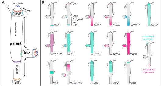
Fig. 1. Schematic overview of morphological boundaries, mass tissue movement and gene expression domains.(A) Morphological boundaries.
(B) Selected expression boundaries of genes in adult Hydra. Dots indicate that only a certain population of epithelial cells (Hym-323, Gata, Cnox2 and
Cnox3) or of gland cells (HyDkk1/2/4C) express the gene. The asterisk indicates that HyTcf is expressed transiently in the budding competent zone prior to bud evagination. Shin guard and manacle are expressed asymmetrically in the early developing bud (x).
HTK-7
HTK-7
shin-guardX
manX
anklet
X
PPOD1 CnNK2 Pedibin HyBMP5-8
HyDkk1/2/4C
HvPKC2 Frizzled
HvPKC1
Hym-323 Gata
HyChdl
HyTcf *
Cnox1 Cnox2 Cnox3
ectodermal expression
endodermal expression tentacle
zone tentacle
gastric region
hypostome
basal disc
el
c
nu
de
p
bud
parent
below the budding zone. This boundary is not visible equally well in every Hydra strain. It marks a decrease in the diameter of the upper body column and the appearance of big, translucent ecto- and entodermal cells. (v) The peduncle -basal disc bound-ary, where proliferating ecto- and endodermal epithelio-muscular cells differentiate into mucous-secreting basal disc cells. (vi) In bud-producing animals, the region from which parent tissue is recruited into the bud protrusion is separated from the parent animal by a morphologically hardly visible boundary (green line in Fig. 1A), When the bud has finished its growth, this boundary becomes very sharp and defines the position, where the bud foot forms and complete separation from parent tissue occurs.
Due to the tissue dynamics of Hydra and the stem cell properties of its three cell lineages, patterning signals are constantly active in the adults. This obviously includes permanent signalling at the tissue boundaries, and it causes the almost unlimited capacity of Hydra for regeneration. During budding and regeneration of adult polyps boundaries have to be formed de novo.
In the following, we will summarize which marker genes and signalling components are expressed at the major tissue bound-aries (and compartments) in Hydra and what is known about boundary-related signalling pathways.
Head and upper body region
The Hydra head consists of a hypostome and mouth opening in its center plus a whorl of tentacles below. Specific gene ex-pression domains surrounding the hypostome can be observed in only a couple of cells at the very tip, in broader rings or in the whole region between mouth opening and the lower tentacle border. Fig. 1B and Fig. 2 give an overview of expression zones within the Hydra head and body column for genes expressed in epithelial cells of the ecto- and entodermal layer as well as, in one case (HyDkk) in gland cells. These gene expression patterns in adult polyps suggest an astonishingly complex regionalization of the Hydra body, which contrasts the morphological simplicity of the animals.
Since expression in stage 9 of bud development mostly cor-responds to the pattern found in adult polyps, the following also referes to patterns shown in Fig. 2 for bud development.
At the very tip of adult polyps and buds, HvPKC2, an e-type
PKC encoding gene (Hassel et al., 1998) and several Wnt path-way genes are strongly expressed (Lengfeld et al., 2009). The smallest expression zone is covered by Wnt3 (Fig. 2), which encompasses only 8-9 cell diameters and is present in both, the ecto- and the endodermal epithelium. Slightly beyond this Wnt3 zone reaches expression of the Wnt-targeted transcription fac-tor, HyTcf (Hobmayer et al., 2000). An even larger area including the entire hypostome and almost touching the bases of tentacles is covered by HyBra expression (Technau and Bode 1999), its expression domain overlaps with HyWnt7 (Lengfeld et al., 2009) and transcription factors like budhead and prdl-a (Martinez et al., 1997; Gauchat et al., 1998; Takahashi et al., 2005). The region where hypostome cells proliferate is marked by the expression of budhead, which is lacking underneath the proliferative region, but is present again in the space between the tentacles.
Especially striking is the expression of the transcription factor goosecoid (Cngsc, Fig. 2). It is found only in non-tentacle cells and the apical-most hypostomal cells, thus comprising a two-stripe
pattern (Broun et al., 1999). These stripes mark morphologically invisible boundaries and seem to correspond to the expression borders of Wnt genes.
At the boundary between tentacles and body column (Fig. 1) proliferating epithelial tissue impinges on tissue made of non-proliferating battery cells. These huge cells have transdifferentiated from body column epithelio-muscular cells and take up mature nematocytes (stinging cells) by transendocytosis.
Genes, which demarcate the basal end of the tentacles and thus the boundary between these two tissues include HyDsh, HvWnt8 and Hmfz2 (Fig. 2) (Minobe et al., 2000; Philipp et al., 2009). A role for non-canonical Wnt in mediating tentacle evagination has been shown recently. Co-expression of BMP5-8b moreover indicates a localized role of BMP and Wnt signalling, which might be complex. Strong expression of a Hydra Chordin-like-encoding gene (HyChdl, Fig. 1B), a putative antagonist of BMP-signalling, furthermore suggests fine-tuned regulation (Reinhardt et al., 2004; Rentzsch et al., 2007). Potential transcriptional regulators at this boundary are the transcription factors CnOtx, HyAlx, Budhead and Prdl-a (Martinez et al., 1997; Gauchat et al., 1998; Smith et al., 1999; Smith et al., 2000).
Unclear is the function of the two epitheliopeptides Hym301 and Pedibin (Takahashi et al., 2005), the role of the RTK Lemon in endodermal basal tentacle cells and in a broader region PKC1 (Hassel et al., 1998; Miller and Steele 2000), as well as that of the Hydra-specific unusual RTK Sweet Tooth, which is charac-terised by an extracellular lectin domain (Reidling et al., 2000). The whole tentacle region is demarcated by scattered expression of the transcription factor Cnox3 in epithelial cells (Gauchat et al., 2000). The endodermal tentacle tissue expresses the Hydra metalloprotease, HMMP, (Fig. 2), which is only weakly expressed in the endoderm of adjacent non-tentacle tissue (Leontovich et al., 2000; Sarras 2012).
In the tissue below the tentacles towards the body column, cells are proliferating. Whole-mount BrdU-labelling (Holstein et al., 1991) as well as the upper expression border of Gata, a transcription factor (Nakamura et al., 2011), mark this boundary.
In summary, the boundaries between tentacles, tentacle zone and hypostome are clearly marked. The apical end of the polyps contains several molecular boundaries surrounding the mouth opening and exemplified by the Cngsc stripes of expression (Fig. 2). The target genes for the transcription factors Cngsc, HyAlx, CnOtx and Cnox3, remain to be elucidated.
Upper body – de novo establishment
Lower body region
The basal end of Hydra where proliferating peduncle cells and non-proliferating differentiated basal disc cells meet shows a clear morphological and molecular boundary between basal disc and
peduncle (Fig. 1B). Expression of the receptor tyrosine kinase shin-guard, the paired like homeobox gene manacle and the Hydra insulin receptor homolog HTK7 (Steele et al., 1996; Bridge et al., 2000) as well as anklet, a gene encoding a novel, secreted perforin-EGF-domain protein (Amimoto et al., 2006) define this
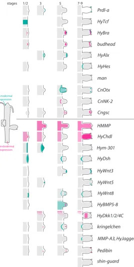
shin-guard Pedibin
MMP-A3, HyJagged kringelchen HyDkk1/2/4C HyBMP5-8 HyWnt8 HyWnt5 HyWnt3 HyDsh Hym-301
HyChdl HMMP Cngsc CnNK-2 CnOtx man HyHes HyAlx budhead HyBra HyTcf Prdl-a
stages 1/2 3 5 7-9
ectodermal expression
endodermal expression
Fig. 2. Schematic representation of expression domains and boundaries in the developing bud. The upper part summarizes transcription factor expression domains, the lower one those of (diffusible) signalling elements, receptors and proteases.
boundary by a narrow expression zone.
As expected for the transition zone towards a terminally differentiated zone, the peduncle - basal disc boundary is marked also by gene expression regions, which are either excluded from the basal disc or expressed exclusively there. PPOD1, for example, is restricted to the basal disc (Fig. 1B) and PPOD2 appears in cells directly adjacent to peduncle cells (Hoffmeister-Ullerich et al., 2002; Thomsen and Bosch 2006). Moreover the basal disc has a very high level of per-oxidase activity, which also clearly separates it from adjacent peduncle cells (Hoffmeister and Schaller 1985). Recent results have shown that PPODs encode extracellular lectins and not peroxidases (Pauly et al., 2007) (Böttger et al., in preparation). Like in the hypostome tip, HvPKC2 expression, is found in a small group of cells in the central basal disc endoderm (Has-sel et al., 1998), indicating that this PKC is a good marker for the extreme positional values in the Hydra body (Fig. 1B).
Excluded from the basal disc but expressed in and above the peduncle are the genes sweet tooth, encoding an atypi-cal RTK (Reidling et al., 2000), the serine-threonine kinase HvPKC1 (Hassel et al., 1998), BMP5-8b (Reinhardt et al., 2004) and Frizzled (Minobe et al., 2000). pedibin, encoding a foot-formation-promoting peptide, CnNK2 and BMP5-8b show expression fading out towards the budding region right above the peduncle (Grens et al., 1999; Hoffmeister-Ullerich 2001; Reinhardt et al., 2004).
In short distance above the sharp peduncle-basal disc boundary, a morphologically unrecognizable border is revealed (Fig. 1B) by the expression boundaries of Gata, Hym323 and Cnox1 (Gauchat et al., 2000; Takahashi et al., 2005; Takahashi and Fujisawa 2009; Nakamura et al., 2011). This diffuse boundary zone seems to correlate with the lower end of the proliferation zone.
A little higher up the body column in the mid-body region, expression domains of the upper and in the lower body half meet as exemplified by CnNK2, Pedibin, HyBMP5-8b, prdl-a and HyDkk1/2/4C expression (Grens et al., 1996; Gauchat et al., 1998; Hoffmeister-Ullerich et al., 2002; Reinhardt et al., 2004; Augustin et al., 2006) (Fig. 2). This border is, again, not a sharp one, nevertheless, the region is extremely interesting with respect to morphogenesis: the budding region (see below) is located here. HyTcf, the Wnt target transcription factor, marks the budding-competent girdle shortly before a bud begins to evaginate. Increasing expression of the transcription factor prdl-A (Gauchat et al., 1998) and the anti-Wnt HyDkk1/2/4C (Augustin et al., 2006) demarcate the region as well.
Lower body region – de novo establishment
expression is initiated precisely at this border region (Bridge et al., 2000). PPOD1 and HvPKC2, which will later be restricted to different regions in the basal disc, are also expressed very early at the tips of foot regenerates. The same is true for HyBMP5-8b, which in contrast to the other two genes is excluded from basal disc cells in mature feet. Thus, similar to head regenerates, genes, which belong to different parts of the final foot, are co-expressed in early regenerating feet and separated only later.
Budding
During budding continuously proliferating tissue of the body column is recruited into the bud. The initially observed thickening of the ectoderm can be noticed morphologically as a placode. By ectodermal India ink labelling it became obvious that Hydra vulgaris tissue marking a concentric ring on the parent animal is recruited: cells in the centre of this ring were shown to later become hypostome whereas cells at the outer circle end up at the basal end of the bud (Otto and Campbell 1977). Except for the ectodermal cells right above or below the bud, all other cells in this region have to change the orientation of muscle fibres. At final stages of this process the bud base constricts and forms a basal disk. Its connection with the parent becomes increasingly smaller until it detaches.
A defined morphological boundary between the early bud and parent cells cannot be observed. However, before first signs of evagination are visible, a molecular boundary defines the budding-competent zone (Fig. 1B, Fig. 2): the Wnt pathway genes HyTcf and
b-catenin are expressed in a broad band covering the region were
buds will evaginate (Hobmayer et al., 2000). Once bud evagina-tion begins, head specific genes including Wnt3a and HyBra are expressed in the centre of the protruding placode (Technau and Bode 1999). Tentacle specific genes are also expressed, but they either only appear transiently (HyAlx) or extend in concentric zones around the centre (budhead, HyDsh, Hmfz2, HyWnt8 and HMMP (Martinez et al., 1997; Leontovich et al., 2000; Smith et al., 2000; Siebert et al., 2005; Philipp et al., 2009).
Almost the whole region from which cells will later be found in the bud is marked by expression of CnOtx, prdl-a and Hym301 (Fig. 2) at budding stage 3 (Gauchat et al., 1998; Smith et al., 1999; Takahashi et al., 2005). However, the boundary between early Cnotx-, prdl-a- and Hym301-positive and -negative tissue is not very sharp. These genes roughly mark the region of the bud’s body column, which will be defined after completion of bud morphogenesis through sharp boundaries apically towards the tentacles and basally towards the foot tissue. At stage 7-8, when morphogenesis begins, CnOtx clearly marks the body-tentacle boundary as well as the basal end of the bud in relatively broad stripes of gene expression.
The homeobox gene CnNK-2, is expressed below the budding zone in adult animals and also at the basal end of progressed bud stages (Fig. 1B, Fig. 2). It is conspicuously absent from the early bud. This indicates that the basal tissue of a bud is not defined early on. CnNK-2 is actually only upregulated in the last set of endodermal cells, which migrates into the bud around stage 6 and ends up in the stalk and basal-most cells of the bud. It appears that a sharp boundary of the bud towards the parent is not clearly determined at the beginning of budding (Siebert et al., 2005). It rather forms late at budding stage 7-8.
The earliest gene demarcating the parent-bud boundary is the Hydra FGFR kringelchen, which becomes upregulated in a ring, 5-6 cells broad and surrounds the bud base from stage 4 onwards. kringelchen marks varying populations of cells while they pass this boundary rather than a certain group of cells. Only late during bud formation, around budding stage 7-8, kringelchen expression is refined into a sharp ring which appears to indicate the boundary separating bud and parent tissue. At this point a number of additional genes change their expression zones from diffuse rings into sharp lines. These include HyDsh and HyWnt8 (Philipp et al., 2009), which are most probably expressed adjacent to each other. Just when these sharp lines of gene expression are produced, HyHes, encoding a target transcription factor of the Notch signalling pathway, is expressed transiently in a single-cell band immediately adjacent to kringelchen. At the same time the metalloprotease MMP-A3 is expressed in the kringelchen positive cells (Münder et al., 2010). From now on, kringelchen, MMP-A3, HyDsh and HyJagged expression are restricted to parent tissue whereas foot specific genes are expressed in the newly forming bud peduncle (Sudhop et al., 2004; Prexl et al., 2011). Interestingly, HyWnt-8 is expressed in cells confining the peduncle at the side of the bud (Philipp et al., 2009).
These data strongly suggests that Notch/Wnt and FGF- signalling pathways are involved in the formation of the parent-bud boundary.
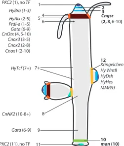
Fig. 3. Summary of epithelial gene expression domains and defined boundaries in Hydra. All boundaries identified by either a sharp line of marker gene expression (right side) or putative boundaries deduced from differential expression zones were numbered. The numbers in brackets indicate, at or between which of these boundaries certain transcription factors are expressed.
HyTcf (7+) Kringelchen Hy Wnt8
FGFR/Notch signalling controls the establishment of
the bud-parent boundary
Support for the hypothesis that FGFR/Wnt/Notch signalling are essential for the establishment of the parent-bud boundary comes from pharmacological inhibition experiments. Inhibition of FGFR-signalling with SU5402 and Notch-FGFR-signalling with DAPT perturbs bud morphogenesis (Sudhop et al., 2004; Münder et al., 2010). Both inhibitors cause the expression zones of kringelchen and HyDsh to completely diffuse. As a consequence the buds fail to form feet and never detach, which yields two-headed, so-called Y-animals. Interestingly, such animals can also be obtained when Src (Fabila et al., 2002) and other potential targets for FGFR signalling are inhibited. Moreover, it is also seen as a result of LiCl treatment, which may increase either Wnt-signalling by inhibition of GSK-3b
or impair PKC/inositol phosphate/calcium signalling and thereby disturb the boundary by shifting the balance of gene expression (Hassel et al., 1993; Fabila et al., 2002). This hypothesis has not been tested experimentally, yet. The expression data (and phar-macological inhibition) clearly show that boundary formation during budding in Hydra relies on Notch and FGFR signalling. Deduced from co-expression data it potentially also involves crosstalk with non-canonical Wnt signalling and HyWnt8.
In summary, a very likely candidate to establish the bud-parent boundary is, like in many other animals, the Notch pathway. Hydra has all major components for bona fide Notch signalling including a well conserved Notch receptor (HvNotch), a putative Notch ligand HyJagged (Prexl et al., 2011), components of the g-secretase
complex including presenilin, metalloproteases of the ADAM fam-ily (Kasbauer et al., 2007; Münder et al., 2010). Genes encoding Su(H) (CBF) and the transcriptional repressor HyHes have been identified as well as modulators of Notch signalling such as Fringe and Numb. It was also shown that the signalling mode in the Notch pathway, which is characterised by a regulated intra-membrane proteolysis mediated by metalloproteases of the ADAM family and presenilin, is conserved. Su(H) binding sites have been identified in the HyHes promoter and it was demonstrated that this promoter responds to NotchICD. Finally, the presenilin inhibitor DAPT is able to inhibit Notch-signalling in hydra cells by blocking the release of NICD from the membrane. Using this inhibitor a role for Notch signalling in interstitial stem cell differentiation was demonstrated (Kasbauer et al., 2007).
What about ephrin signalling?
Genes encoding ephrin receptors have only recently been identified in the genome and as ESTs in Hydra (Reddy et al., 2011). Unpublished results suggest that two of these genes are expressed at the base of the Hydra bud. Moreover, two genes encoding ephrins have also been found, but functional data are not available yet (Tischer and Böttger, unpublished). It will be very interesting to see how and where these molecules act in an organism, which lacks a blood circulation system and in which the nerve net is not visibly centralized.
General considerations and perspectives
The simple Hydra body is subdivided by a few, morphologically distinguishable, boundaries in the head and foot region. On a
mo-lecular level a complex sub-regionalization and additional borders become visible (Fig. 3).
Sharp boundaries in adult Hydra are characterised by mutually exclusive gene expression on either side. Examples are Cngsc in the hypostome (boundaries 2 and 3 in Fig. 3), manacle and PPOD at the basal disc (boundary 10) and kringelchen/HyHes at the buds base (boundary 12). When the corresponding boundaries are formed de novo, in each case, an initial overlap of gene expression zones, which are later separated, is observed. This sequence of events appears as a particularly simple case of creating tissue boundaries.
Differential expression of signalling molecules including BMP-5-8b, Wnt-8 and receptors for FGF and insulin indicate that these molecules might either define intersections for boundary formation or organise the tissue on either side of the boundaries. In agreement with such a view, the transient expression of HyTcf in the budding competent girdle (border region 7+ in Fig. 3) might depend on transiently forming intersections. Notch and FGFR signalling are clearly involved in forming the sharp boundary between bud and parent (Münder et al., 2010; Prexl et al., 2011), a role of ephrin signalling is likely.
In summary, the molecular pathways which are used in all higher animals to form tissue boundaries during development are not only present in prebilaterian animals but have already been recruited for similar tasks. The indicated simplicity of the Hydra system provides an ideal background to further elucidate the cellular and molecular mechanisms controlling the separation of initially identical (embryonic) tissue domains, which is the prerequisite to later form structures of high complexity.
Acknowledgements
We thank Katja Gessner and S. Münder for skillful help with the figures and Bert Hobmayer for critical reading of the manuscript. Our FGFR- and Notch- research was supported by research grants from the DFG (Ha 1732/10 and 11; BO1748-1 and BO1748-5).
References
ACAMPORA, D., BOYL, P. P., MARTINEZ-BARBERA, J. P., ANNINO, A., SIGNORE, M. and SIMEONE, A. (2001). Otx genes in evolution: are they involved in instruct-ing the vertebrate brain morphology? J Anat 199: 53-62.
AMIMOTO, Y., KODAMA, R. and KOBAYAKAWA, Y. (2006). Foot formation in Hydra: a novel gene, anklet, is involved in basal disk formation. Mech Dev 123: 352-361. AUGUSTIN, R., FRANKE, A., KHALTURIN, K., KIKO, R., SIEBERT, S., HEMMRICH, G. and BOSCH, T. C. (2006). Dickkopf related genes are components of the positional value gradient in Hydra. Dev Biol 296: 62-70.
BODE, H. R. (1996). The interstitial cell lineage of hydra: A stem cell system that arose early in evolution. J. Cell Sci. 109: 1155-1164.
BOLOGNESI, R., BEERMANN, A., FARZANA, L., WITTKOPP, N., LUTZ, R., BALA-VOINE, G., BROWN, S. J. and SCHRÖDER, R. (2008). Tribolium Wnts: evidence for a larger repertoire in insects with overlapping expression patterns that suggest multiple redundant functions in embryogenesis. Dev Genes Evol 218: 193-202. BRAY, S. J. (2006). Notch signalling: a simple pathway becomes complex. Nat Rev
Mol Cell Biol 7: 678-689.
BRIDGE, D., STOVER, N. and STEELE, R. (2000). Expression of a novel receptor tyrosine kinase gene and a paired-like homeobox gene provides evidence of dif-ferences in patterning at the oral and aboral ends of hydra. Dev Biol 220: 253-262. BROUN, M., SOKOL, S. and BODE, H. R. (1999). Cngsc, a homologue of goosecoid, participates in the patterning of the head, and is expressed in the organizer region of Hydra. Development 126: 5245-5254.
D. et al. (2010). The dynamic genome of Hydra. Nature 464: 592-596. DAHMANN, C., OATES, A. C. and BRAND, M. (2011). Boundary formation and
maintenance in tissue development. Nat Rev Genet 12: 43-55.
DAVID, C.N. (2012). Interstitial stem cells in Hydra: multipotency and decision-making.
Int J Dev Biol 56: 489-497.
DE CELIS, J. F., TYLER, D. M., DE CELIS, J. and BRAY, S. J. (1998). Notch signal-ling mediates segmentation of the Drosophila leg. Development 125: 4617-4626. DUBRULLE, J. and POURQUIE, O. (2002). From head to tail: links between the
segmentation clock and antero-posterior patterning of the embryo. Curr Opin
Genet Dev 12: 519-523.
FABILA, Y., NAVARRO, L., FUJISAWA, T., BODE, H. and SALGADO, L. (2002). Selective inhibition of protein kinases blocks the formation of a new axis, the beginning of budding, in Hydra. Mech Dev 119: 157-164.
GAUCHAT, D., KREGER, S., HOLSTEIN, T. and GALLIOT, B. (1998). prdl-a, a gene marker for hydra apical differentiation related to triploblastic paired-like head-specific genes. Development 125: 1637-1645.
GAUCHAT, D., MAZET, F., BERNEY, C., SCHUMMER, M., KREGER, S., PAW-LOWSKI, J. and GALLIOT, B. (2000). Evolution of Antp-class genes and differential expression of Hydra Hox/paraHox genes in anterior patterning. Proc Natl Acad
Sci USA 97: 4493-4498.
GRENS, A., GEE, L., FISHER, D. A. and BODE, H. R. (1996). CnNK-2, an NK-2 homeobox gene, has a role in patterning the basal end of the axis in hydra. Dev.
Biol. 180: 473-488.
GRENS, A., SHIMIZU, H., HOFFMEISTER, S. A. H., BODE, H. R. and FUJISAWA, T. (1999). The novel signal peptides, pedibin and Hym-346, lower positional value thereby enhancing foot formation in hydra. Development 126: 517-524. HÄCKER, U., NYBAKKEN, K. and PERRIMON, N. (2005). Heparan sulphate
pro-teoglycans: the sweet side of development. Nat Rev Mol Cell Biol 6: 530-541. HASSEL, M., ALBERT, K. and HOFHEINZ, S. (1993). Pattern-Formation in
Hydra-Vulgaris Is Controlled by Lithium-Sensitive Processes. Dev. Biol. 156: 362-371. HASSEL, M., BRIDGE, D., STOVER, N., KLEINHOLZ, H. and STEELE, R. (1998).
The level of expression of a protein kinase C gene may be an important component of the patterning process in Hydra. Dev Genes Evol 207: 502-514.
HIMANEN, J. P., SAHA, N. and NIKOLOV, D. B. (2007). Cell-cell signaling via Eph receptors and ephrins. Curr Opin Cell Biol 19: 534-542.
HIRTH, F., KAMMERMEIER, L., FREI, E., WALLDORF, U., NOLL, M. and REICHERT, H. (2003). An urbilaterian origin of the tripartite brain: developmental genetic insights from Drosophila. Development 130: 2365-2373.
HOBMAYER, B., JENEWEIN, M., EDER, D., GLASAUER, S., GUFLER, S., HARTL, M. and SALVENMOSER, W. (2012). Stemness in Hydra - a current perspective.
Int J Dev Biol 56: 509-517.
HOBMAYER, B., RENTZSCH, F., KUHN, K., HAPPEL, C., VON LAUE, C., SNYDER, P., ROTHBÄCHER, U. and HOLSTEIN, T. (2000). WNT signalling molecules act in axis formation in the diploblastic metazoan Hydra. Nature 407: 186-189. HOFFMEISTER, S. A. H. and SCHALLER, H. C. (1985). A new biochemical marker
for foot-specific differentiation in hydra. Roux’s Arch. Dev. Biol. 194: 453-461. HOFFMEISTER-ULLERICH, S. A. (2001). The foot formation stimulating peptide
pedibin is also involved in patterning of the head in hydra. Mech Dev 106: 37-45. HOFFMEISTER-ULLERICH, S. A., HERRMANN, D., KIELHOLZ, J., SCHWEIZER,
M. and SCHALLER, H. C. (2002). Isolation of a putative peroxidase, a target for factors controlling foot-formation in the coelenterate hydra. Eur. J. Biochem. 269: 4597-4606.
HOLSTEIN, T., HOBMAYER, E. and DAVID, C. (1991). Pattern of epithelial cell cycling in hydra. Dev Biol 148: 602-611.
KASBAUER, T., TOWB, P., ALEXANDROVA, O., DAVID, C. N., DALL’ARMI, E., STAUDIGL, A., STIENING, B. and BOTTGER, A. (2007). The Notch signaling pathway in the cnidarian Hydra. Dev. Biol. 303: 376-390.
KIM, J., SEBRING, A., ESCH, J. J., KRAUS, M. E., VORWERK, K., MAGEE, J. and CARROLL, S. B. (1996). Integration of positional signals and regulation of wing formation and identity by Drosophila vestigial gene. Nature 382: 133-138. LENGFELD, T., WATANABE, H., SIMAKOV, O., LINDGENS, D., GEE, L., LAW, L.,
SCHMIDT, H., OZBEK, S., BODE, H. and HOLSTEIN, T. (2009). Multiple Wnts are involved in Hydra organizer formation and regeneration. Dev Biol 330: 186-199. LEONTOVICH, A. A., ZHANG, J. S., SHIMOKAWA, K., NAGASE, H. and SARRAS,
M. P. (2000). A novel hydra matrix metalloproteinase (HMMP) functions in
extra-cellular matrix degradation, morphogenesis and the maintenance of differentiated cells in the foot process. Development 127: 907-920.
MARTINEZ, D., DIRKSEN, M., BODE, P., JAMRICH, M., STEELE, R. and BODE, H. (1997). Budhead, a fork head/HNF-3 homologue, is expressed during axis formation and head specification in hydra. Dev Biol 192: 523-536.
MEINHARDT, H. (2012) Modeling pattern formation in hydra: a route to understanding essential steps in development. Int. J. Dev. Biol. 56: 447-462.
MILLER, M. A. and STEELE, R. E. (2000). Lemon encodes an unusual receptor protein-tyrosine kinase expressed during gametogenesis in Hydra. Dev. Biol. 224: 286-298.
MINOBE, S., FEI, K., YAN, L., SARRAS JR, M. and WERLE, M. (2000). Identification and characterization of the epithelial polarity receptor “Frizzled” in Hydra vulgaris.
Dev Genes Evol 210: 258-262.
MÜNDER, S., KÄSBAUER, T., PREXL, A., AUFSCHNAITER, R., ZHANG, X., TOWB, P. and BÖTTGER, A. (2010). Notch signalling defines critical boundary during budding in Hydra. Dev Biol 344: 331-345.
NAKAMURA, Y., TSIAIRIS, C. D., ÖZBEK, S. and HOLSTEIN, T. W. (2011). Auto-regulatory and repressive inputs localize Hydra Wnt3 to the head organizer. Proc
Natl Acad Sci USA 108: 9137-9142.
NIEHRS, C. (2010). On growth and form: a Cartesian coordinate system of Wnt and BMP signaling specifies bilaterian body axes. Development 137: 845-857. OTTO, J. and CAMPBELL, R. (1977). Budding in Hydra attenuata: bud stages and
fate map. J Exp Zool 200: 417-428.
PASQUALE, E. B. (2008). Eph-ephrin bidirectional signaling in physiology and disease. Cell 133: 38-52.
PAULY, B., LASI, M., MACKINTOSH, C., MORRICE, N., IMHOF, A., REGULA, J., RUDD, S., DAVID, C. N. and BOTTGER, A. (2007). Proteomic screen in the simple metazoan Hydra identifies 14-3-3 binding proteins implicated in cellular metabolism, cytoskeletal organisation and Ca2+ signalling. BMC Cell Biol. 8: 31. PHILIPP, I., AUFSCHNAITER, R., OZBEK, S., PONTASCH, S., JENEWEIN, M.,
WATANABE, H., RENTZSCH, F., HOLSTEIN, T. W. and HOBMAYER, B. (2009). Wnt/beta-Catenin and noncanonical Wnt signaling interact in tissue evagination in the simple eumetazoan Hydra. Proc. Natl. Acad. Sci. USA 106: 4290-4295. PREXL, A., MUNDER, S., LOY, B., KREMMER, E., TISCHER, S. and BOTTGER, A.
(2011). The putative Notch Ligand HyJagged is a transmembrane protein present in all cell types of adult hydra and upregulated at the boundary between bud and parent. BMC Cell Biology 12: 38.
REDDY, P. C., BIDAYE, S. S. and GHASKADBI, S. (2011). Genome-wide screening reveals the emergence and divergence of RTK homologues in basal Metazoan Hydra magnipapillata. J Biosci 36: 289-296.
REIDLING, J. C., MILLER, M. A. and STEELE, R. E. (2000). Sweet Tooth, a novel receptor protein-tyrosine kinase with C-type lectin-like extracellular domains. J
Biol Chem 275: 10323-10330.
REINHARDT, B., BROUN, M., BLITZ, I. L. and BODE, H. R. (2004). HyBMP5-8b, a BMP5-8 orthologue, acts during axial patterning and tentacle formation in hydra.
Dev Biol 267: 43-59.
RENTZSCH, F., GUDER, C., VOCKE, D., HOBMAYER, B. and HOLSTEIN, T. W. (2007). An ancient chordin-like gene in organizer formation of Hydra. Proc Natl
Acad Sci USA 104: 3249-3254.
ROHANI, N., CANTY, L., LUU, O., FAGOTTO, F. and WINKLBAUER, R. (2011). EphrinB/EphB signaling controls embryonic germ layer separation by contact-induced cell detachment. PLoS Biol 9: e1000597.
SÁNCHEZ-CAMACHO, C., RODRÍGUEZ, J., RUIZ, J. M., TROUSSE, F. and BO-VOLENTA, P. (2005). Morphogens as growth cone signalling molecules. Brain
Res Brain Res Rev 49: 242-252.
SARRAS, M. P. J. (2012). Components, structure, biogenesis and function of the
Hydra extracellular matrix in regeneration, pattern formation and cell
differentia-tion. Int J Dev Biol 56: 567-576.
SIEBERT, S., THOMSEN, S., REIMER, M. M. and BOSCH, T. C. G. (2005). Control of foot differentiation in Hydra: Phylogenetic footprinting indicates interaction of head, bud and foot patterning systems. Mech. Dev. 122: 998-1007.
SMITH, K. M., GEE, L., BLITZ, I. L. and BODE, H. R. (1999). CnOtx, a member of the Otx gene family, has a role in cell movement in hydra. Dev. Biol. 212: 392-404. SMITH, K. M., GEE, L. and BODE, H. R. (2000). HyAlx, an aristaless-related gene,
SPRINZAK, D., LAKHANPAL, A., LEBON, L., GARCIA-OJALVO, J. and ELOWITZ, M. B. (2011). Mutual inactivation of Notch receptors and ligands facilitates devel-opmental patterning. PLoS Comput Biol 7: e1002069.
SPRINZAK, E., COKUS, S. J., YEATES, T. O., EISENBERG, D. and PELLEGRINI, M. (2009). Detecting coordinated regulation of multi-protein complexes using logic analysis of gene expression. BMC Syst Biol 3: 115.
STEELE, R. E. (2002). Developmental signaling in Hydra: what does it take to build a “simple” animal? Dev Biol 248: 199-219.
STEELE, R.E. (2012). The Hydra genome: insights, puzzles, and opportunities for developmental biologists. Int J Dev Biol 56: 535-542.
STEELE, R. E., LIEU, P., MAI, N. H., SHENK, M. A. and SARRAS, M. P. (1996). Response to insulin and the expression pattern of a gene encoding an insulin receptor homologue suggest a role for an insulin-like molecule in regulating growth and patterning in Hydra. Dev. Genes Evol. 206: 247-259.
SUDHOP, S., COULIER, F., BIELLER, A., VOGT, A., HOTZ, T. and HASSEL, M. (2004). Signalling by the FGFR-like tyrosine kinase, Kringelchen, is essential for bud detachment in Hydra vulgaris. Development 131: 4001-11.
TAKAHASHI, T. and FUJISAWA, T. (2009). Important roles for epithelial cell peptides
in hydra development. Bioessays 31: 610-619.
TAKAHASHI, T., HATTA, M., YUM, S., GEE, L., OHTANI, M., FUJISAWA, T. and BODE, H. R. (2005). Hym-301, a novel peptide, regulates the number of tentacles formed in hydra. Development 132: 2225-2234.
TECHNAU, U. and BODE, H. (1999). HyBra1, a Brachyury homologue, acts during head formation in Hydra. Development 126: 999-1010.
THOMSEN, S. and BOSCH, T. C. (2006). Foot differentiation and genomic plastic-ity in Hydra: lessons from the PPOD gene family. Dev Genes Evol 216: 57-68. WITTLIEB, J., KHALTURIN, K., LOHMANN, J., ANTON-ERXLEBEN, F. and BOSCH,
T. (2006). Transgenic Hydra allow in vivo tracking of individual stem cells during morphogenesis. Proc Natl Acad Sci USA 103: 6208-6211.
YU, S., BURKHARDT, M., NOWAK, M., RIES, J., PETRÁSEK, Z., SCHOLPP, S., SCHWILLE, P. and BRAND, M. (2009). Fgf8 morphogen gradient forms by a source-sink mechanism with freely diffusing molecules. Nature 461: 533-536. ZHANG, X., BOOT-HANDFORD, R. P., HUXLEY-JONES, J., FORSE, L. N., MOULD,
Evolution of angiogenesis Ramon Munoz-Chapuli
Int. J. Dev. Biol. (2011) 55: 345-351
An organizing region in metamorphosing hydrozoan planula larvae - stimulation of axis formation in both larval and in adult tissue Melanie Stumpf, Britta Will, Karola Wittig, Jennifer Kasper, Benjamin Fischer, Jürgen Schmich, Stefanie Seipp and Thomas Leitz
Int. J. Dev. Biol. (2010) 54: 795-802
Induction of reverse development in two marine Hydrozoans
Jürgen Schmich, Yulia Kraus, Doris De Vito, Daria Graziussi, Ferdinando Boero and Stefano Piraino
5 yr ISI Impact Factor (2010) = 2.961 Int. J. Dev. Biol. (2007) 51: 45-56
Wnt signaling in hydroid development: ectopic heads and giant buds induced by GSK-3beta inhibitors
Werner Müller, Uri Frank, Regina Teo, Ofer Mokady, Christina Guette and Günter Plickert Int. J. Dev. Biol. (2007) 51: 211-220
Principles of branch formation and branch patterning in Hydrozoa Stefan Berking
Int. J. Dev. Biol. (2006) 50: 123-134
Cnidarians as a model system for understanding evolution and regeneration Brigitte Galliot and Volker Schmid
Int. J. Dev. Biol. (2002) 46: 39-48
Ca2+-ions and pattern control in Hydra