UP-001
Orthotopic Continent Urinary Diversion vs. Ileal Conduit: Comparison of Intraoperative and In-hospital Complications, Blood Transfusion, Length of Stay, and In-hospital Mortality
Djahangirian, Orchidee; Schmitges, Jan; Tian, Zhe; Karakiewicz, Pierre; Ismail, Salima
Cancer Prognostics and Health Outcomes Unit, University of Montreal Health Centre, Montreal, QC, Canada
Introduction and Objectives: Orthotopic continent urinary diversion (OCUD) after radical cystectomy (RC) may predispose to a higher rate of complications than ileal conduit urinary diversion (ICUD).
Methods: We identified all OCUD and ICUD patients in the Nationwide Inpatient Sample (NIS). We tabulated intraoperative and in-hospital compli-cations, transfusion, length of stay, and in-hospital mortality rates according to urinary diversion. Multivariable analyses were performed, with adjust-ment for age, Charlson comorbidity Index (CCI), gender, race, hospital type and region, annual hospital volume tertiles, and year of surgery. Results: The rate of OCUD was 8% vs. 92% for ICUD. Intraoperative and in-hospital complication rates were 2.5% vs. 2.6% (p=0.8) and 24.7% vs. 28.1% (p=0.04) for OCUD and ICUD patients, respectively. Blood transfusions were administered in 17.9% vs. 26.7% (p<0.001). Length of stay above median (≥9 days) was recorded in 53.5% vs. 54.9% (p=0.4). In-hospital mortality was 0.4% vs. 2.5% (p<0.001). After adjusting for all covariates, OCUD patients were less likely to receive a blood transfusion (odds ratio [OR]: 0.7, p<0.001), more likely to have an increased length of stay (OR: 1.3, p=0.002), and less likely to succumb to in-hospital mortal-ity (OR: 0.3, p=0.03) relative to ICUD patients. Urinary diversion failed to achieve statistical significance for intraoperative (OR: 1.0, p=0.9) and in-hospital complications (OR: 1.0, p=0.8).
Conclusions: After RC, urinary diversion dictates no difference in intraop-erative or in-hospital complications. However, fewer transfusions and a lower in-hospital mortality rate were recorded in OCUD patients. Therefore, broader use of OCUD after RC should be encouraged.
UP-002
Techniques and Agents Used to Conserve Blood During Radical Cystectomy: a Survey of the Society of Urologic Oncology
Punjani, Nahid1; Lavallée, Luke T.2; Momoli, Franco3; Cagiannos, Ilias2;
Morash, Christopher G.2; Fergusson, Dean3; Breau, Rodney H.2
1University of Ottawa, Ottawa, ON, Canada; 2University of Ottawa, Division
of Urology, Ottawa, ON, Canada; 3Ottawa Hospital Research Institute,
Ottawa, ON, Canada
Introduction and Objectives: Radical cystectomy may result in significant blood loss necessitating blood transfusions. The purpose of this study was to determine what intraoperative techniques and agents are currently used by uro-oncologists to prevent blood loss during radical cystectomy. Methods: In August 2011, Society of Urologic Oncology members were solicited to complete an online survey. Respondents were asked to provide demographic information, state opinions on blood loss and transfusion, report techniques used to reduce blood loss and to estimate the propor-tion of cases where they used either systemic or local hemostatic agents. Results: Residents, fellows, and non-urologists were excluded leaving 86 staff urologists who perform radical cystectomies. Of the 86, 73 (85%) had completed an uro-oncology fellowship in the United States, 57 (66%) had been in practice for over 6 years, and 68 (79%) perform over 10 cys-tectomies per year. Forty-nine (57%) of respondents estimated that
trans-fusions were performed in over 20% of patients and 4 (5%) indicated that transfusions were performed in over 50% of patients. Few surgeons reported using CellSaver® (15;17%), autologous blood banking (16;19%), or acute normovolemic hemodilution (22;26%). Topical hemostatic agents were frequently administered with 61 (71%) utilizing oxidized cellulose polymer (Sugicel®), 23 (27%) absorbable gelatin sponge (Gelfoam ®), 44 (51%) gelatin and thrombin matrix (Floseal®), and 15 (17%) thrombin and calcium chloride (Tisseel®). Very few surgeons routinely used systemic anti-hemorrhagics with only 2 (2%) reporting use of factor VII (Novoseven®), 4 (5%) desmopressin (DDAVP®), 4 (5%) tranexamic acid (Cyclokapron®), and 0 (0%) to aminocaproic acid (Amicar®).
Conclusions: There is significant risk of blood loss requiring transfusion during radical cystectomy even among high volume uro-oncologists. Surgeons frequently use topical hemostatic agents and rarely use systemic anti-hemorrhagics.
UP-003
Should CT Urogram Be the First Radiological Investigation to Evaluate Visible Haematuria?
Akhter, Waseem; Sheikh, Mazher; Kilburn, Karen; Peedikayil, Abraham; Palit, Victor
University Hospital of North Tees and Hartlepool NHS Trust, Hartlepool, United Kingdom
Introduction and Objectives: General protocol for investigating visible hae-maturia patients are USS/IVU and flexible cystoscopy. However pooled sensitivity and specificity of CT urogram in diagnosis of urothelial tumors are 96% and 99% respectively (systematic review and meta-analysis EJR Feb 2010) which is better than IVU or USS. So CT urogram became the investigation of choice for visible haematuria at our haematuria clinic. The purpose of this study is to assess the effectiveness and outcome of CT urogram performed as the first radiological investigation for patients with visible haematuria.
Methods: This is a retrospective review of all CT urograms results at hae-maturia clinic from January 2009 to January 2010. Endoscopic findings were also noted in all patients.
Results: This study includes 266 patients with visible haematuria. A total of 76 patients had some abnormal urological findings in CT urogram (Table 1). 31 patients avoided flexible cystoscopy and proceeded directly for resec-tion of bladder tumor/retrograde studies and ureteroscopy. 8 patients were referred to other clinicians for non-urological findings after normal flexible cystoscopy.
Conclusions: CT urogram should be the first line of imaging for visible haematuria as it avoids unnecessary investigations and helps plan effective management of haematuria patients.
Unmoderated Posters
Table 1. UP-003. CT urogram (n=266); abnormal CT urogram (n=76)
RCC 5 (1.87%)
Upper TCC 12 (4.51%)
Bladder TCC 33 (12.40%)
Stones 26 (9.77%)
Other diagnoses 8 (3%)
UP-004
Laparoscopic Robot-assisted Radical Cysto-prostatectomy
Bladou, Franck
McGill University, Jewish General Hospital, Montreal, QC, Canada Introduction and Objectives: Radical cysto-prostatectomy is indicated for non-metastatic infiltrative transitional cell carcinoma of the bladder. During the procedure, an extended lymph node dissection is performed with a control of the vesical and prostatic pedicles, a dissection of the recto-vesical plan and urethral diversion. Due to the anatomic situation of these organs in the deep pelvis, the standart open procedure has an early postoperative morbidity that exceeds a 30% rate and a long length of stay. Methods: A laparoscopic approach has been proposed for this procedure in order to limitate the per-operative blood loss and morbidity rate. It has been shown to be limited to experienced centres, due to the technical difficulty and demanding procedure. Robotic-assistance is an alternative of pure laparoscopic procedure and can be proposed to enhance the sur-geon gesture in a limited anatomic area while keeping oncological safety. Results: The video shows a step-by-step laparoscopic robot-assisted radi-cal cysto-prostatectomy, with an extended lymp node dissection from the aorto-iliac bifurcation, external iliac, obturator and internal iliac dissec-tion. The 3-D surgical vision offered by the DaVinci technology allows a perfect vision of the surgical dissection. Vesical pedicles are cliped at the origin of the internal iliac vessels, allowing a dry dissection with limited blood loss. The complete dissection of the bladder is shown, followed by the control of the Santorini pedicle, urethral and ureteral transections. At the end of the procedure, a running suture is placed on the posterior lip of the urethra to prepare the ileo-urethral anastomosis that will be performed after the construction of the neo-bladder performed in an open approach. Conclusions: Radical cysto-prostatectomy can be performed with the same oncological safety than open procedure, equivalent procedure time and lower blood loss.
UP-005
Survival Impact of Postoperative Expenditures by High Volume Surgeons Following Definitive Surgery for Bladder Cancer
Sandhu, Gurdarshan; Nepple, Kenneth; Yang, Liu; Grubb 3rd, Robert;
Strope, Seth
Washington University School of Medicine, St. Louis, MO, United States Introduction and Objectives: Improved survival has been reported in high volume surgeons, but it is unknown whether variation exists in the outcomes of such surgeons based on surveillance patterns. We evaluated the postoperative expenditures of high volume surgeons and explored the association between these expenditures and survival.
Methods: Using SEER-Medicare records, we identified 2408 patients aged
≥66 years with bladder carcinoma treated with definitive surgery from 1992 to 2005. Surgeons were defined as high volume for performance of ≥10 cystectomies in the cohort. Geography and time (2005) standard-ized outpatient postoperative Medicare expenditures were evaluated for 2 years after surgery. High volume surgeons were stratified into quartiles by mean monthly postoperative expenditures, and survival for the quar-tiles was evaluated with the Kaplan-Meier method. Multivariable Cox proportional hazard regression models were used to estimate mortality hazard ratios by expenditure quartile.
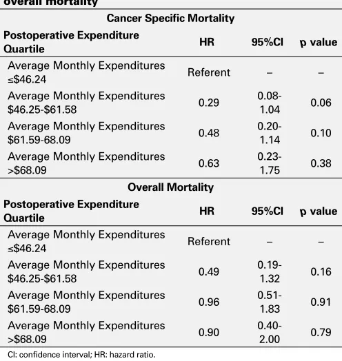
Results: 29 of 833 surgeons were identified as high volume and had operated on 443 patients. Mean monthly postoperative expenditures by individual surgeon ranged from $41.19 to $181.73 amongst the high volume surgeons. No difference in cancer specific (p=0.30) or overall survival (p=0.09) was seen between the surgeon expenditure quartiles on Kaplan-Meier analysis. After adjusting for demographic, socioeconomic, comorbid, treatment, pathologic, hospital and surgeon factors no differ-ence in mortality outcomes were seen between quartiles (Table 1). Conclusions: In high volume surgeons, a broad range in postoperative management was observed, manifested by large differences in postopera-tive expenditures. Despite the increased cost of postoperapostopera-tive care, no difference in survival was seen between surgeon expenditure quartiles implying that improved outcomes in high volume surgeons may be more strongly related to the surgery itself than to strict postoperative surveillance.
UP-006
Is There a Role for the mTOR Pathway in Non-muscle Invasive Bladder Cancer?
Blais, Jean-Philippe; Hovington, Hélène; Brisson, Hervé; Lacombe, Louis; Caumartin, Yves
Centre Hospitalier universitaire de Québec, Quebec, QC, Canada Introduction and Objectives: Non-muscle invasive bladder cancer (NMIBC) is associated with significant risk of recurrence and can progress to an invasive state. Numerous adjuvant intravesical therapies have been used in order to decrease the burden associated with this disease. The mammalian target of rapamycin (mTOR) pathway has been associated with oncogenesis in numerous cancers. We want to evaluate its implica-tion in NMIBC and indirectly, its potential therapeutic role.
Methods: Immunohistochemical analyses were performed on first bladder tumors (BT) resected. We assessed mTOR overexpression with monoclo-nal antibodies. The different tumor specimens were classified according to staining percentage and intensity. Patients and tumors characteristics were reviewed from patients’ charts. We attempted correlation of mTOR expression with clinical outcomes (recurrence and progression). Results: BT with a high intensity staining were associated more often with T1 stage (47.4% vs. 21.6%, p=0.04) and higher grade (31.6% vs. 4.5%, p=0.0009) compared to BT with lower mTOR expression. The proportion of patients who experienced recurrence was more frequent in the high intensity expression group (84.2% vs. 65.9%, p=0.17) as was also the risk of progression (15.8% vs. 9.1%; p=0.41). These results are unfortunately not statistically significant at this point, and for this reason, this study is still ungoing with inclusion of more patients.
Conclusions: Our preliminary results have demonstrated a trend between mTOR overexpression and risk of recurrence and progression in NMIBC. We need to continue the analysis with more patients in order to ascertain this potential pronostic relation. Through this study, we also hope to pro-vide a rationale for the potential mTOR inhibitors utilization in NIMBC.
Table 1. UP-005. Multivariate Cox proportional hazards regression analysis for bladder cancer mortality and overall mortality
Cancer Specific Mortality Postoperative Expenditure
Quartile HR 95%CI p value
Average Monthly Expenditures
≤$46.24 Referent – –
Average Monthly Expenditures
$46.25-$61.58 0.29
0.08-1.04 0.06
Average Monthly Expenditures
$61.59-68.09 0.48
0.20-1.14 0.10
Average Monthly Expenditures
>$68.09 0.63
0.23-1.75 0.38
Overall Mortality Postoperative Expenditure
Quartile HR 95%CI p value
Average Monthly Expenditures
≤$46.24 Referent – –
Average Monthly Expenditures
$46.25-$61.58 0.49
0.19-1.32 0.16
Average Monthly Expenditures
$61.59-68.09 0.96
0.51-1.83 0.91
Average Monthly Expenditures
>$68.09 0.90
0.40-2.00 0.79
UP-007
Meta-analysis of the Efficacy and Safety of Darifenacin and Other Anticholinergics for Managing Overactive Bladder
Elhilali, Mostafa1; Piwko, Charles2; Vincente, Colin2; Sabapathy, Suthacar2;
Tu, Le Mai3; Valiquette, Luc4; Finarson, Thomas R.5; Ahmad, Asma6 1McGill University, Royal Victoria Hospital, Montreal, QC, Canada; 2Pivina
Consulting, Montreal, QC, Canada; 3FRCSC University of Sherbrooke,
Sherbrooke, QC, Canada; 4FRCSC Université de Montréal, Montreal, QC,
Canada; 5University of Toronto, Toronto, ON, Canada; 6Pivina Consulting,
Mississauga, ON, Canada
Introduction and Objectives: Anticholinergics are frequently used to treat Overactive Bladder (OAB) symptoms which impacts quality of life (QoL). They cause adverse events, including dry mouth and constipation. The efficacy and safety of Darifenacin is reported to be similar to other anti-cholinergics with less cardio-vascular and cognitive adverse events (AE). Our objective was to conduct an updated meta-analysis comparing the efficacy, safety, and impact on QoL of Darifenacin and other anticholin-ergics (oxybutynin, tolterodine, solifenacin, and trospium chloride) for managing OAB.
Methods: The search was conducted by 2 independent researchers on Medline, Embase, and Cinhal to identify RCTs reporting safety (dry mouth, constipation, and withdrawals) and efficacy (urgency inconti-nence episodes, number of micturitions frequency, urgency and total incontinence episodes, and volume voided per micturition) of darifenacin and other anticholinergics. Each researcher extracted data independently. Discrepant results were settled through consensus discussion. The meta-analysis was performed using a random effects model.
Results: The literature search resulted in 1,280 citations of which 41 RCTs contained sufficient data for analysis including 3 Darifenacin RCTs (n=1226). All anticholinergics improved clinical outcomes from base-line with high responses to placebo. Trospium chloride and Darifenacin improved all efficacy outcomes compared to placebo. In addition Darifenacin improved QoL compared to placebo. All agents caused some dry mouth and constipation, with the lowest level of total withdrawals noted for Darifenacin.
Conclusions: This meta-analysis demonstrated that Darifenacin is similar in clinical efficacy and safety compared to other anticholinergics, but more effective in terms of QoL.
UP-008
Preoperative Sarcopenia Associated with Renal Function Outcomes in Patients Treated for Renal Masses by Extirpative Surgery
Hennessey, Katherine K.; Michael, Amanda; Elias, Rami; Kapoor, Anil; Shayegan, Bobby; Matsumoto, Edward; Mourtzakis, Marina; Di Sebastiano, Katie; Duivenvoorden, Helga; Pinthus, Jehonathan H. McMaster University, Hamilton, ON, Canada
Introduction and Objectives: Patients with solid tumors and severe skeletal muscle depletion (sarcopenia) have reduced survival. In renal transplants, sarcopenia predicts poorer post-transplant graft and patient survival. Preoperative body mass index (BMI) has been found to be an independent predictor of renal function post-nephrectomy. We hypoth-esize that sarcopenia may also predict renal function following surgery. Methods: We examined the association between preoperative sarcope-nia and postoperative estimated GFR (eGFR). Skeletal muscle area at the 3rd lumbar vertebrae was measured on computed tomography, and analyzed using Slice-O-Matic software. Cut-offs for sarcopenia were set as per standards in the literature. The primary outcome was change in a modified eGFR postoperative, calculated using the Modification of Diet in Renal Disease Study Group equation.
Results: Mean BMI of the sample was 30.1 and mean age was 62.7. Mean preoperative GFR was 91.5 in the sarcopenic patients. The group included 5 open and 13 laparoscopic radical nephrectomies. 10/18 (56%) were sarcopenic preoperative, revealing a high prevalence of sarcope-nia in RCC patients. Mean change in 3 months eGFR was -30.2% and -27.5% in the sarcopenic and non-sarcopenic groups, respectively. Mean change in 12 months eGFR was -29.5% and -28.8% in the sarcopenic and
non-sarcopenic groups, respectively. Unpaired t-test showed change in eGFR in the sarcopenic group greater than the non-sarcopenic, approach-ing significance (p=0.1). A trend between preoperative sarcopenia and decreased postoperative renal function is evident.
Conclusions: Preoperative sarcopenia may be a predictor of renal failure post-nephrectomy. Sarcopenic patients have a lower GFR postoperative. This pilot study is ongoing as we collect data on 100 more patients. With a larger sample, we expect to detect a statistically significant decrease in postoperative eGFR in sarcopenics. Goals of surgery for renal tumors include preservation of function, and thus a nephron-sparing approach could be applied to sarcopenics.
UP-009
Renal Tumor Ablation for pT1 Lesions: Patterns of Recurrence and Follow-up Recommendations
Rutkowski, John; Muruve, Nic
Cleveland Clinic Florida, Weston, FL, United States
Introduction and Objectives: Renal tumor ablation has gained popularity for the treatment of pT1 lesions. We seek to determine the pattern and frequency of recurrence to optimize follow-up.
Methods: We retrospectively analyzed the records of consecutive patients who underwent renal tumor ablation at our institution from 6/2004 to 6/2011. Patients who underwent percutaneous RFA and laparoscopic cryoablation were included. Follow-up imaging studies and clinical notes were reviewed to identify patients who failed therapy. Failure was defined as persistent contrast enhancement or requirement for an additional pro-cedure.
Results: A total of 94 ablations performed in 87 patients with appropriate follow-up were included in our study. Of 65 percutaneous RFAs, there were 11 failures. The average tumor size for failures was 3.5 cm (range 1.5 – 5.5 cm). The average time to failure after treatment was 6 months (range 3-18 months). Failures were treated with repeat percutaneous RFA (6), partial nephrectomy (1) and no treatment (3). One patient developed biopsy proven metastases during follow-up. Of 29 laparoscopic cryoabla-tions, there were 4 failures. The average tumor size for failures was 3.35 cm (2-4 cm). The average time to failure after treatment was 15 months (range 6 – 30 months). Failures were treated with percutaneous RFA (3) and nephrectomy (1). The overall failure rate was 16.9% for percutaneous RFA and 13.7% for laparoscopic cryoablation.
Conclusions: Failure of tumor ablation appears to occur early and in larger tumors suggesting technical failures rather than recurrent disease. We recommend follow-up consist of a CT scan at 6 months to document proper ablation and a second one at either 12 or 18 months from the time of treatment since most failures occur then and progression is rare. Longer intervals can be considered for patients with tumors less than 3 cm as failure in this group was less frequent.
UP-010
Cystatin C for Early Detection of Acute Kidney Injury after Laparoscopic Partial Nephrectomy
Al Esawi, Anwar; Nadeau, Geneviève; Caumartin, Yves; Bergeron, Alain; Guimezap, Jackson; Fradet, Vincent; Caron, André; Dujardin, Thierry; Fradet, Yves; Lacombe, Louis
CHUQ-Laval University, Quebec, QC, Canada
Introduction and Objectives: Partial nephrectomy is the gold standard for the treatment of small renal tumors. Warm ischemia time is one the factor associated with kidney damage after partial nephrectomy but the safe clamping time is still unknown. There is a need to identify biomarkers that would improve the early detection of acute kidney injury (AKI). The objective of this study was to assess whether cystatin C levels obtained at specific timepoints during laparoscopic partial nephrectomy (LPN) could be early predictors of AKI.
cys-tatin C, creatinine, and AKI-related data were measured using Pearson’s correlation statistic.
Results: Clamping time varied between 16 and 44 minutes. Post-operatively, only four of the 25 patients had a 1.5 to 1.9-fold increase in serum creatinine from baseline and were identified with stage 1 AKI according to the AKIN classification. Postoperative cystatin C levels compared to baseline were increased in 13 (52%) of the patients. The differences between post- and preoperative cystatin C and creatinine values were highly correlated (r=0.7697; p<0.0001). Four patients had a
≥1.25-fold increase in cystatin C levels from baseline and three of these were stage I AKI patients. Intraoperative cystatin C levels were increased from baseline in 12 (48%) of the patients; however no association was found with parameters associated to AKI.
Conclusions: High increase in postoperative cystatin C levels from base-line may help identify patients with AKI following LPN. However, intra-operative cystatin C levels do not seem to be predictors of AKI in this small cohort of patients.
UP-011
Ex-vivo Partial Nephrectomy with Autotransplant to Treat Complex Renal Tumors: Case Report and Review of the Literature
Nayak, Jasmir; Archambault, Jason; Koulack, Joshua; McGregor, Thomas B. University of Manitoba, Winnipeg, MB, Canada
Introduction and Objectives: Nephron sparing surgery has become the gold standard for patients with tumors in solitary kidneys, bilateral renal masses, genetic renal masses, or patients with chronic renal dysfunction or risk of future renal impairment. We describe the use of nephrectomy followed by ex-vivo partial nephrectomy, bench renography, and then autotransplantation to treat a complex renal mass in a solitary kidney. Methods: We present our case of management of a complex renal mass in a solitary kidney with 1 year follow-up. The medical literature for ex-vivo partial nephrectomy was then reviewed and analyzed.
Results: Radical nephrectomy was performed through a flank incision-and followed by immediate renal cooling incision-and perfusion with Histidine-tryptophan-ketoglutarate solution, Ex-vivo partial nephrectomy and renog-raphy were then performed, followed by successful autotransplantation in the native renal bed. Cold ischemia time was less then forty minutes and warm ischemia time negligible. Estimated blood loss was 100 cc, with no intra-operative complications. Postoperative course was uneventuful aside from a urinoma that resolved with drain placement. Preoperative Cr was 116 umol/L and peaked at 165 umol/L postoperatively. This patient is disease free with a stable creatinine of 130 umol/L at 1 year follow-up. In accordance with the limited number of reports in the literature, our experi-ence suggests that good outcomes can be achieved with this technique. Conclusions: Ex-vivo partial nephrectomy and autotransplantation is a viable option for patients with a complex renal mass in a solitary kidney. The ex-vivo nature of the procedure allows for excellent exposure, a bloodless field and complex renography.
UP-012
LKB1 Drives Adiponectin Receptor 1 Expression in Clear Cell Renal Cell Carcinoma: from Tumor Development to Disease Characteristics
Zareba, Piotr; Duivenvoorden, Wilhelmina; Beatty, Laura; Lhotak, Sarka; Lu, Jian-Ping; Austin, Richard; Daya, Dean; Pinthus, Jehonathan H. McMaster University, Hamilton, ON, Canada
Introduction and Objectives: Evidence suggests that RCC development is linked to dysregulation of metabolic pathways. The AMPK/mTOR axis is the main energy-sensoring pathway, which, upon activation, results in mTOR inhibition. AMPK is regulated mainly by LKB1. We previously found that adiponectin, through its receptor AdipoR1, induces AMPK activation and consequently tumor suppression. We thus examined the status of AdipoR1 in ccRCC.
Methods: Specimens of ccRCC from 25 patients and their surrounding normal renal parenchyma (NRP) were analyzed for the expression of LKB1 and AdipoR1 by Western blot. A tissue microarray containing tis-sues from 239 ccRCC patients was stained for AdipoR1 or LKB1. Using
ImageScope software, the staining intensity (H-score) was determined. Associations between AdipoR1 H-scores and tumor grade and stage were tested using linear regression. In vitro the AdipoR1-LKB1-AMPK pathway was examined using stable knockdowns of human ccRCC CRL-1932 cells for AdipoR1 or LKB1. Western blot was used to detect AMPK activation and mTOR inhibition.
Results: The expression of LKB1 was significantly reduced in ccRCC com-pared to NRP. AdipoR1 expression was lower (mean reduction 80%) in 24/25 ccRCC compared to NRP. Yet, the expression of both LKB1 and AdipoR1 correlated positively with tumor grade and stage. In vitro AdipoR1 protein expression in ccRCC cells was reduced with LKB1 knockdown. However, LKB1 expression was not reduced with AdipoR1 knockdown. Adiponectin-mediated AMPK activation and mTOR inhibi-tion were disrupted with both AdipoR1 and LKB1 knockdown. Conclusions: The expression of both AdipoR1 and LKB1 is critical for AMPK activation and mTOR inhibition. While ccRCC development was associated with lower LKB1 and hence AdipoR1 expression, the expres-sion of these metabolic mediators correlated reciprocally with ccRCC grade and stage, once the tumor developed. Our results may be explained by compensatory metabolic changes occurring in tumor progression.
UP-013
Kidney Cancer Survivorship Survey: Gap Between Urologist and Survivor Perceptions
Basiuk, Joan1; Jewett, Michael A.S.2; Maskens, Deborah3; Canil, Christina4 1Kidney Cancer Canada, Toronto, ON, Canada; 2Princess Margaret
Hospital, University of Toronto, Toronto, ON, Canada; 3Kidney Cancer
Canada, Guelph, ON, Canada; 4The Ottawa Hospital Cancer Centre/
University of Ottawa, Ottawa, ON, Canada
Introduction and Objectives: The number of survivors with kidney can-cer (KC) in Canada is growing as a result of increasing incidence, ear-lier diagnosis and improvements in therapy. Yet, the recently published “Investment in Research on Survivorship and Palliative and End-of-Life Care”, reported no investment in KC survivorship research between 2005-2008. Kidney Cancer Canada (KCC) has conducted the first Canadian KC survivorship survey. The availability of information pertaining to survivor-ship, the extent to which it is communicated to patients and the interest in more formalized survivorship care plans were the focus of the survey. Methods: Two comparable, online surveys (one for physicians and another for patient/caregivers) were developed to measure knowledge levels regarding KC survivorship issues. Urologists and patient/caregiv-ers across Canada were invited to participate. Forty urologists, 276 KC patients and 45 caregivers of KC patients diagnosed at stages 1 through 3 completed surveys.
Results: Urologists reported that they communicated information regard-ing stage (100%), grade (98%), tumor size (85%) and cell subtype (83%) to their KC patients. In contrast, KC patients/caregivers reported much lower rates for receiving information on their stage (62%), grade (53%), tumor size (80%) and cell type (63%). Furthermore, nearly half (46%) of those affected by KC reported that they received no information from their urologist about possible adverse effects of treatment such as increased risks of chronic kidney disease or hypertension. However, both groups supported the need for a medically endorsed website that would provide patients with an individualized survivorship care plan.
UP-014
Comparison of Renal Function Outcomes Between Laparoscopic Partial Nephrectomy with Early Unclamping versus Laparoscopic Partial Nephrectomy with Zero Ischemia
Tse, Richard1; Karreman, Erwin2; Maslany, Jogan1; Knaus, Russel1; Tse,
Edward1
1University of Saskatchewan, Regina, SK, Canada; 2University of
Saskatchewan, Regina Qu’Appelle Health Region, Regina, SK, Canada Introduction and Objectives: Limiting warm vascular occlusion time to less than 20-30 minutes during partial nephrectomy is vital to minimize renal ischemic damage. Although, both early unclamping and zero isch-emic partial nephrectomy can achieve this time-restraint, only limited data exists comparing their renal function outcomes. We aim to compare renal function outcomes of these two approaches by measuring pre- and postoperative eGFR and post operative MAG 3 renal scans.
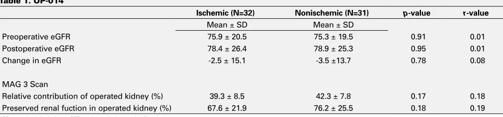
Methods: Between September 2009 to October 2011, 34 consecutive patients underwent laparoscopic partial nephrectomy with early unclamp-ing with mean clamp time of 19.04 minutes. This was followed by 30 consecutives patients undergoing laparoscopic partial nephrectomy with zero ischemia. All cases were performed by a single surgeon. We retro-spectively reviewed records of all 64 patients. Patients with solitary kid-neys and patients with significant renal parenchyma disparity on preop CT scan were excluded from the study. Demographics, tumor pathology, sur-gical margins, perioperative complications, pre- and postoperative eGFR and postoperative MAG 3 Scans were compared between both groups. Results: See Table 1.
Conclusions: Although no statistically significant differences were found, the difference between the two groups was more profound for the MAG 3 Scan variables. Associated r-values show small to medium-sized effects in this regard, which point towards a trend of improved renal function preservation in the zero ischemic group. Larger multicentre trials are required for future research in this area.
UP-015
Reduction of Renal Parenchymal Volume and Renal Function in Patients after Partial Nephrectomy for Renal Cell Carcinoma (RCC)
Ajzenberg, Henry1; Legere, Laura1; Satkunasivam, Raj2; Finelli, Antonio1;
Kachura, John3; O’Malley, Martin3; Tuchscherer, Paul4; Quinn, Paul4; Choi,
Jung4; Jewett, Michael A.S.1
1Princess Margaret Hospital, Toronto, ON, Canada; 2University of Toronto,
Toronto, ON, Canada; 3Toronto General Hospital, Toronto, ON, Canada; 4Toronto Western Hospital, Toronto, ON, Canada
Introduction and Objectives: Nephrectomy and partial nephrectomy (PN) for early-stage RCC produce similar oncological outcomes. However, the loss of normal kidney tissue, or renal parenchymal volume (RPV) may increase the risk of renal dysfunction. The use of PN is increasing and the extent of renal dysfunction attributable to RPV loss is not fully understood. Methods: 31 patients who underwent PN for RCC at the University Health Network were retrospectively studied. Demographic, surgical, and renal functional data were extracted from patient charts. Glomerular
filtra-tion rate (GFR) was estimated using the Modificafiltra-tion of Diet in Renal Disease (MDRD) equation and plasma creatinine concentrations. Changes in pre- and postoperative GFR were quantified (ΔGFR). RPV loss (ΔRPV) was determined using pre- and postoperative CT or MR scans on Vitrea volumetric software. Non-tumor kidney tissue on each axial slice was manually traced. A correlation between ΔRPV and ΔGFR was assessed. Results: There were 18 males and 13 females with a mean age of 57 years (range 28-80). Mean tumor diameter was 3.6 cm (range 1.3-7.5). Mean pre- and postoperative RPV was 160.9 (±33.7, range 81.4-229.5) and 144.2 mL (±36.8, range 61.6-199.8), respectively. Mean ΔRPV was -10.6% (±14.8) or -16.7 mL (±25.1, range -87.1-25.1). Mean preoperative GFR was 86.3 ml/min/1.73m2 (±20.1, range 53.2-128.3). Twelve months
postoperatively, mean GFR was 80.1 ml/min/1.73m2 (±26.5, range
37.7-144.91). Mean ΔGFR was -8.1% (±20.9) or -6.6 ml/min/1.73m2 (±18.1,
range -68.6-25.4). Using a linear regression, we found no correlation between ΔRPV and ΔGFR (r2=.002, p=0.812).
Conclusions: PN does lead to renal dysfunction, but RPV loss does not fully account for this. We found no correlation between ΔRPV and ΔGFR. Patient comorbidity, ischemia/reperfusion injury, and compensatory renal reserve may have a larger impact on GFR than minimal loss of RPV with PN. We are increasing our sample to further explore this relationship.
UP-016
Results of a Survey on the Management of Upper Tract Urothelial Cancer
Zappavigna, Christopher; Rowe, Neal; Morash, Christopher G.; Breau, Rodney H.; Cagiannos, Ilias
University of Ottawa, Division of Urology, Ottawa, ON, Canada Introduction and Objectives: The importance of regional lymphadenec-tomy has been well established in the management of bladder cancer. Considerable uncertainty exists regarding its role and utility in the treat-ment of upper urinary tract transitional cell carcinoma (UTTCC). A recent study has showed that the incidence of lymphatic involvement varied according to stage and grade. Our objective is to survey, study, examine and discuss the current management of UTTCC in Canada. Specically, what are the current practices for the management of UTTCC and when do we utilize lymphadenectomy?
Methods: A 6-page survey was created and sent electronically to all Canadian Urological Association (CUA) members. Data collected include physician demographics, UTTCC management practices and current prac-tices for monitoring tumor recurrence and postoperative renal function. We studied the current practices for the management of UTTCC with a specific focus on lymphadenectomy. Moreover, we will also compile and evaluate data based on the current practices for monitoring tumor recurrence and postoperative renal function.
Results: A total of 27 urologists responded. 32% of respondents would do a lymphadenectomy for Ta/Tis/T1 disease and 64% would do a lymph-adenectomy for T2 or greater disease. If presented with survival data favoring Retroperitoneal Lymph Node Dissection (RPLND) for Ta/Tis/T1 disease, 55% would change their management and 86% would change their management for T2 disease.
Table 1. UP-014
Ischemic (N=32) Nonischemic (N=31) p-value r-value
Mean ± SD Mean ± SD
Preoperative eGFR 75.9 ± 20.5 75.3 ± 19.5 0.91 0.01
Postoperative eGFR 78.4 ± 26.4 78.9 ± 25.3 0.95 0.01
Change in eGFR -2.5 ± 15.1 -3.5 ±13.7 0.78 0.08
MAG 3 Scan
Relative contribution of operated kidney (%) 39.3 ± 8.5 42.3 ± 7.8 0.17 0.18
Preserved renal fuction in operated kidney (%) 67.6 ± 21.9 76.2 ± 25.5 0.18 0.19
Conclusions: Clearly, there is much debate over role of lymphadenectomy in this disease. The relatively low frequency of these lesions and the lack of prospective randomized trials do not permit absolute conclusions about treatment impact on outcomes. Furthermore, it is questionable whether a randomized controlled trial is feasible. Overall, our results suggest the need for a well-defined role for role of lymphadenectomy in the manage-ment of upper tract urothelial carcinoma.
UP-017
Management of Collecting Duct Carcinoma: a Systematic Review, Management Approach, and Case Series
Dason, Shawn1; Sheridan-Jonah, Anna1; Allard, Christopher1; Kajal,
Babita2; Aziz, Tariq2; Gill, Jaskirat3; Jamshaid, Hira3; Kapoor, Anil1 1Division of Urology, McMaster University, Hamilton, ON, Canada; 2Department of Pathology and Molecular Medicine, McMaster University,
Hamilton, ON, Canada; 3McMaster University, Hamilton, ON, Canada
Introduction and Objectives: Collecting duct carcinoma (CDC) is a rare subtype of renal carcinoma. It is aggressive, presents symptomatically at an advanced stage, and has a poor prognosis. Little is known on what constitutes optimal management of CDC. The aim of this study is to develop an evidence-based approach to managing CDC.
Methods: Ovid Medline, The Cochrane Library, EMBASE, MacPLUS FS and conference proceedings (via Web of Science) were searched to iden-tify studies relevant to the management of CDC. A systematic search strategy was developed and applied. Included studies had a minimum of 10 subjects receiving a single intervention. Series in which an evaluation of therapeutic effectiveness was not possible were excluded. An algorithm based on this review for approaching multidisciplinary CDC management is then presented. Four consecutive cases of CDC treated at our tertiary institution between 2006 and 2010 are then related to this algorithm. Results: Our systematic review identified 3 studies relevant to the manage-ment of CDC. Firstly, a gemcitabine/cisplatin or carboplatin (GC) regimen resulted in a 26% partial or complete response rate in a phase II study of 23 patients with metastatic CDC. Two additional studies indicated that 49
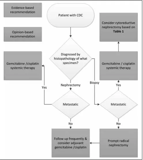
patients treated with immunotherapy achieved no response. A management algorithm based on these findings is presented in Figure 1. Four cases of advanced CDC are then reported. Two patients were unresponsive to MVAC therapy. Cytoreductive nephrectomy (CN) was performed in 2 patients. Performance status and survival were uniformly poor. Management of these cases in relation to our algorithm (Fig. 1) is discussed.
Conclusions: CDC responds to systemic therapy similarly to urothelial carcinoma. Our review suggests that the current standard of care for metastatic CDC is a (GC) regimen. A framework for applying CDC man-agement principles and considering CN based on this review is provided and applied to the 4 reported cases.
UP-018 – WITHDRAWN
UP-019
Improving Outcomes Through the Development of Quality Indicators (QI) in Renal Cell Cancer (RCC)
Finelli, Antonio1; Bjarnason, Georg2; Black, Peter3; Cagiannos, Ilias4; Heng,
Daniel5; Kapoor, Anil6; Kollmannsberger, Christian7; Mohammadzadeh,
Forough8; Moore, Ronald9; Soulieres, Denis10; Tanguay, Simon11; Venner,
Peter12; Jewett, Michael A.S.1; Rendon, Ricardo13; Wood, Lori13
1Princess Margaret Hospital (UHN), Toronto, ON, Canada; 2Sunnybrook
Odette Cancer Centre, Toronto, ON, Canada; 3Department of Urological
Sciences, University of British Columbia, Vancouver, BC, Canada;
4University of Ottawa, Ottawa, ON, Canada; 5Tom Baker Cancer
Centre, University of Calgary, Calgary, AB, Canada; 6Juravinski Cancer
Centre, Hamilton, ON, Canada; 7BCCA Vancouver Cancer Centre, UBC,
Vancouver, BC, Canada; 8UHN, Toronto, ON, Canada; 9Department of
Urology, University of Alberta, Edmonton, AB, Canada; 10Department
of Medicine, Université de Montréal, Montreal, QC, Canada; 11McGill
University Health Centre, McGill University, Montreal, QC, Canada;
12Cross Cancer Institute, University of Alberta, Edmonton, AB, Canada; 13QEII HSC, Dalhousie University, Halifax, NS, Canada
Introduction and Objectives: Optimal quality of care is needed for ideal outcomes, and QI are used to measure quality of care. In RCC, there is a lack of information defining such optimal care. This is especially important as RCC care becomes more complex, with ongoing advances requiring greater expertise. The goal of the study was to identify QI for RCC across the disease spectrum from presentation to palliation.
Methods: A multidisciplinary expert panel of 13 urologic and medical oncologists from across Canada reviewed potential QI. These QI were identified from a systematic literature review. Panel members were also asked to suggest additional potential QI. A modified Delphi technique was used to select QI that were relevant and practical to RCC; this technique incorporated 2 email questionnaires and 1 in-person meeting.
Results: From 233 literature citations, 34 possible QI were identified; 24 additional potential QI were suggested. A final set of 23 QI was estab-lished. These are distributed across the RCC disease spectrum as follows (number of QI in parentheses): screening (1), diagnosis/prognosis (3), surgical management of localized disease (6), surgical management of advanced disease (3), systemic therapy (5), and follow-up (3). These 21 QI focused mostly on the treatment of RCC. In addition, two QI related to survival outcomes (overall and progression-free) were selected. Examples of QI include: the proportion of patients undergoing partial nephrectomy for tumors <4 cm and the proportion of patients with advanced disease who are assessed by a multidisciplinary genitourinary cancer team. The final 23 QI selected will be presented in detail.
Conclusions: A systematic, consensus-based approach was used to deter-mine relevant QI in RCC care. These 23 QIs will provide a means of evaluating the quality of RCC care in an effort to improve outcomes in our patients. The next step will be to establish a means of measuring each of these QI based on defined or yet to be defined benchmarks.
UP-020
Rising Incidence of Upper Tract TCC (UTTCC) in Kettering, United Kingdom
Khan, Faisal1; Payne, David2; Al-Sudani, Mohammed2; Khan, Zeb2;
England, Roland2
1Freeman Hospital, Newcastle, United Kingdom; 2NHS, Kettering, United
Kingdom
Introduction and Objectives: Tumors of the renal pelvis are rare and account for approximately 10% of all renal tumors and approximately 5% of all urothelial tumors. Ureteral tumors are even more uncommon, occurring with one quarter the frequency of renal pelvis tumors. In our hospital we experienced rising incidence of UTTCC as well increase in ureteric TCC. To confirm this we retrospectively analysed the data. Methods: We collected our data retrospectively from 2005 to August 2011.The cases were identified via theatre, histopathology and MDT meeting records. We analyzed known risk factors, investigations, treat-ment, final histology and associated bladder tumor.
Results: Total of 41 patient were diagnosed with UTTCC between 2005 to March 2011.Most of the patients were male (33) with male to female ratio of approx 8:1. Mean age at Ist presentation was 64 years (range 42 to 92 years). Most of the patients were either smoking actively or had stopped smoking (n=28). The analysis revealed rising incidence of UTTCC in Kettering as shown in table form. Of note we also recorded rising incidence of Ureteric TCC then pelvic TCC in these patients.
Conclusions: Our project showed rising incidences of UTTCC especially ureteric tumors. We postulate the rising trend could be secondary to com-bination of strong presence of leather and service industries in Kettering area or secondary to improved diagnostics tests.
UP-021
The Fer Kinase, a Nuclear Effector of Growth-promoting Factors Favoring Castration-resistant Prostate Cancer
Rocha, Joice1; Zouanat, Fatima1; Benidir, Tarik1; Zoubeidi, Amina2; Hamel,
Lucie1; Scarlata, Eleonora1; Aprikian, Armen1; Chevalier, Simone1 1Urologic Oncology Research Group, McGill University, Montreal, QC,
Canada; 2The Vancouver Prostate Cancer, University of British Columbia,
Vancouver, BC, Canada
Introduction and Objectives: Death from prostate cancer (PC) arises from castration-resistant (CR) metastatic disease, developing from an increasing cell ability to survive and thrive in response to a variety of growth-promoting factors (GFs) signaling through tyrosine kinases (TKs). The androgen receptor (AR) itself has appeared as a TK substrate in PC cells responding to epidermal growth factor (EGF), interleukin (IL)-6 and even androgens. As we reported that the Fer TK controls IL-6 signaling, we aimed to verify if Fer further contributes to aberrant signaling by additional GFs controlling AR activation in PC.
Methods: LNCaP cell survival/growth and death were measured by MTT assays and propidium iodide (PI) staining, respectively. Fer down-regula-tion was achieved via siRNAs and PSA was measured by Real Time PCR. Tyrosine (Y) phophorylation, protein interactions and cellular localization were assessed.
Results: Fer was responsible for the LNCaP cell response to IL-6, since fer siRNA reduced growth by 90%. Fer also controlled 50%, 44%, 36% and 32% of the response to FBS (fetal bovine serum), R1881 (androgens), Insulin-like (I) GF-1 and EGF, respectively. Fer knockdown also resulted in 40% less cells when cultured without growth stimuli. Cell death was confirmed by PI staining. Moreover the full R1881 and IL-6 PSA response depended on Fer. IL-6 and R1881 were most potent to increase Fer and AR pY-levels along with their nuclear accumulation as observed in prostate tumors. AR pY-levels were modulated by Fer, both within cells and in kinase assays. Finally, Y223 appeared as a novel functional site preferred by the Fer TK and this AR motif was involved in the interaction with the Fer-SH2 domain.
Conclusions: The up-regulated nuclear Fer in PC appears to intervene in pathways triggered by several GFs contributing to aberrant signaling in CRPC.
UP-022
Single Photon Emission Computed Tomography/CT Imaging of Metastases Using Prostate Specific Membrane Antigen Antibody
Chevalier, Simone1; Derbekyan, Vilma1; Scarlata, Eleonora1; Moffett,
Serge2; Hamel, Lucie1; Zouanat, Fatima1; Aprikian, Armen1; Anidjar,
Maurice1
1McGill University Health Centre Research Institute, Montreal, QC,
Canada; 2ProScan, Montreal, QC, Canada
Introduction and Objectives: Current imaging methods to detect prostate cancer (PC) metastases (mets) lack sensitivity and specificity. Reliable cancer-imaging modalities are needed. Prostate Specific Membrane Antigen (PSMA) is an attractive target for molecular imaging in virtue of its distribution and overexpression in castrate-resistant (CR) PC. We aimed to test a labeled monoclonal antibody (mAb) directed against an extracellular subdomain of human and canine PSMA to detect mets in the dog prostate cancer (DPC)-1 model by single photon emission computed tomography (SPECT)/CT imaging.
Methods: DPC-1 cells were implanted in immuno-suppressed (cyclo-sporine) dogs (n=5). SPECT/CT was repeated during follow-up, 2 days after i.v. injection of 111Indium(In)-PSMA mouse mAb. In some instances,
bone scan (99mTc-MDP) was performed. Controls included imaging
prior (99mTc-MDP, 111In-PSMA) and post (111In-mouse immunoglobulins)
DPC-1 implantation. The prostate, sacroiliac lymph nodes (LNs), lungs and selected bone segments were harvested for gamma counting and pathological analyses.
Results: Four dogs developed prostate tumors and mets in LNs and lungs; 3 had bone mets. SPECT revealed uptake of 111In-PSMA radiotracer in
mets, yet DPC-1 tumors in the prostate remained negative. Mets were imaged as early as by 6-8 weeks and grew during follow-up. CT fusion images indicated enlarged LNs, often necrotic at necropsy, and for which tracer accumulation and tumor positivity were confirmed. Uptake of radiotracer was also detected in lungs and bones (one case studied), confirming a positive bone scan. Controls were negative.
Conclusions: Molecular imaging by SPECT/CT with 111In-PSMA radiotracer
was proven efficient and specific to detect soft tissue and bone mets in the pre-clinical DPC-1 model closely mimicking CRPC, implying feasibility in the clinical setting.
UP-023
PSA Bounce Following Prostate Brachytherapy for Clinically Localized Prostate Adenocarcinoma: a Single Institution Study with Minimum Three Years Follow-up
Nayak, Jasmir; Ong, Aldrich; Bews, Jeff; Chowdhury, Amit; Drachenberg, Darrel
University of Manitoba, Winnipeg, MB, Canada
Introduction and Objectives: Prostate specific antigen (PSA) is a sen-sitive serological marker of outcome following prostate brachytherapy. Following brachytherapy, PSA levels may transiently rise in a phenom-enon known as PSA bounce (PB). We report on PB following permanent radio-iodine (125I) brachytherapy and correlate PB with both clinical and
dosimetric variables.
Methods: We analyzed 145 patients with clinically localized, T1-2c N0 M0 prostate adenocarcinoma treated with brachytherapy with a minimum follow-up of 3 years. Six different PSA thresholds were used to define PB: a increase of ≥0.1 ng/ml (definition I), ≥0.2 ng/ml (definition II), ≥0.3 (definition III) ng/ml, ≥0.4 ng/ml (definition IV), ≥0.5 ng/ml (definition V) and ≥1.0 ng/ml (definition VI) with spontaneous return to ≤pre-bounce levels. Biochemical failure (BF) was defined according to the American Society for Therapeutic Radiology and Oncology Phoenix definition of a rise of ≥2 ng/mL above the nadir.
patients using definitions I, II, III, IV, V and VI respectively. Nine patients (6.2%) had true failure. All definitions of PB occurred earlier than BF (p<0.05). Univariate and multivariate analysis revealed that age <65 and Gleason sum ≤6, were statistically significant predictors of PSA bounce for all definitions, and percent positive biopsies (<25%) for definitions I to IV. Conclusions: PB is a common phenomenon post-brachytherapy. Age <65, Gleason sum 6 and lower volume disease are predictors of PB. The time to first PSA rise can help to distinguish between PB and BF.
UP-024
Is Perineural Invasion in Prostate Biopsies Associated with Adverse Pathological Outcomes? Old Paradigm Revisited
Margel, David1; Elharram, Malik1; Finelli, Antonio1; Zlotta, Alexandre R.1;
Trachtenberg, John1; Evans, Andrew2
1Division of Urology, Department of Surgical Oncology, University Health
Network, University of Toronto, Toronto, ON, Canada; 2Department of
Pathology, University Health Network, University of Toronto, Toronto, ON, Canada
Introduction and Objectives: To determine the role of perineural invasion (PNI) on prostate biopsy in predicting adverse findings at radical prosta-tectomy in a recent cohort of screen detected prostate cancer.
Methods: We analyzed 1041 consecutive patients from a prospectively maintained database. Prostate cancer was diagnosed in 470, and 138 of these patients underwent radical prostatectomy. Pathological specimens were examined, and perineural invasion was identified as carcinoma tracking along or around a nerve in the perineural space. We investigated the predictive value of PNI on biopsy with PNI on radical prostatectomy as well as the ability of PNI on prostate biopsy to predict adverse findings at radical prostatectomy.
Results: Perineural invasion was present in 124 (26%) of biopsy specimens diagnosed with prostate cancer and 38 (27%) of those who chose radical prostatectomy. Perineural invasion on prostate needle biopsy was not predictive of radical prostatectomy Gleason score (p=0.377), pathologi-cal stage (p=0.852), extraprostatic extension (p=0.258), surgipathologi-cal margin (p=0.079), lymphovascular invasion (p=0.499) , and upgrading (p=0.514) or downgrading (p=0.208) at radical prostatectomy. The sensitivity, speci-ficity, positive predictive value, and negative predictive value of PNI on biopsy for PNI on radical prostatectomy were 32%, 82%, 79%, and 37% respectively. The Cohen’s Kappa correlation coefficient was 0.11. Conclusions: Perineural invasion on prostate needle biopsy is not predic-tive of radical prostatectomy outcome. Furthermore, perineural invasion on biopsy has limited predictive value for perineural invasion at radical prostatectomy.
UP-025
The Association between Male Pattern Baldness and Second to Forth Finger Ratio with Prostate Cancer: A Prospective Cohort Study
Margel, David; Venkateswaran, Seetha; Darwish, Abbas; Chadwick, Karen; Fleshner, Neil
Division of Urology, Department of Surgical Oncology, University Health Network, University of Toronto, Toronto, ON, Canada
Introduction and Objectives: Retrospective case control studies have demonstrated an association between male pattern baldness and 2D:4D ratio and prostate cancer. The aim of this study was to validate these findings in a prospective cohort.
Methods: Upon approval from our ethical review board we prospectively enrolled 196 consecutive patients referred to a prostate biopsy. Finger lengths were measured using a digital vernier calliper, and the 2D:4D ratio was calculated. Male pattern baldness was assessed on a scale of 0-4 using the standardized Norwood classification (0= no balding, 1= frontal balding, 2= Mild vortex, 3=moderate vortex and 4=severe vortex). We performed all measurements prior to the biopsy thus blinded to the pathology outcome. We used Univariable and multivariable analysis to associate 2D:4D ratio and male pattern baldness with prostate cancer. The multivariable model included the two main predictors (male pattern baldness and 2D:4D ratio) as well age, digital rectal examination and PSA.
Results: The median (IQR) age and PSA of our cohort was 64 (59-70) and 5.8 (4.1-8.4), respectively. Overall 109 patients (55%) were diagnosed with prostate cancer. On univariable analysis male pattern baldness was associated with prostate cancer (p for trend=0.03). However 2D:4D ratio was not. On multivariable analysis male pattern baldness remained a significant predictor of prostate cancer. Furthermore, we noted a dose response effect- the more severe balding patterns were more strongly associated with prostate cancer (Frontal balding OR 2.0 (95%CI 1.1-6.6); mild vortex OR 2.1 (95%CI 1.5-5.2); moderate vortex OR 2.5 (95%CI 1.2-7.1); severe vortex OR 2.9 (95%CI 1.1-4.3).
Conclusions: In a prospective cohort we found that male pattern bald-ness was an independent predictor of prostate cancer. Further studies are needed in order to assess whether the inclusion of male pattern bald-ness can contribute to existing models to predict prostate cancer prior to biopsy.
UP-026
Coexisting Prostate Cancer Found at the Time of Holmium Laser Enucleation of the Prostate for Benign Prostatic Hyperplasia: Predicting Its Presence and Grade in Analyzed Tissue
Bhojani, Naeem; Boris, Ronald S.; Mandeville, Jessica A.; Lingeman, James E.
Indiana School of Medicine, Department of Urology, Indianapolis, IN, United States
Introduction and Objectives: Since 1996, HoLEP has been a well-accepted surgical option for benign prostatic hyperplasia, mimicking anatomic results of open prostatectomy with removal of the entire tran-sition zone. A portion of these men will harbor prostate cancer (PCa) in their analyzed tissue and may go on to subsequent therapy. Establishing risk factors for PCa at the time of HoLEP may aid in preoperative patient counseling. Our study identifies patients with PCa in a large cohort of men undergoing HoLEP and attempts to identify usable variables to predict either PCa or Gleason score at the time of surgery.
Methods: We performed a retrospective data analysis of HoLEP patients at a single institution between 1998 and 2011. At the discretion of the referring urologist, patients with elevated PSAs and/or abnormal digital rectal exams had prior negative preoperative biopsies. Different preop-erative and postoppreop-erative variables were examined using univariate and multivariate logistic and linear regression models.
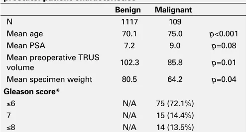
Results: Overall, of 1226 HoLEP patients, 109 (8.9%) had PCa found on tissue analysis (Table 1). Almost ¾ of diagnosed PCa was low grade (≤Gleason 6). After univariate and multivariate analysis, only age and weight of specimen were found to predict the presence of PCa (Table 2). More specifically, the risk of PCa increased with increasing age and decreasing gland size. Only rising PSA was predictive of higher Gleason scores on mulitivariate analysis at the time of HoLEP (Table 3).
Table 1. UP-026. Holmium laser enucleation of the prostate: patient characteristics
Benign Malignant
N 1117 109
Mean age 70.1 75.0 p<0.001
Mean PSA 7.2 9.0 p=0.08
Mean preoperative TRUS
volume 102.3 85.8 p=0.01
Mean specimen weight 80.5 64.2 p=0.04
Gleason score*
≤6 N/A 75 (72.1%)
7 N/A 15 (14.4%)
≤8 N/A 14 (13.5%)
Conclusions: The coexistence of PCa found at the time of HoLEP is low and the majority of patients with cancer will have low grade disease. Older patients with smaller glands appear to be at the highest risk of harboring PCa. In this group of patients only preoperative PSA values appears to influence the presence of more aggressive disease.
UP-027
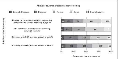
Prostate Cancer Screening: Attitudes and Practices of Family Physicians in Ontario
Allard, Christopher1; Lusis, Janis2; Dason, Shawn1; Kapoor, Anil1 1McMaster Institute of Urology, Hamilton, ON, Canada; 2Brampton
Hospital, Brampton, ON, Canada
Introduction and Objectives: The utility of prostate cancer (PCa) screen-ing is controversial. We sought to determine whether family physicians in
Ontario, Canada, believe PCa screening is beneficial and to characterize their screening protocols.
Methods: A survey was developed with input from urologists, family physicians, and the Ontario Medical Association’s Section on General and Family Practice. Questions covered three domains: demographics, beliefs about screening utility and screening practices. All 7,302 family physicians in Ontario were invited by email to complete the online survey. Results: A total of 969 physicians completed the survey; 955 (52.0% male, 48.0% female) were included. Most (78.9%) use PSA and DRE for screening; 9.1% use DRE alone and 7.0% PSA; 2.4% incorporate transrec-tal ultrasound. 8.3% do not offer PCa screening. Most physicians begin offering screening at age 50 (72.9%) and stop at ages 70 or 80 (68.4%); 4.3% offer screening up to age 90 and 17.9% offer lifelong screening. 54% offer the same amount of screening as they did 5 years ago, while
Table 3. UP-026. Univariate and multivariate analysis predicting Gleason Score
Gleason Score Univariate
Age 0.2 (p=0.05)
PSA 0.29 (p=0.04)
Weight of specimen -0.16 (p=0.1)
Preoperative TRUS volume -0.15 (p=0.21)
History of biopsy -0.07 (p=0.5)
Multivariate
Age 1.49 (p=0.14)
PSA 0.31 (p=0.01)
Weight of specimen -0.36 (p=0.14)
Preoperative TRUS volume 0.17 (p=0.47)
History of biopsy -0.01 (p=0.9)
TRUS: transrectal ultrasound; PSA: prostate-specific antigen.
Statement about screening
Responses in each category
Strongly Disagree
Prostate cancer screening should be routinely recommended to men beginning at age 50
The benefits of prostate cancer screening outweigh the risks
Screening with PSA provides a survival benefit
Screening with DRE provides a survival benefit
Disagree Neutral Agree Strongly Agree
Attitudes towards prostate cancer screening
69 168 170 358 183
45 163 252 364 122
62 292 243 337 73
89 219
0% 25% 50% 75% 100% 280 310 44
Fig. 1. UP-027. PSA: prostate-specific antigen; DRE: digital rectal examination. Table 2. UP-026. Univariate and multivariate analyses predicting presence of cancer
Malignancy Univariate
Age 1.08 (p<0.001)
PSA 1.01 (p=0.1)
Weight of specimen 0.99 (p=0.004)
Preoperative TRUS volume 0.99 (p=0.03)
History of biopsy 0.93 (p=0.85)
Multivariate
Age 1.09 (p<0.001)
PSA 1.02 (p=0.14)
Weight of specimen 0.99 (p=0.01)
Preoperative TRUS volume 1.0 (p=0.97)
History of biopsy 1.62 (p=0.25)
19.5% offer more and 13.8% less. Physician beliefs about the utility of PCa screening are shown in Fig. 1.
Conclusions: Although 91.3% of respondents offer PCa screening, they are divided over its utility, with only 51.4% convinced that the benefits outweigh the harms. The publications in 2009 of two large randomized controlled trials had a negligible impact on the amount of screening per-formed by respondents. There is significant variability between physicians’ screening beliefs and protocols. A limitation of this study is the possibility of selection bias. Nevertheless, this is the largest sample of Ontario family physicians ever surveyed about PCa screening and highlights divergent physician practices and a need for more conclusive evidence on the subject of screening utility.
UP-028
Transrectal Ultrasound with Vibroelastography for the Detection of Prostate Cancer
Gagnon, Louis-Olivier1; Mahdavi, Sara2; Moradi, Mehdi2; Baghani, Ali2;
Jones, Edward3; Salcudean, Septimiu2; Goldenberg, Larry1
1Department of Urologic Sciences, University of British Columbia,
Vancouver, BC, Canada; 2Electrical and Computer Engineering, University
of British Columbia, Vancouver, BC, Canada; 3Department of Pathology
and Laboratory Medicine, University of British Columbia, Vancouver, BC, Canada
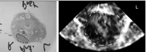
Introduction and Objectives: Vibro-elastography (VE) is a promising tech-nique for imaging soft tissues and relies upon measuring tissue strain in response to a mechanical excitation. The aim of this study is to evaluate the feasibility of detecting cancer within the prostate from VE images. Methods: Transrectal ultrasound with VE was performed intra-operatively, prior to the prostatectomy, on patients diagnosed with prostate cancer. Transfer function images of the prostate, showing the relative stiffness of the tissue within and surrounding the prostate were created. For each case, 9-13 pathology slides extracted from the prostate at approximately 4-mm intervals, with cancer marked, were available. Areas suspected for cancer were marked on the VE images and then compared to the pathology results.
Results: These are preliminary results on 5 patients. Analysis is pending on 5 other patients and recruitment will continue in the coming months. Gleason scores for 51 cancerous areas were available. Twenty of the 31 tumors with Gleason scores of 3+3 (64.5%), 13 of the 16 tumors with Gleason scores of 3+4 (81.25%), both tumors with Gleason scores of 4+3 with tertiary 5 (100%) and both tumors with Gleason scores of 4+5 (100%) were detected. For example, in Figure 1, a tumor of Gleason score 3+4 is identified. The tumor is 12 mm by 6 mm and has an area of 60 mm2. Overall, VE had a sensitivity of 72.5% for detecting prostate cancer, with a false negative and a false positive percentage of 24.4% and 37.3% respectively. The sensitivity of VE for detecting cancer increased as the Gleason score increased, with a sensitivity of 85% for tumors with a Gleason score of 7 and above.
Conclusions: This study shows that the use of additional information from VE has the potential of improving the detection of prostate cancer, espe-cially for cancers of higher grade. This imaging method could aid prostate biopsy by highlighting areas suspicious for cancer, reducing the need for repeated biopsy procedures.
UP-029
Human Prostate Cancer Magnetic Resonance Elastography and Correlation with Histology
Gagnon, Louis-Olivier1; Sahebjavaher, Ramin2; Garteiser, Philippe3;
Sinkus, Ralph3; Baghani, Ali2; Moradi, Mehdi2; Jones, Edward4; Nguan,
Christopher G.1; Goldenberg, Larry1; Salcudean, Septimiu2
1Department of Urologic Sciences, University of British Columbia,
Vancouver, BC, Canada; 2Electrical and Computer Engineering, University
of British Columbia, Vancouver, BC, Canada; 3Hôpital Beaujon, Centre
de Recherche Biomédicale Bichat Beaujon (CRB3), Paris, France;
4Department of Pathology and Laboratory Medicine, University of British
Columbia, Vancouver, BC, Canada
Introduction and Objectives: The aim of this study is to use magnetic reso-nance elastography (MRE) methods to identify cancerous tumors of the prostate and correlate them to the whole mount histopathology marked with the Gleason score.
Methods: Ethics board approval and informed consent was obtained from a patient (first in study of N=20) of age 61 scheduled for radical prosta-tectomy. The experiments were performed on a 3T Achieva scanner. The vibrations were applied to the perineum using a custom made electro-magnetic driver. For the anatomy images, a standard axial T2 weighted FSE sequence was performed. The MRE images were acquired in the axial plane using a novel fast field echo sequence named eXpresso. The wave images were acquired on a matrix with 2 mm isotropic voxel size. Eight vibration phases were encoded at a mechanical excitation of 70 Hz. Results: The peak amplitude of the mechanical wave was 130µm with a mean of 25µm in the prostate. No patient discomfort was reported when specifically asked. In the axial plane, the prostate gland is outlined in the T2W (a), and the reconstructed shear modulus G’ (b) and loss modulus G” (c). The histology is shown in (d) where the outline of the large (Gleason score of 4+3) and smaller (3+3) tumors are shown. A very promising correspondence between reconstructed shear and loss moduli G’ and G” and the matching histology slide can be observed. The mean values of G’ were 3.0, 1.6, and 0.8 kPa for Gleason scores of 4+3, 3+3, and healthy tissue, respectively. Also, the mean values for G” were 1.7, 0.8, and 0.4 kPa for the same regions of interest.
Conclusions: This is, to the best of our knowledge, the first in-vivo pros-tate cancer patient MRE images that are correlated with whole mount histology. These early results confirm that cancerous tissue in prostate specimens has higher stiffness and also higher viscosity compared to healthy tissue. MRE is a promising tool in order to improve diagnosis and staging of prostate cancer tumors.
UP-030
Changes in Positive Surgical Margins over Time Reflect a Risk Migration in Patients Undergoing Radical Prostatecomy at a Tertiary Care Centre
Longpre, Michelle; Gleave, Martin; Harriman, David; Hurtado-Coll, Antonio
University of British Columbia, Vancouver, BC, Canada
Introduction and Objectives: Positive surgical margin (PSM) rates after radical prostatectomy (RP) have been shown to be as low as 4% and as high as 48% in high volume centres. The incidence of PSM varies by pathological stage but can also be influenced by surgical techniques and by pathologist interpretation. PSM are predictive of biochemical recur-rence but a significant proportion will not recur. In this study we sought to determine changes in incidence of PSM rates over time as lower risk patients opt for active surveillance and whether this was related to a shift in pathological stage and grade.
Methods: PSM, clinical and pathological staging, as well as PSA recur-rence in almost 2,000 patients who underwent RP from 1993 to present was extracted from a prospectively recorded prostate cancer database at Vancouver General Hospital, BC, Canada. Patients were grouped by their date of surgery into either remote (1993-2004) or recent (2005-2011) cohorts. All pathological pT2 and greater patients were included. Regression models were developed to predict the rate PSM with adjust-ment for known confounders.
Results: Remote (765) and recent (1,216) radical prostatectomies were compared. The PSM rate was 21.7% from the remote cohort and 27% from the recent cohort (p=0.027). The postoperative Gleason grades shifted significantly from 45.97% Gleason 6, 43.83% Gleason 7, and 10.20% Gleason greater than 8 in the remote cases to 17.4%, 67.46% and 15.30% respectively in the recent cases (p≤0.0005). The percentage of patients with clinically high-risk disease was also significantly greater in the recent cohort (p≤0.0005).
Conclusions: The PSM rates in our tertiary care high volume centre are similar to other reported centres. The increase in PSM rates is likely due to a shift in patient population from lower risk patients to higher risk patients as lower risk patients opt for active surveillance.
UP-031
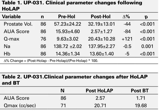
Holmium Laser Ablation of the Prostate Gland (HoLAP) Followed by Brachytherapy (BT) for Treatment of Patients with Clinically Localized Prostate Carcinoma (PC) and Obstructive Urinary Symptoms (LUTS)
Kumar, Surendra
Oakwood Annapolis Hospital, Wayne, MI, United States
Introduction and Objectives: Patients with localized prostate carcinoma and LUTS who undergo BT with permanent seed implants have a 40 to 50% chance of going into urinary retention or requiring a catheter for intermittent self-catheterization (ISC) due to worsening of their obstruc-tive urinary symptoms. The purpose of this study was to assess the role of HoLAP prior to BT in preventing post BT urinary complications. Methods: 86 patients age 50-83 (mean 67.6) years with LUTS and clinically localized prostate carcinoma underwent HoLAP with a 100W holmium laser under spinal anesthesia. The end point of the procedure involved complete vaporization of obstructing prostate tissue down to the capsular fibers and appearance of an open prostate cavity. Patients under-went BT 7-45 (mean 16) weeks after HoLAP when they had recovered from the procedure. BT was done by real-time interactive ultrasound-guided (Iodine-125 or Palladium-103) seed placement with peripheral loading under general anesthesia.
Results: HoLAP significantly reduced prostate volume an average 25cc (44%), reduced the mean AUA symptom score by 13 points (84%) and increased mean Q-max by 11cc/sec (121%) (Table 1). There was no clini-cally significant change in AUA symptom score or Q-max following BT with a follow-up of 0.43-6.91 (mean 3.65) years (Table 2). No patient experienced prolonged urinary retention after BT and none has required ISC. No patient developed stress incontinence after HoLAP and BT. Conclusions: Patients with prostate carcinoma and LUTS who have under-gone HoLAP to relieve their obstructive urinary symptoms prior to BT
do not experience prolonged urinary retention, worsening of their LUTS or stress incontinence following real-time interactive ultrasound-guided radiation seed placement with peripheral loading. With elimination of obstructive urinary symptoms and an approximately 44% reduction in the prostate volume by HoLAP a larger pool of patients with prostate carcinoma can benefit from modern brachytherapy.
UP-032
Identification of Thrombotic Risk for Men with Advanced Prostate Cancer: a Pilot Study Evaluating Hemostatic Status Using Thromboelastography
Siemens, D. Robert; Toukh, Mazen; Othman, Maha; Black, Angela; Graham, Charles
Queen’s University, Kingston, ON, Canada
Introduction and Objectives: Coagulopathy is the second most common cause of death from cancer, and thrombotic complications are amplified in prostate cancer with systemic therapy. We aim to help identify patients at higher risk for thrombotic events in patients with prostate cancer with well-defined hemostatic tests, novel in their application to patients with advanced prostate cancer.
Methods: We performed intensive haemostatic studies in 27 patients (age range 59-88 years) at various stages (non-metastatic, metastatic, castration resistant) as compared to an age-matched control group (biopsy negative, n=9). Thromboelastography (TEG) is a global haemeostatic test that quan-tifies a vesicoelastic trace that reflects the kinetics of clotting. The study included whole blood TEG and flow cytometry analysis of microprticles (MPs) in plasma using Annexin V- FITC and anti-tissue factor - PE. Results: Analysis of the data revealed hypercoagulable state in all patients with advanced disease. The mean values for TEG parameters in the patients were: R: 6.01 vs. 9.8 minutes in the control group (p=0.009), alpha angle: 68.3 (controls 53.1 degrees), MA: 69.3 vs. 57.9 mm in controls (p=0.053), and CI: 3.32 vs. 0.7 in controls (p=0.05). Microparticle