Abstract
Metastasis of a primary osteosarcoma to the muscles is extremely rare. As there have been few reported cases, the necessity of surgical treatment for such metastatic lesions remains controversial. We present the case of a primary osteosarcoma with development of a solitary metastasis to the trapezius muscle during chemotherapy for pulmonary metastasis. The patient was a 51-year-old man diagnosed with osteosarcoma of the right tibia. After undergoing chemotherapy and femoral amputation, he developed pulmonary metastasis. Chemotherapy was reinitiated, however, after approximately 1 year a palpable tumor was identified in the patient’s right shoulder. This tumor grew and was associated with pain in the right shoulder. It was surgically removed 3 years after the re-initiation of chemotherapy. The pathological diagnosis was osteosarcoma with metastasis to the trapezius muscle. Although the patient died of respiratory failure due to pulmonary metastasis 14 months after resection of the metastatic lesion in the trapezius muscle, no new extrapulmonary metastasis was observed after the resection.
Keyword:Osteosarcoma, Skeletal muscle metastasis, Resection, Chemotherapy
Background
The metastasis of osteosarcoma is mainly hematogenous and it has been demonstrated that, at the time of diag-nosis, approximately 80% of osteosarcomas have already metastasized if micrometastases are included [1]. The vast majority of metastases occur in the lungs, with me-tastasis to the soft tissue being extremely rare. We were only able to identify nine reported cases of metastasis to skeletal muscle in our review of the English literature. Here, we present a case in which the patient developed metastasis to the trapezius muscle while undergoing treatment for osteosarcoma.
Case presentation
The patient, a 51-year-old man, consulted a local hospi-tal after noticing idiopathic pain in his lower right thigh. The patient had no medical history of note. A radiogra-phy revealed osteolysis with partial ossification (Figure 1a and b) in the region from the proximal tibial epiphysis
to the metaphysis. The patient was referred to our department where a magnetic resonance imaging (MRI) scan revealed a bone tumor located in the proximal tibia. A tissue biopsy was performed, revealing atypical spindle-shaped tumor cells and multi-nucleated giant cells, as well as a neoplastic osteoid formation in the interstitial tissue, which led to the diagnosis of osteosarcoma (Figure 1c).
At this time no apparent metastatic lesions were ob-served. The administration of preoperative neoadjuvant chemotherapy with cisplatin (120 mg/m2 on day 1) and doxorubicin (30 mg/m2on days 1 to 2) was not effective and the tumor continued to grow. On completion of two courses of chemotherapy a pathological fracture occurred and an amputation of the right femur was performed. The histological response rate of the resected specimen was less than 10% of the entire lesion. Follow-ing surgery, five courses of adjuvant chemotherapy with ifosfamide (3 g/m2on days 1 to 4) and VP-16 (75 mg/m2 on days 1 to 4) were administered and the patient was discharged as disease free.
During follow-up, a computed tomography (CT) scan at 9 months after discharge revealed multiple metastases in both lungs (Figure 2a), and chemotherapy was re-initiated. The chemotherapy did not result in any marked
* Correspondence:masahiro@m3.kufm.kagoshima-u.ac.jp
1Department of Orthopedic Surgery, Graduate School of Medical and Dental
Sciences, Kagoshima University, 8-35-1 Sakuragaoka, Kagoshima 890-8520, Japan
Full list of author information is available at the end of the article
change in the pulmonary metastases. Although the pa-tient’s general condition continued to be good, a tumor was palpated in the right shoulder approximately 1 year later. At approximately 3 years after recommencing che-motherapy, the tumor had grown to 7 cm in size and was causing intense pain. An MRI scan revealed a well-defined and markedly enhanced mass located in the trapezius muscle (Figure 2b, c, and d). Although positron emission tomography-CT (PET-CT) scan revealed an insignificant accumulation of the tracer in the pulmonary metastatic le-sions (Figure 3), the accumulation was comparatively high (standardized uptake value (SUV) of 4.6) in the tumor within the trapezius muscle (Figure 3). With the exception of the trapezius muscle, any accumulation suggestive
of metastasis was not found on the PET-CT scan. A thallium-201-scintigraphy revealed a high and even accu-mulation of the tracer within the tumor in the trapezius muscle. A thermography revealed a 1.5 degree increase in temperature inside the tumor compared with the contra-lateral side, which was suggestive of high activity.
Based on these results, the tumor within the trapezius muscle was deemed to be malignant. As no growth of the pulmonary metastases or development of new le-sions was observed, the tumor in the trapezius muscle was surgically removed. An intraoperative review of the tissue specimen by a pathologist showed atypical cell proliferation and osteoid formation, which led to a diag-nosis of metastatic osteosarcoma. Consequently, wide and Figure 1Plain radiography and histological findings of primary osteosarcoma. (a)Anteroposterior and(b)lateral radiographs show a central osteolytic lesion with partial ossification in the region from the proximal tibial epiphysis to the metaphysis.(c)Microscopic examination of the biopsy specimen revealed atypical spindle-shaped tumor cells and multi-nucleated osteoclast-like giant cells associated with a neoplastic osteoid formation in the interstitial tissue.
tumor-free resection was performed. Macroscopically, the resected tissue consisted of a solid tumor with bleed-ing and necrosis (Figure 4a). As with the biopsy spe-cimen and the total cleavage hematoxylin and eosin stain (H & E) specimen, metastatic osteosarcoma was diagnosed (Figure 4b).
The integrity of other tissues around the surgery area was damaged (Figure 5a) and the range of motion of the patient’s shoulder joint was limited after surgery (flexion: 120°, abduction: 105°) compared with the non-operated shoulder joint. However, the patient’s pain was eased after resection of the trapezius muscle metastatic lesion, and a good quality of life was maintained between chemo-therapy treatments. Despite chemochemo-therapy, at 14 months post-resection the pulmonary metastases began to grow rapidly (Figure 5b), and the patient died from respiratory failure. No local recurrence (Figure 5a) or new extrapul-monary metastases were observed after resection of the metastatic lesion in the trapezius muscle.
Discussion
It has been demonstrated that the metastasis of malig-nant tumors to skeletal muscle is extremely rare, with an incidence of approximately 1% [2,3]. According to Sridhar
et al. [4], muscular metastasis is rare because of the fol-lowing: (1) the high permeability of tumor cells and great variation in blood flow make it difficult for tumor cells to implant; (2) the movement of skeletal muscles physically destroys tumor cells; and (3) lactate metabolism within muscles and pH-dependent protease activity inhibit tumor cell proliferation in this type of tissue. Lung cancer is the most common primary malignancy to cause skeletal mus-cle metastasis, followed by hematological malignancies and gastrointestinal cancer [5,6], with muscular metastasis of bone and soft tissue tumors is extremely rare.
actual rate of skeletal muscle metastasis is expected to be much lower. In our review of the English literature pertaining to osteosarcoma with skeletal muscle metasta-sis, only nine cases have been reported to date [7,12-18], mostly involving patients who were in a pre-terminal condition with multiple metastases. Contrastingly, in the
present case, the pulmonary metastases were controlled and the patient had a good performance status. We be-lieve that this represents an extremely rare case with soli-tary muscle metastasis localized to the trapezius muscle.
At present, conservative treatment with the systemic administration of anticancer agents along with radiation
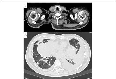
Figure 5CT imaging 14 months after resection of the muscle metastasis. (a)No local recurrence was observed.(b)The pulmonary metastases began to grow rapidly.
validity of the metastatic lesion resection that we per-formed with radiation therapy. However, as a result of the resection the patient was relieved from severe pain, and did not experience any subsequent localized recurrence in the time leading up to his death from respiratory failure more than one year after the tumor resection. We believe that resection of the muscular metastasis in this patient was an effective treatment that enabled him to maintain a good quality of life.
Our patient received systemic chemotherapy for both pulmonary and muscle metastases. Despite achieving good control of the pulmonary metastases, we were unable to control the metastatic lesion in the muscle with chemo-therapy. The resected muscle lesion revealed living cells in the majority of the tissue. A previous study demonstrated differing sensitivity patterns to cytostatic agents in cell culture studies of metastatic pulmonary and muscular osteosarcoma, suggesting that therapy may have resul-ted in positive selection for a tumor clone which is resistant to therapy and/or more metastatic than other subpopulations within the tumor [17]. Although treat-ment experience is limited owing to the rarity of this condition, our case report illustrates that, depending on the patient’s overall condition, resection might be con-sidered as a therapeutic option when treating osteosar-coma with muscle metastasis that does not respond to chemotherapy.
Conclusions
We have presented an extremely rare case of primary osteosarcoma with solitary metastasis to the trapezius muscle that developed during chemotherapy for pul-monary metastasis.
Consent
Written informed consent was obtained from the patient for publication of this case report and any accompanying images. A copy of the written consent is available for review by the Editor-in-Chief of this journal.
Abbreviations
CT:Computational tomography; H & E: Hematoxylin and eosin stain; MRI: Magnetic resonance imaging; PET-CT: Positron emission tomography-CT; SUV: Standardized uptake value.
Japan.2The Near-Future Locomotor Organ Medicine Creation Course,
Graduate School of Medical and Dental Sciences, Kagoshima University, 8-35-1 Sakuragaoka, Kagoshima 890-8520, Japan.3Department of Medical
Joint Materials, Graduate School of Medical and Dental Sciences, Kagoshima University, 8-35-1 Sakuragaoka, Kagoshima 890-8520, Japan.4Department of
Molecular and Cellular Pathology, Kagoshima Graduate School of Medical and Dental Sciences, Kagoshima University, 8-35-1 Sakuragaoka, Kagoshima 890-8520, Japan.
Received: 14 February 2014 Accepted: 16 May 2014 Published: 4 June 2014
References
1. Weis L:Common malignant bone tumors: osteosarcoma.InSurgery for Bone and Soft-Tissue Tumors.Edited by Simon MA, Springfield D. Philadelphia: Lippincott-Raven; 1998:265–274.
2. Willis RA:The Spread of Tumours in the Human Body.London: Butterworths; 1973:282.
3. Berge T, Lundberg S:Cancer in Malmö 1958-1969: an autopsy study.
Acta Pathol Microbiol Scand Suppl1977,260:1–235.
4. Sridhar KS, Rao RK, Kurhardt B:Skeletal muscle metastases from lung cancer.Cancer1987,59:1530–1534.
5. Darmon TA, Heiner J:A series of 30 new patients and 91 cases from the literature.Ann Surg Oncol2000,7:526–534.
6. Menard O, Parache RM:Muscle metastases of cancers.Ann Med Interne 1991,142:423–428.
7. Kim SJ, Choi JA, Lee SH, Choi JY, Hong SH, Chung HW, Kang HS:Imaging findings of extrapulmonary metastases of osteosarcoma.Clin Imaging 2004,28:291–300.
8. Kempf-Bielack B, Bielack SS, Jürgens H, Branscheid D, Berdel WE, Exner GU, Göbel U, Helmke K, Jundt G, Kabisch H, Kevric M, Klingebiel T, Kotz R, Maas R, Schwarz R, Semik M, Treuner J, Zoubek A, Winkler K:Osteosarcoma relapse after combined modality therapy: an analysis of unselected patients in the Cooperative Osteosarcoma Study Group (COSS).J Clin Oncol 2005,23:559–568.
9. Bacci G, Briccoli A, Longhi A, Ferrari S, Mercuri M, Faggioli F, Versari M, Picci P:Treatment and outcome of recurrent osteosarcoma: experience at Rizzoli in 235 patients initially treated with neoadjuvant
chemotherapy.Acta Oncol2005,44:748–755.
10. Hawkins DS, Arndt CA:Pattern of disease recurrence and prognostic factors in patients with osteosarcoma treated with contemporary chemotherapy.Cancer2003,98:2447–2456.
11. Daw NC, Billups CA, Rodriguez-Galindo C, McCarville MB, Rao BN, Cain AM, Jenkins JJ, Neel MD, Meyer WH:Metastatic osteosarcoma.Cancer2006,
106:403–412.
12. Yamada K, Yatabe Y, Sugiura H:Osteosarcoma with skeletal muscle metastasis.Arch Orthop Trauma Surg2008,128:695–699.
13. Wolf R, Wolf RF, Hoekstra HJ:Recurrent, multiple, calcified soft tissue metastases from osteogenic sarcoma without pulmonary involvement.
Skeletal Radiol1999,28:710–713.
14. Peh WC, Shek TW, Wang SC, Wong JW, Chien EP:Osteogenic sarcoma with skeletal muscle metastases.Skeletal Radiol1999,
28:298–304.
16. Arrington ER, Eisenberg B, Orrison WW Jr, Williamson MR:Scintigraphic appearance of uncommon soft-Tissue osteogenic sarcoma metastases.
J Nucl Med1990,31:679–681.
17. Miki T, Yamamuro T, Kotoura Y, Matsushita M, Shimizu Y, Nakamura T:
Osteosarcoma with multiple intramuscular metastases.Acta Orthop Scand 1985,56:92–95.
18. Pace WM, Ross MDI:Tc-99 m MDP uptake in soft tissue extraskeletal metastasis from osteogenic sarcoma.Clin Nucl Med2000,25:333–334.
doi:10.1186/1477-7819-12-176
Cite this article as:Sakamotoet al.:Metastasis of osteosarcoma to the trapezius muscle: a case report.World Journal of Surgical Oncology
201412:176.
Submit your next manuscript to BioMed Central and take full advantage of:
• Convenient online submission
• Thorough peer review
• No space constraints or color figure charges
• Immediate publication on acceptance
• Inclusion in PubMed, CAS, Scopus and Google Scholar
• Research which is freely available for redistribution