285 | P a g e
SYNTHESIS AND CHARACTERIZATION OF Fe-Ni-Co
MAGNETIC THIN FILMS AT DIFFERENT BATH
TEMPERATURE
K. Mohan Rangam Kadiresan
1, Dr.V. Senthil Kumar
21Research Scholar, Department of Physics, Karpagam University, Coimbatore-641021, TamilNadu , (India) 2
Associate Professor, Department of Physics, Karpagam University, Coimbatore-641021, TamilNadu , (India)
ABSTRACT
The magnetic alloy thin films Fe-Ni-Co were deposited on the copper substrateby electrodeposition method at different temperature. Electro deposited Fe-Ni-Co thin films were subjected to the morphological, structural, and mechanical characterization analysis. The chemical composition of the coated films was analysed by EDAX. The surface and structural morphology of the coated film were analysed by using SEM and XRD. The mechanical properties of Fe-Ni-Co thin films have been analysed by VHT. The electroplated Fe-Ni-Co thin films were strongly adherent to the copper substrate. The SEM pictures of Fe-Ni-Co thin films show that, the deposits of thin films are crack free, uniform and bright surface with fine grain size. All the electro deposited Fe-Ni-Co films exhibit FCC crystalline structure. The VHN result of Fe-Ni-Co thin films shows that the Fe-Ni-Co thin films coated at high bath temperature have highest saturation hardness value.Fe-Ni-Co thin films can be used for the manufacturing of MEMS and NEMS devices.
Keywords: Thin Films, Characterization, Electrodeposition, Crystalline Size , Temperature ,X-Ray
Diffraction, Micro Hardness, , Surface Morphology
.
I INTRODUCTION
Electrodeposited nickel is one of the most widely used materials in the fabrication of micro machines such as micro
cantilevers, micro gears and their components (14,22,26). Electrodeposition is the dominant manufacturing
technology in many new applications such as MEMS devices, NEMS devices, data storage media and magnetic
recording head. The most commonly used magnetic materials in MEMS and NEMS are soft magnetic materials,
such as NiCo ,NiFe and NiP (1-6)
.
The combination of good mechanical properties and high corrosion resistance286 | P a g e
as the soft film can be improved by adding a third element with NiFe alloy. Electro depositedPermalloy[NiFe] is
thebestknownthinfilm alloyinMEMS applications(13,19). In this current investigation, the electrodeposition method
has been chosen for coating Fe-Ni-Co thin films. In this present work, we have analysedthe effect of different
temperatureon FeNiCothin films with. This paper summarizes the synthesis and characterizations of electroplated
FeNiCo thin films.
II EXPERIMENTAL PART
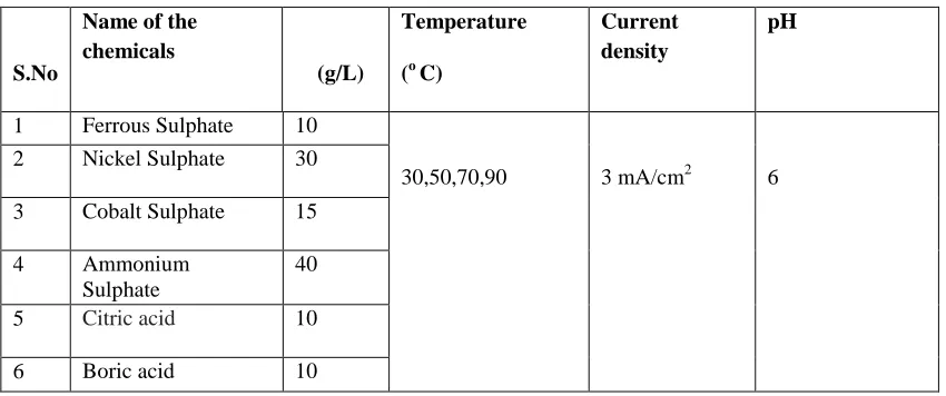
The working conditions and bath composition of Fe-Ni-Co alloy thin film are shown in Table 1.The Fe-Ni-Co thin
films are successfully coated by electrodeposition method. In this investigation, Copper and stainless steel substrates
act as cathode and anode respectively.A copper plate and stainless steel of size 1.5 cm as breath and 7.5cm as length
were used as substrates. Both cathode and anode were washed with soap and soaking in 15% H2SO4 for 2 minutes.
The reagent grade chemicals and triple distilled water were used to prepare electroplating bath . The pH value of the
bath was adjusted to 6 by adding few drops of ammonia solution(10-14). The Fe-Ni-Co thin films were electro
deposited on the copper substrate by applying a current of 15 mA for 15 minutes at30˚C,50˚C ,70˚C and 90˚C. The
cathode was carefully removed from the bath after 15 minutes and dried for few minutes. The surface morphology
of the Fe-Ni-Co thin films was analysed with the help of Scanning electron microscope (SEM). The film
composition and structural characters of thin films were measured by Energy-dispersive X-ray Spectroscopy
(EDAX) andX-ray diffraction (XRD) respectively. The hardness of Fe-Ni-Co thin films was measured by Vickers
Hardness Test (VHN).The magnetic property of Fe-Ni-Co thin films film was measured by Vibrating Sample
Magnetometer (VSM).The thicknesses of the films were determined by cross sectional view of SEM images. The
electrodeposition bath details of Fe-Ni-Co thin films are given in table 1.
Table 1. Electroplating bath details of FeNiCo thin films
S.No
Name of the chemicals
(g/L)
Temperature (o C)
Current density
pH
1 Ferrous Sulphate 10
30,50,70,90 3 mA/cm2 6
2 Nickel Sulphate 30
3 Cobalt Sulphate 15
4 Ammonium
Sulphate
40
5 Citric acid 10
287 | P a g e
III RESULTS AND DISCUSSION
3.1 Composition of Electrodeposited Thin Films
The chemical composition of the electroplated thin films is analysed by EDAX spectrum. The EDAX data‟s of thin
films are shown in Table 2.EDAX result showed that the films obtained at higher temperature have high ferrous
content. The highest ferrous content of 22.13 wt% was obtained at temperature 90˚C.EDAX result showed that Ni
content increases with increasing the bath temperature. The maximum Ni content of 47.92 wt% was obtained for
Fe-Ni-Co thin films at 90˚C bath temperature. The weight percentage of Co decreaseswhile increasing the bath
temperature.Ammonia solution is used to correct the pH value of the bath solution only and its effect on the film was
ignored.
Table 2: EDAX analysis of thin films
S. No Temperature Co Wt%
Ni Wt%
Fe Wt%
1. 30˚C 68.45 19.87 11.68
2 50˚C 56.16 29.56 14.28
3 70˚C 40.23 40.08 19.69
4 90˚C 29.95 47.92 22.13
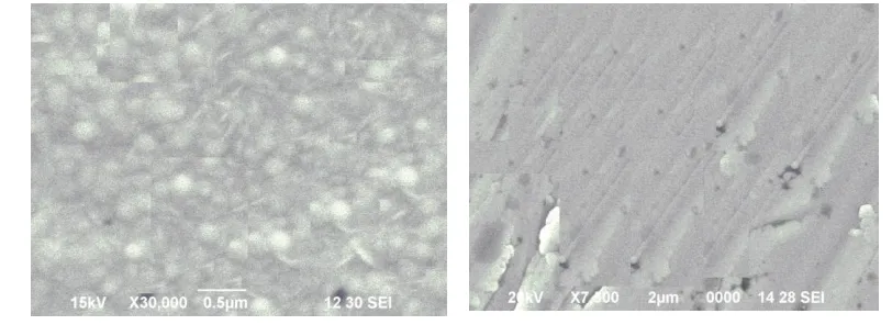
3.2 Morphological Observation
The surface morphology of the electroplated Fe-Ni-Cothin films with different temperature is analysed by using
SEM pictures and are shown in fig 1.The electroplated thin films are smooth and uniform. The thin films are bright,
crack free and uniform. From SEM analysis we conclude that the formation of thin films on the copper substrate is
288 | P a g e
0 20 40 60 80 100
0 1000 2000 3000 4000 5000
(a)
(220) (111)
(200)
Inten
sity
(co
un
ts)
Two Theata(degree) 0 20 40 60 80 100
0 1000 2000 3000 4000 5000
(b)
(220) (200)
(111)
Inten
sity
(co
un
t)
Two Theata(degree)
Figure 1: SEM images for Electro deposited Fe-Ni-Cothin film for different bath temperatures
(a) 30˚C (b) 50˚C (c) 70˚C (d) 90˚C
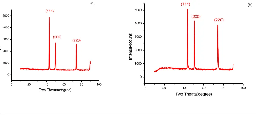
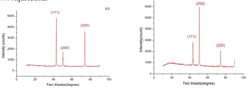
3.3 Structural Analysis
The crystal structure of the electro deposited Fe-Ni-Co alloy thin films was determined by XRD analysis. X- ray
diffraction patterns of Fe-Ni-Co films obtained at different temperatures are shown in fig 3. The presence of sharp
peaks in XRD pattern reveals that the films are crystalline in nature. The crystalline size of the deposits was
calculated from XRD usingScherrer‟s formula
D=0.954λ/βcosθ
Where, θ is the Bragg‟s angle , λ is the X-ray wavelength, β is the full width at half maximum intensity of the diffraction peak located at 2θ and.The XRD patterns of NiCoFe filmsrevealedtheexistence ofFCCphasewith (111),
(200) and (220) diffraction peaks.The result shows that the crystalline sizes of the Fe-Ni-Co deposits obtained by
289 | P a g e
0 20 40 60 80 100
0 1000 2000 3000 4000 5000 (c) (200) (220) (111) Inten sity (co un ts) Two theata(degree)
0 20 40 60 80 100
0 1000 2000 3000 4000 5000 6000 (220) (200) (111) (d) Inten sity (co un t) Two theata(degree)
30 40 50 60 70 80 90
18 19 20 21 22 23 24 Particl e Size Bath Temprature
Fig.2 XRD patterns of Fe-Ni-Co thin films at (a) 300C (b) 500C (c) 700C (d) 900C
The crystal size of Fe-Ni-Co alloy films is tabulated and shown in table 3.When the bath temperature is increased
the crystalline size of thin films decrease due to onset orientation of crystals during electrodeposition
Table.3 :Structural characteristics of NiCoFe alloy thin films
S. No
Bath Temperature
(0C)
2Ɵ
(deg)
d (A0)
Particle size, D
(nm)
Strain (10-3)
Dislocation density (1014 / m2)
1 30 43.310 1.5634 23.32 1.578 21.87
2 50 43.681 1.4376 21.23 1.765 22.43
3 70 43.108 1.7650 19.67 1.854 23.76
4 90 50.412 1.4538 18.02 1.897 26.34
290 | P a g e
30 40 50 60 70 80 90
75 80 85 90 95 100 105 110
Ha
rd
ne
ss
Bath Temprature
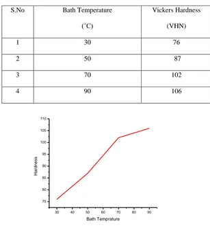
3.4 Mechanical Properties
Hardness of the films was examined by using Vickers hardness tester ( the diamond intender method). The results
show that the hardness increases with increasing bath temperature.This may be due to lower stress associated with
electrodeposited Fe-Ni-Co films. The hardness of Fe-Ni-Co thin films have been shown in table 4.
Table.4: Mechanical Properties of electro deposited Ni-Co-Fe thin films
S.No Bath Temperature
(˚C)
Vickers Hardness
(VHN)
1 30 76
2 50 87
3 70 102
4 90 106
Figure 4. Vickers Hardness as a function of bath temperature
IV CONCLUSION
The Ni-Co-Fe magnetic thin films were successfully prepared by electro deposition at different bath temperatures
30˚C, 50˚, 70˚C and 90˚C .The crystalline sizes of the deposits obtained by electro deposition process are in the nano
scale. The thin films obtained at different temperature are uniform, bright andcrack free. FCC was the dominant
structure of electro deposited Fe-Ni-Cothin films(14,22). Hardness is increases with increasing bath temperature.
When the bath temperature was increasedfrom 30 ˚C to 90 ˚C, the particle size values decreases from 23.32 nm to
291 | P a g e
summaries the optimized operating condition of electroplated bath. The Ni-Co-Fe thin films can be used in various
electronic devices, MEMS and NEMS.
REFERENCES
1. Hamid Z.A., “Electrodeposition of Cobalt- Tungsten Alloys from Acidic Bath Containing Cationic Surfactants”,
Materials Letters.,2003,57, 2558.
2. Ho Soon Min., “Metal Selenide semiconductor thin films: A Review”, International Journal of ChemTech
Research 2016, 9, 390-395.
3. Manjulavalli.T.E, Kannan.A.G, “Structural and optical properties of ZnS thin films preparedby chemical bath
deposition method”, International Journal of ChemTech Research 2015, 8, 396-402.
4. Sivasankar.G ,Ramajothi.J., “Aluminium Doped Zinc Oxide (ZnO) Thin Film Fabricated for Semiconductor by
Spray Pyrolysis Technique”, International Journal of ChemTech Research 2015, 8, 497-501.
5. Kannan, R, Ganesan, S,Selvakumari, „‟Structural and Magnetic properties of electrodeposited NiFeWS thin
films‟‟, Optoelectronics and advanced materials-Rapid Communication 2012,3-4, 383-388.
6. Thangaraj.N, Tamilarasan.K ,Sasikumar.D., “Effect of Phosphorous Acid on the Ferrous TungstenPhosphorous
Magnetic Thin Film”, International Journal of ChemTech Research 2014, 6, 384-390.
7. Kannan, R, Ganesan, S ,Selvakumari, TM „‟Synthesis and characterization of nano crystalline NiFeWS thin films
in diammonium citrate bath‟‟, Digest journal of nanomaterials and biostructures, 2012,7, 1039-1050.
8. Nosang V, Park D.Y, Yoob B.Y. and Paulo T.A., „„Development of electroplated magnetic materials for MEMS”,
Journal of Magnetism and Magnetic Materials., 2003, 265 , 189-198
9.Ravindranadh.K, Sridhar Kumar.D,DurgaVenkata Prasad,.K, Rao.M.C., “Luminescent Properties of Cu2+ Doped
SnO2 Thin Films by Spray Pyrolysis”, International Journal of ChemTech Research 2016, 9, 598-603.
10. Kavitha.N, Manohar.P., “Magnetic and Dielectric studies of Ni-Co-Zn Ferrites synthesized by Nonconventional
combustion method”, International Journal of ChemTech Research 2015, 8, 308-315.
11. Pushpalatha.H.L, Ganesha.R
.,
“Growth and characterization of CdS thin films by photochemical and chemicalbath deposition”, International Journal of ChemTech Research 2015, 7, 185-189.
12. Iwasaki S., Nakamura Y., “An analysis for the magnetization mode for high density magnetic recording”,
Journal of Magnetism and Magnetic Materials., 1977, 200, 634-648.
13. Emerson R.N., Kennady C.J.,Ganesan S., “Effect of Organic additives on the Magnetic properties of
Electrodeposition of CoNiP Hard Magnetic Films”, Thin solid films, 2007,515, 3391-3396.
14. Baskar.T, Rajni.K.S, “Effect of bath temperature on structural and magnetic properties of electrodeposited
NiCoS magnetic thin films”, International Journal of ChemTechResearch ,2015,8, 234-239
.
15. Rajni.K.S., “Studies on the Structural and Conduction mechanism (ac) in thermally Evaporated CdSe thin
292 | P a g e
16. Chidambara Kumar K N, KhadeerBasha S K, Shakil Muhammad G, “X Ray line profile analysis of Mn doped
PbS thin films bySuccessive Ionic Layer Adsorption and Reaction Method”, International Journal of ChemTech
Research 2015, 7, 2257-2264.
17. Pushpalatha H.L, Ganesha.R., “X-ray photoelectron spectroscopic studies of CdSsemiconductor thin films
deposited by photochemical deposition”, International Journal of ChemTech Research 2015, 7, 2171-2175.
18. Manjulavalli.T.E, Kannan.A.G., “Effects of deposition time on structural, optical and electricalproperties of
chemically deposited Cu2S thin films”, International Journal of ChemTech Research 2015, 8, 607-616.
19. Baskar.T, Rajni.K.S, “Effect of different Sulfur concentration on structural and magnetic properties of
electrodeposited NiCoS magnetic thin films”, International Journal of ChemTechResearch ,2016,5 ,317-324.
20. Cho H. J, Bhansali S. ,Ahn C. H., “Electroplated thick permanent magnet arrays with controlled direction of
magnetization for MEMS application”, Journal of Applied Physics, 2000,87, 6340- 6342.
21. Esther. P , Joseph Kennady.C., „‟Effect of sodium tungstate on the properties of Electrodeposited nanocrystalline