Eye and Brain
Dove
press
R E v i E w
open access to scientific and medical research
Open Access Full Text Article
Current screening and treatments in retinopathy
of prematurity in the US
Ana M Suelves Julia P Shulman
Department of Ophthalmology, New York Medical College, valhalla, NY, USA
Correspondence: Julia P Shulman Department of Ophthalmology, New York Medical College, 20 E 9th Street, New York, NY 10003, USA
Tel +1 212 203 0999 Fax +1 212 202 4884 Email jshulman03@gmail.com
Abstract: Retinopathy of prematurity (ROP) is a complex disease characterized by an aberrant developmental retinal angiogenesis in preterm infants and can carry significant visual morbidity, including retinal detachment and blindness. Though large scale, randomized clinical trials have improved our understanding of the pathophysiology and progression of the disease, the management of ROP remains a challenge for ophthalmologists. This review addresses the up-to-date screening approach, diagnosis, and treatment guidelines for ROP in the US.
Keywords: retinopathy of prematurity, retinal vascular development, vascular endothelial growth factor, ETROP, BEAT-ROP, eROP
Introduction
Retinopathy of prematurity (ROP) was first described in 1940s when high saturation of oxygen supplementation improved the survival of preterm infants but contributed to blindness.1 At that time, Terry2 used the term “retrolental fibroplasia,” which
we now know corresponds to end-stage ROP. ROP is characterized by a delay in physio logic retinal vascular development followed by abnormal proliferation into the vitreous.
Limiting the inspired oxygen concentration has decreased the incidence of ROP but has caused a higher mortality and cerebral palsy in the surviving newborns. Though unregulated oxygen is a clear risk factor for ROP, in modern neonatal practice, with tight regulation and control of oxygen saturation, the relationship between oxygen and development of ROP is complex and incompletely understood. Today, young ges-tational age (GA) and low birth weight are the uniformly recognized risk factors for developing ROP, and other variables such as low insulin-like growth factor 1 (IGF-1) levels, postnatal weight gain, hyperglycemia, blood transfusions, use of surfactant, and prolonged artificial ventilation have also been identified.3–6
Improvement of neonatal care has led to the survival of younger preterm infants of very low GA and birth weight7,8 who are at high risk for developing ROP. Moreover,
more premature infants are also surviving in developing countries, where uncontrolled oxygen delivery is still being used, causing an increase in ROP cases in developing countries around the world.
Despite improved screening and treatment guidelines, ROP remains a leading cause of visual impairment in premature infants in the US.9
Eye and Brain downloaded from https://www.dovepress.com/ by 118.70.13.36 on 22-Aug-2020
For personal use only.
Number of times this article has been viewed
This article was published in the following Dove Press journal: Eye and Brain
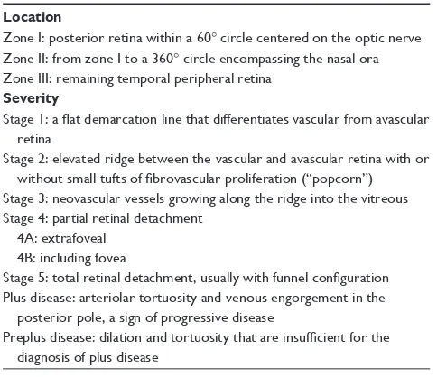
Table 1 Classification of retinopathy of prematurity Location
Zone i: posterior retina within a 60° circle centered on the optic nerve Zone ii: from zone i to a 360° circle encompassing the nasal ora Zone iii: remaining temporal peripheral retina
Severity
Stage 1: a flat demarcation line that differentiates vascular from avascular retina
Stage 2: elevated ridge between the vascular and avascular retina with or without small tufts of fibrovascular proliferation (“popcorn”)
Stage 3: neovascular vessels growing along the ridge into the vitreous Stage 4: partial retinal detachment
4A: extrafoveal 4B: including fovea
Stage 5: total retinal detachment, usually with funnel configuration Plus disease: arteriolar tortuosity and venous engorgement in the posterior pole, a sign of progressive disease
Preplus disease: dilation and tortuosity that are insufficient for the diagnosis of plus disease
Dovepress Suelves and Shulman
Pathophysiology of ROP
Until the fourth month of gestation, the retina remains avas-cular and the developing retina obtains its nutrients from the hyaloid vasculature. At 16 weeks, the angioblasts near the hyaloid artery invade the nerve fiber layer. The first retinal vessels sprout from the optic nerve head and migrate toward the periphery extending to the ora serrata nasally by 36 weeks and temporally by 40 weeks. The microglia and astrocytes secrete vascular endothelial growth factor (VEGF) and IGF-1. The delicate balance and gradient of growth factors play an important role in the development of the superficial and deep layers of the retinal vasculature.9
Increasing evidence points to the fact that the mechanism of retinal vascularization is similar to that of vascularization of the brain during development. Retina and central nervous system vascular networks originate from existing vessels by angiogenesis; periventricular leukomalacia and ROP may develop in preterm infants and altered oxygen conditions is a risk factor for both.10
During the second half of pregnancy, there is relative intrauterine hypoxia (oxygen tissue tension of 25–35 mmHg) compared to 95–100 mmHg in room air after birth. ROP can be seen as a delay of normal retinal neuronal and vascular development in preterm infants, which causes pathological compensatory mechanisms that lead to aberrant vascular-ization. Pathogenesis can be separated into two different postnatal phases that differ in the expression of VEGF. The first phase is characterized by an arrest of the normal retinal development because of relative environmental hyperoxia and downregulation of VEGF, IGF-1, erythropoietin, and ω-3 polyunsaturated fatty acids. The second phase occurs between 32 and 36 weeks post-GA. As the infant retina matures and metabolic demands increase, relative hypoxia of the avascular retina results, causing an overexpression of VEGF, IGF-1, and oxidative damage. Vasoproliferation is the hallmark of the second phase, but is a disorganized vascular growth into the vitreous at the border of the avascular retina. If ROP contin-ues to progress, it can lead to a tractional retinal detachment, considered the fibrovascular phase.
Animal models have helped in the understanding of the pathogenic mechanism of the disease. In 1953, Patz et al11
described for the first time the association between ROP and administration of high doses of oxygen. Aston12 was
the first in reporting the two postnatal phases in premature infants and further described through experimental work in kittens the closure of newly formed capillaries related to oxygen-toxicity (Phase I) followed by hypoxia-mediated vasoproliferation (Phase II) when kittens were transferred to
room air.13 However, animal models have several limitations.
First, animal retinas normally vascularize after birth, unlike human retinas that vascularize prenatally. Second, animal models have no other comorbidities such as lung pathology, sepsis, and anemia.
Classification
The original International Classification of ROP was a consensus of 15 international experts in ROP, published in 198414 and was last updated in 2005.15 It has facilitated the
development of large multicenter clinical trials, helped in the understanding of the natural course of the disease as well as classified infants with ROP into prognostic subgroups of higher risk of unfavorable outcomes.
The classification of ROP describes three locations (zones I–III), five stages (stages 0–5), and the presence of plus disease (Table 1).
The zone is the extent of physiologic vascularization completed in utero; therefore, lower zone implies worse disease. The stage is defined by the appearance of the junc-tion between the vascular and avascular retina. If more than one stage is present, the staging is determined by the most severe stage identified. Plus disease is the dilation and tortuosity of retinal arteries and veins in the posterior pole compared to a standard photograph.15 The presence of plus
disease increases the likelihood of disease progression and risk of poor visual outcome.
Aggressive posterior ROP is a subtype of ROP that behaves very aggressively and unpredictably. It describes posterior disease (in zone I or posterior zone II) with plus disease out of proportion to that characteristically presenting
Eye and Brain downloaded from https://www.dovepress.com/ by 118.70.13.36 on 22-Aug-2020
Table 2 Clinical phenotypes of retinopathy of prematurity (ROP) as per the Early Treatment of Retinopathy of Prematurity (ETROP) cooperative group classification
Type 1 ROP (high-risk prethreshold disease) – treat (within 48–72 hours) Zone i, any stage with plus disease
Zone i, stage 3 without plus disease Zone ii, stage 2 or 3 with plus disease
Type 2 ROP (low-risk prethreshold disease) – watch closely (within 1 week)
Zone i, stage 1 or 2 without plus disease Zone ii, stage 3 without plus disease
Dovepress ROP screening and treatment
with a flat neovascularization at the vascular–avascular junction. This ROP phenotype is seen in the lowest birth weight infants and carries a higher risk of poor visual out-come and retinal detachment.15
The Early Treatment for Retinopathy of Prematurity (ETROP) study reclassified ROP into type 2 (to be closely monitored) and type 1 (requires treatment)16 (Table 2).
Diagnosis and screening
recommendations
Fortunately, between 92% and 96% of ROP regresses spon-taneously without any intervention. It is estimated that less than 10% of infants screened develop severe forms of the disease and require treatment.17
Current US guidelines, updated in 2013, recommend screening for infants with birth weight of 1,500 g or less or GA of 30 weeks or less.18 A third criterion for ROP screening
includes more mature infants with a complicated postnatal course or at risk for ROP at the discretion of the neonatolo-gist, especially in the presence of necrotizing enterocolitis, intraventricular hemorrhage, sepsis, or bronchopulmonary dysplasia.
The first screening examination should take place at 4–6 weeks postpartum or at 31 weeks GA, whichever is later. Special attention to those infants born before 25 weeks GA with screening at 6 weeks postpartum permits the early detec-tion of aggressive posterior ROP.
The fundus exam in a preterm infant may reveal one of three findings: mature retina, immature retinal vasculature, or ROP. ROP screening can be discontinued if: 1) Zone III retinal vascularization is present without previous zone I or zone II ROP. 2) There is no evidence of prethreshold disease or worse ROP by 50 weeks postmenstrual age. 3) There is regressing ROP in zone III without abnormal vascular tissue that can reactivate in zone II or III.18
Screening preterm infants at risk of ROP requires serial funduscopic examinations by skilled ophthalmologists. This
is done after pupillary dilation using a binocular indirect ophthalmoscope and a scleral depressor, and may be stressful to a preterm infant even in the hands of an experienced examiner.
A national survey revealed that more than a third of the providers who perform ROP examinations are general ophthalmologists, nonfellowship trained.19 As more
pre-mature babies are surviving, there is a growing demand for ROP evaluations. The overall paucity of skilled examiners linked to the increasing frequency of ROP examinations as per revised recommendation guidelines18 can make a timely
evaluation a real challenge, especially in underserved areas of the US and middle-income countries. An appropriately timed examination and treatment of ROP has a major impact on visual outcome. Despite improving screening guidelines and increased number of therapies, the main cause of child-hood blindness in middle-income countries is the shortage of trained ophthalmologists willing to screen and treat ROP infants.20
In order to decrease the burden of examinations and detect infants who will progress to treatment requiring ROP, the WINROP algorithm was developed. It incorporates the weekly weigh gain postpartum, GA, and birth weight to predict progression to type 1 ROP.21
Telemedicine is another promising methodology that has gained popularity in recent years. Infants’ retinas are photo-graphed by nonphysicians using a RetCam System (Clarity Medical Systems, Pleasanton, CA, USA) and interpreted at an off-site reading center. Remote ROP screening has been shown to be an alternative to an ROP specialist examining every infant at risk for ROP using indirect ophthalmoscopy, especially in underserved areas of the US22 and
middle-income countries. It is reproducible and decreases the inter-observer discrepancy. The disadvantages of retinal imaging are that it requires an excellent pupillary dilation (more than 7–8 mm); difficulty visualizing zone III; and the difficulty photographing older infants.
The use of telemedicine was validated in the recent eROP study, which was a multicenter trial funded by the National Eye Institute that compared screening results from wide-field digital retinal images obtained by nonphysician certified ROP imagers using a standardized imaging protocol and conventional retinal exams by trained ophthalmologists for acute ROP screening. This study showed that wide field imaging has an excellent inter- and intragrader agreement for ROP diagnosis compared to standard of care23 and may
provide an objective way to standardize ROP protocols and guidelines.24,25
Eye and Brain downloaded from https://www.dovepress.com/ by 118.70.13.36 on 22-Aug-2020
Dovepress Suelves and Shulman
In rural areas of India, the availability of ophthalmologists experienced in ROP management is especially pronounced. The KIDROP program initiated in 2008 has the aim of training technicians to perform and interpret retinal imaging obtained by RetCam or cell phones. Infants that fulfill criteria for laser ablation as per the ETROP study undergo laser locally without the need to travel to the city.26
Multiple prospective and retrospective studies have shown that digital photography may be a valuable tool to detect clinically significant ROP and referral-warranted ROP,27–29 although it does not replace indirect
ophthal-moscopy, the gold standard.30 The different sensitivity,
specificity, and positive predictive value of the RetCam compared to ophthalmic examination may reflect dis-crepancy in training level of examiners and differences in methodology.31
Prevention and risk factors
Prevention of preterm birth is the most important preventive factor for ROP development.
Current research efforts are focused on the detection of pre- and postnatal risk factors for ROP progression.
The STOP-ROP multicenter study showed no significant difference between maintaining an oxygen saturation level of 96%–99% versus 89%–94% for prethreshold ROP.32
A higher survival rate of infants younger than 28 weeks with 91%–95% oxygen saturation was found in the BOOST II study, although there is an increase of incidence of ROP shown at this oxygen rate in other studies.33
A recent study showed significant differences in IGF levels in black mothers compared to other races.34
Genetic factors may explain difference in incidence in twins. Several gene variants such as those of the wnt pathway (frizzled 4, lipoprotein-related receptor-related protein 5, and Norrie Disease Protein) have been implicated.35
Treatment
Prior to the 1980s, the only standardized modality for treat-ment of ROP was surgery for retinal detachtreat-ment in the advanced stages of disease. Cryotherapy emerged in the treatment of ROP around this time as it ablates peripheral ischemic retina; however, there were no guidelines for its use based on severity of disease.
The Cryotherapy for Retinopathy of Prematurity (CRYO-ROP) study sought to determine the point at which retinal neovascularization was equally likely to progress to retinal detachment or regress. Carried out from the mid-1980s, this seminal trial in ROP and ophthalmology involved 23 centers
and followed patients for 20 years.36 CRYO-ROP introduced
a unified classification of ROP, including a standardized photograph of plus disease and the concept of threshold disease, defined as five contiguous clock hours or eight noncontiguous clock hours of stage 3 ROP with plus disease in zone I or zone II. The study demonstrated a significant reduction in unfavorable anatomic outcome, such as retinal detachment, posterior folds in the retina, or development of retrolental fibrous tissue (primary outcome). At 15-year follow-up, the final report from CRYO-ROP, 30% of treated eyes and 52% of control eyes had unfavorable structural outcomes. Unfavorable visual acuity outcomes (defined as Snellen visual acuity equivalent of 20/200 or worse) were seen in 45% of treated eyes versus 64% of control eyes at 15-year follow-up.37
The CRYO-ROP study was terminated early because of the significant reduction in unfavorable outcomes in treated eyes36 and it was felt that treatment should not be withheld
in the control group. Much of our current understanding of the natural history of ROP derives from the control group in the CRYO-ROP study.
The CRYO-ROP established the benefits of treating ROP at threshold, and a subsequent study, the ETROP, was designed to investigate if treatment prior to threshold disease would further decrease unfavorable anatomic out-comes. The ETROP defined “prethreshold disease” as type 1 and type 2 ROP. It was calculated that type 1 eyes had a 15% or greater chance of unfavorable anatomic outcome versus less than 15% chance for type 2 eyes. ETROP dem-onstrated the benefit of peripheral laser ablation in type 1 ROP and frequent observation in type 2 disease. At this point, ablation of the avascular retina was done with diode laser as opposed to cryotherapy.38 Though types 1 and 2
disease were defined in the ETROP study by a multivari-ate logistic regression model based on natural history data from CRYO-ROP, clinically, type 1 disease was found to correspond to zone I ROP of any stage with plus disease, zone 1 stage 3 disease without plus, and zone 2 stage 2 or 3 disease with plus. Type 2 ROP was found to correspond to zone 1, stage 1 or 2 without plus, or zone 2 stage 3 without plus. At 6-year follow-up, 9% of early treated eyes had unfavorable structural outcomes compared to 15% in the conventional treatment group.16
The results of the ETROP trial form the framework for modern surveillance and treatment of ROP in the US and worldwide. Current guidelines in the US recommend peripheral laser ablation to the avascular retina for type 1 ROP, performed within 72 hours of diagnosis. Complete laser
Eye and Brain downloaded from https://www.dovepress.com/ by 118.70.13.36 on 22-Aug-2020
Dovepress ROP screening and treatment
treatment from the ridge to the ora serrata is administered, with half burn width apart spacing of laser spots. After treat-ment, infants are followed closely for resolution of disease or additional laser to any “skip areas” or areas that were not adequately treated initially.
Laser treatment has many ocular and systemic advan-tages over cryotherapy39: laser decreases the requirements
for general anesthesia, allows treating more posterior retina, and has a lower rate of systemic complications (eg, apnea, bradycardia, cardiopulmonary arrest requiring resuscita-tion).40 Ocular complications include vitreous hemorrhage,
cataract, elevated intraocular pressure, choroidal effusions, and postoperative inflammation. Topical cycloplegics and steroids may be administered for a short time after treatment to minimize these complications.
With the recent increase in intravitreal anti-VEGF therapy in adults for the treatment of various vasoprolifera-tive disorders and given the role of VEGF in angiogenesis, anti-VEGF therapies have been successfully utilized in the treatment of ROP. The Bevacizumab Eliminates the Angio-genic Threat of Retinopathy of Prematurity study random-ized ROP patients to receive a single intravitreal injection of bevacizumab at 0.625 mg in 0.025 mL. This study concluded that intravitreal bevacizumab reduced the recurrence of stage 3 ROP in zone 1 as compared to laser; 42% in the group of laser therapy compared to 6% in the bevacizumab group. Posteriorly, Reynolds et al suggested that bevacizumab may be superior to laser therapy for stage 3+ zone I but not for zone II posterior disease.41,42 Interestingly, recurrence of ROP
in patients treated with intravitreal bevacizumab was found to occur later than recurrence after laser ablation therapy (16±4.6 weeks with bevacizumab versus 6.2±5.7 weeks with laser), necessitating longer follow-up. Once bevacizumab is administered, patients need to be followed up until full vascularization of the retina. Intravitreal bevacizumab is not US Food and Drug Administration-approved for the treat-ment of ROP. Although anti-VEGF therapy is a promising therapy in infants with ROP, its use is still controversial given the unknown systemic risks and cases of poor clinical outcome with bevacizumab treatment.43 Systemic inhibition
of VEGF for up to 8 weeks after a single intravitreal injec-tion has been reported44 and the effects of systemic VEGF
inhibition on other developing vascular beds such as the lungs are unknown. Treatment failure and adverse events such as persistent avascular retina, retinal detachment, and blindness have also been reported.41,45,46 The selection of
anti-VEGF agent and optimal dosage are unknown and remain the subject of investigation.
Despite treatment, ∼16% of patients with type 1 ROP developed a retinal detachment.16 Once an advancing
stage 4 detachment is diagnosed, treatment with lens spar-ing vitrectomy (LSV) is recommended. In one large series, the reattachment rate after one LSV was 82% for stage 4A disease, 70% for stage 4B, and 43% for stage 5.47 Scleral
buckles can also be used in the treatment of ROP detachments but often need to be divided to avoid significant induced refractive changes.
In a small series, the combination of LSV and scleral buckling was not superior to LSV alone.48 The visual
outcomes in stage 5 detachments are extremely poor; there-fore, the goal of ROP care is to prevent their development by screening appropriately and treating progressing disease.49
Patients with ROP can also develop effusive or serous detachments, which are caused by leakage from vascular structures. These are usually posterior to the ridge and convex shaped. Effusive detachment may resolve spontaneously, and there are reports of successful resolution after intravitreal bevacizumab administration50; however, surgical intervention
may be needed in select cases.
Summary
Our understanding of the pathophysiology of ROP and approach to management is evolving but many questions remain. Knowledge from ongoing studies will hopefully lead to the decrease of vision loss in ROP as well as better screening methods, better predictive algorithms, and new treatments.
Disclosure
The authors report no conflicts of interest in this work.
References
1. Campbell K. Intensive oxygen therapy as a possible cause of retrolental fibroplasias: a clinical approach. Med J Aust. 1951;2:48–50.
2. Terry TL. Retrolental fibroplasia. J Pediatr. 1946;29:770–773. 3. Perez-Manuzuri A, Fernandez-Lorenzo JR, Couce-Pico ML,
Blanco-Teijeiro MJ, Fraga-Bermúdez JM. Serum levels of IGF-1 are a useful predictor of ROP. Acta Paediatr. 2010;99(4):519–525.
4. Brown MS, Baron A, France E, Hamman RF. Association between higher cumulative doses of recombinant erythropoietin and risk of retinopathy of prematurity. J AAPOS. 2006;10:143–149.
5. Hesse L, Eberi W, Schlaud M, Poets CF. Blood transfusion: iron load and retinopathy of prematurity. Eur J Pediatr. 1997;156:465–470. 6. Tin W, Miligan DW, Pennefalther P, Hey E. Pulse oximetry, severe
retin-opathy and outcome at one year in babies of less than 28 weeks gestation. Arch Dis Child Fetal Neonatal. 2001;84:F106–F110.
7. Stenson B, Brocklehurst P, Tarnow-Mordi W. Increased 36-week survival with high oxygen saturation target in extremely preterm infants. N Engl J Med. 2010;362:1959–1969.
8. Horbar JD, Carpenter JH, Badger GJ, et al. Mortality and neonatal mor-bidity among infants 501 to 1500 grams from 2000 to 2009. Pediatrics. 2012;129:1019–1026.
Eye and Brain downloaded from https://www.dovepress.com/ by 118.70.13.36 on 22-Aug-2020
Dovepress Suelves and Shulman
9. Drenser KA, Capone A. Retinopathy of prematurity. In: Yanoff M, Duker JS, editors. Yanoff and Duker Ophthalmology. 3rd ed. St Louis: Mosby Inc; 2008:606–612.
10. Steck J, Blueml C, Kampmann S, Greene B. Retinal vessel pathologies in a rat model of periventricular leukomalacia: a new model for retinopathy of prematurity? Invest Ophthalmol Vis Sci. 2015;56:1830–1841. 11. Patz A, Eastham A, Higginbotham DH, Kleh T. Oxygen studies in
retrolental fibroplasia. Am J Ophthalmol. 1953;36:1511–1522. 12. Ashton N. Pathological basis of retrolental fibroplasia. Br J Ophthalmol.
1954;38:385–396.
13. Ashton N, Ward B, Serpell G. Effect of oxygen on developing retinal vessels with particular reference to the problem of retrolental fibroplasia. Br J Ophthalmol. 1954;38397–38432.
14. [No authors listed]. An international classification of retinopathy of prematurity. The committee for the classification of retinopathy of prematurity. Arch Ophthalmol. 1984;102:1130–1134.
15. The International Classification of Retinopathy of Prematurity. Inter-national committee for the classification of retinopathy of prematurity revisited. Arch Ophthalmol. 2005;123(7):991–999.
16. Good W, on behalf of the ETROP Cooperative Group. Final Results of the Early Treatment for Retinopathy of Prematurity (ETROP) Random-ized Trial. Trans Am Ophthalmol Soc. 2004;102:233–250.
17. Chiang M, Arons R, Flynn JT, Starren JB. Incidence of retinopathy of prematurity from 1996 to 2000: analysis of a comprehensive New York state database. Ophthalmology. 2004;111(7):1317–1325.
18. American Academy of Pediatrics Section on Ophthalmology, American Academy of Ophthalmology, American Association for Pediatric Ophthalmology and Strabismus, American Association of Certified Orthoptists. Screening examination of premature infants for retinopathy of prematurity. Pediatrics. 2013;131:189–195.
19. Kemper AR, Freedman SF, Wallace DK. Retinopathy of prematurity care: patterns of care and workforce analysis. J AAPOS. 2008;12: 344–348.
20. Vinekar A, Jayadev C, Mangalesh S, et al. Role of tele-medicine in retinopathy of prematurity screening in rural outreach centers of India – a report of 20,214 imaging sessions in the KIDROP program. Semin Fetal Neonatal Med. 2015;20(5):335–345.
21. Lundgren P, Sjostom E, Domellof M, et al. WINROP identifies severe retinopathy of prematurity at an early stage in a nation-based cohort of extremely preterm infants. PLoS One. 2013;8(9):e73256.
22. Weaver DT, Murdock TJ. Telemedicine detection of type 1 ROP in a distant neonatal intensive care unit. J AAPOS. 2012;16(3):229–233. 23. Daniel E, Quinn GE, Hildebrand PL, et al. Validated system for
central-ized grading of retinopathy of prematurity: telemedicine approaches to evaluating acute-phase retinopathy of prematurity (e-ROP) study. JAMA Ophthalmol. 2015;133:675–682.
24. Gschließer A, Stifter E, Neumayer T, et al. Inter-expert and intra-expert agreement on the diagnosis and treatment of retinopathy of prematurity. Am J Ophthalmol. 2015;160:553–560.
25. Wang SK. Callaway N, Wallenstein MB, et al. Sundrops: six years of screening for retinopathy of prematurity with telemedicine. Can J Ophthalmol. 2015;50(2):101–106.
26. Vinekar A, Gilbert C, Dogra M, et al. The KIDROP model of combin-ing strategies for providcombin-ing retinopathy of prematurity screencombin-ing in underserved areas in India using wide-field imaging, tele-medicine, non-physician graders and smart phone reporting. Indian J Ophthalmol. 2014;62(1):41–49.
27. Ells AL, Holmes JM, Astle WF, et al. Telemedicine approach to screen-ing for severe retinopathy of prematurity: a pilot study. Ophthalmology. 2003;110(11):2113–2117.
28. Photographic Screening for Retinopathy of Prematurity (Photo-ROP) Cooperative Group. The photographic screening for retinopathy of prematurity study (photo-ROP). Primary outcomes. Retina. 2008; 28(Suppl 3):S47–S54.
29. Chiang MF, Melia M, Buffenn AN, et al. Detection of clinically sig-nificant retinopathy of prematurity using wide-angle digital retinal photography: a report by the American Academy of Ophthalmology. Ophthalmology. 2012;119(6)1272–1280.
30. Slidsborg C, Forman JL, Fieder AR, et al. Experts do not agree when to treat retinopathy of prematurity based on plus disease. Br J Ophthalmol. 2012;96:549–553.
31. Fierson WM, Blocker RJ, Bradford GE, et al. American Academy of Pediatrics Section on Ophthalmology; American Academy of Ophthalmology; American Association for Pediatric Ophthalmology and Strabismus; American Association of Certified Orthoptists. Screening examination. Pediatrics. 2015:135(1):238–254.
32. [No authors listed]. Supplemental Therapeutic Oxygen for Prethreshold Retinopathy of Prematurity (STOP-ROP), a randomized, controlled trial. Pediatrics. 2006;105(2)295–310.
33. SUPPORT Study Group of the Eunice Kennedy Shriver NICHD Neonatal Research Network. Target ranges of oxygen saturation in extremely preterm infants. N Engl J Med. 2010;362:1959–1969. 34. Reddy MA, Patel HI, Karim SM, et al. Reduced utility of serum IGF-1
levels in predicting retinopathy of prematurity reflects maternal ethnic-ity. Br J Ophthalmol. Epub 2015 Aug 24.
35. Bizarro MJ, Hussain N, Johnson B, et al. Genetic susceptibility to retinopathy of prematurity. Pediatrics. 2006;118:1858–1863. 36. Mills MD. Evaluating the Cryotherapy for Retinopathy of Prematurity
Study (CRYO-ROP). Arch Ophthalmol. 2007;125(9):1276–1281. 37. Palmer EA, Hardy RJ, Dobson V, et al. 15-year outcomes following
threshold retinopathy of prematurity: final results from the multicenter cryotherapy for retinopathy of prematurity trial. Arch Ophthalmol. 2005;123(3):311–318.
38. Connolly BP, Ng EY, McNamara JA, Regillo CD, Vander JF, Tasman W. A comparison on laser photocoagulation with cryotherapy for threshold retinopathy of prematurity at 10 years: part 2. Refractive outcome. Ophthalmology. 2002;109:936–941.
39. Simpson JL, Melia M, Yang MB, Buffenn AN, Chiang MF, Lambert SR. Current role of cryotherapy in retinopathy of prematurity: a report by the American Academy of Ophthalmology. Ophthalmology. 2012; 119(4):873–877.
40. White JE, Repka MX. Randomized comparison of diode laser photo-coagulation versus cryotherapy for threshold retinopathy of prematurity: 3-year outcome. J Pediatr Ophthalmol Strabismus. 1997;34:83–87. 41. Mintz-Hitter HA, Kennedy KA, Chuang AZ. BEAT-ROP Cooperative
group. Efficacy of intravitreal bevacizumab for stage 3+ retinopathy of prematurity. N Engl J Med. 2011;364:603–615.
42. Renolds ND. Bevacizumab for retinopathy of prematurity. N Engl J Med. 2011;364(7):677–678.
43. Hartnett ME. Vascular endothelial growth factor antagonist therapy for retinopathy of prematurity. Clin Perinatol. 2014;41(4):925–943. 44. Hong YR, Kim YH, Kim SY, Nam GY, Cheon HJ, Lee SJ. Plasma
concentrations of vascular endothelial growth factor in retinopathy of prematurity after intravitreal bevacizumab injection. Retina. 2015;35(9):1772–1777.
45. Patel RD, Blair MP, Shapiro MJ, Lichtenstein SJ. Significant treatment failure with intravitreous bevacizumab for retinopathy of prematurity. Arch Ophthalmol. 2012;130:801–802.
46. Jalali S, Balakrishnan D, Zeynalova Z, Padhi TR, Rani PK. Serious adverse events and visual outcomes of rescue therapy using adjunct bevacizumab to laser and surgery for retinopathy of prematurity. The Indian Twin Cities Retinopathy of Prematurity Screening database Report number 5. Arch Dis Child Fetal Neonatal Ed. 2013;98:F327–F333. 47. Nudleman E, Robinson J, Rao P, et al. Long term outcomes on lens
clarity after lens sparing vitrectomy for retinopathy of prematurity. Ophthalmology. 2015;122(4):755–759.
48. Sears JE, Sonnie C. Anatomic success of lens-sparing vitrectomy with and without scleral buckle for stage 4 retinopathy of prematurity. Am J Ophthalmol. 2007;143(5):810–813.
49. Quinn GE, Dobson V, Barr CC, et al. Visual acuity of eyes after vitrectomy for retinopathy of prematurity: follow-up at 5 1/5 years. The cryotherapy for retinopathy of prematurity cooperative group. Ophthalmology. 1996;103:595–600.
50. Ehmann D, Greve M. Intravitreal bevacizumab for exudative retinal detachment post laser therapy for retinopathy of prematurity. Can J Ophthalmol. 2014;49(2):228–231.
Eye and Brain downloaded from https://www.dovepress.com/ by 118.70.13.36 on 22-Aug-2020
Eye and Brain
Publish your work in this journal
Submit your manuscript here: http://www.dovepress.com/eye-and-brain-journal Eye and Brain is an international, peer-reviewed, open access journal focusing on clinical and experimental research in the field of neuro- ophthalmology. All aspects of patient care are addressed within the jour nal as well as basic research. Papers covering original research, basic science, clinical and epidemiological studies, reviews and evaluations,
guidelines, expert opinion and commentary, case reports and extended reports are welcome. The manuscript management system is completely online and includes a very quick and fair peer-review system, which is all easy to use. Visit http://www.dovepress.com/testimonials.php to read real quotes from published authors.
Dovepress
Dove
press
Dove
press
ROP screening and treatment
Eye and Brain downloaded from https://www.dovepress.com/ by 118.70.13.36 on 22-Aug-2020