Proceedings of The National Conference On Undergraduate Research (NCUR) 2017 University of Memphis Memphis Tennessee April 7-9, 2017
Repeated Laser Doppler Flowmetry Procedures in Bone Do Not Alter Perfusion,
Localized Inflammation, or Gait Patterns
Elizabeth Easter
Biomedical Engineering
North Carolina State University
Faculty Advisor: Dr. Jacqueline Cole
Abstract
Bone is a highly vascularized tissue that requires adequate blood perfusion to remodel in response to loading and heal after fracturing. Laser Doppler flowmetry (LDF) is a technique that has been validated to measure blood flow in mouse hindlimb bones in vivo, but to date it has only been used for measurements at a single timepoint, because the invasive surgery required to place the probe on the bone surface induced prolonged inflammation. The goal of this study was to determine if a modified, minimally invasive LDF technique could be performed repeatedly at the same bone site without altering gait patterns or causing substantial inflammation at the wound site, which would confound bone measurements in studies using exercise therapy. Twenty 14-week-old C57Bl/6J mice were divided into two activity groups and two LDF groups for 4 weeks prior to sacrifice. The exercise group performed daily treadmill exercise (30 min/day, 5 days/week), while the sedentary group spent a matched time on a stationary treadmill. Mice were further divided into either a repeated LDF group, which received an LDF procedure weekly (n=5 per activity group), or an endpoint LDF group, which underwent the LDF procedure only once (n=5 per activity group). LDF measurements were taken on the anteromedial surface of the right proximal tibial metaphysis. The effect of the LDF procedure was assessed with measures of blood perfusion, wound healing, and gait kinematics. Repeated LDF procedures did not alter blood perfusion measurements across weeks, either in the exercise or sedentary group. Wound area was measured from weekly photographs to determine the extent of inflammation. All wounds were fully closed within one week by the time of the next procedure. Changes in mouse gait kinematics were analyzed using weekly high-speed video of treadmill running. Duty cycle and phase dispersion remained constant from week to week with repeated LDF procedures. These results validated that the modified, minimally invasive LDF technique can be used for serial measurements of intraosseous blood perfusion without inducing potentially confounding effects of inflammation or altered gait or undesired changes in the perfusion itself. These findings will enable future longitudinal studies of bone perfusion with various pathologies, such as diabetes and stroke.
1.
Introduction
Vasculature within bone (osteovasculature) provides nutrients, oxygen, cells, and chemical signals to bone tissue and removes waste products [1,2]. Adequate vascular perfusion is required for bone to develop, remodel in response to loading, and heal after fracture [3,4]. Vascular dysfunction from pathologies such as peripheral artery disease, atherosclerosis, and ischemic stroke has been associated with bone loss, reduced bone strength, and increased fracture risk [5–8].Interventions such as exercise and drug therapies may improve vascular integrity, increase blood flow, and prevent bone loss. Although mouse models are commonly used to determine the effect of pathologies on bone properties, measuring blood perfusion within bone in mice is complicated due to their small size. Current methods are either experimentally difficult, provide poor resolution, or require the animal to be sacrificed [9,10]. Endpoint and ex vivo measurements only provide a snapshot of the vascular network, missing the timing of vascular changes and any transient changes to vascular supply. A technique that could be used for longitudinal measurements of bone blood flow in vivo would enable the examination of temporal changes in bone perfusion for individual animals, which would improve understanding of disease progression and intervention effectiveness.
A method was developed by Dr. Lafage-Proust and her team to measure bone perfusion in mice using laser Doppler flowmetry (LDF) [11]. In LDF, a monochromatic light source illuminates the tissue, and backscattered light caused by flowing red blood cells is assessed by a photodetector, providing a relative measure of blood perfusion. Dr. Lafage-Proust’s technique for measuring intraosseous blood flow involved a relatively large incision, which resulted in inflammation at the incision site three months after the LDF procedure [11]. Inflammation and gait alterations caused by this procedure could change how limbs are loaded and alter bone cell metabolism, confounding the main effects of a study design or intervention.
A less invasive LDF protocol was developed and has been used to measure significant changes in tibial blood flow in mice due to obesity, stroke, and treadmill exercise [12,13]. The objective of this study was to determine if a less invasive LDF procedure could be performed repeatedly without modifying bone perfusion, inducing inflammation, or altering gait kinematics, which could confound bone metrics of interest. Developing new in vivo techniques to measure bone blood perfusion is a critical step needed to make animal work more translatable to humans.
2. Materials and Methods
2.1 animals
The protocol for this project was approved by the North Carolina State University Institutional Animal Care and Use Committee. Twenty 14-week-old male C57Bl/6J mice (The Jackson Laboratory, Bar Harbor, ME) were randomized into four groups of 5 mice each based on combinations of activity (exercise, sedentary) and LDF procedure (repeated, endpoint), as described below. Prior to the start of the study, the mice were acclimated to a rodent treadmill (Exer-3/6, Columbus Instruments, Columbus, OH) for 2 days before the study began. On the first acclimation day the mice ran at 5 m/min for 10 min, 9 m/min for 10 min, and 12 m/min for 10 min. On the second acclimation day they ran at 5 m/min for 5 min, 9 m/min for 5 min, and 12 m/min for 20 min. The exercise groups performed daily exercise (30 min/day, 5 days/week, 12 m/min, 5-degree incline), while the sedentary groups were placed on a stationary treadmill for the same amount of time to control for the stress of handling. LDF procedures were performed on the exercise and sedentary groups either weekly (repeated-exercise, repeated-sedentary) or at a single timepoint at the end of the study (endpoint-exercise, endpoint-sedentary) for 4 weeks. After the last LDF procedure, mice were euthanized using CO2
inhalation.
On procedure days, laser Doppler flowmetry was used to collect measures of blood perfusion. For the repeated LDF groups, starting on the second week, wound images were taken right before the LDF procedure to assess inflammation and wound healing from the procedure performed in the preceding week. Immediately after the LDF procedure blood was collected for serum analysis of systemic inflammation. At the same time, blood samples were also collected for endpoint groups that did not receive the LDF procedure. Five days after each LDF procedure gait kinematics were assessed using high-speed video.
2.2 laser Doppler flowmetry
tibial metaphysis to expose the bone, and the periosteum was gently removed. LDF measurements were taken using a laser Doppler flowmetry monitor (MoorVMS-LDF, Moor Instruments Ltd, Axminster, UK) with a VP4 Needle Probe (0.8 mm outer diameter, 0.25 mm fiber separation, 3 kHz sampling frequency) placed directly on the exposed bone surface (Fig. 1). The probe was held in place using a micromanipulator (MM3-ALL, World Precision Instruments, Sarasota, FL) and was positioned at 3 different locations in the tibial metaphysis for 30 seconds each. The three measurements were averaged. Wounds were closed using VetBond™ glue (3M Company, St. Paul, MN) and covered with triple antibiotic cream.
The probe was placed on the surface of the tibia after a small incision over the anteromedial surface of the proximal tibia was made and the periosteum was removed. The probe emitted 785-nm light, which scattered through tissue and experienced Doppler shifts when scattered, some of which returned to the collection probe, where it was measured.
2.3 wound size
Wounds were photographed weekly before the LDF procedures were performed to determine the extent of inflammation and healing progression. Wound area was determined by tracing the edge of the wound in ImageJ (version 1.51k, National Institutes of Health, Bethesda, MD). The wound was considered closed if no moist granulation tissue was visible and the wound was covered with new epithelium [14].
2.4 gait analysis
Alterations in treadmill running gait for the affected right hindlimb were analyzed for all mice weekly on the fifth day after the LDF procedures. Alterations in gait could change the forces experienced by bone tissue and confound bone
outcome measures since bone is mechanically reactive. High-speed video surveillance was collected in the sagittal plane at 240 frames per second (HERO4, GoPro, Inc., San Mateo, CA). Gait kinematics were measured using Kinovea (version 0.8, Kinovea Open Source Project) to quantify duty cycle for the affected limb and phase dispersions for ipsilateral, diagonal, and contralateral limbs (Fig. 2) [15–17]. Duty cycle is the amount of time a limb is on the ground, measured from foot strike to lift off. Phase dispersion is a measure of the time between placement of two paws within a gait cycle. Duty cycle and phase dispersion were averaged over 5 gait cycles for each mouse.
Figure 1. LDF setup for bone blood flow measurements in the proximal tibia.
Figure 1. LDF setup for bone blood flow measurements in the proximal tibia. The probe was placed on the surface of the tibia after a small incision over the anteromedial surface of the proximal tibia was made and the
Each relevant phase dispersion involving the affected paw (RH) are highlighted in red. L=left, R=right, F=forelimb, H=hindlimb.
2.5 statistical analysis
The effect of repeated LDF procedures and activity on LDF blood perfusion, and gait kinematics metrics was examined in SAS (SAS Statistical Software, Cary, NC) using various models described below with a significance level set at 0.05. For the LDF blood perfusion data, two analyses were performed: 1) repeated measures for Weeks 1-4 and 2) endpoint analysis at Week 1-4 only. For the first analysis, a mixed effects linear model was used, where the repeated measure was measurement week (Weeks 1-4), and the factor was activity (exercise or sedentary). For the second analysis, Week 4 data only were analyzed using two-way ANOVA with two factors (LDF procedure and activity). For gait kinematics data, the repeated measure was measurement week (Weeks 1-4 for inflammation, Weeks 1-3 for gait), and the factors were LDF procedure (repeated or endpoint) and activity (exercise or sedentary).
3. Results
3.1 laser Doppler flowmetry
There was no significant effect of exercise on perfusion measurements for the repeated LDF groups (p=0.78). No significant differences were observed across time for Weeks 1-4 within either the exercise or repeated-sedentary groups. No differences were found between any of the groups at Week 4 (p=0.54) (Fig. 3).
No significant longitudinal effects were measured in the repeated LDF groups. Similarly, no significant differences were observed due to exercise. There were no significant differences between any of the groups at Week 4.
Figure 2. Schematic of the ipsilateral, diagonal, and contralateral phase dispersions used for gait analysis.
3.2 wound size
Wound images taken one week after the LDF procedure showed full wound closure for all wounds (Fig. 4) and minimal inflammation. Wound area could not be measured, since wounds were closed, and the area could not be clearly identified. Both exercise and sedentary groups healed completely.
The marks on the ruler are 1mm long.
3.3 gait analysis
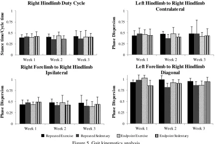
The percent of gait cycle that the right hindlimb was on the ground (duty cycle) did not differ by activity (p=0.92) or repeated LDF procedures (p=0.69) for any of the weeks (Fig. 5). Similarly, no significant differences were observed due to activity or repeated LDF procedures for contralateral (p=0.87 for exercise and p=0.56 for repeated LDF), ipsilateral (p=0.25 for exercise and p=0.56 for repeated LDF), or diagonal phase dispersion (p=0.76 for exercise and p=0.09 for repeated LDF).
Right hindlimb duty cycle and phase dispersion showed no differences between groups.
Figure 4. Example wound images from repeated groups one week following the LDF procedure.
Week 2
Week 1
Week 3
Week 4
Figure 5.Gait kinematics analysis.
Repeated Exercise Repeated Sedentary Endpoint Exercise Endpoint Sedentary
0 0.25 0.5 0.75 1
Week 1 Week 2 Week 3
Right Hindlimb Duty Cycle
S ta n c e t im e/ C y c le t ime 0 0.25 0.5 0.75 1
Week 1 Week 2 Week 3
Left Hindlimb to Right Hindlimb Contralateral P h a s e D is p e rs io n 0 0.25 0.5 0.75 1
Week 1 Week 2 Week 3
Left Forelimb to Right Hindlimb Diagonal P h a s e D is p e rs io n 0 0.25 0.5 0.75 1
Week 1 Week 2 Week 3
4. Discussion
Laser Doppler flowmetry procedures repeated weekly did not alter blood perfusion measurements across weeks for either exercise or sedentary groups. All incisions from the procedure were fully closed within one week and duty cycle and phase dispersion remained constant from week to week with repeated LDF procedures.
Osteovasculature is known to play an important role in bone development and remodeling [18,19]. The goal of this research was to validate laser Doppler flowmetry as a method to assess longitudinal changes in blood perfusion within bone. LDF was capable of measuring bone blood perfusion over a 4-week period in mice. A previous study demonstrated that laser Doppler flowmetry could be used to measure blood perfusion accurately; however, this study was limited to a single endpoint measurement and reported inflammation at the incision site three months following the procedure [11]. Reducing the incision size and closing the wound limited the amount of inflammation and resulted in no changes to gait kinematics, allowing repeated measurements to be performed. Inflammation was of significant concern, since systemic inflammation has been shown to promote bone resorption, while localized inflammation can increase blood perfusion to the region [20]. Wound images were taken weekly in repeated LDF groups to assess wound healing. In all cases, the wounds were fully closed one week following the procedure. Healthy C57Bl/6J mice with a 6-mm diameter wound were shown to have partial epithelialization after 7 days [21]. Incisions for the LDF procedure were 2-mm long slits and were closed with surgical glue, therefore wound closure before 7 days is likely. Exercise and sedentary groups showed similar extents of wound healing; however, wounds were only observed at weekly time points, whereas healing may have proceeded at different rates within that weekly period. The wound assessments here are consistent with a previous study, where wound healing was similar between exercise and sedentary groups, at least in young mice [22]. In aged mice, the study found that sedentary groups had slower wound healing than exercising mice. Therefore, the repeated LDF procedure would need be validated in older animals if using an aged model. In addition to inflammation, a primary concern was that the mice would limp following the repeated procedure. Any changes to gait kinematics would alter loading on the bones, which would affect bone remodeling and could induce changes to the blood supply within bone [22].
This study showed that Laser doppler flowmetry can be used to measure blood perfusion in bone over subsequent weeks in mice with no significant changes to blood perfusion, minimal local inflammation and no change to gait kinematics. This new method of measuring blood flow in bone has the potential to allow researchers to determine how disease states in bone are correlated with changes in perfusion, which will improve understanding of the underlying mechanisms involved and thus could enable the development of improved interventions and therapeutics.
4.1 shortcomings and future work
Several technical difficulties were encountered during this study that delayed the official start of the study. During the first week (two weeks before the official start), the LDF probe broke, and only four mice from the repeated-exercise group and three mice from repeated-sedentary group received the procedure. During the following week, the treadmill malfunctioned, and thus the exercise regimen could not be completed in any of the exercise groups. Because a replacement LDF probe had not yet arrived, no LDF measurements were collected, although an incision was made and the periosteum was removed to simulate the incision made during the LDF procedure. Because of these issues, the study duration was extended so that data were collected consistently over a four-week period. These interventions prior to the official start of the study may have affected the outcomes, although the data were consistent for the four-week duration of the study. In future studies, to improve the accuracy of blood perfusion measurements, the thickness of the cortical bone of the proximal tibia could be measured. As demonstrated in a previous study, this step would allow us to determine if differences in LDF measurements are correlated with cortical thickness. If so, then blood perfusion measurements could be adjusted accordingly, since a thicker bone could decrease the amount of light reaching the blood cells and thus impact the amount of Doppler shifted light reaching the detector within the LDF probe [11].
5. Conclusions
determine how diseases and therapies affect bone perfusion over time, which is an important step in making models more clinically relevant.
6. Acknowledgments
The author thanks Nicholas Hanne and Dr. Jacqueline Cole for their training and mentorship. Support for this work was provided by the Eunice Kennedy Shriver National Institute of Child Health & Human Development of the National Institutes of Health under Award Number K12HD073945. The content is solely the responsibility of the author and does not necessarily represent the official views of the National Institutes of Health. Additional support was provided by the North Carolina State University Office of Undergraduate Research.
7. References
[1] J. Trueta, The Role of the Vessels in Osteogenesis, Bone Jt. J. 45–B (1963) 402–418.
[2] A.M. Parfitt, The mechanism of coupling: a role for the vasculature, Bone. 26 (2000) 319–323. [3] R.E. Tomlinson, J.A. McKenzie, A.H. Schmieder, G.R. Wohl, G.M. Lanza, M.J. Silva, Angiogenesis is
Required for Stress Fracture Healing in Rats, Bone. 52 (2013) 212–219.
[4] R. Ellman, J. Spatz, A. Cloutier, R. Palme, B.A. Christiansen, M.L. Bouxsein, Partial reductions in mechanical loading yield proportional changes in bone density, bone architecture, and muscle mass, J. Bone Miner. Res. 28 (2013) 875–885.
[5] S. Ziegler, S. Kudlacek, A. Luger, E. Minar, Osteoprotegerin plasma concentrations correlate with severity of peripheral artery disease, Atherosclerosis. 182 (2005) 175–180.
[6] M.T. Vogt, J.A. Cauley, L.H. Kuller, M.C. Nevitt, Bone Mineral Density and Blood Flow to the Lower Extremities: The Study of Osteoporotic Fractures, J. Bone Miner. Res. 12 (1997) 283–289.
[7] J. Bernhardt, C. English, L. Johnson, T.B. Cumming, Early Mobilization After Stroke, Stroke. 46 (2015) 1141– 1146.
[8] D.B. Burr, M.R. Forwood, D.P. Fyhrie, R.B. Martin, M.B. Schaffler, C.H. Turner, Bone Microdamage and Skeletal Fragility in Osteoporotic and Stress Fractures, J. Bone Miner. Res. 12 (1997) 6–15.
[9] R.E. Tomlinson, M.J. Silva, Skeletal Blood Flow in Bone Repair and Maintenance, Bone Res. 1 (2013) 311– 322.
[10] M.-H. Lafage-Proust, B. Roche, M. Langer, D. Cleret, A. Vanden Bossche, T. Olivier, L. Vico, Assessment of bone vascularization and its role in bone remodeling, BoneKEy Rep. 4 (2015).
[11] B. Roche, A. Vanden-Bossche, M. Normand, L. Malaval, L. Vico, M.-H. Lafage-Proust, Validated Laser Doppler protocol for measurement of mouse bone blood perfusion — Response to age or ovariectomy differs with genetic background, Bone. 55 (2013) 418–426.
[12] N.J. Hanne, E.D. Easter, A.J Stewart, S.V. Pinnamaraju, J.H. Cole, Diet-Induced Obesity Deteriorates Cancellous Bone Structure Despite Increased Blood Perfusion, in: Orthop. Res. Soc. Annu. Meet., 2017: p. 1699.
[13] N.J. Hanne, A.J. Steward, S.V. Pinnamaraju, S.D. Teeter, J.H. Cole, Exercise Therapy Mitigates Reductions in Tibial Blood Flow during Acute Stroke Recovery, in: Orthop. Res. Soc. Annu. Meet., 2017: p. 0337.
[14] M.T. Goova, J. Li, T. Kislinger, W. Qu, Y. Lu, L.G. Bucciarelli, S. Nowygrod, B.M. Wolf, X. Caliste, S.F. Yan, D.M. Stern, A.M. Schmidt, Blockade of Receptor for Advanced Glycation End-Products Restores Effective Wound Healing in Diabetic Mice, Am. J. Pathol. 159 (2001) 513–525.
[15] H. Leblond, M. L’Espérance, D. Orsal, S. Rossignol, Treadmill Locomotion in the Intact and Spinal Mouse, J. Neurosci. 23 (2003) 11411–11419.
[16] A.D. Kloos, L.C. Fisher, M.R. Detloff, D.L. Hassenzahl, D.M. Basso, Stepwise motor and all-or-none sensory recovery is associated with nonlinear sparing after incremental spinal cord injury in rats, Exp. Neurol. 191 (2005) 251–265.
[17] E. Redondo-Castro, A. Torres-Espín, G. García-Alías, X. Navarro, Quantitative assessment of locomotion and interlimb coordination in rats after different spinal cord injuries, J. Neurosci. Methods. 213 (2013) 165–178.. [18] S.C. Marks Jr., P.R. Odgren, Chapter 1 - Structure and Development of the Skeleton, in: J.P. Bilezikian, L.G.
Raisz, G.A. Rodan (Eds.), Princ. Bone Biol. Second Ed., Academic Press, San Diego, 2002: pp. 3–15. [19] H.-P. Gerber, T.H. Vu, A.M. Ryan, J. Kowalski, Z. Werb, N. Ferrara, VEGF couples hypertrophic cartilage
[20] J.E. Fonseca, M.J. Santos, H. Canhão, E. Choy, Interleukin-6 as a key player in systemic inflammation and joint destruction, Autoimmun. Rev. 8 (2009) 538–542.
[21] S.A. Eming, S. Werner, P. Bugnon, C. Wickenhauser, L. Siewe, O. Utermöhlen, J.M. Davidson, T. Krieg, A. Roers, Accelerated Wound Closure in Mice Deficient for Interleukin-10, Am. J. Pathol. 170 (2007) 188–202. [22] H.M. Frost, Skeletal structural adaptations to mechanical usage (SATMU): 1. Redefining Wolff’s law: the bone