ABSTRACT
BUTTON, KHAI ALLEN. Soft Tissue Reconstruction and Ecomorphology of Beaks in Extant and Extinct Theropod Dinosaurs. (Under the Direction of Drs. Lindsay Zanno and Theodore Simons).
We are seldom able to directly observe evidence of soft tissue morphology or feeding behavior of extinct species; thus, reconstructing their ecologies is notoriously difficult, and inferences based on the most common types of fossils (teeth and bone) are regularly limited by the strength of analogies to living species. Here I reconstruct the ecology and morphology of Theropoda, whose extant members (birds) represent the most speciose clade of terrestrial vertebrates. In particular, I focus on the anatomy and dietary ecomorphology of the cornified beak.
Within Archosauria alone, beaks are hypothesized to have independently arisen a minimum of fifteen times, alluding to the ubiquitous adaptive significance of this. In Theropoda, the
rhamphotheca has been linked to dietary transitions and diversification in clades most closely related to birds. Thus, to understand the success and paleoecology of theropod dinosaurs, the beak is a vital, yet relatively understudied structure of interest. Fortunately, the correlation between beak morphology and ecology in extant birds has a long history of study. Landmark work on Darwin’s finches in the Galápagos and others has demonstrated a tight linkage between
the shape of the beak and diet. However, new research may indicate that this pattern does not extend to birds as a whole, necessitating additional testing of hypothesized morphology-ecology correlation in birds. Here I quantify beak shape in a wide array of extant and extinct theropod dinosaurs and use phylogenetically-informed analysis of diet-morphology correlation to test predictions of feeding ecology in extinct Theropoda. My statistical model using geometric morphometric landmarks to quantify beak shape achieved moderate success in predicting dietary ecology, but more work remains to be done. Reconstruction of extinct theropod paleoecology is further hampered by difficulties in reconstructing the soft tissue morphology of the beak.
dinosaurs, to develop testable hypotheses about the specific dimensions and properties of the beak in extinct taxa. I investigated the correlation between rhinotheca and premaxilla
morphology in avian and non-avian dinosaurs to reconstruct the presence and shape of the beak in extinct taxa. I find that the presence, extent, and curvature of the rhamphotheca are
© Copyright 2018 by Khai Button
Soft Tissue Reconstruction and Ecomorphology of Beaks in Extant and Extinct Theropod Dinosaurs
by
Khai Allen Button
A dissertation submitted to the Graduate Faculty of North Carolina State University
in partial fulfillment of the requirements for the degree of
Doctor of Philosophy
Biology
Raleigh, North Carolina
2018
APPROVED BY:
_______________________________ _______________________________
Lindsay Zanno Theodore Simons
Co-Chair of Advisory Committee Co-Chair of Advisory Committee
_______________________________ _______________________________
ii
DEDICATION
For my parents, who believed in and encouraged me, For my wife, who loved me and pushed me,
And
iii
BIOGRAPHY
Khai Button first discovered a love of dinosaurs as a four-year-old. It was the year that Jurassic Park came out, which was convenient timing. He was probably too young to see it at that age, but it irreversibly changed him. Just like most kids, it was his dream to become a paleontologist. But unlike many other children with whom he played dinosaurs on the playground, he never grew out of it. Still, he didn’t honestly believe that it could be a viable career path until he got to college. He studied biology in the hope of becoming a doctor or a biomedical researcher until he attended a lecture by a visiting paleontologist. That was a light bulb moment, when he realized that paleontology really was an actual job, and he saw the path unfolding before him that would lead him there.
Graduate school in North Carolina afforded him the opportunity to complete a number of lifelong goals, including becoming a published author and participating in field work. However, the most important discovery he made was a deep passion and talent for teaching and educating others. Be it students in the classroom or audiences young and old at museum outreach events, I strived to share my passion and knowledge for the study of life.
iv
ACKNOWLEDGMENTS
After close to six years working toward my doctorate, I discovered the real reason that so few people ever complete a Ph.D. It’s not just that this degree was the most challenging thing I’ve
ever attempted in my academic career; completing your doctoral degree is simply too much for one person—I gave 100% of myself toward completing my Ph.D., but that wasn’t enough, and I never would have succeeded without the unending support and enduring love of my friends and family to fill the gap.
I want to thank my parents for teaching me the value of education, and of completing what you begin. They were always there for me when I needed advice or financial support, which they offered without question, and there was never a single moment of doubt in their minds that I would succeed. I want to thank my wife. When she married me, she married my academic aspirations, with all that they entailed. From day one, she truly amazed me with the huge sacrifices she made to ensure that I had every chance for success: she moved across the country for me, she missed out on precious time with her family, and she worked tirelessly to support me, and I know as absolute fact that I would not be here without her. Lastly, I want to thank my daughter. She doesn’t yet know how dearly I wish I could get back the time I was away from her, or how much she inspired me every day to push forward and learn new things. But I hope that one day she’ll learn that as much as I did this for myself and for the pursuit of scientific
knowledge, I did this for her—for her future. My dearest wish is that I can inspire her to pursue her dreams and complete her goals, whatever they may be. It is my hope that she is lucky enough to have the opportunity to dedicate herself to something she believes in, the way I did.
v
TABLE OF CONTENTS
LIST OF TABLES ... vii
LIST OF FIGURES ... viii
Chapter 1: Introduction ... 1
Beak Ecomorphology ... 3
The Extant Phylogenetic Bracket ... 6
Soft Tissue Preservation and Reconstruction ... 7
References ... 10
Figures ... 20
Chapter 2: Osteological Correlates of Soft Tissue Beak Morphology in Extant Birds ... 21
Introduction ... 22
Methods ... 24
Data Collection and Sampling Protocol ... 24
Keratin Thickness ... 25
Rhamphotheca Extent ... 25
Rhamphotheca Presence ... 26
Homologous Regions ... 26
Curvature... 26
Definitions ... 26
Statistical Analysis ... 26
Results ... 29
LPA Change from Rostral to Caudal ... 29
Porosity Index ... 29
Average Keratin Thickness ... 29
Extent ... 30
Rhamphotheca Presence ... 30
Discussion ... 31
Conclusion ... 35
References ... 37
Figures & Tables ... 41
Chapter 3: Shape Covariation between the Keratinous and Bony Components of the Avian Maxillary Rostrum ... 51
Introduction ... 52
Methods ... 53
Curvature... 53
Geometric Morphometrics ... 54
Phylogenetic Signal ... 56
Results ... 56
Curvature... 56
Geometric Morphometrics ... 56
Discussion ... 57
Conclusion ... 60
vi
Figures & Tables ... 66
Chapter 4: Soft Tissue Reconstruction in Recently Extinct and Ancient Theropoda ... 72
Introduction ... 73
Methods ... 74
Data Collection and Sampling Protocol ... 75
Homologous Regions ... 76
Definitions ... 76
Statistical Analysis ... 77
Results ... 80
Curvature... 80
Lateral Pore Area ... 80
Rhamphotheca Extent ... 81
Presence of the Rhamphotheca ... 81
Discussion ... 82
Reconstructing Rhamphotheca Morphology ... 85
Conclusion ... 86
References ... 86
Figures & Tables ... 93
Chapter 5: Dietary Ecomorphology in Extant and Extinct Avian and Non-Avian Theropods ... 112
Introduction ... 114
Methods ... 116
Data Collection and Sampling Protocol ... 116
Time-Calibrated Phylogeny ... 117
Diet & Ecology... 117
Linear Measurements... 118
Geometric Morphometric Landmarks ... 118
Statistical Analysis ... 119
Results ... 119
Linear Morphometrics ... 119
Geometric Morphometrics ... 120
Discussion ... 121
Conclusion ... 127
References ... 128
Figures & Tables ... 138
vii
LIST OF TABLES
Table 1. Raw data ... 48
Table 2. Curvature summary statistics ... 71
Table 3. Ancient theropods sampled ... 103
Table 4. Recently extinct bird species sampled ... 105
Table 5. Curvature, edentulism, and rhinotheca extent in extinct theropods ... 107
Table 6. Surface texture and rhamphotheca presence per transect ... 109
viii
LIST OF FIGURES
Figure 1. Repeated independent evolution of a keratinous rhamphotheca in Archosauria ... 20
Figure 2. Keratin thickness diagram ... 41
Figure 3. Measuring rhamphotheca extent ... 41
Figure 4. Homologous regions of the beak ... 42
Figure 5. Diagram of curvature measurements ... 42
Figure 6. LPA per beak region... 43
Figure 7. Breakdown of beak transects by homologous region ... 43
Figure 8. Beak length vs. LPA in birds ... 44
Figure 9. Phenogram showing relative porosity across extant Aves ... 45
Figure 10. Keratin thickness in birds ... 46
Figure 11. Unsupported extent of the rhamphotheca ... 47
Figure 12. Geometric morphometric landmarks placed in lateral view ... 66
Figure 13. Geometric landmarks placed in dorsoventral view ... 66
Figure 14. Beak curvature by avian clade ... 67
Figure 15. Curvature phenograms... 68
Figure 16. Bone vs. keratin shape (lateral) ... 69
Figure 17. Consensus shapes of beak bone and keratin in lateral view ... 69
Figure 18. Bone vs. keratin shape (dorsoventral) ... 70
Figure 19. Consensus shapes of beak bone and keratin in dorsoventral view... 70
Figure 20. Homologous regions of the premaxilla ... 93
Figure 21. Time-calibrated phylogeny of extinct Theropoda ... 95
ix
Figure 23. Measuring rhamphotheca extent ... 96
Figure 24. Lateral pore area vs. premaxilla length in Theropoda ... 96
Figure 25. Time-scaled traitgram of porosity indices ... 97
Figure 26. Gross rhamphotheca extent vs. porosity index in extant and extinct Theropoda .... 98
Figure 27. Rostroventral extent of the rhamphotheca vs. maxillary curvature ... 99
Figure 28. Rhinotheca presence vs. bone surface texture per transect in Theropoda ... 100
Figure 29. Relative premaxilla size in theropods ... 101
Figure 30. Rhamphotheca reconstruction in Erlikosaurus andrewsi (Maniraptora: Therizinosauria) ... 102
Figure 31. Breakdown of extant birds sampled by dietary ecology ... 138
Figure 32. Homologous landmarks and sliding semilandmarks ... 139
Figure 33. Consensus configuration of GM landmarks on theropod rostra ... 140
Figure 34. Consensus shapes of theropod rostra in dorsoventral view ... 141
1
Chapter 1: Introduction
Paleontology, though it is an imperfect lens through which to view the past, remains the most effective means of understanding life on the planet in the eons preceding the present day. Through our study of fossils, we are able to reconstruct the climate and ecology of past
environments, and these reconstructions are a crucial foundation upon which our understanding of the world is based. Models predicting how modern species will adapt to things like
anthropogenic climate change require a consistent baseline using climate data from deep time to improve accuracy. To be data-driven, predictive models of changing modern ecology must incorporate an understanding of the fossil record. It is therefore vital to study the paleoecology of extinct species, particularly those with extant relatives for direct application of observed patterns. However, reconstructing the ecology of extinct taxa is notoriously difficult proposition, primarily because, with very few exceptions, we are unable to directly observe evidence of soft tissue morphology or feeding behavior, and inferences based on the most common types of fossils (teeth and bone) are regularly limited by the strength of analogies to living species.
The overarching goal of my investigation is to develop a novel, statistically robust methodology for reconstructing the dietary ecology of extinct theropods, the only clade of dinosaurs with extant representatives. To strengthen ecological inferences, I target theropod dinosaurs with beaks, the best functional analog to modern birds. I focus on two key aspects of their biology: 1) diet-morphology correlation and 2) soft tissue morphology, largely focusing on the rostrum. This research is an essential first step to understanding paleoecology in dinosaurs, as diet and
morphology underpin our knowledge of behavior, predator-prey interactions, and community as well as ecosystem structure. Laying the foundations of theropod dinosaur paleoecology will potentially open many new avenues for research, including the evolution and diversification of beak morphology in birds and the interaction between edentulism, growth of a keratinous rhamphotheca, and diet in dinosaurs and other beaked taxa.
2 Silesauridae (Dzik, 2003), Ornithischia, Ceratosauria (Wang et al., 2017), Ornithomimosauria (Barsbold, 1981), Therizinosauroidea (Lautenschlager et al., 2013, 2014), Oviraptorosauria (Norell, Makovicky & Currie, 2001; Barrett, 2005), multiple times within Pterosauria (Frey & Martill, 1998), Suchia (Rauhut, 1997; Göhlich et al., 2005; Nesbitt, 2007; Holliday & Nesbitt, 2013), Avialae, and tentatively in Sauropodomorpha (Apesteguía, 2004; Gallina & Apesteguía, 2011; Wiersma & Sander, 2016) (Figure 1). However, among these groups, Testudinata and Theropoda are the only extant forms that have beaked representatives, and among these, Theropods demonstrate the greatest diversity and disparity of bill morphologies (Mitchell & Makovicky, 2014). Birds have adapted beaks into chisels (Piciformes), probes (Scolopacidae), siphons (nectivorous birds such as trochillid hummingbirds), fishnets (Pelecanidae), spears (Ardeidae), nutcrackers (Psittaciformes), filters (Phoenicopteriformes), and myriad other forms (Gill, 2007). I have therefore chosen bird beaks as the target of this investigation. Within
Avialae, there were numerous independent reductions in dentition and concomitant development of a rostral rhamphotheca that partially or completely covers the premaxilla/dentary (Zhou & Li, 2010; Louchart & Viriot, 2011). Although molecular evidence supports a single common
toothless common ancestor for modern birds (Meredith et al., 2014), complete edentulism developed a minimum of six separate times throughout Avialae: in Confuciusornithiformes, Gobipteryx (O’Connor & Chiappe, 2011), Archaeorhynchus, Zhongjianornis, Schizooura (Zhou, Zhou & O’Connor, 2012), and Neornithes. Given that this versatile composite of bony and soft
tissue has arisen so many times, it is clear that this evolutionary trajectory is strongly favored by natural selection. Time and again, it has been hypothesized that the beak is a key structural component allowing clades to exploit new ecological niches and undergo adaptive radiations (Grant, 1999; Bhullar et al., 2015). If we aim to understand the evolution and diversification of avian and non-avian dinosaurs, the cornified rhamphotheca is a vital structure of interest.
Adaptive radiations within Aves have resulted in a staggering array of beak morphologies that birds employ for nearly all activities of daily life, including: food acquisition and processing, preening (Clayton et al., 2005), making and using tools, display/courtship and species
3 adaptability and multifunctionality of the beak that has often been cited as a defining
characteristic of the clade and a key evolutionary innovation allowing for the successful dispersal of birds to nearly every habitat on every continent across the globe (Zusi, 1993; Zhou, Zhou & O’Connor, 2012; Kissling, Sekercioglu & Jetz, 2012).
Beak shape has long been observed to vary between species and sexes of birds (Herrel et al., 2010), and along dietary, environmental, and geographic gradients (Price, 1991; Foster, Podos & Hendry, 2008). Although many examples exist of adaptive radiations resulting in vastly different beak morphologies, the most well-known and thoroughly documented are Darwin’s finches in the Galápagos (Schluter & Grant, 1984; Grant & Grant, 1989, 1996; Grant, 1999; Foster, Podos & Hendry, 2008) and the honeycreepers of the Hawaiian archipelago (Richards & Bock, 1973; Lovette, Bermingham & Ricklefs, 2002). Both groups descend from small ancestral populations with intermediate beak phenotypes that became geographically isolated and subsequently speciated, diversifying to fill the available niches.
BEAK ECOMORPHOLOGY
Given the close relationship between feeding and beak shape, it is hardly surprising that
numerous studies have attempted to identify correlations between beak morphology and ecology. Notably, BR. & PR. Grant, in their landmark studies of Galápagos finches, observed that mean beak phenotype shifted in response to prolonged drought which altered food availability (Grant & Grant, 1989; Grant, 1999). However, in recent years, other studies have come to question the utility of analyzing beak shape to reconstruct diet. A study by Bright et al. (2016) on extant raptors, which they defined as polyphyletic assemblage of diurnal predatory birds, was unable to demonstrate a correlation between beak shape and diet. Additionally, the authors were unable to separate out the beak and braincase as independent developmental modules, suggesting that natural selection was not directly acting on beak morphology, but on the skull as a whole (Bright et al., 2016). However, these and other studies rarely separate out the effect of degrees of
relatedness between taxa when attempting to reconstruct diet-morphology correlation.
4 Because so few fossil beds preserve extrinsic evidence of diet (sensu Zanno & Makovicky, 2011), dietary reconstruction in dinosaurs has historically been conducted primarily by analogy. If gross anatomy or tooth morphology of extinct taxa closely resemble living species, then a similar diet is proposed for the extinct representative. An important application of this principle can be seen in Norell et al., 2001. Two ornithomimid specimens possessing putative fossilized keratinous tissue of the rhamphotheca were hypothesized to be sediment-straining based on soft tissue structures of the bill that resemble the lamellae of extant anseriforms. But, as demonstrated by Barrett (2005) this interpretation is problematic from the standpoint of feeding efficiency and energy intake rate, and so it is clear that more work needs to be done to determine diet in this clade. To date, no rigorous and widely applicable metric exists to establish which coupling of extant and extinct species are similar enough to make strong inferences about the diet of a given extinct taxon, and inferences are generally made in a piecemeal, taxon-by-taxon, fashion. Additional difficulties arise when trying to reconstruct the diets of many extinct theropod
dinosaurs, as no suitable modern analogs exist; there are no living birds that possess mineralized teeth as well as a beak, and even the largest extant ratites (Smith et al., 2006) are smaller than large-bodied maniraptorans by more than an order of magnitude (Paul, 2010; Zanno &
Makovicky, 2012). One final consideration is that previous studies have often focused on broad categories of diet (e.g. carnivory vs. herbivory) in extinct taxa (Zanno & Makovicky, 2011; Brusatte, 2012; Foth & Rauhut, 2013), which introduces a priori bias by forcing complex diets into semi-arbitrary bins and does not reflect the way living animals utilize food resources (Button & Zanno, 2016). Clearly, different approaches for dietary reconstruction are required, and there is a need for a statistically robust methodology for quantifying beak morphology in both extant and extinct taxa and correlating it with diet (Barrett, 2014).
5 statistically unsound (Adams, Rohlf & Slice, 2004). In response to these and other issues, new techniques have been devised. One that has gained popularity in recent years is Geometric morphometrics (GM). GM is a technique that differs from traditional linear morphometrics in that it is based on homologous landmarks rather than size-based metrics. The coordinates of these landmarks in space are subjected to Procrustes superimposition to separate out the effects of rotation and size to focus solely on shape (Zelditch, Swiderski & Sheets, 2012). This shape data can then be correlated with another data array such as a categorical representation of dietary ecology. This technique is an important step forward in the quantification of biological shape because it focuses on biologically meaningful structures that are consistent across taxa and takes size into account (Mitteroecker & Gunz, 2009). GM has been used in numerous studies and together they have been dubbed the “Geometric Morphometrics revolution” (Adams, Rohlf &
Slice, 2004) and is demonstrably more statistically sound than previous morphometric techniques. GM can be applied either in 2D or in 3D, and studies have shown that vastly different results can be garnered depending on whether the landmarks are 2D or 3D (Gould, 2014). It is clear from these studies that 3D is preferable to fully capture the fine scale details of biological shape, but this is not always possible, particularly when working on extinct taxa (Mitchell & Makovicky, 2014). Many fossil forms are only known from incomplete, crushed, or even completely flattened specimens, making reconstruction of 3D landmarks impossible (Hedrick et al., 2014). Work on retrodeforming the taphonomically warped skulls of extinct animals may have applications for increasing the possible number of samples (Arbour & Currie, 2012; Cuff & Rayfield, 2015), but more work is required. Given the dearth of
three-dimensionally preserved fossils and the fact that the smooth tapered surface of a bird bill possess few clearly defined landmarks (Foster, Podos & Hendry, 2008; Cooney et al., 2017), other approaches may be required to quantify beak morphology in the fossil record. Recent work has employed sliding semi-landmarks to quantify outlines when there is a deficit of homologous points (Bookstein, 1997; Mallon & Anderson, 2014). The outline of a fossil is digitized and then equidistant semi-landmarks are distributed along that outline. They are then slid to reduce either the bending energy or Procrustes distance, so although the landmarks themselves are not
6 Mitchell & Makovicky (2014), uses a combination of discrete and continuous traits that describe the ecomorphology of the beak and postcrania to calculate ecological diversity and disparity in extant and Cretaceous birds (Mitchell & Makovicky, 2014; Cooney et al., 2017). However, this work does not extend to non-avian theropods, and so direct comparisons between the efficacy of this technique and other morphometrics-based techniques remains to be conducted.
None of these approaches is perfect in every situation. One of my goals in the following chapters is to compare LM and GM approaches for generating a morphological dataset that can used in a phylogenetically-informed regression on dietary ecology. This is the first study to consistently apply and compare these techniques to such a large and taxonomically diverse morphological dataset encompassing avian and non-avian theropods. I examine which, or what combination of these techniques is most useful for describing diet-morphology correlation in extant birds, and use this information to reconstruct diet in extinct theropods. By analyzing the relationship between morphology and diet in extant animals, we begin to establish a basis for reconstructing paleoecology. This approach can be further extended to the reconstruction of soft tissues in dinosaurs.
THE EXTANT PHYLOGENETIC BRACKET
Vertebrate paleontologists have long been limited in the anatomical study of ancient life to information that can be gleaned directly from bone and teeth, which are by far the most commonly preserved biological tissues (Moyer, Zheng & Schweitzer, 2016a). To make inferences about soft tissue, a technique known as the “extant phylogenetic bracket” (EPB),
proposed by Bryant and Russell (1992) and Witmer (1995), has been developed. This
methodology allows for parsimonious inference of the presence/absence or extent of soft tissue structures by bracketing the extinct taxon of interest with two extant members of the same clade and it has become a staple of paleobiology. If a structure or trait is present in both extant forms, then we hypothesize that it was present in their common ancestor and all its descendants. This approach is further strengthened by the presence of osseous correlates in the extant members that are also present ancestrally. A canonical example of this is respiration; both birds and
7 possessed by only one extant member of the clade and where direct fossil evidence is absent in the extinct species, (e.g., feathers in some extinct birds), this is a second order inference (Witmer, 1995).
It has previously been proposed that the presence of dense vascularization on the surface of the maxillae/premaxillae and dentaries of theropod dinosaurs and on the skull roofs of some ceratopsians (Hieronymus, 2009; Hieronymus et al., 2009; Carr et al., 2017) are osteological correlates of a keratinous covering for those bones. However, extant crocodilians also possess numerous neurovascular foramina on the rostral aspect of the snout, and no species of living crocodilians possess any form of beak, making this hypothesis less parsimonious and
confounding previous attempts to reconstruct cranial soft tissue morphology. Additionally, only a handful of studies have examined the relationship between bony and soft tissue in living dinosaurs (birds), to identify robust osteological correlates of keratin structure and morphology for the purpose of making inferences about extinct dinosaur species (Knutsen, 2007; Jansen, 2008). My goal is to examine extant birds to identify bony correlates of soft tissue beak
morphology, and to quantify the covariation of hard and soft tissue shape of the avian beak. This will allow me to make predictions of the shape and extent of beaks in extinct taxa, which can then be tested against those fossil specimens that preserve direct evidence of rhamphotheca morphology, be it as an impression or natural cast, or as actual preserved soft tissue (Hou et al., 1999; Norell, Makovicky & Currie, 2001; Pan et al., 2016).
SOFT TISSUE PRESERVATION AND RECONSTRUCTION
Two primary families of keratin have been identified: alpha and beta. Alpha-keratin is
8 proteins have been identified from claw and feather material in avian and-non avian dinosaurs (Schweitzer et al., 1999; Moyer, Zheng & Schweitzer, 2016b; Pan et al., 2016). However, despite the relative resistance of keratinous soft tissue to degradation and the plethora of remarkably preserved feathered avian and non-avian dinosaurs recovered from Lagerstätten in China and elsewhere, few specimens that preserve the morphology of the rhamphotheca have ever been found, and birds remain a relatively small proportion of fossil discoveries (Birn-Jeffery et al., 2012; Ksepka & Boyd, 2012; Mitchell, 2015b). These factors make it difficult to reconstruct the soft tissue of the beak in extinct taxa. Additionally, there are soft tissue structures have been discovered in the fossil record that were unknown to exist solely from investigation of fossil bone. A “mummified” specimen of the hadrosaurid Edmontosaurus regalis was found to possess a previously undiscovered soft tissue “cocks comb” that has changed the way we reconstruct this
saurolophine (Bell et al., 2014). Furthermore, there are soft tissue structures in the bills of extant birds such as the tomia, keratinous “pseudoteeth” found in mergansers and other piscivores (Barrett, 2005), or the flexible bill-tip organ in probing birds such as avocets and stilts
(Cunningham et al., 2013) that, if they existed in extinct dinosaurs, would be unlikely to preserve in the fossil record (Hieronymus & Witmer, 2010). Taken together, these factors pose a serious challenge for researchers attempting to reconstruct the shape, structure, and extent of the soft tissue beak and the presence of other cranial integumentary appendages in extinct animals.
Although edentulism is, generally speaking, readily evident in the fossil record (but see (Zheng et al., 2014)), it is a mistake to assume a 1:1 correlation between degrees of edentulism and the existence of a keratinous rhamphotheca, as the presence of both partial dentition with a soft tissue beak is not reflected in any extant taxa, but is widespread in the fossil record (Elzanowski, 1991; Zhou & Martin, 2011). Additionally, there are numerous factors related to beak size and shape that must be explored in order to accurately reconstruct the soft tissue of extinct taxa in which it is not preserved. Although the morphology of the keratinous covering of the beak generally follows that of the underlying bone (Stettenheim, 2000; Button & Zanno, 2016), this is not always the case, and the soft tissue thickness is not uniformly distributed across the beak. For example, the rhamphotheca will occasionally extend far beyond the rostralmost tips of the
9 feeding, and the structure and spatial geometry of the keratinous sheath contribute to the
biomechanics of feeding (Soons et al., 2012; Lautenschlager et al., 2013). In particular, a coating of keratin has been shown to help dissipate bite forces (reducing strain) more efficiently than an edentulous jaw with keratin absent, as demonstrated by Seki and colleagues using finite element analysis in toucans (Seki, Schneider & Meyers, 2005; Seki, Mackey & Meyers, 2012). An additional study using the CT-scanned skull of Erlikosaurus andrewsi corroborates these results in an extinct maniraptoran (Lautenschlager et al., 2013). However, this study simply tested artistic interpretations of bill morphology, rather than attempting an accurate reconstruction based on the morphology of the underlying bone. More work is needed to describe the
relationship between the bony beak and the rhamphotheca in a taxonomically broader and more ecologically diverse sample of modern birds in order to make reconstruction of the structure and mechanical properties of beaks in extinct animals possible. Reconstructing the shape and extent of keratinous soft tissue in dinosaur beaks is a necessary precursor to future exploration of theropod dinosaur development and ecology. To unravel the evolution of the avian
rhamphotheca, analogous structures in outgroup taxa must be reliably reconstructed. The
techniques used in the following chapters have potential applications to the study of beaked taxa across Archosauria, including Ornithischia, Pterosauria, and Pseudosuchia.
10
REFERENCES
Adams DC, Rohlf FJ, Slice DE. 2004. Geometric morphometrics: Ten years of progress following the “revolution.” Italian Journal of Zoology 71:5–16. DOI:
10.1080/11250000409356545.
Apesteguía S. 2004. Bonitasaura salgadoi gen. et sp. nov.: A beaked sauropod from the Late Cretaceous of Patagonia. Naturwissenschaften 91:493–497. DOI: 10.1007/s00114-004-0560-6.
Arbour V, Currie P. 2012. Analyzing taphonomic deformation of ankylosaur skulls using retrodeformation and finite element analysis. PLOS ONE 7:e39323. DOI:
10.1371/journal.pone.0039323.
Barrett PM. 2005. The diet of ostrich dinosaurs (Theropoda: Ornithomimosauria). Palaeontology 48:347–358. DOI: 10.1111/j.1475-4983.2005.00448.x.
Barrett PM. 2014. Paleobiology of herbivorous dinosaurs. Annual Review of Earth and Planetary Sciences 42:207–230. DOI: 10.1146/annurev-earth-042711-105515.
Barsbold R. 1981. Toothless dinosaurs of Mongolia. Fossil Vertebrates of Mongolia:26.
Bell PR, Fanti F, Currie PJ, Arbour VM. 2014. A mummified duck-billed dinosaur with a soft-tissue cock’s comb. Current Biology 24:70–75. DOI: 10.1016/j.cub.2013.11.008.
Bhullar B-AS, Hanson M, Fabbri M, Pritchard A, Bever GS, Hoffman E. 2016. How to make a bird skull: Major transitions in the evolution of the avian cranium, paedomorphosis, and the beak as a surrogate hand. Integrative and Comparative Biology 56:389–403. DOI: 10.1093/icb/icw069.
11 Birn-Jeffery AV, Miller CE, Naish D, Rayfield EJ, Hone DWE. 2012. Pedal claw curvature in
birds, lizards and Mesozoic dinosaurs - Complicated categories and compensating for mass-specific and phylogenetic control. PLOS ONE 7:e50555. DOI:
10.1371/journal.pone.0050555.
Bookstein FL. 1997. Landmark methods for forms without landmarks: Morphometrics of group differences in outline shape. Medical Image Analysis 1:225–43.
Bright JA., Marugán-Lobón J., Cobb SN., Rayfield EJ. 2016. The shapes of bird beaks are highly controlled by nondietary factors. Proceedings of the National Academy of Sciences 113:5352–5357. DOI: 10.1073/pnas.1602683113.
Brusatte SL. 2012. Feeding and diet. In: Dinosaur Paleobiology. Oxford: Wiley-Blackwell, 159– 190. DOI: 10.1002/9781118274071.ch7.
Bryant HN, Russell AP. 1992. The role of phylogenetic analysis in the inference of unpreserved attributes of extinct taxa. Philosophical Transactions of the Royal Society B: Biological Sciences 337:405–418. DOI: 10.1098/rstb.1992.0117.
Butler RJ, Benson RBJ., Barrett PM. 2013. Pterosaur diversity: Untangling the influence of sampling biases, Lagerstätten, and genuine biodiversity signals. Palaeogeography, Palaeoclimatology, Palaeoecology 372:78–87. DOI: 10.1016/j.palaeo.2012.08.012.
Button K, Zanno LE. 2016. Linear morphometric analysis of beak ecomorphology in avian and non-avian dinosaurs [Abstract]. In: Journal of Vertebrate Paleontology. Salt Lake City, 107.
Clayton DH, Moyer BR, Bush SE, Jones TG, Gardiner DW, Rhodes BB, Goller F. 2005.
Adaptive significance of avian beak morphology for ectoparasite control. Proceedings of the Royal Society B: Biological Sciences 272:811–7. DOI: 10.1098/rspb.2004.3036.
12 Crawford NG, Faircloth BC, McCormack JE, Brumfield RT, Winker K, Glenn TC. 2012. More
than 1000 ultraconserved elements provide evidence that turtles are the sister group of archosaurs. Biology Letters 8:783–786. DOI: 10.1098/rsbl.2012.0331.
Cuff AR, Rayfield EJ. 2015. Retrodeformation and muscular reconstruction of
ornithomimosaurian dinosaur crania. PeerJ 3:e1093. DOI: 10.7717/peerj.1093.
Cunningham SJ, Corfield JR, Iwaniuk AN, Castro I, Alley MR, Birkhead TR, Parsons S. 2013. The anatomy of the bill tip of kiwi and associated somatosensory regions of the brain: Comparisons with shorebirds. PLOS ONE 8:1–17. DOI: 10.1371/journal.pone.0080036.
Davit-Béal T, Tucker AS, Sire J-Y. 2009. Loss of teeth and enamel in tetrapods: Fossil record, genetic data and morphological adaptations. Journal of Anatomy 214:477–501. DOI: 10.1111/j.1469-7580.2009.01060.x.
Dial KP. 1992. Avian forelimb muscles and nonsteady flight: Can birds fly without using the muscles in their wings? The Auk 109:874–885. DOI: 10.2307/4088162.
Dzik J. 2003. A beaked herbivorous archosaur with dinosaur affinities from the early Late Triassic of Poland. Journal of Vertebrate Paleontology 23:556–574. DOI:
10.1671/A1097.
Elzanowski A. 1991. New observations on the skull of Hesperornis with reconstructions of the bony palate and the otic region. Postilla 207:1–20.
Farmer CG, Sanders K. 2010. Unidirectional airflow in the lungs of alligators. Science 327:338– 340. DOI: 10.1126/science.1180219.
Foster D, Podos J, Hendry A. 2008. A geometric morphometric appraisal of beak shape in Darwin’s finches. Journal of Evolutionary Biology 21:263–75. DOI:
10.1111/j.1420-9101.2007.01449.x.
13 Frey E, Martill DM. 1998. Soft tissue preservation in a specimen of Pterodactylus kochi
(Wagner) from the Upper Jurassic of Germany. Neues Jahrbuch für Geologie und Paläontologie 210:421–441.
Gallina PA, Apesteguía S. 2011. Cranial anatomy and phylogenetic position of the titanosaurian sauropod Bonitasaura salgadoi. Acta Palaeontologica Polonica 56:45–60. DOI:
10.4202/app.2010.0011.
Gill FB. 2007. Ornithology. New York: W. H. Freeman and Company.
Göhlich UB, Chiappe LM, Clark JM, Sues H. 2005. The systematic position of the Late Jurassic alleged dinosaur Macelognathus (Crocodylomorpha : Sphenosuchia). Canadian Journal of Earth Sciences 42:307–321. DOI: 10.1139/E05-005.
Gould FDH. 2014. To 3D or not to 3D, that is the question: Do 3D surface analyses improve the ecomorphological power of the distal femur in placental mammals? PLOS ONE 9:1–12. DOI: 10.1371/journal.pone.0091719.
Grant PR. 1999. Ecology and Evolution of Darwin’s Finches. Princeton: Princeton University
Press.
Grant B, Grant P. 1989. Natural selection in a population of Darwin’s finches. American Naturalist 133:377–393.
Grant B, Grant P. 1996. High survival of Darwin’s finch hybrids: Effects of beak morphology and diets. Ecology 77:500–509.
Hedrick BP, Chunling G, Omar GI, Fengjiao Z, Caizhi S, Dodson P. 2014. The osteology and taphonomy of a Psittacosaurus bonebed assemblage of the Yixian Formation (Lower Cretaceous), Liaoning, China. Cretaceous Research 51:321–340. DOI:
10.1016/j.cretres.2014.06.015.
Herrel A, Soons J, Aerts P, Dirckx J, Boone M, Jacobs P, Adriaens D, Podos J. 2010. Adaptation and function of the bills of Darwin’s finches: Divergence by feeding type and sex. Emu -
14 Hieronymus TL, Witmer LM. 2010. Homology and evolution of avian compound
rhamphothecae. The Auk 127:590–604. DOI: 10.1525/auk.2010.09122.
Holliday CM, Nesbitt SJ. 2013. Morphology and diversity of the mandibular symphysis of archosauriforms. Geological Society, London, Special Publications 379:555–571. DOI: 10.1144/sp379.2.
Hou LL, Martin LLD, Zhou Z, Feduccia A, Zhang F. 1999. A diapsid skull in a new species of the primitive bird Confuciusornis. Nature 399:679–682. DOI: 10.1038/21411.
Jansen SOK. 2008. Beak morphology in oviraptorids based on extant birds and turtles. M. Sci. Thesis, University of Oslo.
Kissling WD, Sekercioglu CH, Jetz W. 2012. Bird dietary guild richness across latitudes, environments and biogeographic regions. Global Ecology and Biogeography 21:328– 340. DOI: 10.1111/j.1466-8238.2011.00679.x.
Knutsen EM. 2007. Beak morphology in extant birds with implications on beak morphology in ornithomimids. M. Sci. Thesis, University of Oslo. DOI: 10.1525/jer.2007.2.4.toc.
Ksepka DT, Boyd CA. 2012. Quantifying historical trends in the completeness of the fossil record and the contributing factors: An example using Aves. Paleobiology 38:112–125. DOI: 10.5061/dryad.k7t00.
Lautenschlager S, Witmer LM, Altangerel P, Rayfield EJ. 2013. Edentulism, beaks, and biomechanical innovations in the evolution of theropod dinosaurs. Proceedings of the National Academy of Sciences 110:20657–62. DOI: 10.1073/pnas.1310711110.
Lautenschlager S, Witmer LM, Altangerel P, Zanno LE, Rayfield EJ. 2014. Cranial anatomy of Erlikosaurus andrewsi (Dinosauria, Therizinosauria): New insights based on digital reconstruction. Journal of Vertebrate Paleontology 34:1263–1291. DOI:
10.1080/02724634.2014.874529.
15 Lovette IJ, Bermingham E, Ricklefs RE. 2002. Clade-specific morphological diversification and
adaptive radiation in Hawaiian songbirds. Proceedings of the Royal Society B: Biological Sciences 269:37–42. DOI: 10.1098/rspb.2001.1789.
Mallon JC, Anderson JS. 2014. Implications of beak morphology for the evolutionary paleoecology of the megaherbivorous dinosaurs from the Dinosaur Park Formation (upper Campanian) of Alberta, Canada. Palaeogeography, Palaeoclimatology, Palaeoecology 394:29–41. DOI: 10.1016/j.palaeo.2013.11.014.
Meredith RW, Zhang G, Gilbert MTP, Jarvis ED, Springer MS. 2014. Evidence for a single loss of mineralized teeth in the common avian ancestor. Science 346:1254390. DOI:
10.1126/science.1254390.
Mitchell JS. 2015a. Extant-only comparative methods fail to recover the disparity preserved in the bird fossil record. Evolution 69:2414–2424. DOI: 10.1111/evo.12738.
Mitchell JS. 2015b. Preservation is predictable: Quantifying the effect of taphonomic biases on ecological disparity in birds. Paleobiology 41:1–15. DOI: 10.1017/pab.2014.23.
Mitchell JS., Makovicky PJ. 2014. Low ecological disparity in Early Cretaceous birds. Proceedings of the Royal Society B: Biological Sciences 281:1–6. DOI:
10.1098/rspb.2014.0608.
Mitteroecker P, Gunz P. 2009. Advances in geometric morphometrics. Evolutionary Biology 36:235–247. DOI: 10.1007/s11692-009-9055-x.
Moyer AE, Zheng W, Schweitzer MH. 2016a. Keratin durability has implications for the fossil record: Results from a 10 year feather degradation experiment. PLOS ONE 11:1–19. DOI: 10.1371/journal.pone.0157699.
16 Nesbitt S. 2007. The anatomy of Effigia okeeffeae (Archosauria, Suchia), theropod-like
convergence, and the distribution of related taxa. Bulletin of the American Museum of Natural History 302:1. DOI: 10.1206/0003-0090(2007)302[1:TAOEOA]2.0.CO;2.
Norell MA, Makovicky PJ., Currie PJ. 2001. The beaks of ostrich dinosaurs. Nature 412:873– 874. DOI: 10.1038/35091139.
O’Connor JK, Chiappe LM. 2011. A revision of enantiornithine (Aves: Ornithothoraces) skull
morphology. Journal of Systematic Palaeontology 9:135–157. DOI: 10.1080/14772019.2010.526639.
Pan Y, Zheng W, Moyer AE, O’Connor JK, Wang M, Zheng X, Wang X, Schroeter ER, Zhou Z,
Schweitzer MH. 2016. Molecular evidence of keratin and melanosomes in feathers of the Early Cretaceous bird Eoconfuciusornis. Proceedings of the National Academy of
Sciences 113:E7900–E7907. DOI: 10.1073/pnas.1617168113.
Paul GS. 2010. The Princeton Field Guide to Dinosaurs. Princeton: Princeton University Press.
Perez SI, Bernal V, Gonzalez PN. 2006. Differences between sliding semi-landmark methods in geometric morphometrics, with an application to human craniofacial and dental variation. Journal of Anatomy 208:769–84. DOI: 10.1111/j.1469-7580.2006.00576.x.
Price T. 1991. Morphology and ecology of breeding warblers along an altitudinal gradient in Kashmir, India. Journal of Animal Ecology 60:643–664.
Rauhut OWM. 1997. On the cranial anatomy of Shuvosaurus inexpectatus (Dinosauria; Theropoda). Terra Nostra 7:17–21.
Richards LP, Bock WJ. 1973. Functional anatomy and adaptive evolution of the feeding apparatus in the Hawaiian honeycreeper genus Loxops (Drepanididae). Ornithological monographs 15:1–174.
Ruben JA, Jones TD, Geist NR. 2013. Respiratory and reproductive paleophysiology of dinosaurs and early birds. Physiological and Biochemical Zoology : PBZ 76:141–164.
17 Schluter D, Grant P. 1984. Ecological correlates of morphological evolution in a Darwin’s finch,
Geospiza difficilis. Evolution 38:856–869.
Schweitzer MH, Marshall M. 2012. Claws, scales, beaks, and feathers: Molecular traces in the fossil record. In: Brett-Surman MK, Thomas R. Holtz J, Farlow JO, eds. The Complete Dinosaur. Bloomington: Indiana University Press, 273–284.
Schweitzer MH, Watt JA, Avci R, Forster CA, Krause DW, Knapp L, Rogers RR, Beech I, Marshall M. 1999. Keratin immunoreactivity in the Late Cretaceous bird Rahonavis ostromi. Journal of Vertebrate Paleontology 19:712–722. DOI:
10.1080/02724634.1999.10011183.
Seki Y, Mackey M, Meyers MA. 2012. Structure and micro-computed tomography-based finite element modeling of Toucan beak. Journal of the Mechanical Behavior of Biomedical Materials 9:1–8. DOI: 10.1016/j.jmbbm.2011.08.003.
Seki Y, Schneider M, Meyers M. 2005. Structure and mechanical behavior of a toucan beak. Acta Materialia 53:5281–5296. DOI: 10.1016/j.actamat.2005.04.048.
Smith NC, Wilson AM, Jespers KJ, Payne RC. 2006. Muscle architecture and functional anatomy of the pelvic limb of the ostrich (Struthio camelus). Journal of Anatomy 209:765–779. DOI: 10.1111/j.1469-7580.2006.00658.x.
Soons J, Herrel A, Genbrugge A, Adriaens D, Aerts P, Dirckx J. 2012. Multi-layered bird beaks: A finite-element approach towards the role of keratin in stress dissipation. Journal of The Royal Society Interface 10:1787–1796. DOI: 10.1098/rsif.2012.0733.
Stettenheim PR. 2000. The integumentary morphology of modern birds--An overview. Integrative and Comparative Biology 40:461–477. DOI: 10.1093/icb/40.4.461.
Van Hemert C, Handel CM, Blake JE, Swor RM, O’Hara TM. 2012. Microanatomy of passerine hard-cornified tissues: Beak and claw structure of the black-capped chickadee (Poecile atricapillus). Journal of Morphology 273:226–40. DOI: 10.1002/jmor.11023.
18 changes in a ceratosaurian theropod. Current Biology 27:144–148. DOI:
10.1016/j.cub.2016.10.043.
Wiersma K, Sander PM. 2016. The dentition of a well-preserved specimen of Camarasaurus sp.: Implications for function, tooth replacement, soft part reconstruction, and food intake. Paleontologie 91:145–161. DOI: 10.1007/s12542-016-0332-6.
Witmer LM. 1995. The extant phylogenetic bracket and the importance of reconstructing soft tissues in fossils. In: Thompson JJ, ed. Functional Morphology in Vertebrate
Paleontology. New York: Press Syndicate of the University of Cambridge, 19–33.
Zanno LE, Makovicky PJ. 2011. Herbivorous ecomorphology and specialization patterns in theropod dinosaur evolution. Proceedings of the National Academy of Sciences 108:232– 237. DOI: 10.1073/pnas.1011924108.
Zanno LE, Makovicky PJ. 2012. No evidence for directional evolution of body mass in
herbivorous theropod dinosaurs. Proceedings of the Royal Society B: Biological Sciences 280:1–9.
Zelditch M, Swiderski D, Sheets H. 2012. Geometric morphometrics for biologists: A primer. London: Academic Press.
Zheng X-T, O’Connor J, Wang X-L, Zhang X-M, Wang Y. 2014. New information on
Hongshanornithidae (Aves: Ornithuromorpha) from a new subadult specimen. Vertebrata PalAsiatica 52:217–232.
Zhou Z, Fucheng Z, Zhiheng L. 2010. A new Lower Cretaceous bird from China and tooth reduction in early avian evolution. Proceedings of the Royal Society B: Biological Sciences 277:219–227. DOI: 10.1098/rspb.2009.0885.
Zhou Z, Martin LD. 2011. Distribution of the predentary bone in Mesozoic ornithurine birds. Journal of Systematic Palaeontology 9:25–31. DOI: 10.1080/14772019.2010.504080.
20
FIGURES
Figure 1. Repeated independent evolution of a keratinous rhamphotheca in Archosauria
Beaks are hypothesized to have arisen a minimum of 15 times independently within Archosauria, and at least 9 times within Maniraptoriformes.
†Silesauridae ≥ 1
†Pterosauria ≥ 1
†Ornithischia ≥ 1 Ornithodira
Suchia ≥ 2
Dinosauria
†Ceratosauria ≥ 1
21
Chapter 2: Osteological Correlates of Soft Tissue Beak
Morphology in Extant Birds
ABSTRACT
Previous research has examined the correlation between bone surface texture and morphology and the presence of cephalic skin structures in amniotes. Neurovascular sulci and obliquely-oriented neurovascular foramina have been identified as osteological correlates of a cornified sheath in the horns of extant bovids and the beaks of birds. However, no work has attempted to correlate properties of premaxillary surface texture with the specific dimensions of the keratinous rhamphotheca, rendering quantitative reconstruction of rhamphotheca morphology in extinct taxa impossible. Here I assess the porosity and surface texture of the upper mandible of a wide
22
INTRODUCTION
The keratinous rhamphotheca is often the first tissue substrate through which birds interact with their environments (Homberger & Brush, 1986); not only does the beak serve as the mouth, but it is an evolutionary replacement for the avian forelimb used for, among myriad other functions, prey capture, object manipulation, and social signaling (Stettenheim, 2000; Van Hemert et al., 2012; Bhullar et al., 2012, 2016). It is therefore a vital structure of interest in the study of intraspecific and ecological interactions. Bird beaks have been adapted to a plethora of food sources and lifestyles resulting in a vast array of different beak morphologies, yet the underlying gross anatomy remains consistent across crown Aves (Van Hemert et al., 2012). The avian beak is a multi-layered composite structure comprised of bone coated in a keratinous rhamphotheca (Soons et al., 2012). The bone forms a lightweight framework upon which the continuously-growing cornified sheath is supported (Wang et al., 2016). The rhamphotheca itself is comprised of two primary soft tissue layers. The dermal layer, anchored directly to the bone of the beak via Sharpey’s fibers, contains nerve bundles and vasculature as well as mechanoreceptors (Gandry
and Herbst corpuscles) (Cunningham et al., 2013; Crole & Soley, 2016). The dermis is overlain by the epidermal layer, which contains actively dividing keratinocytes that cornify outward toward the dead surface of the beak sheath (Genbrugge et al., 2012). As these cells are, by and large, living tissue (unlike mammalian hair, where only the follicle of the hair contains living cells), they require both nerve innervation and blood supply (Hausman, 1920; Genbrugge et al., 2012; Cunningham et al., 2013). A more detailed review of the structural organization of beak tissues can be found in Genbrugge et al. (2012) and van Hemert et al. (2012). While the majority of extant birds possess a simple, continuous sheath of keratin forming the rhinotheca (on the upper beak) and another forming the gnathotheca (on the lower beak), some birds such as Procellariiformes and Casuariiformes possess compound rhamphothecae made up of distinct regions or ‘plates ‘ of keratin (Hieronymus & Witmer, 2010). Although the beak consists of the
23 The maxillary rostrum is primarily innervated by the trigeminal and facial nerves (CNs V & VII) (Van Hemert et al., 2012; Crole & Solely, 2016). Numerous neurovascular foramina on the bone surface allow for the passage of both nerves and blood vessels. Larger foramina contain greater cross-sectional area through which nerve bundles may innervate epidermal structures, which in turn are associated with increased tactile sensitivity (Crole & Soley, 2016). These trigeminal nerves fibers are found to decrease in number in the rostra of de-beaked chickens and turkeys (Dubbeldam, De Bakker & Bout, 1995; Gentle, Thorp & Hughes, 1995), which is evidence of a physiological link between the presence of rostral innervation and that of soft tissue structures of the rhinotheca. The hypothesis that the presence of a cornified sheath is correlated with bone surface morphology was supported in previous work by Hieronymus (2009; Hieronymus et al., 2009). In particular, dense neurovascular grooves on the outer surface of bones like the horn cores of bovids or the beaks of birds were regularly associated with the presence of kerat inous covering. Additionally, the orientation of neurovascular foramina has also been applied as an osteological correlate of cephalic skin structures. When obliquely-oriented, both neurovascular foramina and rugosity at the bone surface were found to be robust osteological correlates of the presence of a cornified sheath in extant bovids, birds and turtles (Hieronymus, 2009). By contrast, bone overlain by cornified pads of tissue will have neurovascular foramina oriented normal to the bone surface. Despite the characterization of qualitative categories of bone surface texture and their association with a keratinous soft tissue covering, it is unknown if this
relationship can be used to predict specific attributes of rhamphotheca morphology such as keratin thickness or rostroventral extent in extant birds, and no work has attempted to quantify the degree of porosity of beaks across the avian crown. Furthermore, it is unknown if the degree of vascularization of the upper mandible is correlated with the development or structure of the rhinotheca. Here I assess the relative porosities of a broad sample of avian premaxillae and test for a correlation with the presence of the rhinotheca along the beak, as well as keratin thickness and extent in extant birds. Understanding the relationship between the bony and soft tissues of the bird beak provides much-needed insights into the development and physiology of this remarkable and highly versatile biological structure. Determination of how bone surface
24 predicting the morphology of the beak as well as other keratinous structures, especially in rare or extinct taxa where the rhamphotheca is removed or absent.
METHODS
Institutional Abbreviations: NCSM, North Carolina Science Museum (NC Museum of Natural Sciences), Raleigh, NC; USNM, US National Museum (National Museum of Natural History), Washington, D.C.; FMNH, Field Museum of Natural History, Chicago, IL.
Data Collection and Sampling Protocol
For this study, I photographed 66 specimens (41 species) of extant Aves from various museum collections (including 5 NCSM specimens, 52 FMNH specimens, and 9 USNM specimens) and combined these with 37 images (32 additional species) downloaded from the Syntopic Skeletons Special Collections of the USNM Division of Birds online database. I downloaded images of an additional 41 avian specimens from Skullsite.com, for a total sample size of 144 specimens and 73 extant avian species (63 neoavians, 6 galliforms, 1 anseriform, and 3 palaeognaths) covering 61 extant bird families. Complete sampling protocol per specimen is detailed in Appendix C.
To examine both the surface texture of the bone and the thickness, presence, and extent of the keratin of a given taxon, I sampled 2 FMNH specimens that had previously had the keratinous rhamphotheca removed and placed in the box with the prepared skeleton, allowing me to simultaneously examine bone surface texture and keratin properties within the same specimen. For the remaining taxa in my sample, I used a pair of specimens of the same sex and species (two per data point), one with keratin intact, and the other fully prepared with the keratin removed. I elected not to use study skins in my assessment of rhamphotheca morphology, as drying of the skins could result in alterations in texture or dimensions (Hieronymus, 2009; Pers. obs.). In total, I sampled 2 individual and 70 pairs of specimens.
keratin-25 removed specimens, the first transect was placed on the rostralmost extent of the premaxilla. The last transect intersected with the caudalmost extent of the nasal process of the premaxilla.
Neurovascular foramina on the premaxilla, visible in lateral images, were manually colored red in Paint.NET for all keratin-removed specimens. The area of each visible pore was measured in ImageJ 1.49v and summed for each transect. Pores that fell on the boundary between two
transects were counted as belonging to the more caudal of two the transects. Bone surface texture characteristics were scored in each transect as follows:
1. Texture: (0) smooth; (1) rugose. I defined smooth texture as lacking visible pits or grooves.
2. Neurovascular canals: (0) absent; (1) present. I identified neurovascular canals as branching grooved structures on the lateral surface of the premaxilla.
3. Transect location: (0) caudal to rostral extent of naris; (1) rostral to naris. Going from rostral to caudal, the first transect to be coded as “caudal” was the first transect that was
completely caudal to the rostral extent of the naris.
Keratin Thickness
Fifteen of the keratin-intact specimens and one keratin in-box specimen were digitally x-rayed in dorsal view. X-rays differentiate the bone from the keratinous rhamphotheca by density.
Mediolateral keratin thickness on the left side of the premaxilla was then measured at the border of each 5 mm transect (corresponding to the transects on keratin-removed specimens) on x-ray images in ImageJ. For the remaining 55 keratin-intact and 4 keratin in-box specimens, I digitally overlaid keratin-removed specimens onto their keratin-intact counterparts in ventral view and placed the same 5 mm transects in Photoshop, then increased the transparency of the top layer, allowing me to view bone and keratin similar to in an x-ray (Figure 2). I then measured
mediolateral keratin thickness in ImageJ.
Rhamphotheca Extent
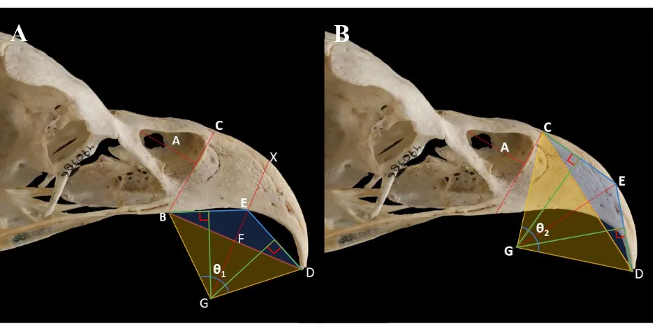
26 distance from the apex of the bony beak to the apex of the rhamphotheca. I define extent
percentage as the gross extent divided by total beak length. Bending of the maxillary rostrum (such as in raptorial birds with hooked beaks) resulted in shorter measured total beak length and thus longer extent percentage (Figure 3).
Rhamphotheca Presence
Transects in lateral view images of keratin-intact specimens were scored for rhamphotheca presence using two dummy variables: presence of keratin on the rostral dorsal extent of the transect (Y1) and presence of keratin on the rostral ventral extent of the transect (Y2).
Homologous Regions
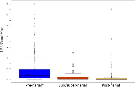
Transects were then binned (Figure 4) as being:
1. Pre-narial: rostralmost extent of transect was rostral to the narial opening.
2. Sub/super-narial: rostralmost extent of transect was caudal to the rostralmost extent of the naris. Caudalmost extent of transect was rostral to the caudalmost extent of the naris. 3. Post-narial: Caudalmost extent of transect was caudal to the caudalmost extent of the
naris.
Curvature
I measured the angle of curvature on the inner (Figure 5A: θ1) and outer surfaces (Figure 5B: θ2) of the pre-narial region of the beak on all x-rays and keratin-removed images in lateral view, applying the method used by Feduccia (1993) and Birn-Jeffery et al. (2012) on claw sheaths to curvature in bird beaks. More extreme angles of curvature resulted in higher values for and
.
Definitions
I define Lateral pore area (LPA) as the sum of areas of neurovascular foramina in lateral view on keratin-removed specimens across all transects. To control for beak length, I divided LPA by beak length (in number of 5 mm transects) to calculate Porosity Index (PI) for each species.
Statistical Analysis
27 listed in Appendix A. Of 94 studies queried, 65 (69%) utilized fossils for minimum age
constraints. I manually constructed the base tree with 72 tips and 63 internal nodes in Mesquite version 3.31 using consensus divergence times from TimeTree.org. I then generated 1000 time-scaled phylogenies by randomly sampling node ages from 95% confidence intervals provided by TimeTree.org for each inner node in my phylogeny. For nodes that had less than 4 separate primary literature sources, node ages were sampled using minimum and maximum reported ages. Edge lengths were calculated from the difference between node ages, and then zero and negative edge length branches were collapsed and polytomies were randomly resolved using the ‘ape’
(version 5.0, Paradis, Claude & Strimmer, 2004) package in RStudio version 1.1.383.0. Randomly resolving polytomies resulted in changes to tree topology from the base tree. I then coerced the trees to be ultrametric via extension using the ‘phytools’ package (version 0.6-44)
(for a sample of 6 time-calibrated phylogenies, see Appendix B). Extension resulted in total node height at the tips greater than the root time, so edge lengths were rescaled by multiplying them by the ratio of root time to total node height. I calculated the least squares consensus tree (using 1000 trees generated above) via quadratic path differences, using my original base timetree as the starting topology for optimization. All phylogenetic regressions used this consensus timetree, and all analyses were performed in RStudio. See Electronic Supplement 2 for annotated R script. Raw data compiled in Table 1.
Porosity Index
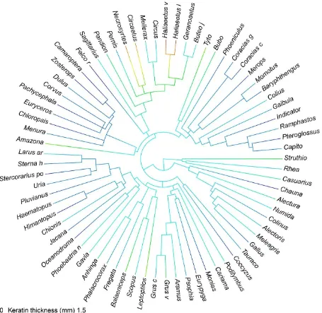
I generated a phenogram of PI using ‘phytools’ and then used an ANOVA test and Tukey
multiple comparisons test (α=0.05) to compare relative porosity across taxa separated into one of
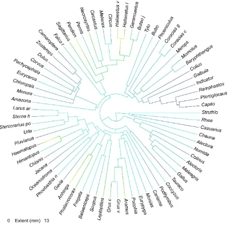
8 clades: Accipitriformes, Aequorlitornithes, Australaves, Coraciimorphae, Galloanserae, Gruiformes, Palaeognathae, and Strigiformes. Clade names based on Prum et al. (2015).
Average keratin thickness -vs. PI-
Mediolateral keratin thickness was averaged for each specimen and was plotted against PI for all 73 bird species. I performed a phylogenetic least squares regression (pGLS) using ‘ape’ and
‘nlme’ (version 3.1-131) packages in RStudio. Linearity assumptions were checked using a
28 testing free-floating lambda against lambda fixed at 1 (Brownian motion) and 0 (independence) using two separate ANOVAs.
-vs. Beak length-
Using a second phylogenetic regression, I tested for a correlation between keratin thickness and beak length. After removing two influential outliers for beak length (Leptoptilos and
Balaeniceps), I re-ran the analysis.
Rostroventral keratin extent
I also created a phenogram showing rostroventral extent of the rhamphotheca beyond the bony beak in birds. Rhamphotheca extent (both gross and as a percentage of beak length) for each species was plotted against PI. I again performed a pGLS. No outliers were found in the tests for linearity. Following the procedure described above, I again tested for phylogenetic signal in the data. I also used a Pearson’s correlation and pGLS test to assess the correlation between beak
length in transects and rhamphotheca extent. I generated a third phenogram showing rhamphotheca extent in birds. I separated taxa into the same clades as above to compare
percentage extent across taxa using a single-factor ANOVA test and Tukey multiple comparisons test.
Rhamphotheca presence per transect
I performed a MANOVA to assess the amount of variance explained in my two variables of rhamphotheca presence (Y1 & Y2) by surface texture and transect location. I then used two multivariate multiple regression models to predict rhamphotheca presence in a given transect. The first was an omnibus test using both presence dummy variables as dependent variables and 5 independent variables as predictors: X1) surface texture, X2) neurovascular canals, X3) LPA, and X4 & X5) two dummy variables representing transect position corresponding to the homologous regions described above. For the second model, I removed surface texture as a predictor and re-ran the analysis.
LPA change across beak length
29 Curvature
I used a Pearson’s correlation test to compare curvature on the inner and outer surfaces of the
beak in lateral view. I then averaged inner and outer curvatures for each specimen and plotted this against both raw keratin extent and extent as a percentage of total beak length. Using a phylogenetic regression similar to those described above, I calculated the correlation between percentage extent and beak curvature, and recorded the phylogenetic signal present.
RESULTS
LPA Change from Rostral to Caudal
LPA was significantly related to homologous region of the beak. LPA was significantly greater in the pre-narial region than in either the sub/super-narial or post-narial regions (Figure 6). There was no significant difference in LPA between sub/super-narial and post-narial regions. Of the three homologous regions, 54.3% of transects fell in the pre-narial region, followed by sub/super-narial region with 27.5% of transects, with the remaining 18.2% falling into the post-narial region (Figure 7).
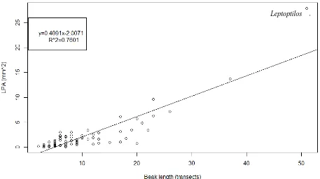
To explore whether LPA represents a size-independent quantification of porosity, I tested for a possible correlation between LPA and beak length. LPA ranged from 0.024 mm2 (Pachycephala) to 27.891 mm2 (Leptoptilos) and was strongly correlated (Adjusted R-squared: 0.7601) with beak length (Figure 8), and the correlation was highly significant (p<0.0001). LPA is therefore not a size-independent measure of porosity. In order to control for beak size, I calculated Porosity Index (PI) (LPA/beak length in transects) for use in subsequent analyses in this chapter.
Porosity Index
PI ranged from a minimum of 0.008 mm in the Yellow-bellied whistler (Pachycephala
philippensis) to a maximum of 0.5469 mm in the Maribou stork (Leptoptilos crumenifer) (Figure 9). Mean PI was 0.1682 mm.
Average Keratin Thickness
Porosity Index was significantly (p=0.003) linearly correlated (r=0.478) with average keratin thickness. I estimate a λ of 0.7443 (Approximate 95% confidence interval 0.1495–1.3391),
30 (p=0.0101) correlated r=0.432) with beak length. However, after two influential outliers for beak length are removed, the correlation is no longer significant (p=0.2134).
Extent
When rostral extent of rhamphotheca beyond the premaxilla is expressed as raw length, it was significantly (p=0.0014) linearly correlated (r=0.518) with PI. Therefore, I reject the hypothesis of phylogenetic independence (p=0.0031), as well as the hypothesis of Brownian motion (p= 0.0064). I estimate a λ of 0.5736 (Approximate 95% confidence interval 0.1398–1.0074),
indicating intermediate phylogenetic influence on the linear correlation of PI and raw rhamphotheca extent (Figure 11).
When expressed as a percent of total beak length, rhamphotheca extent is not significantly correlated with PI (p=0.0579), and the correlation is weaker (r=0.428). Percent extent was not significantly correlated with beak length, using either the Pearson’s correlation test or the phylogenetic least squares regression (P>0.05).
Mean beak curvature was not a significant predictor of gross rhamphotheca extent (P=0.9109), but it was correlated (r=0.423) with extent when extent is measured as a percentage of total beak length, and the correlation was significant (p=0.0060). I calculate a strong phylogenetic signal in the correlation between curvature and percent extent, with λ = 0.8976.
Rhamphotheca Presence
31 rostral ventral extent of transect). AIC was 397.9228 for the omnibus model, and 396.2968 for model 2.
DISCUSSION
The rhamphotheca, as a living keratinous soft tissue structure that closely adheres to the surface of the bone, requires both blood supply and nerve innervation (Crole & Solely, 2016). It is therefore reasonable to posit that the surface texture morphology is correlated with soft tissue presence and/or specific dimensions of epithelial tissues; indeed, the presence of neurovascular sulci and obliquely oriented foramina at the bone surface are known osteological correlates of the presence of a cornified sheath. However, this study attempts to test whether specific dimensions of the keratinous rhamphotheca can be predicted from relative porosity of the premaxilla in birds. Porosity Index (PI) was significantly correlated with key aspects of soft tissue morphology in birds, including average keratin thickness and rhamphotheca extent. These results indicate the action of functional and/or physiological constraints on the relationship between the bone and keratin of the beak. However, the precise developmental or physiological links between
keratinous soft tissue morphology and the relative porosity of the premaxilla are not yet known and will require future work to uncover.
Although phylogeny was a significant predictor of PI, there were no pairwise differences
between any of the clades in my sample, indicating that despite moderately strong influence from phylogeny, relative porosity of the premaxilla is an important variable in determining the
relationship between bony and soft tissue across a broad range of living birds. Because of this, it may be possible to extend the results of this study beyond the realm of birds to other cornified soft tissue structures in more distantly related taxa. Although the keratinous beak rarely survives the process of fossilization intact (Hou et al., 1999), the lateral surface texture of skeletal
32 One important pitfall to discuss is the source of these data, particularly for keratin thickness. Due to time constraints, only a small subsample of birds were x-rayed in dorsoventral view, and of these, only those that had the mandible removed could be used to measure keratin thickness. The remaining samples were assessed from digital photographs taken in ventral view with partially transparent images of keratin-removed specimens digitally overlying them. This inconsistency may result in some measurement bias in my data. I justify this bias as it allowed me to greatly increase my sample size. However, as my analysis required two specimens for each data point (excluding those few that had the keratinous sheath removed but preserved in the box), if any taxon had only one specimen available, I had to remove it from consideration, resulting in a smaller sample size than I had originally intended. Taxonomic sampling bias must also be considered. Although it was my goal to sample as broadly as possible within a realistic timeframe, my chosen taxa were not selected randomly, and were based on both on specimen availability and the completeness of the associated specimen data. Certain taxa with highly unusual morphologies that ultimately I had to drop from my analysis such as the ground hornbill (Bucorvus), which has a keratinized casque that extends nearly to the back of the skull, may have had a significant influence on PI-Keratin thickness correlation.
There were several notable outliers for keratin thickness, including, Circaetus, Necrosyrtes, and Haliaeetus, which had the thickest keratin coating of all the birds I sampled. Despite their relatively large body masses (Prange, Anderson & Rahn, 1979; Buij et al., 2013), beak length in these accipitriform raptors was well within the range observed for much smaller-bodied avian species such as the European roller (Coracias garrulus), which is evidence that these taxa are truly outliers for keratin thickness even when beak length is accounted for. This could indicate a potential functional or phylogenetic signal, as each of these taxa are mostly terrestrial vertebrate predators. As their hunting style often involves subduing living prey that is a significant
proportion of the bird’s own body size (Slagsvold & A. Sonerud, 2007), increased keratin