1556-6811/06/$08.00⫹0 doi:10.1128/CVI.13.4.525–529.2006
Copyright © 2006, American Society for Microbiology. All Rights Reserved.
NOTES
A Decrease in the Immunoglobulin G Antibody Response against the
VlsE Protein of
Borrelia burgdorferi
Sensu Lato Correlates with
the Resolution of Clinical Signs in Antibiotic-Treated
Patients with Early Lyme Disease
Antonella Marangoni,
1Vittorio Sambri,
1* Silvia Accardo,
1Francesca Cavrini,
2Valeria Mondardini,
3Alessandra Moroni,
1Elisa Storni,
2and Roberto Cevenini
1Section of Microbiology-DMCSS, University of Bologna, Bologna,1Centro di Riferimento Regionale per le Emergenze Microbiologiche,
Ospedale Policlinico S. Orsola, Bologna,2and Division of Infectious Diseases, Ospedale di Belluno, Belluno,3Italy
Received 4 November 2005/Returned for modification 30 December 2005/Accepted 2 February 2006
The purpose of this study was to evaluate the diagnostic performance of the LIAISON Borrelia Screen (Diasorin, Saluggia, Italy), a new automated immunoassay based on the chemiluminescent technology (chemiluminescence immunoassay). To assess whether a decrease in a negative value in the anti-VlsE immunoglobulin G (IgG) antibody titer was correlated with a positive response to treatment, a group of serially collected serum samples from 67 patients with culture-confirmed erythema migrans was retrospectively studied. All the patients had been treated with antibiotics and were free of disease within 3 to 6 months of follow-up. All the 15 patients who were found to be IgG positive at the time of enrollment and who were bled at least four times during the follow-up became IgG seronegative at 2 to 6 months posttreatment. These results indicate that a decline in the anti-VlsE antibody titer coincides with effective antimicrobial therapy in patients with early localized Lyme disease.
Lyme disease, caused by the tick-borne spirochetes belong-ing to the speciesBorrelia burgdorferisensu lato, is a multistage infection that has become the most common vector-borne dis-ease in North America and Europe (7, 38). In Europe three different species of B. burgdorferi sensu lato pathogenic for humans (namely,B. burgdorferisensu stricto,B. garinii, andB. afzelii) (3, 5, 36, 39, 40) are known and demonstrate both inter-and intraspecies heterogeneity (3, 41). The illness usually be-gins with a characteristic, expanding skin lesion, erythema mi-grans (EM) (26, 27, 37). The diagnosis of Lyme disease is based on clinical and laboratory findings. Serologic testing is the most commonly used corroborative laboratory method, but serology also harbors several problems. The occurrence of cross-reacting antibodies may result in false-positive findings (1, 17–19). Furthermore, patients may still be seronegative in the early stages of the infection and the humoral immune response can be diminished after the early onset of antibiotic treatment (1, 2, 28, 39). Several attempts to standardize the serological tests for Lyme disease have been done so far (10– 13, 33), but considerable variations in test results are still present among different laboratories even when they use the same strategy (33). The recommendations of the Centers for Disease Control and Prevention and the German Society for Hygiene and Microbiology (DGHM) (4, 42) have relied for years on the use of a second-tier, confirmatory test for Lyme disease
when the first test yielded a positive or equivocal result. More recently, a comparison of classic two-tiered testing and a VlsE (Vmp-like sequence, expressed)-based enzyme immunoassay (EIA) reported higher values of sensitivity for the latter one, which also maintained very good specificity (2). This is a new immunoassay that uses as the antigen a 26-mer synthetic pep-tide (the C6 peppep-tide) based on invariable region 6 (IR6) of the VlsE lipoprotein ofB. burgdorferi(43). IR6 is a highly immu-nogenic peptide (15) that has been shown to remain un-changed during antigenic variation and is antigenically con-served among pathogenicB. burgdorferisensu lato strains (9, 16, 35), although it was recently showed to be structurally hetero-geneous (8). Philipp et al. (32) demonstrated that a decline in the C6 antibody titer significantly correlated with a successful treatment outcome in patients with early localized or early disseminated Lyme disease. The purpose of this study was to evaluate the diagnostic performance of LIAISON Borrelia Screen (Diasorin, Saluggia, Italy), a new automated immuno-assay based on chemiluminescent technology (chemilumines-cence immunoassay [CLIA]). Since a recombinant VlsE anti-gen obtained from the PBi strain ofB. gariniiis used to coat magnetic particles in the immunoglobulin G (IgG) assay, our hypothesis was that this new immunoassay could be useful for the follow-up of patients with early Lyme disease. To assess whether a decrease to a negative anti-VlsE antibody titer was correlated with a positive response to treatment, a group of serially collected serum samples from patients with culture-confirmed EM was retrospectively studied.
In this study a total of 387 human serum specimens were retrospectively studied. A total of 177 serum specimens were
* Corresponding author. Mailing address: Section of Microbiology, DMCSS, University of Bologna, St. Orsola Hospital, via Massarenti 9, Bologna 40138, Italy. Phone: 39 051 4290 913. Fax: 39 051 636 4516. E-mail: [email protected].
525
on August 17, 2020 by guest
http://cvi.asm.org/
obtained from 67 patients (27 men and 40 women) with cul-ture-confirmed Lyme disease. These patients were between the ages of 19 and 78 years (mean age, 42.8 years) and had EM following a tick bite. The EM diameter ranged from 3 cm to 25 cm (mean diameter, 15 cm). All the patients came from an area of endemicity in the northeast of Italy. Only 71.6% of patients (48 of 67) recalled the tick bite, but all 67 subjects had an occupational or recreational risk of exposure toIxodes ricinusticks. A skin punch biopsy specimen (diameter, 0.25 cm) was obtained from each patient when the patient entered the study and was cultivated in Barbour-Stoenner-Kelly medium modified (ATCC medium 1914) plus ciprofloxacin (0.4g/ml) and rifampin (40g/ml); the tubes were examined weekly by dark-field microscopy for motile spirochetes over a period of at least 45 days, as described previously (21). All 67 cultures of the specimens from the patients showed positivity for Lyme disease spirochetes within 1 month. At the initial clinical eval-uation, each patient was bled and given specific antibiotic ther-apy for Lyme borreliosis. The follow-up study was performed by taking additional serum samples at 30, 60, 120, and 180 days after enrollment. In particular, 2 patients were bled five times, 17 patients were bled four times, 18 patients were bled three times, and 15 patients were bled twice; finally, 15 patients were lost to follow-up. Patients entered into the study after having had a mean duration of EM of 20 days (range, 1 to 150 days). Two hundred ten additional serum samples were obtained from the blood bank of the St. Orsola Hospital in Bologna, Italy, and 24 samples were obtained from healthy blood donors from an area of endemicity for Lyme disease in northeastern Italy (Trento). Furthermore, a panel of 40 serum samples was obtained from patients with some of the most common biolog-ical conditions that possibly result in false-positive reactivity in Lyme disease serology. The following specimens were included in this group: sera from patients withStreptococcus pyogenes
acute infection (streptolysin O antibody response, ⬎400 IU/ ml) (n ⫽ 10), serum samples from subjects with a clinical diagnosis of infectious mononucleosis and found to be positive by Paul-Bunnel-Davidsohn agglutination (n⫽10), sera from patients with hepatitis A virus acute infection (IgM positive) (n⫽10), and, finally, sera from syphilis patients (primary and secondary stage) (n⫽10).
LIAISON Borrelia Screen (DiaSorin) is a qualitative fully automated method for determination of specific antibodies to
B. burgdorferisensu lato in human serum or plasma. This new method is a one-step sandwich CLIA. Recombinant-specific VlsE antigen obtained from the PBi strain ofB. gariniiis used to coat magnetic particles (solid phase) in the IgG assay, and recombinant OspC obtained from B. afzelii Pko is used in the IgM assay; the same antigens are linked to an isoluminol derivative (isoluminol-antigen conjugate) in the IgG and IgM tests, respectively. During incubation, antibodies toB. burgdor-feripresent in calibrators, samples, or controls bind to the solid phase and to the antigen conjugate. The unbound material is removed with a wash cycle. Subsequently, the starter reagents are added and a flash chemiluminescence reaction is thus in-duced. The light signal, and, hence, the amount of isoluminol-antibody conjugate, is measured by a photomultiplier as rela-tive light units and is indicarela-tive of the concentration of antibodies toB. burgdorferi present in calibrators, samples, or controls. The global results of the IgM test are reported in index units
and are evaluated by using a cutoff value of 1.0 index unit, with a gray zone of⫾10%. The results are interpreted as follows: samples scored with an index value⬍0.9 are considered neg-ative; samples with an index valueⱖ1.1 are considered posi-tive; and finally, samples with a value that falls in the gray range (i.e., 0.9⬍ index value⬍1.1) are boundary cases and, following the manufacturer’s instructions, were tested again. The IgG concentration was expressed, instead, as arbitrary units (AU)/ml; the measurement range was from 0 to 240 AU/ml. Sample results were interpreted as follows: samples with IgG concentrations below 10 AU/ml were graded nega-tive; and samples with IgG concentrations ranging from 10 to 15 AU/ml were graded equivocal, and, following the manufac-turer’s instructions, were tested again. Finally, samples with IgG concentrations equal to or greater than 15 AU/ml were graded positive.
PCR was performed by using five different sets of primers whose sequences have been obtained from the literature (25, 29): FL6-FL7 (which amplifies a fragment of the flagellin gene that is conserved in allB. burgdorferisensu lato strains); LD (which amplifies a 16S rRNA genomic fragment common to the three genospecies); and BB, BG, and BA (each of which amplifies a species-specific 16S rRNA genomic fragment). DNA was extracted from the spirochetes for PCR as described previously (34). These primer sets generated amplification products of 276, 357, 574, 574, and 591 bp, respectively. All the PCR reagents except the primers were from the GeneAmp kit (Perkin-Elmer Cetus). A total of 50 pmol of the appropriate primer set and 25l of the boiled spirochete suspension were used in each 50-l reaction mixture. All amplifications were carried out with an automatic Eppendorf Mastercycler per-sonal DNA thermal cycler.
Anti-BorreliaPlus VlsE enzyme-linked immunosorbent as-say (ELISA) (Euroimmun, Lu¨beck, Germany) is a quantitative enzyme-linked immunoassay based on a mixture of whole-antigen extracts ofB. burgdorferisensu stricto,B. afzelii, andB. garinii and recombinant VlsE lipoprotein of B. burgdorferi
sensu stricto. Three calibrators and positive and negative con-trols were used. The results were scored as negative (ratio,
⬍1.0) or positive (ratio, ⱖ1.0), following the manufacturer’s instructions.
In an attempt to improve Lyme disease diagnosis in Europe, two different panels of sera were evaluated: the first consisted of samples obtained from Italian patients with culture-con-firmed erythema migrans, whereas the second was composed of sera collected from blood donors and patients with infec-tions that could interfere with Lyme disease serology. All 67 cultures obtained from the biopsy specimens were identified as
B. afzeliiby PCR, confirming previous data from our laboratory (23), namely, that in the northeast of Italy, the prevalent geno-species ofB. burgdorferisensu lato isB. afzelii. As shown in Fig. 1, 49 samples obtained at enrollment were identified to be positive by the LIAISON Borrelia Screen IgG or IgM assay (in partic-ular, 25 serum samples were IgG positive and 42 serum ples were IgM positive), whereas the remaining 18 serum sam-ples were both IgG and IgM seronegative. Similar results were obtained by the Anti-BorreliaPlus VlsE ELISA. The sensitivity of the LIAISON Borrelia Screen assay with this group of serum specimens was 73.1%, comparable to that of Anti- Bor-reliaPlus VlsE ELISA (72.6%). It is noteworthy that no
on August 17, 2020 by guest
http://cvi.asm.org/
conversion was observed during the follow-up of the 18 pa-tients initially found to be IgG and IgM negative by the LIAISON Borrelia Screen assay. This could be consistent with the rapid evanescence of the antibody response to VlsE after antibiotic treatment (30–32). As a confirmation of this hypoth-esis, we further studied the 15 patients who were initially found to be IgG positive by the LIAISON Borrelia Screen assay and
who were bled at least four times during the follow-up: all of them became IgG seronegative at 2 to 6 months posttreatment (Fig. 2). A similar effect was not observed when Anti-Borrelia
Plus VlsE ELISA IgG was used: 14 of 18 patients initially IgG positive still remained reactive when they were analyzed by this EIA method during the follow-up period.
All the tests studied showed good results when the
cross-react-FIG. 1. Results obtained by LIAISON Borrelia Screen assay when the 67 serum specimens drawn at the enrollment were tested. Thexaxis shows the duration of EM at the time that the patients entered the study. The number of samples is indicated by the number above each individual bar in the graph.
FIG. 2. Decrease in IgG response observed in the 15 patients studied during the follow-up, as detected by LIAISON Borrelia Screen IgG assay. Samples with IgG concentrations below 10 AU/ml were negative, samples with IgG concentrations ranging from 10 to 15 AU/ml were equivocal (as indicated by the horizontal bar), and samples with IgG concentrations equal to or greater than 15 AU/ml were positive.
on August 17, 2020 by guest
http://cvi.asm.org/
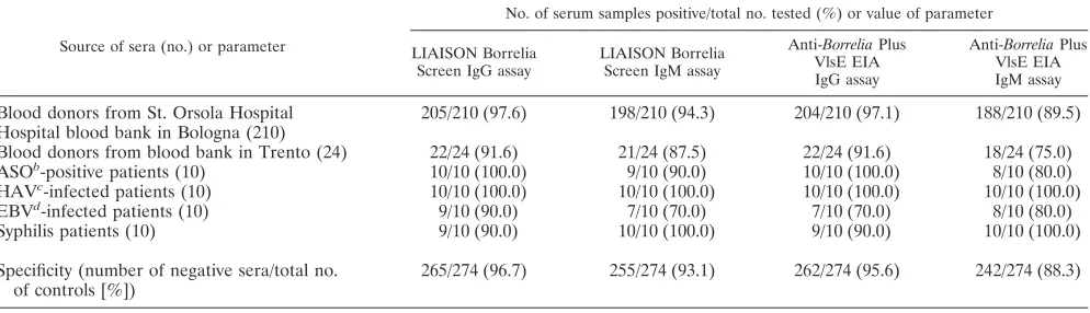
ing sera were tested (Table 1). In particular, the LIAISON Bor-relia Screen IgG assay had a specificity of 96.7%, whereas the Anti-BorreliaPlus VlsE ELISA IgG assay had a specificity of 95.6%. The LIAISON Borrelia Screen IgM assay was shown to be 93.1% specific, whereas the specificity of the IgM EIA was 88.3%.
The findings reported in this paper confirm and expand the observations made previously both by us and by others.
First, chemiluminescent immunoassays have been evaluated in recent years (6, 24) and have been shown to be as sensitive and specific as the immunoenzymatic technique. Moreover, this method is very simple to perform and cost saving. In the present study, the diagnostic performance of a new chemilu-minescent assay, LIAISON Borrelia Screen, was compared with those obtained by a commercial immunoenzymatic assay. The LIAISON Borrelia Screen was shown to be a very good alternative to the traditional EIAs, since it was shown to be at least as sensitive as the Anti-BorreliaPlus VlsE ELISA when samples from patients with EM were tested. Moreover, the LIAISON Borrelia Screen was slightly more specific than the Anti-BorreliaPlus VlsE ELISA; the most problematic sera for both methods seemed to be the samples obtained from mono-nucleosis patients, whereas EIA IgM showed a poor value of specificity when sera from blood donors from Trento, Italy, were tested.
Second, when the C6 peptide was used in a diagnostic EIA with serum samples from U.S. patients, the assay performed with a good sensitivity and a good specificity (15, 20, 28). In Europe, a few papers on the diagnostic use of the C6 peptide have been published (9, 14, 16, 22, 23). Moreover, Bacon and coworkers (2) reported that the test with the C6 peptide per-formed as well as or better than the two-tiered algorithm. The LIAISON Borrelia Screen IgG assay is the first immunoassay based exclusively on the whole recombinant form of the VlsE antigen. In this study all sera from patients initially found to be IgG positive became nonreactive 2 to 6 months after treat-ment. This test confirmed what Philipp and coworkers had previously reported, i.e., that the C6 test performed well as a predictor of treatment outcome (30–32). It is noteworthy that all the Italian patients studied had early localized Lyme disease
and that they promptly received antibiotic therapy. This can explain the difference of our results from those obtained by Peltomaa and coworkers (28) in a retrospective analysis of sera obtained from patients with early or late Lyme disease. Their patients showed positive anti-VlsE antibodies 8 to 15 years after treatment, but it was hypothesized that the persistence could be due to the generation of both memory T and B cells. Our hypothesis is that our patients did not develop T cells be-cause of the prompt therapy that they received. The LIAISON Borrelia Screen IgG assay was shown to be a useful support for clinical diagnosis, since it could be used for the follow-up of Lyme disease patients. A possible disadvantage is that this IgG CLIA method does not allow the detection of IgG seroconversion in patients who scored negative before treatment. On the contrary, one major advantage of the LIAISON Borrelia Screen assay is that it can discriminate between the IgG and the IgM responses (the commercial C6 antigen-based EIA is not able to do so), and this could be very useful for clinicians. In particular, when sera drawn from patients with EM lasting less than 10 days were tested, we found that the sensitivity of IgG detection was quite low (21.2%) but that the sensitivity of IgM detection was much more acceptable (66.7%). Similar results were obtained in a pre-vious study (22), in which the Quick ELISA C6 Borrelia assay (Immunetics, Cambridge, Mass.) was demonstrated to have a low sensitivity (33.3%) if it was used to test sera collected from Italian patients with EM.
This study was supported in part by grant from the University of Bologna (fondi RFO; ex quota 60%; 2004) to V.S. and in part by grant Centro di Riferimento Regionale per le Emergenze Microbiologiche from Regione Emilia Romagna (2004) to V.S.
We thank Danila Bassetti for providing the blood donor samples from Trento.
REFERENCES
1.Aguero-Rosenfeld, M. E., J. Nowakowski, S. Bittker, D. Cooper, R. B. Nadel-man, and G. P. Wormser.1996. Evolution of the serologic response to
Borrelia burgdorferiin treated patients with culture-confirmed erythema
mi-grans. J. Clin. Microbiol.34:1–9.
2.Bacon, R. M., B. J. Biggerstaff, M. Schriefer, R. D. Gilmore, M. T. Philipp, A. Steere, G. P. Wormser, A. R. Marques, and B. J. B. Johnson.2003. Improved serodiagnosis of Lyme disease by kinetic EIAs using recombinant
TABLE 1. Number of negative specimens detected in the panel of control seraa
Source of sera (no.) or parameter
No. of serum samples positive/total no. tested (%) or value of parameter
LIAISON Borrelia Screen IgG assay
LIAISON Borrelia Screen IgM assay
Anti-BorreliaPlus
VlsE EIA IgG assay
Anti-BorreliaPlus
VlsE EIA IgM assay
Blood donors from St. Orsola Hospital 205/210 (97.6) 198/210 (94.3) 204/210 (97.1) 188/210 (89.5) Hospital blood bank in Bologna (210)
Blood donors from blood bank in Trento (24) 22/24 (91.6) 21/24 (87.5) 22/24 (91.6) 18/24 (75.0) ASOb-positive patients (10) 10/10 (100.0) 9/10 (90.0) 10/10 (100.0) 8/10 (80.0) HAVc-infected patients (10) 10/10 (100.0) 10/10 (100.0) 10/10 (100.0) 10/10 (100.0)
EBVd-infected patients (10) 9/10 (90.0) 7/10 (70.0) 7/10 (70.0) 8/10 (80.0)
Syphilis patients (10) 9/10 (90.0) 10/10 (100.0) 9/10 (90.0) 10/10 (100.0)
Specificity (number of negative sera/total no. of controls [%])
265/274 (96.7) 255/274 (93.1) 262/274 (95.6) 242/274 (88.3)
a
The results were obtained by testing the sera once.
b
ASO, streptolysin O antibody titer⬎400 IU/ml.
c
HAV, hepatitis A virus acute infection (IgM positive).
d
EBV, Epstein-Barr virus (Paul-Bunnel-Davidsohn agglutination assay positive).
on August 17, 2020 by guest
http://cvi.asm.org/
VlsE1 or peptide antigens ofBorrelia burgdorfericompared with two-tiered
testing. J. Infect. Dis.187:1187–1199.
3.Baranton, G., D. Postic, I. Saint Girons, P. Boerlin, J. C. Piffaretti, M. Assous, and P. A. D. Grimont.1992. Delineation ofBorrelia burgdorferisensu
stricto,Borrelia gariniisp. nov., and group VS461 associated with Lyme
borreliosis. Int. J. Syst. Bacteriol.42:378–383.
4.Centers for Disease Control and Prevention.1995. Recommendations for test performance and interpretation from the Second National Conference
on Serologic Diagnosis of Lyme Disease. Morb. Mortal. Wkly. Rep.44:590–
591.
5.Ciceroni, L., S. Ciarrochi, A. Ciervo, V. Mondarini, F. Guzzo, G. Caruso, R. Murgia, and M. Cinco.2001. Isolation and characterization ofBorrelia burg-dorferisensu lato strains in an area of Italy where Lyme borreliosis is
en-demic. J. Clin. Microbiol.39:2254–2260.
6.Deguchi, M., N. Yamashita, M. Kagita, S. Asari, Y. Iwatani, T. Tsuchida, K. Iinuma, and I. K. Mushahwar.2004. Quantitation of hepatitis B surface antigen by an automated chemiluminescent microparticle immunoassay.
J. Virol. Methods115:217–222.
7.Gern, L., A. Estrada-Pena, F. Frandsen, J. S. Gray, T. G. Jaenson, F. Jongejan, O. Kahl, E. Korenberg, R. Mehl, and P. A. Nuttall.1998.
Euro-pean reservoir hosts ofBorrelia burgdorferisensu lato. Zentbl. Bakteriol.
Parasitenkd. Infektkrankh. Hyg. Abt. 1 Orig.287:196–204.
8.Goettner, G., U. Schulte-Spechtel, and B. Wilske.2004. Heterogeneity of the
immunodominant surface protein VlsE among the three genospecies ofBorrelia
burgdorferipathogenic for humans. Int. J. Med. Microbiol.293(Suppl. 37):172– 173.
9.Goettner, G., U. Schulte-Spechtel, R. Hillermann, G. Liegl, B. Wilske, and V. Fingerle.2005. Improvement of Lyme borreliosis serodiagnosis by a newly developed recombinant immunoglobulin G (IgG) and IgM line immunoblot
assay and addition of VlsE and DbpA homologues. J. Clin. Microbiol.43:
3602–3609.
10.Hauser, U., G. Lehnert, R. Lobentanzer, and B. Wilske.1997. Interpretation
criteria for standardized Western blots for three European species of
Bor-relia burgdorferisensu lato. J. Clin. Microbiol.35:1433–1444.
11.Hauser, U., H. Krahl, H. Peters, V. Fingerle, and B. Wilske.1998. Impact of strain heterogeneity on Lyme disease serology in Europe: comparison of
enzyme-linked immunosorbent assays using different species ofBorrelia
burg-dorferisensu lato. J. Clin. Microbiol.36:427–436.
12.Hauser, U., G. Lehnert, and B. Wilske.1999. Validity of interpretation criteria for standardized Western blots (immunoblots) for serodiagnosis of Lyme borreliosis based on sera collected throughout Europe. J. Clin.
Mi-crobiol.37:2241–2247.
13.Heikkila, T., I. Seppala, H. Saxen, J. Panelius, H. Yrjanainen, and P. Lah-denne.2002. Species-specific serodiagnosis of Lyme arthritis and
neurobor-reliosis due toBorrelia burgdorferisensu stricto,B. afzelii, andB. gariniiby
using decorin binding protein A. J. Clin. Microbiol.40:453–460.
14.Jansson, C., S. A. Carlsson, H. Granlund, P. Wahleberg, and D. Nyman.
2005. Analysis ofBorrelia burgdorferiIgG antibodies with a combination of
IgG ELISA and VlsE C6 peptide ELISA. Clin. Microbiol. Infect.11:147–
150.
15.Liang, F. T., A. C. Steere, A. R. Marques, B. J. B. Johnson, J. N. Miller, and M. T. Philipp.1999. Sensitive and specific serodiagnosis of Lyme disease by enzyme-linked immunosorbent assay with a peptide based on an
immuno-dominant conserved region ofBorrelia burgdorferiVlsE. J. Clin. Microbiol.
37:3990–3996.
16.Liang, F. T., E. Aberer, M. Cinco, L. Gern, C. M. Hu, Y. N. Lobet, M. Ruscio, P. E. Voet, Jr., V. E. Weynants, and M. T. Philipp.2000. Antigenic conser-vation of an immunodominant invariable region of the VlsE lipoprotein
among European pathogenic genospecies ofBorrelia burgdorferiSL. J. Infect.
Dis.182:1455–1462.
17.Magnarelli, L. A., J. N. Miller, J. F. Anderson, and G. F. Riviere.1990. Cross-reactivity of nonspecific treponemal antibody in serologic tests for
Lyme disease. J. Clin. Microbiol.28:1276–1279.
18.Magnarelli, L. A., J. F. Anderson, R. C. Johnson, R. B. Nadelman, and G. P. Wormser.1994. Comparison of different strains ofBorrelia burgdorferisensu lato used as antigens in enzyme-linked immunosorbent assays. J. Clin.
Mi-crobiol.32:1154–1158.
19.Magnarelli, L. A., J. W. Ijdo, S. J. Padula, R. A. Flavell, and E. Fikrig.2000. Serologic diagnosis of Lyme borreliosis by using enzyme-linked
immunosor-bent assays with recombinant antigens. J. Clin. Microbiol.38:1735–1739.
20.Magnarelli, L. A., M. Lawrenz, S. J. Norris, and E. Fikrig.2002.
Compar-ative reactivity of human sera to recombinant VlsE and otherBorrelia
burg-dorferiantigens in class-specific enzyme-linked immunosorbent assays for
Lyme borreliosis. J. Med. Microbiol.51:649–655.
21.Marangoni, A., V. Sambri, C. Cimmino, G. Caruso, V. Mondardini, and R. Cevenini.1999. Evaluation of the immune response in culture-confirmed Lyme borreliosis erythema migrans patients. Zentbl. Bakteriol. Parasitenkd.
Infektkrankh. Hyg. Abt. 1 Orig.289:736–739.
22.Marangoni, A., M. Sparacino, V. Mondardini, F. Cavrini, E. Storni, M.
Donati, R. Cevenini, and V. Sambri.2005. Comparative evaluation of two enzyme linked immunosorbent assay methods and three Western Blot meth-ods for the diagnosis of culture-confirmed early Lyme borreliosis in Italy.
New Microbiol.28:37–43.
23.Marangoni, A., M. Sparacino, F. Cavrini, E. Storni, V. Mondardini, V. Sambri, and R. Cevenini.2005. Comparative evaluation of three different EIA methods for the diagnosis of early culture-confirmed Lyme disease in
Italy. J. Med. Microbiol.54:361–367.
24.Marangoni, A., V. Sambri, S. Accardo, F. Cavrini, A. D’Antuono, A. Moroni, E. Storni, and R. Cevenini. 2005. Evaluation of LIAISON Treponema Screen, a novel recombinant antigen-based chemiluminescence immunoas-say for the laboratory diagnosis of syphilis. Clin. Diagn. Lab. Immunol.
12:1231–1234.
25.Marconi, R. T., and C. F. Garon.1992. Development of chain reaction primer sets for diagnosis of Lyme disease and for species-specific identifica-tion of Lyme disease isolates by 16S rRNA signature nucleotide analysis.
J. Clin. Microbiol.30:2830–2834.
26.Nadelman, R. B., J. Nowakowski, G. Forseter, N. S. Goldberg, S. Bittker, D. Cooper, M. Aguero-Rosenfeld, and G. P. Wormser.1996. The clinical spec-trum of early Lyme borreliosis in patients with culture-confirmed erythema
migrans. Am. J. Med.100:502–508.
27.Nadelman, R. B., and G. P. Wormser.1998. Lyme borreliosis. Lancet352:
557–565.
28.Peltomaa, M., G. McHugh, and A. C. Steere.2003. Persistence of the
anti-body response to the VlsE sixth invariant region (IR6) peptide ofBorrelia
burgdorferiafter successful antibiotic treatment of Lyme disease. J. Infect.
Dis.187:1178–1186.
29.Picken, R. N.1992. Polymerase chain reaction primers and probes derived from flagellin gene sequences for specific detection of the agents of Lyme
disease and North American relapsing fever. J. Clin. Microbiol.30:99–114.
30.Philipp, M. T., L. C. Bowers, P. T. Fawcett, M. B. Jacobs, F. T. Liang, A. R. Marques, P. D. Mitchell, J. E. Purcell, M. S. Ratterree, and R. K. Straubinger.
2001. Antibody response to IR6, a conserved immunodominant region of the
VlsE lipoprotein, wanes rapidly after antibiotic treatment ofBorrelia
burg-dorferi infection in experimental animals and in humans. J. Infect. Dis.
84:870–878.
31.Philipp, M. T., A. R. Marques, P. T. Fawcett, L. G. Dally, and D. S. Martin.
2003. C6 test as an indicator of therapy outcome for patients with localized
or disseminated Lyme borreliosis. J. Clin. Microbiol.41:4955–4960.
32.Philipp, M. T., G. Wormser, A. D. Marques, S. Bittker, D. S. Martin, J. Nowakowski, and L. G. Dally.2005. A decline in C6 antibody titer occurs in successfully treated patients with culture-confirmed early localized or early
disseminated Lyme borreliosis. Clin. Diagn. Lab. Immunol.12:1069–1074.
33.Robertson, J., E. Guy, N. Andrews, B. Wilske, P. Anda, M. Granstro¨m, U. Hauser, Y. Moosmann, V. Sambri, J. Schellekens, G. Stanek, and J. S. Gray.
2000. A European multicenter study of immunoblotting in serodiagnosis of
Lyme borreliosis. J. Clin. Microbiol.38:2097–2102.
34.Sambri, V., A. Marangoni, C. Eyer, C. Reichhuber, E. Soutschek, M. Negosanti, A. D’Antuono, and R. Cevenini.2001. Western immunoblotting
with fiveTreponema pallidumrecombinant antigens for serologic diagnosis of
syphilis. Clin. Diagn. Lab. Immunol.8:534–539.
35.Schulte-Spechtel, U., G. Lehnert, G. Liegl, V. Fingerle, C. Heimerl, B. J. Johnson, and B. Wilske.2003. Significant improvement of the recombinant
Borrelia-specific immunoglobulin G immunoblot test by addition of VlsE and
a DbpA homologue derived from Borrelia garinii for diagnosis of early
neuroborreliosis. J. Clin. Microbiol.41:1299–1303.
36.Stanek, G., and F. Strle.2003. Lyme borreliosis. Lancet362:1639–1647. 37.Steere, A. C.1994. Lyme disease: a growing threat to urban populations.
Proc. Natl. Acad. Sci. USA91:2378–2383.
38.Steere, A. C.2001. Lyme disease. N. Engl. J. Med.345:115–125. 39.Strle, F., J. A. Nelson, E. Ruzic-Sabljic, J. Cimperman, V. Maraspin, S.
Lotric-Furlan, Y. Cheng, M. M. Picken, G. M. Trenholme, and R. N. Picken.
1996. European Lyme borreliosis: 231 culture-confirmed cases involving
patients with erythema migrans. Clin. Infect. Dis.23:61–65.
40.Wang, G., A. P. van Dam, I. Schwartz, and J. Dankert.1999. Molecular
typing ofBorrelia burgdorferisensu lato: taxonomic, epidemiological, and
clinical implications. Clin. Microbiol. Rev.12:633–653.
41.Wilske, B., U. Busch, H. Eiffert, V. Fingerle, H. W. Pfister, D. Ro¨ssler, and V. Preac-Mursic.1996. Diversity of OspA and OspC among cerebrospinal
fluid isolates ofBorrelia burgdorferisensu lato from patients with
neurobor-reliosis in Germany. Med. Microbiol. Immunol.184:195–201.
42.Wilske, B., L. Zo¨ller, V. Brade, M. Eiffert, U. B. Go¨bel, G. Stanek, and H. W. Pfister.2000. MIQ 12 Lyme-Borreliose, p. 1–59.InH. Mauch and R.
Lu¨tticken (ed.), Qualita¨tsstandards in der mikrobiologisch-infektiologischen
Diagnostik. Urban and Fischer Verlag, Munich, Germany.
43.Zhang, J. R., J. M. Hardham, A. G. Barbour, and S. J. Norris. 1997. Antigenic variation in Lyme disease borreliae by promiscuous recombination
of VMP-like sequence cassettes. Cell89:275–285.