In-Silico Analysis of Natural Products that Modulates Enzymes of Diabetic Target
Babita Aryal1, Saroj Basnet2,Bishnu P. Marasini3, Karan Khadayat1, Darbin Kumar Poudel1, Ganesh Lamichhane1, Prakriti Budhathoki1, Purushottam Niraula1, Sonika Dawadi1, Rishab Marahatha1, Sitaram Phuyal1, and Niranjan Parajuli1*
Specifications Table
Subject Pharmaceutical Science
Specific subject area Interdisciplinary field includes organic chemistry, biochemistry, and
biology. Drug design and discovery from plant sources. Type of data Tables
Figures
How data were acquired MOE 2009 and GOLD V 4.51 Data format Raw and Analysed
Parameters for data collection
Gold Fitness score, energetic values and interactions of protein with the ligand.
Description of data collection
The Proteins were collected from RCSB protein bank. The secondary metabolites structures were obtained from Pubchem online database. The docking was done using GOLD software.
Data source location https://www.rcsb.org/
https://pubchem.ncbi.nlm.nih.gov/
Data accessibility PDB files of the chosen enzyme targets are publically available at
https://www.rcsb.org/ Tables and Figures of the docking are accessible in the article.
Value of the Data
● The screening procedure enable the researchers to rapidly identify active natural compounds
which can modulate a particular biochemical pathway.
● The screening results help to study the interaction/role of active metabolites in a particular biochemical process at cellular level and provide preliminary ideas for drug design development
● By using this in-silico docking data, novel synthetic analogues with improved bioactivity and minimized side effects can be developed against these targets and research time can be minimized considerably.
● We select these 23 metabolites because these are abundant in nature and well explored.
Among these metabolites, the compounds which show best affinity for various targets are
shortlisted.
● The data is also useful for research scholars who does not have the sufficient software and
hardware requirements which not affordable by them.
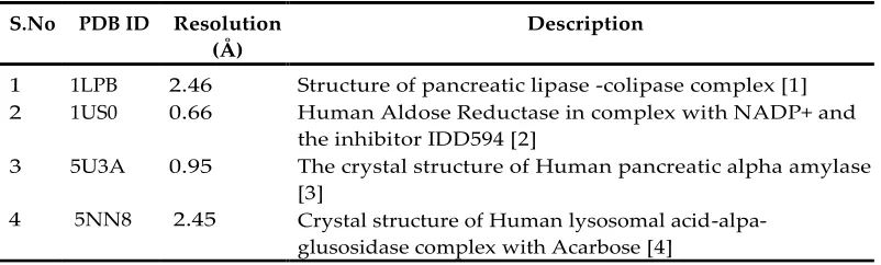
Table 1S. List of Targets showing the PDB ID, resolution and description of the proteins selected for docking with complexed inhibitor.
S.No PDB ID Resolution (Å)
Description
1 1LPB 2.46 Structure of pancreatic lipase -colipase complex [1] 2 1US0 0.66 Human Aldose Reductase in complex with NADP+ and
the inhibitor IDD594 [2]
3 5U3A 0.95 The crystal structure of Human pancreatic alpha amylase [3]
4 5NN8 2.45 Crystal structure of Human lysosomal acid-alpa-glusosidase complex with Acarbose [4]
Table 2S. Inhibition of lipase by secondary metabolites
Natural Source Secondary metabolite IC50 (µM) Reference
Penicillium
purpurogenum IMM 003
Purpurolide F 1.22 [1]
Purpurolide D 6.5
Purpurolide E 7.88
Xestospongia testudinaria (sponges)
Methyl xestospongic ester 3.11 [2]
Caralluma hexagona Lavranos(aerial part)
12,20-di-O-benzoyl-3β,8β,12β,14β,20 -pentahydroxy-(20R)-pregn-5-ene-3-O-β -D-glucopyranosyl-(1→4)β -D-digitalosiden
23.59 [3]
Polygonum aviculare L. whole
Quercetin (competitive inhibition) 53.05 [4] Kaempferol (competitive
inhibition)
79.38
Myricitrin (non-competitive or mixed)
92.85
Quercitrin (non-competitive or mixed)
100.56
Avicularin (non-competitive or mixed)
141.84
Trigonella foenum-graecum (seeds)
Schaftoside 230.29 [5]
Vicenin-1 336.7
Isoschaftoside 584.59
Table 3S. Inhibition of aldose reductase by secondary metabolites
Enzyme Natural Source Secondary metabolite IC50
(μM) Reference
Rat lens aldose reductase
Cuminum cyminum
(seeds) Cuminaldehyde 5.37 [6] Rat lens aldose
reductase
Melastoma
sanguineum 2,″4″-O-diacetylquercitrin 0.077 [7] Rat lens aldose
reductase
Artemisia montana (whole plant)
Luteolin 0.19
[8] Hyperoside 1.85
Chlorogenic acid 4.36 3,5-Di-O-caffeoylquinic
acid 5.37
Rat lens aldose reductase (RLAR)
Xanthium strumarium (fruit)
Methyl-3,5-di-O-Caffeoylquinate 0.3 [9] Human recombinant aldose reductase(rhAR) 0.67
Rat lens aldose reductase Human
recombinant
aldose reductase Sophora flavescens (roots)
Desmethylanhydroicaritin 0.45
[10] 0.95
8-lavandulylkaempferol 0.79
3.8 Recombinant
human aldose reductase
Rhus verniciflua Butein 0.5 [11]
Rat lens aldose reductase
Smilax china L. (stems)
Quercitrin 0.56
[12] 3-O-Caffeoylquinic acid 0.6
Isoscutellarein-8-O-rhamnoside 17 4-O-Caffeoylquinic acid 20.1
Rat lens aldose reductase
Belamcanda
chinensis Tectorigenin 1.08 [13] (rhizomes) Tectoridin 1.12
Rat lens aldose reductase
Abeliophyllum
distichum (leaves) Acteoside 1.39 [14]
Rat lens aldose reductase
Chrysanthemum indicum
(2R)-eriodictyol 7-O-β -D-glucopyranosiduronic
acids
1.5
[15] (2S)-eriodictyol 7-O-β
-D-glucopyranosiduronic acids
2.1
reductase Isoorientin 1.92
Rat lens aldose reductase
Artemisia princeps 1,3,5-tri-O-caffeoylquinic
acid 1.78
[17] (Aerial part) 3,4,5-tri-O-caffeoylqunic
acid 1.95
3,4-di-O-caffeoylquinic
acid 2.4
Rat lens aldose reductase
Glycyrrhiza
uralensis (roots) Semilicoisoflavone B
1.8
[18] 10.6
Bovine lens aldose reductase
Ocimum basilicum (Aerial)
7-(3-hydroxypropyl)-3-methyl-8-β -O-d-
glucoside-2H-chromen-2-one
2.095 [19]
Rat lens aldose reductase
Maackia amurensis
(bark) Chlorogenic acid 4.2 [20]
Rat lens aldose
reductase Zingiber zerumbet Afzelin 5.54 [21] Rat lens aldose
reductase
Cuminum cyminum
(seeds) Cuminaldehyde 5.9 [6]
Recombinant human aldose reductase
Nephelium
lappaceum Geraniin 7.34 [22]
Rat lens aldose
reductase Cassia tora (seeds)
Chryso-obtusin-2-O-β
-D-glucoside 8.8
[23] Aurantio-obtusin 13.6
Emodin 15.9 Recombinant
human aldose reductase
Paulownia coreana Isocampneoside II 9.72 [24]
Rat lens aldose
reductase Paeonia suffruticosa
Palbinone 11.4
[25] 30-norhederagenin 28.8
Bovine lens aldose reductase
Ganoderma lucidum (fruiting
body)
Lucidumol A 19.1 [26]
Table 4S: Inhibition of α-amylase by secondary metabolites
Natural Source Secondary metabolite IC50value (μM) Reference
Ficus deltoidea Vitexin 0.046 [27]
Isovitexin 0.138
Solenostemma argel Kaempferol-3-O-neohesperidoside
0.08 [28]
Olea europaea Oleanolic acid 0.219 [29]
Eruca vesicaria Erucin 0.315 [30]
Ruellia tuberosa Betulin 0.316 [31]
Citrus paradise Pectin 2.11 [32]
Abelmoschus esculentus Proanthocyanidins 3.88 [33]
Phyllanthus amarus Oleanolic acid+ Ursolic acid
4.4 [34]
Vitex glabrata ß amyrin 32.33 [35]
Rheum turkestanicum Daucosterol 46.4 [36]
Himatanthus drasticus Plumieride 71.69 [37]
Vaccinium arctostaphylos (Ericaceae)
Malvidin-3-O-beta-glucoside
329 [38]
Setosphaeria rostrata Rostratazine B 578 [39]
Psidium guajava Linn. (Myrtaceae)
Myricetin 4300 [40]
Kaempferol 5300
Current medication: Acarbose, biguanides, miglitol, voglibose, and 1-deoxynojirimycin.
Note: Some IC50 values are adjusted in terms of molarity to make the comparison convenient.
Table 5S: Inhibition of α-glucosidase by secondary metabolites
Natural Source Secondary metabolite IC50 (µM) Reference
Salacia hainanensis Chun 2,3-seco-lup-20(29)en-2,3-dioic acid
0.01 [41]
Lup-20(29)-en-3,21-dione 0.07 30-hydroxy-friedelan-3one 0.08
3α -hydroxy-lup-20(29)-en-2-one
0.09
Salvia miltiorrhiza Bge Isosalvianolic acid C methyl ester
111.9 x10⁻³ [42]
Dihydrotanshinone 320.1 x10⁻³ Isocryptotanshinone 452.1 x10⁻³
Polygonum hyrcanicum N-trans-Caffeoyl-tyramine 0.3 [43] Myricitrin 0.6
(-) Gallocatechin 1
Harungana madagascariensis
Kenganthranols B 6.3 [44] Kenganthranols C 21.9
Punicatannins B 12.39 ± 0.05
Quercus gilva Tiliroside 28.36 [46]
Euonymus alatus (Twig) Betulinic acid 83.6 [47] Hederagenin 85.3
Naringenin 96.8
Kaempferol 107.8
Dryopteris cycadina 3, 5, 7-trihydroxy-2-(p-tolyl) chorman-4-one
133 [48]
β-Sitosterol 143 5,7,4/
-Trihydroxyflavon-3-glucopyranoid
146
Brickellia cavanillesii Calein C 280 [49]
6-hydroxyacetyl-5-hydroxy-2,2-dimethyl-2H-chromene
420
Aspergillus aculeatus Aspergillusol A 465 [50]
Uncaria cordata Querecetin 1840.82 [51] 2,4-Hydroxybenzoic acid 3652.16
Ligusticum porter 3-(Z)-butylidenephthalide 2350 [52]
Current medication: Acarbose, miglitol, Voglibose, genistein, quercitrin, and 1-deoxynojirinmycin
Table 6S. Prediction of toxicity of secondary metabolites inhibiting metabolic enzymes using ProTox-II
Compound LD50mg/Kg Toxicity
class
Active target Probability
(-) gallocatechin 10000 6 - -
(2R)-eriodictyol 7-O-β -D-glucopyranosiduronic acids
2300 5 Immunotoxicity 0.84
(2S)-eriodictyol 7-O-β -D-glucopyranosiduronic acids
2300 5 Immunotoxicity 0.84
1,3,5-Tricaffeoylquinic acid 5000 5 Immunotoxicity 0.97 1,7-bis(4-hydroxyphenyl)-3,5-diol 1600 4 - -
2-(4-hydroxyphenyl)-ethyl-3,4,5-trihydroxybenzoate
1700 4 Estrogen Receptor Ligand Binding
Domain
0.7
2,″4″-O-diacetylquercitrin 5000 5 Immunotoxicity 0.98 2,3-seco-lup-20(29)en-2,3-dioic acid 2500 5 - - 2,4-hydroxybenzoic acid 2000 4 Carcinogenicity 0.72
3-oxolupenal 5000 5 - -
3,4,5-tri-O-caffeoylqunic acid 5000 5 Immunotoxicity 0.98 3,4-di-O-caffeoylquinic acid 5000 5 Immunotoxicity 0.99
3,5,7-trihydroxy-2-(p-yolyl)chroman-4-one
2000 4 Aryl hydrocarbon Receptor
0.98
Aromatase 0.79 Estrogen Receptor
Alpha
0.95
Estrogen Receptor Ligand Binding
Domain
0.88
Mitochondrial Membrane Potential
0.98
3α-hydroxy-lup-20(29)-en-2-one 2500 5 - - 3,5-Di-O-caffeoylquinic acid 5000 5 Immunotoxicity 0.99
3-(z)-butylidenepthalide 1850 4 - -
4-O-Caffeoylquinic acid 5000 5 Immunotoxicity 0.99 5,7,4-trihydroxyflavon-3-glucopyranoid 5000 5 Immunotoxicity 0.98
6-gingerol 250 3 Immunotoxicity 0.96
6-hydroxyaceyl-5-2,2-dimethyl-2H-chromene
500 4 Immunotoxicity 0.9
12,20-di-O-benzoyl-3β,8β,12β,14β,20 -
pentahydroxy-(20R)-pregn-5-ene-3-O-β-D-glucopyranosyl-(1→4)β -D-digitaloside
650 4 Immunotoxicity 0.99
(20R)-pregn-5-ene-3-O-β -D-glucopyranosyl-
(1→4)β-D-digitaloside
30-hydroxy-friedelan-3-one 3265 5 Immunotoxicity 0.84 30-N0rhederagenin 2000 4 Immunotoxicity 0.72 Acarbose (commercial drugs for
diabetes)
24000 6 Immunotoxicity 0.9
Acteoside 5000 5 Immunotoxicity 0.99
Afzelin 5000 5 Immunotoxicity 0.92
Aurantio-obtusin 5000 5 Immunotoxicity 0.87 Mitochondrial
Membrane Potential (MMP)
0.85
Avicularin 5000 5 - -
Azadiradione 600 4 Immunotoxicity 0.85
B2-3'-O-gallate 1000 4 Immunotoxicity 0.98
Β- amyrin 7000 6 Immunotoxicity 0.93
β-sitosterol 890 4 Immunotoxicity 0.99
Betulin 2000 4 - -
Butein 1000 4 Immunotoxicity Aryl hydrocarbon
Receptor(ArH) Estrogen Receptor
Alpha (ER) Estrogen Receptor
Ligand Binding
Domain Mitochondrial
Membrane potential
0.95 0.72 0.96 0.91 0.97
Calein C 7 2 Immunotoxicity 0.99
Chlorogenic acid 5000 5 Immunotoxicity 0.99 chryso-obtusin-2-O-β -D-glucoside 3000 5 Immunotoxicity 0.99
Cuminaldehyde 1320 4 - -
Daucosterol 8000 6 Immunotoxicity 0.99
Desmethylanhydroicaritin 3919 5 Mitochondrial Membranz Potential
(MMP)
0.77
Dihydrotanshinone 260 3 Immunotoxicity 0.98
Emodin 5000 5 Mutagenicity 0.93
Aryl hydrocarbon Receptor
1
Estrogen Receptor Alpha
1
Estrogen Receptor Ligand Binding
Domain
1
Mitochondrial Membrane Potential
0.99
Phosphoprotein (Tumor Supressor)
p53
1
Epalrestat (Commercial drugs for aldol reductase)
5 2 ATPase family AAA domain-containing
protein 5
0.98
Erucin 39800 6 No -
Ganomycin I 2000 4 No -
Gedunin 2744 3 Immunotoxicity 0.99
Geraniin 122 3 Mutagenicity 0.98
Hederagenin 2000 4 - -
Hyperoside 5000 5 - -
Isocryptotanshinone 2000 4 Immunotoxicity 0.89 Isosalvianolic acid C methyl ester 5000 5 Immunotoxicity 0.99
Isoschaftoside 536 4 - -
Isoscutellarein-8-O-rhamnoside 5000 5 Immunotoxicity 0.86
Isovitexin 159 3 - -
Kaempferol 3919 5 Aryl hydrocarbon
receptor
1
Aromatase 0.96 Estrogen Receptor
Alpha (ER)
1
Estrogen Receptor Ligand Binding
Domain
0.95
Mitochondrial Membrane Potential
1
kaempferol-3-O-neohesperidoside 5000 5 Immunotoxicity 0.98
katononic acid 5000 5 - -
kenganthranols B 5000 5 Immunotoxicity 0.97
Mitochondrial Membrane Potential
0.8
kenganthranols C 1340 4 Immunotoxicity 0.96
Lucidumol A 1860 4 Carcinogenicity 0.72
lup-20(29)-en-3,21-dione 5000 5 - -
Lycopene 5000 5 Androgen receptor
ligand binding
1
Estrogen receptor 1 Alpha Estrogen
receptor ligand binding domain
1
Malvidin-3-O-beta-glucoside 5000 5 Immunotoxicity Aryl hydrocarbon
0.98
Melacolin 600 4 Immunotoxicity 0.87
Mitochondrial Membrane Potential
0.79
Methyl xestospongic ester 338 4 - -
methyl-3,5-di-O-caffeoylquinate 5000 5 Immunotoxicity 0.99
Myricetin 159 3 Aryl hydrocarbon
Receptor (AhR)
0.91
Estrogen Receptor Alpha (ER)
0.87
Estrogen Receptor Ligand Binding
Domain
0.95,
Mitochondrial Membrane Potential
(MMP)
1
Myricitrin 159 3 Immunotoxicity 0.95
Mitochondrial Membrane Potential
0.76
Naringenin 2000 4 Estrogen Receptor
Alpha
0.74
Membrane Potential
N-trans-caffeoyl-tyramine 500 4 - -
Oleanolic acid 159 4 Immunotoxicity 0.79
Nuclear factor (erythroid-derived 2)-like 2/antioxidant
responsive element (nrf2/ARE)
0.7
Heat shock factor response element
(HSE)
0.7
Orientin 5000 5 - -
Orlistat (commercial drugs for lipase) 1300 4 Hepatotoxicity 0.7
Papyriflavonol A 5000 5 Immunotoxicity 0.92
Mitochondrial Membrane Potential
0.73
Palbinone 4000 Immunotoxicity 0.95
Androgen receptor (AR)
0.75
Pectin 10000 6 - -
Plumieride 2000 4 Immunotoxicity 0.93
Proanthocyanidins 2500 5 Immunotoxicity 0.98
punicatannins A 419 4 Immunotoxicity 0.99
purpurolide D 3220 5 - -
purpurolide E 3220 5 - -
purpurolide F 3220 5 - -
Quercetin 159 3 Aryl hydrocarbon
Receptor (AhR)
0.91
Estrogen Receptor Alpha (ER)
0.87
Estrogen Receptor Ligand Binding
Domain
0.95
Mitochondrial Membrane Potential
1
Quercitrin 5000 5 Immunotoxicity 0.97
Immunotoxicity 0.97 Rosamarinate acid 5000 5 Immunotoxicity 0.93
Rostratazine B 3549 5 - -
Schaftoside 2000 4 - -
Semilicoisoflavone B 3850 5 - -
Sinigrin 15 2 - -
Tectoridin 5000 5 Immunotoxicity 0.95
Tectorigenin 2500 5 Immunotoxicity 0.71
Aryl hydrocarbon Receptor (AhR)
0.97
Estrogen Receptor Alpha (ER)
0.88
Estrogen Receptor Ligand Binding
Domain Mitochondrial Membrane Potential
(MMP)
0.92
Tiliroside 5000 5 Immunotoxicity 0.97
Ursolic acid 2000 4 Immunotoxicity 0.95
Nuclear factor (erythroid-derived 2)-like 2/antioxidant
responsive element (nrf2/ARE)
0.7
Heat shock factor response element
(HSE)
0.7
Vicenin-1 536 4 - -
Vitexin 1213 4 - -
Reference
[1] G.-Y. Xia et al., “Three new polyoxygenated bergamotanes from the endophytic fungus Penicillium purpurogenum IMM 003 and their inhibitory activity against pancreatic lipase,” Chin. J. Nat. Med., vol. 18, no. 1, pp. 75–80, Jan. 2020, doi: 10.1016/S1875-5364(20)30007-8.
[2] L.-F. Liang et al., “Brominated polyunsaturated lipids from the Chinese sponge Xestospongia testudinaria as a new class of pancreatic lipase inhibitors,” Eur. J. Med. Chem., vol. 79, pp. 290–297, May 2014, doi:
10.1016/j.ejmech.2014.04.003.
[3] A. A. Shalabi et al., “New pregnane glycosides from Caralluma hexagona Lavranos and their in vitro α
-glucosidase and pancreatic lipase inhibitory effects,” Phytochem. Lett., vol. 36, pp. 49–57, Apr. 2020, doi: 10.1016/j.phytol.2020.01.015.
[4] J.-Y. Park, C. S. Kim, K.-M. Park, and P.-S. Chang, “Inhibitory characteristics of flavonol-3-O-glycosides from
Polygonum aviculare L. (common knotgrass) against porcine pancreatic lipase,” Sci. Rep., vol. 9, no. 1, p. 18080, Dec. 2019, doi: 10.1038/s41598-019-54546-8.
[5] W. I. T. Fernando et al., “Isolation, identification and characterization of pancreatic lipase inhibitors from
Trigonella foenum-graecum seeds,” South Afr. J. Bot., vol. 121, pp. 418–421, Mar. 2019, doi: 10.1016/j.sajb.2018.10.023.
[6] H.-S. Lee, “Cuminaldehyde: Aldose Reductase and α-Glucosidase Inhibitor Derived from Cuminum cyminumL. Seeds,” J. Agric. Food Chem., vol. 53, no. 7, pp. 2446–2450, Apr. 2005, doi: 10.1021/jf048451g.
[7] I.-S. Lee, I. S. Kim, Y. M. Lee, Y. Lee, J.-H. Kim, and J. S. Kim, “2″,4″-O-Diacetylquercitrin, a Novel Advanced Glycation End-Product Formation and Aldose Reductase Inhibitor from Melastoma sanguineum,” Chem. Pharm. Bull. (Tokyo), vol. 61, no. 6, pp. 662–665, 2013, doi: 10.1248/cpb.c12-00877.
[8] H. A. Jung et al., “Extraction and identification of three major aldose reductase inhibitors from Artemisia
montana,” Food Chem. Toxicol., vol. 49, no. 2, pp. 376–384, Feb. 2011, doi: 10.1016/j.fct.2010.11.012.
[9] H. N. Yoon, M. Y. Lee, J.-K. Kim, H.-W. Suh, and S. S. Lim, “Aldose Reductase Inhibitory Compounds from
Xanthium strumarium,” Arch. Pharm. Res., vol. 36, no. 9, pp. 1090–1095, Sep. 2013, doi: 10.1007/s12272-013-0123-5.
[10] H. A. Jung, N. Y. Yoon, S. S. Kang, Y. S. Kim, and J. S. Choi, “Inhibitory activities of prenylated flavonoids from Sophora flavescensagainst aldose reductase and generation of advanced glycation endproducts,” J. Pharm. Pharmacol., vol. 60, no. 9, pp. 1227–1236, Sep. 2008, doi: 10.1211/jpp.60.9.0016.
[11] E. H. Lee, D.-G. Song, J. Y. Lee, C.-H. Pan, B. H. Um, and S. H. Jung, “Inhibitory Effect of the Compounds Isolated from Rhus verniciflua on Aldose Reductase and Advanced Glycation Endproducts,” Biol. Pharm. Bull., vol. 31, no. 8, pp. 1626–1630, 2008, doi: 10.1248/bpb.31.1626.
[12] H. Lee, J. Kim, and W. Whang, “Chemical Constituents of Smilax china L. Stems and Their Inhibitory Activities against Glycation, Aldose Reductase, α-Glucosidase, and Lipase,” Molecules, vol. 22, no. 3, p. 451, Mar. 2017, doi: 10.3390/molecules22030451.
[13] S. H. Jung, Y. S. Lee, S. Lee, S. S. Lim, Y. S. Kim, and K. H. Shin, “Isoflavonoids from the rhizomes
ofBelamcanda chinensis and their effects on aldose reductase and sorbitol accumulation in streptozotocin induced
diabetic rat tissues,” Arch. Pharm. Res., vol. 25, no. 3, pp. 306–312, Jun. 2002, doi: 10.1007/BF02976631.
[14] H. M. Li, J. K. Kim, J. M. Jang, C. B. Cui, and S. S. Lim, “Analysis of the inhibitory activity of Abeliophyllum
distichum leaf constituents against aldose reductase by using high-speed counter current chromatography,” Arch. Pharm. Res., vol. 36, no. 9, pp. 1104–1112, Sep. 2013, doi: 10.1007/s12272-013-0127-1.
[15] H. Matsuda, T. Morikawa, I. Toguchida, S. Harima, and M. Yoshikawa, “Medicinal Flowers. VI. Absolute
Stereostructures of Two New Flavanone Glycosides and a Phenylbutanoid Glycoside from the Flowers of
[16] H. Li, S. Hwang, B. Kang, J. Hong, and S. Lim, “Inhibitory Effects of Colocasia esculenta (L.) Schott Constituents on Aldose Reductase,” Molecules, vol. 19, no. 9, pp. 13212–13224, Aug. 2014, doi:
10.3390/molecules190913212.
[17] C.-B. Cui, “Inhibitory Activity of Caffeoylquinic Acids from the Aerial Parts of Artemisia princeps on Rat
Lens Aldose Reductase and on the Formation of Advanced Glycation End Products,” J. Korean Soc. Appl. Biol. Chem., vol. 52, no. 6, pp. 655–662, Dec. 2009, doi: 10.3839/jksabc.2009.109.
[18] Y. S. Lee, S. H. Kim, S. H. Jung, J. K. Kim, C.-H. Pan, and S. S. Lim, “Aldose Reductase Inhibitory
Compounds from Glycyrrhiza uralensis,” Biol. Pharm. Bull., vol. 33, no. 5, pp. 917–921, 2010, doi: 10.1248/bpb.33.917.
[19] H. A. Bhatti et al., “Identification of newpotent inhibitor of aldose reductase from Ocimum basilicum,”
Bioorganic Chem., vol. 75, pp. 62–70, Dec. 2017, doi: 10.1016/j.bioorg.2017.08.011.
[20] S. B. Kim, S. H. Hwang, Z. Wang, J. M. Yu, and S. S. Lim, “Rapid Identification and Isolation of Inhibitors of Rat Lens Aldose Reductase and Antioxidant in Maackia amurensis,” BioMed Res. Int., vol. 2017, pp. 1–10, 2017, doi: 10.1155/2017/4941825.
[21] K. R. Ajish et al., “Studies on α-glucosidase, aldose reductase and glycation inhibitory properties of
sesquiterpenes and flavonoids of Zingiber zerumbetSmith,” Nat. Prod. Res., vol. 29, no. 10, pp. 947–952, May 2015, doi: 10.1080/14786419.2014.956741.
[22] U. D. Palanisamy, L. T. Ling, T. Manaharan, and D. Appleton, “Rapid isolation of geraniin from Nephelium
lappaceum rind waste and its anti-hyperglycemic activity,” Food Chem., vol. 127, no. 1, pp. 21–27, Jul. 2011, doi: 10.1016/j.foodchem.2010.12.070.
[23] D. S. Jang et al., “Anthraquinones from the Seeds of Cassia tora with Inhibitory Activity on Protein Glycation
and Aldose Reductase,” Biol. Pharm. Bull., vol. 30, no. 11, pp. 2207–2210, 2007, doi: 10.1248/bpb.30.2207.
[24] J. K. Kim, Y. S. Lee, S. H. Kim, Y. S. Bae, and S. S. Lim, “Inhibition of Aldose Reductase by Phenylethanoid
Glycoside Isolated from the Seeds of Paulownia coreana,” Biol. Pharm. Bull., vol. 34, no. 1, pp. 160–163, 2011, doi: 10.1248/bpb.34.160.
[25] D. T. Ha et al., “Inhibitors of Aldose Reductase and Formation of Advanced Glycation End-Products in Moutan Cortex ( Paeonia suffruticosa),” J. Nat. Prod., vol. 72, no. 8, pp. 1465–1470, Aug. 2009, doi: 10.1021/np9002004.
[26] B. Chen et al., “Triterpenes and meroterpenes from Ganoderma lucidum with inhibitory activity against HMGs reductase, aldose reductase and α-glucosidase,” Fitoterapia, vol. 120, pp. 6–16, Jul. 2017, doi:
10.1016/j.fitote.2017.05.005.
[27] A. R. Abu Bakar, T. Manaharan, A. F. Merican, and S. B. Mohamad, “Experimental and computational
approaches to reveal the potential of Ficus deltoidealeaves extract as α-amylase inhibitor,” Nat. Prod. Res., vol. 32, no. 4, pp. 473–476, Feb. 2018, doi: 10.1080/14786419.2017.1312393.
[28] R. A. El-shiekh, D. A. Al-Mahdy, M. S. Hifnawy, and E. A. Abdel-Sattar, “In-vitro screening of selected traditional medicinal plants for their anti-obesity and anti-oxidant activities,” South Afr. J. Bot., vol. 123, pp. 43–50, Jul. 2019, doi: 10.1016/j.sajb.2019.01.022.
[29] E. Komaki et al., “Identification of Anti-.ALPHA.-Amylase Components from Olive Leaf Extracts,” Food Sci. Technol. Res., vol. 9, no. 1, pp. 35–39, 2003, doi: 10.3136/fstr.9.35.
[30] F. Hichri, A. Omri, A. S. M. Hossan, and H. Ben Jannet, “Alpha-glucosidase and amylase inhibitory effects of Eruca vesicaria subsp. longirostris essential oils: synthesis of new 1,2,4-triazole-thiol derivatives and 1,3,4-thiadiazole
[31] D. Ratna Wulan, E. Priyo Utomo, and C. Mahdi, “Antidiabetic Activity of Ruellia tuberosa L., Role of α -Amylase Inhibitor: In Silico , In Vitro , and In VivoApproaches,” Biochem. Res. Int., vol. 2015, pp. 1–9, 2015, doi: 10.1155/2015/349261.
[32] A. L. de la Garza et al., “Helichrysum and Grapefruit Extracts Inhibit Carbohydrate Digestion and
Absorption, Improving Postprandial Glucose Levels and Hyperinsulinemia in Rats,” J. Agric. Food Chem., vol. 61, no. 49, pp. 12012–12019, Dec. 2013, doi: 10.1021/jf4021569.
[33] Y. Lu, M. F. Demleitner, L. Song, M. Rychlik, and D. Huang, “Oligomeric proanthocyanidins are the active compounds in Abelmoschus esculentus Moench for its α-amylase and α-glucosidase inhibition activity,” J. Funct. Foods, vol. 20, pp. 463–471, Jan. 2016, doi: 10.1016/j.jff.2015.10.037.
[34] H. Ali, P. J. Houghton, and A. Soumyanath, “α-Amylase inhibitory activity of some Malaysian plants used to treat diabetes; with particular reference to Phyllanthus amarus,” J. Ethnopharmacol., vol. 107, no. 3, pp. 449–455, Oct. 2006, doi: 10.1016/j.jep.2006.04.004.
[35] S. Thengyai, P. Thiantongin, C. Sontimuang, C. Ovatlarnporn, and P. Puttarak, “α-Glucosidase and α -amylase inhibitory activities of medicinal plants in Thai antidiabetic recipes and bioactive compounds from Vitex
glabrata R. Br. stem bark,” J. Herb. Med., vol. 19, p. 100302, Feb. 2020, doi: 10.1016/j.hermed.2019.100302.
[36] H. Dehghan, P. Salehi, and M. S. Amiri, “Bioassay-guided purification of α-amylase, α-glucosidase
inhibitors and DPPH radical scavengers from roots of Rheum turkestanicum,” Ind. Crops Prod., vol. 117, pp. 303–309, Jul. 2018, doi: 10.1016/j.indcrop.2018.02.086.
[37] F. S. Morais et al., “Chemical profiling of secondary metabolites from Himatanthus drasticus (Mart.) Plumel
latex with inhibitory action against the enzymes α-amylase and α-glucosidase: In vitro and in silico assays,” J. Ethnopharmacol., vol. 253, p. 112644, May 2020, doi: 10.1016/j.jep.2020.112644.
[38] B. Nickavar and G. Amin, “Enzyme Assay Guided Isolation of an α-Amylase Inhibitor Flavonoid from
Vaccinium arctostaphylos Leaves,” Iran. J. Pharm. Res. IJPR, vol. 10, no. 4, pp. 849–853, 2011.
[39] R. M. Centko, P. B. Ratnaweera, C. Tysoe, S. G. Withers, E. D. de Silva, and R. J. Andersen, “Alpha -glucosidase and alpha-amylase inhibiting thiodiketopiperazines from the endophytic fungus Setosphaeria rostrata
isolated from the medicinal plant Costus speciosus in Sri Lanka,” Phytochem. Lett., vol. 22, pp. 76–80, Dec. 2017, doi: 10.1016/j.phytol.2017.09.004.
[40] H. Wang, Y.-J. Du, and H.-C. Song, “α-Glucosidase and α-amylase inhibitory activities of guava leaves,”
Food Chem., vol. 123, no. 1, pp. 6–13, Nov. 2010, doi: 10.1016/j.foodchem.2010.03.088.
[41] H.-Y. Gao, Z.-H. Guo, P. Cheng, X.-M. Xu, and L.-J. Wu, “New triterpenes from Salacia hainanensis Chun et
How with α-glucosidase inhibitory activity,” J. Asian Nat. Prod. Res., vol. 12, no. 10, pp. 834–842, Oct. 2010, doi: 10.1080/10286020.2010.503653.
[42] H.-Y. Ma, H.-Y. Gao, L. Sun, J. Huang, X.-M. Xu, and L.-J. Wu, “Constituents with α-glucosidase and advanced glycation end-product formation inhibitory activities from Salvia miltiorrhiza Bge.,” J. Nat. Med., vol. 65, no. 1, pp. 37–42, Jan. 2011, doi: 10.1007/s11418-010-0453-2.
[43] F. Moradi-Afrapoli et al., “In vitro α-glucosidase inhibitory activity of phenolic constituents from aerial parts
of Polygonum hyrcanicum,” DARU J. Pharm. Sci., vol. 20, no. 1, p. 37, Dec. 2012, doi: 10.1186/2008-2231-20-37.
[44] S. F. Kouam et al., “α-Glucosidase Inhibitory Anthranols, Kenganthranols A−C, from the Stem Bark of
Harunganamadagascariensis,” J. Nat. Prod., vol. 69, no. 2, pp. 229–233, Feb. 2006, doi: 10.1021/np050407n.
[45] T. Yuan et al., “Antidiabetic Ellagitannins from Pomegranate Flowers: Inhibition of α-Glucosidase and
[46] A. W. Indrianingsih, S. Tachibana, R. T. Dewi, and K. Itoh, “Antioxidant and α-glucosidase inhibitor
activities of natural compounds isolated from Quercus gilva Blume leaves,” Asian Pac. J. Trop. Biomed., vol. 5, no. 9, pp. 748–755, Sep. 2015, doi: 10.1016/j.apjtb.2015.07.004.
[47] C.-I. Choi, S. R. Lee, and K. H. Kim, “Antioxidant and α-glucosidase inhibitory activities of constituents
from Euonymus alatus twigs,” Ind. Crops Prod., vol. 76, pp. 1055–1060, Dec. 2015, doi: 10.1016/j.indcrop.2015.08.031.
[48] S. Amin et al., “Potent in Vitro α-Glucosidase Inhibition of Secondary Metabolites Derived from Dryopteris
cycadina,” Molecules, vol. 24, no. 3, p. 427, Jan. 2019, doi: 10.3390/molecules24030427.
[49] S. Escandón-Rivera, M. González-Andrade, R. Bye, E. Linares, A. Navarrete, and R. Mata, “α-Glucosidase Inhibitors from Brickellia cavanillesii,” J. Nat. Prod., vol. 75, no. 5, pp. 968–974, May 2012, doi: 10.1021/np300204p.
[50] N. Ingavat, J. Dobereiner, S. Wiyakrutta, C. Mahidol, S. Ruchirawat, and P. Kittakoop, “Aspergillusol A, an α-Glucosidase Inhibitor from the Marine-Derived Fungus Aspergillus aculeatus,” J. Nat. Prod., vol. 72, no. 11, pp. 2049–
2052, Nov. 2009, doi: 10.1021/np9003883.
[51] N. Abdullah, F. Salim, and R. Ahmad, “Chemical Constituents of Malaysian U. cordata var. ferruginea and Their in Vitro α-Glucosidase Inhibitory Activities,” Molecules, vol. 21, no. 5, p. 525, Apr. 2016, doi:
10.3390/molecules21050525.
[52] F. Brindis, R. Rodríguez, R. Bye, M. González-Andrade, and R. Mata, “( Z )-3-Butylidenephthalide from