Review
Dictyostelium discoideum: a model system for
differentiation and patterning
RICARDO ESCALANTE* and JUAN J. VICENTE
Instituto de Investigaciones Biomédicas. C.S.I.C/U.A.M., Madrid, Spain
CONTENTS
Introduction... 819 The morphological development and genome of Dictyostelium discoideum... 820 Tools used to study Dictyostelium - Biochemistry and Molecular Genetics... 821
Transformation
Restriction enzyme-mediated integration (REMI) Disruption of genes by homologous recombination Suppressor screening
In situ hybridization and lacZ staining for gene expression studies
Mixing experiments for detection of subtle phenotypes and extracellular signaling
Regulatory pathways in Dictyostelium development... 823 Chemotaxis and aggregation
Regulation of post-aggregative cell type differentiation Regulation of cell type proportion
Terminal differentiation
Summary... 831
KEY WORDS:
Dictyostelium discoideum, signal transduction pathways, sporulation, differentiation,
culmination, slug migration, aggregation.
0214-6282/2000/$20.00 © UBC Press
Printed in Spain
www.ehu.es/ijdb
*Address correspondence to: Ricardo Escalante. Instituto de Investigaciones Biomédicas. C.S.I.C/U.A.M., C/Arturo Duperier, 4, 28029 Madrid, Spain. FAX: +34-91-5854587. e-mail: [email protected]
Abbreviations used in this paper: ACA, adenylyl cyclase; ACR, adenylyl cyclase and response regulator; ALC, anterior-like cell; cAMP, cyclic-AMP; cAR, cAMP receptor; CRAC, cytosolic regulator of adenylyl cyclase; Dd, Dictyostelium discoideum; Dhk, Dictyostelium histidine kinase; ECM, extracellular matrix; GBF, G-box binding factor; GCA, guanylyl cyclase; GEF, guanine nucleotide exchange factor; GSK, glycogen synthase kinase; hk, histidine kinase; MAP kinase, mitogen activating protein kinase; PDE, extracellular phosphodi-esterase; PDI, phosphodiesterase inhibitor; PKA, protein kinase A; pst, prestalk; REMI, restriction enzyme-mediated integration; Srf, serum re-sponse factor; STAT, signal transducers and activators of transcription; ZAK1, a novel tyrosine kinase.
Introduction
Dictyostelium, often referred to as “slime mould” or “social amoeba”, was first described in the mid-1800s and initially classi-fied as a fungus (Brefeld, 1869). It soon became clear that Dictyostelium is not a true fungus since it lacks vegetative hyphae. Phylogenetic analysis of the sequence of its proteins led to the classification of Dictyostelium, together with Physarum and Planoprotostelium, as a monophyletic group which is different to animals, fungi, plants and protists but more closely related to animals and fungi (Loomis and Smith, 1995; Baldauf and Doolittle, 1997). Multicellular morphogenesis of this simple eukaryote pro-vides excellent opportunities to investigate several basic principles of developmental biology. Cell movement, cell type determination, spatial patterning, coordination between morphogenesis and gene expression, etc., are among the central aspects that are shared by any developmental process. The availability of an extensive reper-toire of well-developed biochemical and molecular genetic tech-niques has permitted an elucidation of the molecular mechanisms
that coordinate these aspects of multicellular development. The sequencing of the Dictyostelium genome, which is nearing comple-tion, will also provide the opportunity to study the function of genes that are shared with more complex organisms.
sys-tem. On the other hand, many of the molecular components underlying several aspects of Dictyostelium multicellular develop-ment have been identified. The regulatory networks in which they participate are slowly being elucidated and we shall discuss some of these in detail. The conservation of these signaling components throughout evolution means that many of the principles learned from Dictyostelium can be easily applied to more complex organisms.
The morphological development and genome of
Dictyostelium discoideum
Dictyostelium is a unicellular microorganism that feeds on bacteria and divides by simple binary division every 8-10 hours. Under starvation, cells become chemotactically sensitive to gradi-ents of cAMP which is released from aggregation centers in a pulsatile fashion. Cells eventually converge into aggregates of approximately 100,000 cells. A complex extracellular matrix of protein, cellulose and polysaccharides (Freeze and Loomis, 1977a,b; Zhou-Chou et al., 1995) surrounds the cells to form a
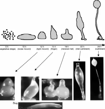
sheath that isolates the developing structure. In the laboratory, Dictyostelium cells can be grown in axenic media that allows the recovery of large quantities of cells (Sussman, 1987). Develop-ment can be induced synchronously by washing the cells free of nutrients, and laying them on agar plates or nitrocellulose filters supported by buffered pads (Spudich, 1987) (Fig. 1).
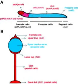
After aggregation, several cell types arise that will organize themselves spatially in the developing structure (Fig. 2). A tip is formed in the aggregate that elongates to give rise to a finger-shaped structure. This finger may initiate a transient period of migration named slug stage (see below). The anterior 20% region of the finger (and also the slug) is composed of prestalk cells that will eventually form the stalk at culmination. Prestalk cells are not a homogeneous population. The study of the promoter activity of the gene encoding the extracellular matrix protein EcmA defined two subpopulations of prestalk (pst) cells: pstA cells, located at the most anterior part, and pstO cells, that lie at the posterior part of the prestalk region (Early et al., 1993). A central core of cells in the prestalk region expresses another extracellular matrix protein, EcmB. The expression of this
Fig. 1. Dictyostelium development. Under conditions of starvation, Dictyostelium cells aggregate in response to extracellular cAMP pulses to form a fruiting body containing spores. A cartoon of the timing and different developmental stages is shown at the top, together with representative pictures below. The slug, a transient migratory structure, can be formed after the finger stage under appropriate conditions and is shown at the bottom of the figure. After a variable period of migration, the structure resumes culmination at the mexican hat stage. This developmental program can be easily reproduced in the laboratory. Thus, Dictyostelium is a very useful model which can help to answer important scientific questions regarding cell type differentiation and morphogenesis.
Moreover, a recent report by Kellerman and McNally has shown how cell density and buffered conditions also affect mound-cell motion and modes of slug formation. The tendency to the formation of migrating slugs is even different between laboratory strains (Kellerman and McNally, 1999).
Slugs are phototactic and thermotactic and their migration is achieved by the coordinated movement of individual cells that make traction on the surface sheath, which is continuously produced and left behind as the slug moves. Cells in the posterior region have linear trajectories, but prestalk cells in the anterior zone rotate around the long axis (Siegert and Weijer, 1992; Siegert and Weijer, 1995). It has been proposed that spiral scroll waves of cAMP in the prestalk zone organize cell movement at the mound and slug stage (Dormann et al., 1998), in addition to chemotactic movement during aggregation. Orchestrating culmination requires coordination of cell movement and terminal differentiation of each cell type. The prestalk cells from the tip build the stalk tube, which grows from the top downwards to lift the spore head over the substratum, thus favoring the dispersal of spores. It has been proposed that during culmination, there are at least two signaling centers that must be coordinated. One is com-posed of the prestalk cells at the tip, which will eventually form the stalk. The second center is made up of the anterior-like cells, which form the basal disc and permit the attachment of the structure to the substratum (Dormann et al., 1996).
The Dictyostelium genome is haploid, and has a size of approxi-mately 34 Mb organized in six chromosomes (Loomis et al., 1995; Kuspa and Loomis, 1996). The ribosomal RNAs are encoded by copies of extrachromosomal DNA which comprises nearly 18% of the nuclear DNA mass (Cockburn et al., 1976; Cockburn et al., 1978). The mitochondrial genome also comprises a third of the total cell DNA and has already been sequenced (Cole and Williams, 1994). The A+T content of the DNA in Dictyostelium is very high. In protein coding regions the base composition is skewed to an average of 60-70% (A+T) but the regions that do not code for proteins contain a much higher A+T content (around 80-95%). These regions of high A+T content include the 5´ and 3´-untranslated sequences of mRNAs and the introns. It is therefore, relatively easy to predict the organiza-tion of the genes. Introns are usually short (approximately 100-200 bp) and the average number of introns per gene is about 1 to 2.
The Dictyostelium genome is in the process of being sequenced by an international consortium (DFG at Jena, the NIH sequencing at Baylor College and the EU sequencing at the Sanger Center). The strategy is based on a chromosomal shotgun methodology and the raw data are now available (http://www.sdsc.edu/mpr/ dicty/). The number of the genes estimated to be present in the Dictyostelium genome is about 8,000-10,000 (Loomis and Kuspa, 1997). If confirmed, this number would be between the 6,000 of yeast and 13,000 of Drosophila. A cDNA sequencing project, in progress in Japan over the last few years, is also providing invaluable sequence data on genes which are specifically ex-pressed at different developmental stages (http:// www.csm.biol.tsukuba.ac.jp/cDNAproject.html).
Tools used to study Dictyostelium - Biochemistry and
Molecular Genetics
Transformation
There are several available selection markers, including neo-mycin, blasticidin, hygromicyn, bleomicyn, uracil and thymidine,
but the former two are the most commonly used. Transformation is generally achieved with high efficiency by electroporation (Pang et al., 1999). The selection can be performed in culture plates with liquid media supplemented at the appropriate concen-tration with antibiotic. The cells attach to the plastic and grow as independent foci. Hundreds of independent transformant foci are routinely obtained per plate. After 10 days of selection there are enough cells for subsequent studies if the transformants are desired to be analyzed as a pool. Alternatively, clonal isolation can be obtained by plating dilutions of cells in association with bacteria.
The constructs are integrated randomly in the genome as tandem repeats of variable number of copies, but also non-integrative extra-chromosomal plasmids are available. Several well characterized promoters can be used to drive the expression of the gene under study to the appropriate time and place of the developing structure. The prestalk ecmA and ecmB promoters and the prespore cotB promoter are routinely used for cell type specific expression. Several actin promoters are also available for high level of expression throughout the vegetative stage and development in all the cell types. Inducible promoters, such as the one controlled by tetracycline, provide tight control of the level of gene expression (Blaauw et al., 2000).
Restriction-enzyme-mediated integration (REMI)
In Dictyostelium many mutants showing alterations in several aspects of multicellular development have been obtained by chemi-cal mutagenesis (Loomis, 1975), but the isolation of the mutated gene is very difficult and must rely on genome mapping or func-tional complementation. Inserfunc-tional mutagenesis by Restriction Enzyme-Mediated Integration (REMI) is a powerful technique (Kuspa and Loomis, 1992) that enables the rapid identification of the disrupted gene. REMI was first applied successfully in yeast
and is basically described in Fig. 3. The introduction of a restriction enzyme, together with the linearized plasmid, will create compat-ible integration sites in the genome increasing the efficiency of transformation. For example, the restriction enzyme DpnII (which recognizes the 4-base sequence GATC and generates GATC overhangs) will allow the integration of a plasmid cut with the compatible restriction enzyme BamHI (that recognizes the 6-base sequence GGATCC). The frequency of DpnII-sites in the genome is much higher than that of BamHI covering a wider spectrum of potential target genes. After electroporation, some plasmids will integrate into the appropriate restriction sites of the genome. If the insertion interrupts a gene necessary for development, the result-ing phenotype can be easily identified, and the strain recovered for further manipulation. The identification of the mutated gene is accomplished by the isolation of the regions flanking the insertion site by plasmid rescue. These DNA regions provide sequence information that may identify the gene, and also may provide probes to screen cDNA libraries for its complete characterization. A subsequent replica of the disruption must be performed by homologous recombination in a fresh wild type strain to assure that the observed phenotype is caused exclusively by the knockout event and to rule out the possibility of secondary mutations.
Disruption of genes by homologous recombination
Homologous recombination in Dictyostelium is very efficient and is commonly used to knockout genes by gene disruption. Since Dictyostelium is haploid the consequences of the mutation can be determined directly in the clonal isolate without further manipula-tion. Vegetative growth and multicellular development are some-what independent in Dictyostelium, so many genes that are neces-sary for development are dispensable for growth, expanding the number of potential genes available by knock-out analysis. Blasticidin is the most common and efficient selection for knockout experiments (Adachi et al., 1994). The size requirements of the flanking regions surrounding the resistance cassette are variable but in general 0.3-0.4 Kb should be sufficient to render the required specificity. The result of a typical knockout experiment may be within reach in just a couple a weeks.
Suppressor screening
Second-site suppressor analysis is a powerful genetic method that allows the identification of components of complex signal transduction pathways. It is based on the identification of a second mutation that overcomes the defect of the primary mutation. The method is only feasible if there is a way to screen for the revertants. In summary, second site mutagenesis is performed on the mutant background using REMI, that later will allow the recovery of the gene acting as suppressor. After antibiotic selection, the transformants must be screened for clones that display total or partial recovery of the phenotype. The disrupted gene is expected to act as a negative component downstream of the primary defect in a given signal transduction pathway (Shaulsky et al., 1996). For example, the mutation of tagB, which encodes a multidrug resist-ance transporter (MDR), blocked sporulation. A second-site sup-pressor screening performed on the tagB mutant revealed several genes whose disruption partially bypassed the sporulation defect. One of these suppressors of tagB mutation was found to be a phosphodiesterase, that plays an essential role in the control of terminal differentiation, and will be described later in detail (Shaulsky et al., 1996).
In situ hybridization and lacZ staining for gene expression studies
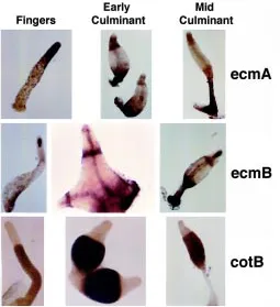
During Dictyostelium development cell type differentiation and gene expression patterns are tightly controlled. Many mutants display abnormalities on these aspects that can be studied by following the pattern of expression of several cell type specific markers. The in situ hybridization technique for mRNA detection (Escalante and Loomis, 1995) and lacZ staining to follow promoter activity (Dingermann et al., 1989), are the most used tools to enable accurate observation at a single cell level. Nucleic acid probes and well-characterized promoters fused to lacZ are available and cover most Dictyostelium cell types. As an example of the most common markers, structures hybridized with specific riboprobes are shown in Fig. 4 (compare the patterns to those represented in the cartoon of Fig. 2). EcmA is an extracellular matrix protein which represents a specific marker for prestalk and stalk cells. EcmB, another extracellular matrix protein is expressed in a subset of prestalk cells at the core of the prestalk region and in anterior-like cells. CotB is a spore coat protein specific for prespore and spore cells. Similarly, the pattern of expression of newly identified genes can be easily determined by in situ hybridization. The lacZ technique can also be used but requires the cloning of the relevant promoter and the isolation of transformants carrying the con-struct. On the other hand, this technique is essential when a detailed study of complex promoters (i.e. alternative promoters) has to be performed. Due to its higher sensitivity, lacZ is also the technique of choice when the gene of interest is expressed at very low levels. The interpretation of LacZ experiments must take into account the stability of β-galactosidase at the protein level. This may raise the question of whether the expression seen at a certain developmental time is real or the reflection of a previous active expression of the gene that was subsequently turned off. It is therefore desirable to use short-life forms of β-galactosidase (Detterbeck et al., 1994) or confirm the pattern observed by the detection of mRNA by in situ hybridization. The use of the green-fluorescence protein (GFP) as a cell marker or fused to the protein of interest is also providing excellent opportunities to study in vivo cell movement in the aggregate and subcellular localization of proteins (Jin et al., 2000).
Mixing experiments for detection of subtle phenotypes and extracellular signaling pathways
It is frequently of interest to challenge a mutant strain to compete with the wild type during development. For example, gene mutation experiments can result in an apparently normal phenotype due to the fact that Dictyostelium exhibits developmental plasticity which allows the morphogenetic program to overcome such defects. Such “masked” phenotypes as they are termed can be sometimes revealed by mixing and competition with wild type or under special conditions (Ponte et al., 1998). In Dictyostelium such experiments are easy to perform and can be very informative. Cells from both strains are grown separately and mixed at the desired proportions to initiate development. One of them is usually labeled genetically with lacZ to determine the distribution of each strain in the devel-oping structures. Some subtle defects may only be revealed when the mutant cells have to compete with wild type for the formation of the different cell types and structures. Figure 5 displays an exam-ple of such experiments in which migA mutant cells, with a defect in chemotaxis, are relegated to the edge of the streams that eventually form the aggregate (Escalante et al., 1997).
The mixing experiments may also answer questions about the cell autonomy of a certain defect and also reveal the presence of extracellular signaling. For example, the block on sporulation generated by the mutation in the MDR transporter TagB can be efficiently overcome by the presence, in the developing structures, of a small proportion of wild type cells (Shaulsky et al., 1995). This defect in sporulation is therefore non cell-autonomous, suggesting that wild type cells were providing an extracellular signal necessary for sporulation that was absent in the mutant (this will be described later in more detail).
Regulatory pathways in Dictyostelium development
Chemotaxis and aggregation
Chemotaxis is a process by which cells display directional movement towards the source of diffusible chemicals and plays important roles in a wide variety of phenomena, including the migration of mammalian phagocytic cells such as neutrophils and macrophages, axonal targeting and morphogenesis. During growth, Dictyostelium cells show chemotaxis to folate, a bacterial by-product, to locate their food source (Pan et al., 1972). During starvation, Dictyostelium amoebae become responsive to cAMP, another chemoattractant which they release in pulses and which governs the process of aggregation.
During growth, cells continuously secrete the autocrine factor PSF (prestarvation factor), a 68-kDa protein. Its concentration serves to monitor cell density (Clarke et al., 1988). The response
of the cells to PSF is inhibited by the bacteria used as a food supply, and this allows the cells to determine their own density in relation to bacteria and to prepare themselves for imminent starvation (reviewed by Clarke and Gomer, 1995). Discoidins are 26 kDa and 28 kDa lectins which are believed to play a role in cytoskeletal organization and cell morphology during aggregation (Alexander et al., 1992). Genes encoding these discoidins are among the first to be induced by PSF (Rathi et al., 1991; Rathi and Clarke, 1992). Later, following food depletion, another protein, CMF (condition medium factor) (Clarke and Gomer, 1995), is released into the medium. This protein is essential to initiate the cAMP relay system, which is necessary for the cells to aggregate (Yuen et al., 1995; van Haastert et al., 1996; Brazill et al., 1998). Components of the cAMP relay system, like cAMP receptors (cARs) and adenylyl cyclase (ACA), are maximally expressed to allow the production of cAMP and the subsequent chemotactic response. The signaling pathways activated by cAMP and CMF are necessary to relay the signal, regulate chemotaxis and control developmental gene expression.
There are four receptors for extracellular cAMP (cAR1-4) that are sequentially expressed during Dictyostelium development. These receptors display different cell type specificities and differ-ent affinities for cAMP. Thus cAR 1 and 3 have high affinity for cAMP, whereas cAR 2 and 4 have low affinities for this ligand. (Klein et al., 1988; Saxe III et al., 1993; Johnson et al., 1993; Louis et al., 1994). cARs belong to the seven-transmembrane (7TM) receptor family, together with a wide variety of neurotransmitter receptors and hormone receptors of higher eukaryotes. The high-affinity cAR1 receptor is the first to be expressed and it is the main receptor involved in aggregation (Sun and Devreotes, 1991). The other high-affinity cAMP receptor, cAR3, is partially redundant and can mediate most cAR1-dependent signaling (Insall et al., 1994). Upon cAMP binding to the receptor, the signal is transduced into the cell through heterotrimeric Gproteindependent and also -independent pathways (see Fig. 6 for a schematic description of the pathways controlling aggregation).
tion of a guanylyl cyclase required for chemotaxis and rearrangement of the actin and myosin cytoskeleton; (c) PLC activation and (d) activation of the protein kinase Akt/PKB necessary for sensing and responding to the chemoattractant gradient (Meili et al., 1999).
(a) The activation of ACA is mediated by the βγ-subunit com-plex (Wu et al., 1995) with the requirement of two cytosolic regulators: the PH (pleckstrin homology) domain-containing pro-tein CRAC (Cytosolic Regulator of ACA) and Pianissimo (Insall et al., 1994; Chen et al., 1997). CRAC is transiently translocated to the membrane of the leading edge of chemotactic cells and it is believed to act as an adaptor between the βγ-complex and ACA (Parent et al., 1998). No homologs of CRAC have been identified so far in other systems. Pianissimo is also essential for the activation of ACA and homologs have been found in S. cerevisiae and S. pombe. It is not clear at what point Pianissimo functions. It may be required to mediate the binding of CRAC, or act after CRAC has bound to the membrane (Parent et al., 1998). It is interesting to note that while CRAC binds to the membrane within seconds after cAMP stimulation, the production of cAMP takes a minute, suggesting that there must be additional steps between CRAC translocation and the activation of the adenylyl cyclase (Parent et al., 1998).
Surprisingly, another component necessary for this receptor-mediated activation of ACA is the MAP Kinase ERK2 (Segall et al., 1995). It is not known whether ERK2 acts directly on any of the components of the signal transduction pathway discussed above, or more indirectly, on the expression of other unknown essential elements. As expected, the mutants on CRAC, Pianissimo or ERK2 are unable to aggregate. However, they respond chemotac-tically to exogenously added cAMP to some extent, suggesting that the primary defect in these mutants is the absence of ACA activation, rather than an alteration in the ability of the cells to move. Other components of the Ras pathway are also necessary for ACA activation but they also lead to additional chemotaxis defects.
Fig. 5. Dictyostelium mixing experiments. When two different strains develop in the same structure, any slight abnormality may become evident due to competition between them. In this example, a mutant in chemotaxis (in the gene migA) was mixed with wild type and the mix was allowed to develop. The mutant strain contains a β-galactosidase expression plasmid to facilitate detection. (A) Several aggregating structures show how the mutant blue cells are relegated to the edges of the streams of cells that are converging in the mounds. (B) A finger shows the anterior region exclusively formed by wild type cells.
G-protein independent pathways have been revealed by the analysis of cAMP-dependent re-sponses in mutants lacking Gα2 or Gβ subunits. These responses include calcium uptake (Milne et al., 1995), tyrosine phosphorylation of ERK2 (Maeda et al., 1996) and post aggregative gene expression mediated by the G-box binding factor (GBF) transcription factor (Schnitzler et al., 1995; see later). The molecular mechanisms of the transmission of the signal for these G-protein independent effects are unknown.
activa-These include Aimless, a Ras-guanine nucleotide exchange factor (GEF) (Insall et al., 1996) and the Ras-interacting protein RIP3, which shares no general homology with any known protein (Lee et al., 1999). It is possible that these are components of the same Ras-regulated pathway. RIP3 interacts in vitro with RasG but the phenotype of the RasG null mutant is not the same as that of Aimless and RIP3, suggesting that these components interact with another not-yet-identified Ras protein. Alternatively, they may have other unknown functions independent of their interaction with Ras (Lee et al., 1999).
To generate the oscillatory waves of extracellular cAMP and the outward propagation of the signal, which are necessary for the formation of the aggregation territories, this cAMP relay pathway must adapt rapidly (reviewed by Parent and Devreotes, 1996) so that the adenylyl cyclase (ACA), which generates the cAMP, can not be further activated until extracellular cAMP has been degraded by a phosphodiesterase (PDE) (Podgorski et al., 1988; Franke et al., 1991; Hall et al., 1993). The cells regain sensitivity in a few minutes and these cycles of refractory and responsive states govern the pulses of extracellular cAMP every 5-6 minutes for several hours. The chemotactic movement of the cells towards these cAMP gradi-ents eventually leads to the formation of the aggregates. The activity of the PDE is also regulated by an inhibitor (PDI).
A fraction of the cAMP produced by the ACA remains within the cell to activate the cAMP-dependent protein kinase A (PKA). It has been proposed that the regulation of the level of intracellular cAMP is also involved in pathways required for the pulses of ACA activation during aggregation. According to this model, phospho-rylation of the internal phosphodiesterase RegA by ERK2 will inhibit RegA activity and consequently, intracellular cAMP will rise and activate PKA. PKA in turn will inhibit ERK2 in an inhibitory loop that may also involve Ras (Aubry et al., 1996; Laub and Loomis,
1998). As also proposed in this model, PKA may either directly or indirectly phosphorylate the receptor cAR1 causing loss of ligand binding. When the levels of internal cAMP are decreased by the activity of RegA, PKA is inhibited and protein phosphatases return cAR1 to its high-affinity state (Laub and Loomis, 1998).
(b)The activation of guanylyl cyclase; the gene encoding this enzyme has not yet been identified, but defects in the production of cGMP were observed in several mutants obtained by chemical mutagenesis. They were found to have abnormal chemotactic directionality and amoeboid movement leading to aggregation-less phenotypes (Kuwayama et al., 1993). Similarly, mutants of StmF, a cGMP phosphodiesterase, display an extended cGMP response and abnormal chemotactic orientation (van Haastert et al., 1982). The activation of guanylyl cyclase by cAR1 requires the G-protein Gα2 and interestingly, the MAP kinase kinase DdMEK1 (Ma et al., 1997). The loss of function of this MAP kinase kinase does not affect the activation of the MAP kinase ERK2 mentioned above, suggesting the presence of two different MAP kinase cascades involved in aggregation: one contains ERK2, required for cAMP-mediated activation of adenylyl cyclase as described in (a) and the other, contains DdMEK1, essential for the cAMP-mediated activation of guanylyl cyclase.
(c) PLC activation in response to cAMP pulses elevates IP3 levels and this response is also mediated by Gα2 (Bominaar and van Haastert, 1994), but the relevance of IP3 signaling in aggre-gation remains controversial. Cells in which PLC has been knocked out show no distinctive phenotype (Drayer et al., 1994) but it is important to point out that this mutant can generate IP3 by a different mechanism (the breakdown of IP5). The knockout of the prolyl oligopeptidase gene enhances the conversion of IP5 to IP3. The increase in IP3 through this pathway counteracts the effects of lithium which inhibits aggregation by reducing IP3 levels
(Williams et al., 1999). This indicates that IP3 signaling is impor-tant for aggregation. The opposite idea comes from the analysis of mutants of the IP3 receptor IplA. This mutant shows no calcium entry in response to cAMP. Nevertheless, the cells did not show any defect in chemotaxis suggesting that IP3-dependent calcium signaling may not be required during aggregation (Traynor et al., 2000). Finally, the interaction between cAMP and CMF signaling involves PLC activity and IP3. The CMF protein binds to its G-protein coupled receptor causing the activation of PLC through Gα1. Through an unknown mechanism, PLC inhibits the GTPase activity of Gα2 (the α-subunit coupled to cAR1), thus prolonging the life-time of the active form Gα2-GTP, which is necessary to activate its downstream effectors (Brazill et al., 1998).
(d) Activation of the protein kinase Akt/PKB; this Dictyostelium protein kinase, which is a homologue of the mammalian Akt/PKB, is also essential for chemotaxis. Cells with mutated kinase are unable to polarize properly in cAMP gradients and they move slower than wild type cells (Meili et al., 1999). Activation of this kinase involves the cAMP receptor cAR1 and is dependent on the coupled heterotrimeric G-protein and PI3-kinase. The fusion of the PH domain of Akt/PKB to green fluorescent protein (GFP) demon-strated a transient translocation of the protein to the leading edge of chemotactic cells upon receptor stimulation.
The cAMP pulses during aggregation induce, in a feed back loop, the maximal level of gene expression of the components involved in the signal relay and chemotaxis. The intracellular pathways leading to the regulation of gene expression from the receptor to the nucleus are largely unknown. However, it is known that this aggregation-stage gene expression mainly depends on the cAMP-dependent protein kinase A (PKA), which also plays multiple functions in Dictyostelium development, as will be described throughout the review. PKA is composed of a single regulatory subunit and a single catalytic subunit (de Gunzburg et al., 1984). The binding of cAMP to the regulatory subunit dissociates the heterodimer, activating the catalytic subunit. Null mutants in the PKA catalytic subunit do not express ACA, which is essential for cAMP production, and therefore generates an aggregation-defective mutant (Mann et al., 1997). However, PKA must display additional functions during aggregation besides its role in ACA expression, since the constitutive expression of the non-G-protein coupled adenylyl cyclase ACG in PKA null cells does not rescue aggregation, although ACG can complement the aggregation-less phenotype of ACA (Mann et al., 1997). PKA also regulates discoidin gene expression during growth and early devel-opment (Primpke et al., 2000).
ACA expression is also dependent on two other components: the Myb-related transcription factor DdMyb2 (Otsuka and van Haastert, 1998) and AmiB , a novel protein of unknown biochemical function (Kon et al., 2000). Cells bearing null mutants of these components do not express detectable levels of ACA and display an aggregation-less phenotype. The overexpression of the cata-lytic subunit of PKA or DdMyb2 in the amiB null mutant rescues ACA expression and development suggesting the possibility that these three components may function in the same or related pathways to control ACA expression.
Several experimental observations on the role of extracellular and intracellular cAMP in the regulation of gene expression were controversial before the discovery of a third adenylyl cyclase. ACA null cells were thought to have no internal cAMP and therefore no active PKA, since the other adenylyl cyclase known at that time, ACG, is only expressed during the germination of spores (Pitt et al., 1992). Nevertheless, stage and post aggregation-stage gene expression can be induced by artificial extracellular pulses of cAMP in ACA null mutants (Pitt et al., 1993). Moreover, the ACA nulls can also be induced to develop into viable spores by synergy with wild-type cells in mixing experiments to form small but well-proportioned fruiting bodies (Pitt et al., 1993). This paradox between the requirement for PKA activation and the apparent dispensability for intracellular cAMP led to the idea that PKA might be activated in a way independent of cAMP binding to the regula-tory subunit. This concept was challenged by the discovery of another adenylyl cyclase activity (ACB) that may provide intracel-lular cAMP to induce PKA activation even in the absence of ACA (Kim et al., 1998). This adenylyl cyclase is not regulated by G-proteins and its activity can be detected during growth and early development. Recently, this adenylyl cyclase has been cloned and renamed as ACR (Söderbom et al., 1999). This gene encodes a composite protein with an adenylyl cyclase domain and a response regulator domain (Söderbom et al., 1999). The response regulator domain of ACR suggests that the activity of this enzyme might be regulated by phosphorylation of the conserved aspartate through phosphorelay from a histidine kinase that remains to be identified. The activity of ACR during growth and aggregation does not seem to be essential, since the phenotype of ACR null mutants is only apparent during culmination.
The molecular basis of how eukaryotic cells sense and interpret a chemical gradient is poorly understood. A chemotactic mutant, isolated by REMI, shows a very specific defect in the assessment of cAMP gradients without any abnormal random cell movement behaviour. The gene responsible for this phenotype encodes a novel protein, MigA, with a BTB domain near the N-terminus that may be involved in protein-protein interactions (Escalante et al., 1997). However, the biochemical role of this protein remains unknown. More recently, several reports have shown that gradi-ents of chemoattractants elicit asymmetrical signaling evgradi-ents in the cell, even though the receptors are uniformly distributed over the cell surface. The specific translocation of CRAC to the leading edge of chemotactic cells (Parent et al., 1998) and the anterior-posterior localization of G-protein βγ-subunits suggest that local-ized activation of receptor-mediated heterotrimeric G-proteins may be part of a general mechanism for gradient sensing (Jin et al., 2000).
Little is known about the mechanisms that govern regulation of size during metazoan development. Dictyostelium has developed
a cell-counting mechanism essential to monitor and adjust the size of its structures during aggregation. Cells bearing mutated smlA, which codes for a novel protein found in the cytoplasm of all cells during growth and early development, form very small aggregates (Brock et al., 1996). This mutant was found to overproduce an extracellular complex of at least 6 polypeptides, named “counting factor”. When wild-type cells were exposed to mutant conditioned medium or mixed with a small proportion of mutant cells, the size of the structures was reduced. This is consistent with a mechanism to monitor the size of the aggregate based on the concentration of the counting factor. One of the subunits of this extracellular complex was cloned and knocked out and the consequence, as expected, was the generation of abnormally large aggregates (Brock and Gomer, 1999).
Regulation of post aggregative cell type differentiation After mound formation, the aggregation-stage pathways are turned off and other pathways leading to cell type differentiation and morphogenesis must be activated. Some molecular compo-nents controlling this transition are known but the mechanism of their action is still far from elucidated. It is believed that during mound formation, the extracellular cAMP concentration rises to reach a constant µM level. This contrasts with the pulsatile concen-tration in the nM range, to which the cells were exposed during aggregation. At this high concentration of cAMP, the high affinity receptors cAR1/cAR3 are expected to be saturated and in a fully adapted state. Nevertheless, two transcription factors, the G-box binding factor (GBF) (Schnitzler et al., 1994; Schnitzler et al., 1995) and DdSTATa, the Dictyostelium “Signal Transducers and Activa-tors of Transcription”, (Araki et al., 1998) are activated by high cAMP levels via cAR1, through a poorly understood, G-protein independent mechanism. GBF is a zinc finger transcription factor that recognizes an 8-base sequence referred to as G-Box that is found in the regulatory regions of many post aggregative genes, including both prestalk and prespore-specific ones (Pears and Williams, 1987; Pavlovic et al., 1989; Hjorth et al., 1990; Haberstroh et al., 1991; Ceccarelli et al., 1992; Powell-Coffman et al., 1994). This transcription factor is essential for the switch between early and late development since null mutants are arrested at the loose aggregate stage, and they show no induction of cell type specific gene expression, even after exposure to cAMP. Cells of the mutant strain aggregate to the loose mound stage and then disaggregate. After disaggregation the cells start emitting again pulses of cAMP that allow another cycle of aggregation. Successive cycles of aggregation and disaggregation take place, which may reflect the inability of the structure to maintain the multicellular state without the expression of post aggregative genes. LagC is a membrane glycoprotein that mediates cell-cell adhesion (Dynes et al., 1994; Sukumaran et al., 1998; Wang et al., 2000). Mutation in the lagC gene generates the same phenotype. Since lagC expression is dependent on GBF, it is very likely that the phenotype of GBF is the consequence of the lack of lagC expression. The premature expression of the GBF gene from the actin15 promoter does not induce premature expression of post-aggregative genes (Schnitzler et al., 1995) indicating that it is not sufficient for post aggregative gene expression. This suggests that other factors are also re-quired.
One of these factors might be the gene spalten, which is also essential to induce cell type differentiation and development past
the loose aggregate stage. However, spalten expression is not controlled by GBF. Spalten encodes a novel signaling protein with an amino-terminal domain which has very high homology with Gα -protein subunits, and a carboxy-terminal domain that encodes a functional PP2C-type protein phosphatase (Aubry and Firtel, 1998). The Gα-like domain of Spalten might act as a molecular switch by GDP/GTP interchange that may regulate the function of the effec-tor phosphatase domain. It will be essential to discover the targets of this regulated phosphatase on order to understand better this molecular pathway.
Another central question in developmental biology is how cell type choice is determined in a population of undifferentiated cells. In Dictyostelium, the initial cell type divergence into prestalk and prespore cells takes place very early, at the loose mound stage. Cells expressing the prestalk marker ecmA or the prespore marker cotB are visible as a few scattered cells randomly distributed in the aggregate (Gomer et al., 1986). Subsequent sorting of these cell types leads to the formation of the antero-posterior pattern in which the prestalk cells occupy the front 1/3 and the prespore cells the rest of the slug. Two mechanisms have been proposed to explain this initial cell type determination. According to the first, positional information is generated by a gradient of a putative morphogen that would induce the initial differentiation of the cells located in a certain region of the aggregate. This idea comes essentially from the observation of a preferential localization of cells expressing ecmA at the periphery of the loose mound (Early et al., 1995). This
localization, however, does not rule out the possibility of a previous random differentiation followed by sorting. Differentiation-inducing factor 1 (DIF-1), a chlorinated hexaphenone, was proposed to be the morphogen controlling this initial prestalk differentiation (Kay et al., 1989). The second possibility, supported by several experi-mental data, suggests that the decision to differentiate into a particular cell type is based on cell autonomous factors (Clay et al., 1995). When cells were plated at low density, starved and exposed to DIF-1, only 25% of the cells ended up expressing the prestalk marker ecmA even though every cell was exposed to the same concentration of DIF. Additional data supports the hypothesis that the cell-cycle phase at the time of starvation is the key factor that influences the initial cell type choice in a cell-autonomous manner (McDonald and Durston, 1984; Gomer and Firtel, 1987; Ohmori and Maeda, 1987; Zimmerman and Weijer, 1993). Prestalk cells differentiate preferentially from those cells in S or early G2 phases at starvation and prespore cells from cells in late G2 or M phase (the Dictyostelium cell-cycle lacks a G1 phase (Weijer et al., 1984)). The novel gene rtoA shares no significant homology with any known protein. Analysis of the corresponding mutant has shown alterations in the latter mechanism since both prestalk and prespore cells originate randomly from cells in any phase of the cell cycle, resulting in an abnormally high proportion of prestalk cells (Wood et al., 1996). The initial cell type choice does not seem to definitely determine cell fate, since artificial alterations of cell type propor-tions are corrected by means of transdifferentiation, suggesting additional mechanisms to control differentiation and cell type proportion at later stages.
The mechanism by which cell type sorting of prestalk and prespore cells, necessary for the establishment of the antero-posterior pattern, is accomplished is only poorly understood (re-viewed by Firtel and Meili, 2000). Chemotaxis to cAMP at the mound stage has been proposed to mediate the sorting of prestalk cells to the tip (Matsukuma and Durston, 1979; Sternfeld and David, 1981; Mee et al., 1986; Traynor et al., 1992). Differential cell adhesion may also play a role in this process since prespore cells have been found to be more adhesive than prestalk cells (Lam et al., 1981; Siu et al., 1983). Sorting of the initial cell types has been described to be affected in a mutant with altered tipA, a gene which codes for a protein of unknown biochemical function. tipA mutant cells show, however, normal chemotactic responses to cAMP and normal cell-cell adhesion suggesting that other mechanisms may be required for cell sorting that are absent in this mutant (Stege et al., 1997).
Assays in monolayer cell culture, in the absence of cell contact, have shown that cell type differentiation is under a combinatorial control of at least two extracellular molecules: cAMP and DIF-1 (Kay et al., 1989). The prestalk-specific marker ecmA is induced synergistically by cAMP and DIF (Berks and Kay, 1990). However, cAMP inhibits the inducing effect of DIF-1 on the expression of the other prestalk-specific gene, ecmB (Berks, 1988). In contrast, the expression of prespore genes is induced by cAMP and repressed by DIF-1 (Early, 1988). However, the prestalk-specific gene cAR2 is induced by cAMP but repressed by 1 suggesting that DIF-1 is not positively regulating all prestalk-specific genes (Saxe III et al., 1996).
It is not known which cell type secretes DIF-1 but the concentra-tion of this morphogen is higher in the prespore region than in the prestalk region. This is believed to be caused by the secretion of an enzyme responsible for the degradation of DIF-1 by the prestalk
cells (Insall et al., 1992). The mechanism of action of DIF-1 is currently unknown but a DIF binding protein has been detected in nuclei and cytoplasmic fractions of Dictyostelium cells (Berks et al., 1991). Recently, a strain which is mutant in the biosynthesis of DIF-1 has been isolated. This strain still forms slugs with prestalk cells of the pstA type. However, the other prestalk cell type, pstO, is not present (Thompson and Kay, 2000).
Extracellular cAMP functions through the cAR receptors to regulate cell type differentiation and morphogenesis. Both the high affinity receptors (cAR1/3) and the low affinity receptors (cAR2/4) may mediate different responses. cAR2 is expressed in a subset of prestalk cells (Saxe III et al., 1996) and the corresponding null mutants are arrested at the tight mound stage, exhibiting an enhanced expression of prespore-specific mRNAs (Saxe III et al., 1993). cAR1, cAR3 and cAR4 are involved in the regulation of the anterior (prestalk) / posterior (prespore) pattern together with the kinase GSK3 and will be discussed in the next section.
Intracellular cAMP, through the activity of PKA, is also a key component in the control of cell type differentiation. Expression of a dominant-negative PKA regulatory subunit (PKA-Rm), which inhibits the catalytic subunit even in the presence of cAMP, affects prespore and prestalk-gene expression. When PKA-Rm expres-sion is controlled by the prespore-specific promoter of pspA (whose expression is not dependent on PKA) the expression of the other known prespore genes is seriously affected (Hopper et al., 1993; Hopper et al., 1995). Similarly, the expression of PKA-Rm in prestalk cells reduces the expression of the prestalk-specific genes ecmA and ecmB (Harwood et al., 1992a,b) and other prestalk-specific genes (Zhukovskaya et al., 1996).
Regulation of the synthesis and degradation of intracellular cAMP (and therefore regulation of PKA activity) is tightly controlled in the different cell types throughout development. The activities of an intracellular phosphodiesterase (RegA) and two different adenylyl cyclases (ACA and ACR) are regulated by different signal trans-duction pathways that may function at different stages. These pathways have been better characterized during terminal differen-tiation and will be described in the last section.
Regulation of cell type proportion
The ability of Dictyostelium cells to reestablish normal prestalk/ prespore proportioning by redifferentiation was first demonstrated by Raper’s classic microdissection experiments (Raper, 1940). More recently, the conversion of prestalk cells into prespore cells has been demonstrated when prespore cells are poisoned by the expression of the inhibitor of protein synthesis ricinA (Shaulsky and Loomis, 1993). Conversely, the regulative capacity of prespore cells has also been observed in a prespore cell population obtained by fluorescence-activated cell sorting. This population of pure prespore cells is able to form well proportioned fruiting bodies, confirming the developmental totipotency of prespore cells (Nadin et al., 2000).
the regulation of GSK3 (Ginsburg and Kimmel, 1997) (see Fig. 7). Activation of GSK3 in turn promotes prespore differentiation and inhibits prestalk differentiation (Ginsburg and Kimmel, 1997). As mentioned above, assays in monolayer cell culture in the absence of cell contact have shown that exogenous cAMP induces prespore gene expression, and also inhibits the induction that DIF-1 exerts on the prestalk-specific gene ecmB. These cAMP-dependent effects on gene expression are abolished simultaneously in gskA null mutants. Consistent with the model, gskA null mutants form aggregates with an abnormally high number of prestalk cells and reduced prespore expression. The extra number of stalk cells derives from the population of cells that express ecmB and form the basal disc (the pstB cells) (Harwood et al., 1995).
Genetic and biochemical evidence indicate that the cAMP receptor cAR3 is one of the principal upstream activators of GSK3. Thus, a) cAR3 null mutants have a phenotype which is essentially similar to that of gskA mutants, contrary to previous observations (Johnson et al., 1993); b) cAR3 null mutants, like gskA mutants do not show cAMP repression of DIF-1-induction of prestalk differen-tiation and c) loss of cAR3 attenuates GSK3 kinase activation. It is interesting to note that the phenotype of the cAR3 mutant is less severe than that of the gskA mutant, suggesting a functional redundancy with other cARs, possibly cAR1, since cAR1 is also redundant with cAR3 during aggregation.
Recently, a novel tyrosine kinase, ZAK1, has been shown to directly phosphorylate and activate GSK3 in response to cAR3 activation by cAMP (Kim et al., 1999). Consistent with this pro-posed pathway, cAR3 null cells are defective in ZAK1 activation by cAMP, and ZAK1 null cells have reduced levels of GSK3 activity. As in cAR3 and GSK3 mutants, the ZAK1 null mutants show reduction of prespore/spore pattern and a loss of inhibition of stalk formation.
It has been proposed that the cAMP receptor cAR4 is likely to participate in a pathway which inhibits GSK3 activity. cAR4 null mutants show characteristics which clearly contrast with those of gskA mutants, such as the reduction of prestalk expression and the enhancement of prespore expression. In cell monolayer assays of cAR4 null mutants, the specific inhibition of GSK3 by lithium reduces the abnormally high prespore expression of cotB, and increases the abnormally low prestalk expression of ecmB (Ginsburg and Kimmel, 1997).
In metazoans, homeobox-containing transcription factors play essential roles in the regulation of the anterior-posterior pattern. In Dictyostelium, the homeobox-containing gene DdHBx1 (named Wariai) is involved in the regulation of the size of the pstO compartment. This gene is expressed in the most anterior pstA region and in the ALC, and seems to exert its function over the contiguous pstO region in a non-cell-autonomous manner (Han and Firtel, 1998). Cell type proportion can be regulated during terminal differentiation as happens in the mutants of the putative transcription factor Stalky. This mutant shows normal prespore differentiation but during culmination, these prespore cells fail to encapsulate and instead, they switch their pathway of differentia-tion to develop into stalk cells (Chang et al., 1996).
Terminal differentiation
The choice between slug migration and culmination seems to be controlled by a variety of signals including light, humidity, tempera-ture, and the concentration of ammonia (Slifkin and Bonner, 1952; Newell et al., 1969; Schindler and Sussman, 1977). The signal
transduction pathways that regulate the sensing and the re-sponses to these environmental cues are largely unknown. Muta-tion of the gene encoding the nuclear protein CudA (whose only known homologue is from Entamoeba histolyca) generates slugs which are unable to culminate (known as the “slugger” phenotype) (Fukuzawa et al., 1997). A wide variety of experimental data indicate that culmination and terminal differentiation of stalk and spores depend on PKA. The activation of PKA is required to trigger culmination since the expression in prestalk cells of PKA-Rm, a dominant inhibitor of the PKA catalytic subunit, leads to slugs that undergo prolonged migration in conditions that promote culmina-tion in the wild type (Harwood et al., 1992b). When the catalytic subunit of PKA is overexpressed, the strain develops faster than the wild type, and is thus called the rapid developing phenotype. This strain shows a sporogeneous phenotype in which cells form spores under monolayer culture conditions, which only lead to a prespore state in the wild type (Anjard et al., 1992; Mann et al., 1994). Similarly, when cells are disaggregated at the mound stage and treated with 8-Br-cAMP, a permeable analog of cAMP that binds to the regulatory subunit of PKA, sporulation takes place very efficiently in the absence of cell contact. In the same kind of in vitro experiments, stalk differentiation can be achieved if the cells are exposed to 8-Br-cAMP and DIF-1.
The existence of other extracellular signals that regulate termi-nal differentiation is suggested from the study of tagB and tagC mutants. These genes code for proteins that contain a region of homology to serine proteases, and another region of homology to multidrug resistance (MDR) transporters, suggesting a mecha-nism that could include proteolysis and the export of peptide signals (Shaulsky et al., 1995). These genes are expressed only in prestalk cells, and the tagB knockout strain presents a cooperative (non-cell-autonomous) defect in prestalk differentiation at the mound stage. There is also a cooperative defect in spore formation during terminal differentiation. The tagB-null cells express normal levels of the prespore-specific gene cotB, but low levels of the prestalk-specific gene ecmA. However, this strain does not ex-press the ecmO::lacZ construct (ecmO is a subregion of the ecmA promoter) which is active in the cells located at the base of the tip behind the pstA cells (see Fig. 2). Both the ability to express ecmO::lacZ and the block in sporulation can be rescued when the mutant cells are co-developed with wild type cells in mixing experiments. This cooperative behavior suggests that TagB may be essential for the processing and secretion of an extracellular signal, that may first enhance prestalk cell differentiation in an autoregulatory loop at the mound stage, and later, might induce neighboring prespore cells to become spores at terminal differen-tiation (Shaulsky et al., 1995).
Since a secondary mutation of the phosphodiesterase RegA rescues the block in sporulation of tagB/C mutants, another path-way involving RegA inhibition has been proposed (Shaulsky et al., 1998). The TagB-dependent secretion of a peptide would promote sporulation by activating a receptor histidine kinase. This would transduce the signal to RegA and inhibit its phosphodiesterase activity, elevating the intracellular cAMP level and activating PKA (Anjard et al., 1998b). While the activating pathway described above leads to a rapid developing phenotype when any of the components are mutated, alterations in the RegA inhibitory path-way are expected to generate a block in sporulation, as in the TagB mutant. Null mutants of another histidine kinase, DhkA, were found to have long fragile stalks and severely affected sporulation (Wang et al., 1996). Sporulation in DhkA mutants can be rescued by activation of PKA or mutation of the phosphodiesterase RegA, suggesting that DhkA could be the kinase that exerts the inhibitory effect on RegA (Wang et al., 1999). However, since the phospho-rylation of RegA was found to activate (not inhibit) its phosphodi-esterase domain (Thomason et al., 1998), the putative mechanism of RegA inactivation by the histidine kinase remains obscure.
Two secreted peptides, SDF1 and SDF2 (spore differentiation factors), have been identified as inducers of sporulation and stalk cell terminal differentiation (Anjard et al., 1998a). SDF-1 accumu-lates during the slug stage and is released in a single burst at the onset of culmination, while SDF-2 accumulates during early culmi-nation and is released in a single burst in mid-culminants (Anjard et al., 1998b). When prespore cells are exposed to SDF-2, encap-sulation of spores takes place in a short time and this effect is dependent on PKA and the histidine kinase DhkA. Similarly, modification of the extracellular domain of DhkA by insertion of a myc-tag reduces the sensibility of the cells to SDF-2. These data are consistent with the idea that DhkA is the receptor for the SDF-2 ligand (Wang et al., 1999).
Little is known about the mechanisms, downstream of PKA activation, which control gene expression and cell differentiation.
Dictyostelium SrfA, a transcription factor which is homologous to the mammalian Serum Response Factor (SRF), has been found to be required for spore terminal differentiation. The SRF-like group, the most evolutionary conserved subfamily of MADS-box tran-scription factors, includes the human, Xenopus and Drosophila SRF homologs and the yeast proteins MCM1 and ARG80. There is increasing evidence for the requirement of SRF in cell differen-tiation and morphogenesis in metazoans. In Drosophila, SRF is essential for the differentiation of the wing intervein cells (Montagne et al., 1996) and the formation of the terminal branches of the traqueal system (Affolter et al., 1994; Guillemin et al., 1996). In mammals, the analysis of a mouse knockout strain has shown a severe defect in mesoderm differentiation (Arsenian et al., 1998). In Dictyostelium, srfA null mutants display a severe defect in spore differentiation in addition to loss of viability (Escalante and Sastre, 1998). Interestingly, the PKA-dependent expression of the spore coat gene spiA was greatly reduced in this mutant. The spore viability phenotype of spiA nulls is much weaker than that gener-ated by mutations in srfA, suggesting that other genes essential for sporulation must be under the control of SrfA besides spiA. Activation of PKA by 8-BrcAMP (Escalante and Sastre, 1998) or overexpression of the PKA catalytic subunit (Escalante and Sastre, unpublished results) does not restore spore formation, suggesting that srfA may function downstream of PKA. The artificial activation of PKA partially restores spiA expression. This is consistent with a model in which PKA and SrfA regulate the expression of spiA in parallel pathways (Fig. 8). The mechanism of activation of srfA during sporulation remains to be determined. A gradient of expres-sion of spiA in the sorus reveals the progresexpres-sion of an inductive signal that seems to emanate from the anterior prestalk cells (Richardson et al., 1994) and may reflect the activation of PKA in response to secreted molecules like SDF-2. The distal promoter of srfA that directs its expression to the spores, shows a pattern reminiscent of the spiA gradient of expression (Escalante and Sastre, unpublished results) suggesting that regulation of srfA-dependent spore-specific gene expression may be regulated by the induction of the expression of this transcription factor in the correct spatio-temporal context.
differentiated stalk cells (Mohanty et al., 1999). To interpret these results, it has been proposed that stalk differentiation requires two steps that are controlled by STATa: first, the release of the repression exerted by STATa, which agrees with the observed results in terms of ecmB expression and DIF-1 hypersensitivity and second, a later induction step required for final stalk differentiation.
Summary
In Dictyostelium, development begins with the aggregation of free living amoebae, which soon become organized into a relatively simple organism with a few different cell types. Coordinated cell type differentiation and morphogenesis lead to a final fruiting body that allows the dispersal of spores. The study of these processes is having increasing impact on our understanding of general develop-mental mechanisms. The availability of biochemical and molecular genetics techniques has allowed the discovery of complex signaling networks which are essential for Dictyostelium development and are also conserved in other organisms. The levels of cAMP (both intracellular and extracellular) play essential roles in every stage of Dictyostelium development, regulating many different signal trans-duction pathways. Two-component systems, involving histidine kinases and response regulators, have been found to regulate intracellular cAMP levels and PKA during terminal differentiation. The sequence of the Dictyostelium genome is expected to be completed in less than two years. Nevertheless, the available sequences that are already being released, together with the results of expressed sequence tags (ESTs), are providing invaluable tools to identify new and interesting genes for further functional analysis. Global expression studies, using DNA microarrays in synchronous development to study temporal changes in gene expression, are presently being developed. In the near future, the application of this type of technology to the complete set of Dictyostelium genes (approximately 10,000) will facilitate the discovery of the effects of mutation of components of the signaling networks that regulate Dictyostelium development on changes in gene expression.
Acknowledgments
We thank Leandro Sastre, Rosa Calvo and Margarita Fernandez for helpful discussions and critical reading of the manuscript. The English version of our paper was revised and corrected by ACTS ([email protected]). This work has been supported by grants from the Direccion General de Enseñanza Superior (grants: PB95-0096 and PB98-0517). RE received financial support from the Comunidad Autonoma de Madrid.
References
ADACHI, H., HASEBE, T., YOSHINAGA, K., OHTA, T. and SUTOH, K. (1994). Isolation of Dictyostelium discoideum cytokinesis mutants by restriction enzyme-mediated integration of the Blasticidin S resistance marker. Biochem. Biophys. Res. Commun. 205: 1808-1814.
AFFOLTER, M., MONTAGNE, J., WALLDORF, U., GROPPE, J., KLOTER, U., LAROSA, M. and GEHRING, W.J. (1994). The Drosophila SRF homolog is expressed in a subset of tracheal cells and maps within a genomic region required for tracheal development. Development 120: 743-753.
ALEXANDER, S., SYDOW, L.M., WESSELS, D. and SOLL, D.R.(1992). Discoidin proteins of Dictyostelium are necessary for normal cytoskeletal organization and cellular morphology during aggregation. Differentiation 51: 149-161.
ANJARD, C., CHANG, W.T., GROSS, J. and NELLEN, W. (1998)a. Production and activity of spore differentiation factors (SDFs) in Dictyostelium. Development 125: 4067-4075.
ANJARD, C., PINAUD, S., KAY, R.R. and REYMOND, C.D. (1992). Overexpression of DdPK2 protein kinase causes rapid development and affects the intracellular cAMP pathway of Dictyostelium discoideum. Development 115: 785-790.
ANJARD, C., ZENG, C., LOOMIS, W.F. and NELLEN, W. (1998)b. Signal transduc-tion pathways leading to spore differentiatransduc-tion in Dictyostelium discoideum. Dev. Biol. 193: 146-155.
ARAKI, T., GAMPER, M., EARLY, A., FUKUZAWA, M., ABE, T., KAWATA, T., KIM, E., FIRTEL, R.A. and WILLIAMS, J.G. (1998). Developmentally and spatially regulated activation of a Dictyostelium STAT protein by a serpentine receptor. EMBO J. 17: 4018-4028.
ARSENIAN, S., WEINHOLD, B., OELGESCHLAGER, M., RUTHER, U. and NORDHEIM, A. (1998). Serum response factor is essential for mesoderm forma-tion during mouse embryogenesis. EMBO J. 17: 6289-6299.
AUBRY, L. and FIRTEL, R.A. (1998). Spalten, a protein containing G alpha-protein-like and PP2C domains, is essential for cell-type differentiation in Dictyostelium. Genes Dev. 12: 1525-1538.
BALDAUF, S.L. and DOOLITTLE, W.F. (1997). Origin and evolution of the slime molds (Mycetozoa). Proc. Natl. Acad. Sci. USA 94: 12007-12012.
BERKS, M. and KAY, R.R. (1988). Cyclic AMP is an inhibitor of stalk cell differentiation in Dictyostelium discoideum. Dev. Biol. 126: 108-114.
BERKS, M. and KAY, R.R. (1990). Combinatorial control of cell differentiation by cAMP and DIF-1 during development of Dictyostelium discoideum. Development 110: 977-984.
BERKS, M., TRAYNOR, D., CARRIN, I., INSALL, R.H. and KAY, R.R. (1991). Diffusible signal molecules controlling cell differentiation and patterning in Dictyostelium. Development Suppl. 1: 131-139.
BLAAUW, M, LINSKENS, M.H. and VAN HAASTERT, P.J. (2000). Efficient control of gene expression by a tetracycline-dependent transactivator in single Dictyostelium discoideum cells. Gene 252: 71-82.
BOMINAAR, A.A. and VAN HAASTERT, P.J.M. (1994). Phospholipase C in Dictyostelium discoideum - identification of stimulatory and inhibitory surface receptors and G-proteins. Biochem. J. 297: 189-193.
BRAZILL, D.T., LINDSEY, D.F., BISHOP, J.D. and GOMER, R.H. (1998). Cell density sensing mediated by a G protein-coupled receptor activating phospholipase C. J. Biol. Chem. 273: 8161-8168.
BREFELD, O. (1869) Dictyostelium mucoroides. Ein neuer Organismus aus der Verwandtschaft der Myxomyceten. Abhandlungen der Senckenbergischen Naturforschenden Gesellschaft Frankfurt 7: 85-107.
BROCK, D.A. and GOMER, R.H. (1999). A cell-counting factor regulating structure size in Dictyostelium. Genes Devel. 13: 1960-1969.
BROCK, D.A., BUCZYNSKI, G., SPANN, T.P., WOOD, S.A., CARDELLI, J. and GOMER, R.H. (1996). A Dictyostelium mutant with defective aggregate size determination. Development 122: 2569-2578.
CECCARELLI, A., MAHBUBANI, H. and WILLIAMS, J.G. (1991). Positively and negatively acting signals regulating stalk cell and anterior-like cell differentiation in Dictyostelium. Cell 65: 983-989.
CECCARELLI, A., MAHBUBANI, H.J., INSALL, R., SCHNITZLER, G., FIRTEL, R.A. and WILLIAMS, J.G. (1992). A G-rich sequence element common to Dictyostelium genes which differ radically in their patterns of expression. Dev. Biol. 152: 188-193.
CLARKE, M. and GOMER, R.H. (1995). PSF and CMF, autocrine factors that regulate gene expression during growth and early development of Dictyostelium. Experientia 51: 1124-1134.
CLARKE, M., YANG, J. and KAYMAN, S. (1988). Analysis of the prestarvation response in growing cells of Dictyostelium discoideum. Dev. Genet. 9: 315-326.
CLAY, J.L., AMMANN, R.R. and GOMER, R.H. (1995). Initial cell-type choice in a simple eukaryote: Cell- autonomous or morphogen-gradient dependent?. Dev. Biol. 172: 665-674.
COCKBURN, A.F., FRANKEL, G.A., FIRTEL, R.A., KINDLE, K.L. and NEWKIRK, M.J. (1976). Organization of the ribosomal genes of Dictyostelium. In Molecular mechanisms in the control of gene expression. [ICN-UCLA Symp. Mol. Cell. Biol.] (Eds. D.P. Nierlich, W.J. Rutter and C.F. Fox). Acad. Press, New York, pp. 599-603.
COCKBURN, A.F., TAYLOR, W.C. and FIRTEL, R.A. (1978). Dictyostelium rDNA consists of nonchromosomal palindromic dimers containing 5S and 36S coding regions. Chromosoma 70: 19-29.
genome: A primordial system using the universal code and encoding hydrophilic proteins atypical of metazoan mitochondrial DNA. J. Mol. Evol. 39: 579-588.
CHANG, W., Newell, P.C. and Gross, J.D. (1996). Identification of the cell fate gene stalky in Dictyostelium. Cell 87: 471-481.
CHEN, M.Y., LONG, Y. and DEVREOTES, P.N. (1997). A novel cytosolic regulator, pianissimo, is required for chemoattractant receptor and G protein-mediated activation of the 12 transmembrane domain adenylyl cyclase in Dictyostelium. Genes Dev. 11: 3218-3231.
DAVIES, L., SATRE, M., MARTIN, J.B. and GROSS, J.D. (1993). The target of ammonia action in Dictyostelium. Cell 75: 321-327.
DE GUNZBURG, J., PART, D., GUISO, N. and VERON, M. (1984). An unusual adenosine 3',5'-phosphate dependent protein kinase from Dictyostelium discoideum. Biochem. 23: 3805-3812.
DETTERBECK, S., MORANDINI, P., WETTERAUER, B., BACHMAIR, A., FISCHER, K. and MACWILLIAMS, H.K. (1994). The ‘prespore-like cells’ of Dictyostelium have ceased to express a prespore gene: Analysis using short-lived beta-galactosidases as reporters. Development 120: 2847-2855.
DINGERMANN, T., REINDL, N., WERNER, H., HILDEBRANDT, M., NELLEN, W., HARWOOD, A., WILLIAMS, J. and NERKE, K. (1989). Optimization and in situ detection of Escherichia coli beta-galactosidase gene expression in Dictyostelium discoideum. Gene 85: 353-362.
DORMANN, D., SIEGERT, F. and WEIJER, C.J. (1996). Analysis of cell movement during the culmination phase of Dictyostelium development. Development 122: 761-769.
DORMANN, D., VASIEV, B. and WEIJER, C.J. (1998). Propagating waves control Dictyostelium discoideum morphogenesis. Biophys. Chem. 72: 21-35.
DRAYER, A.L., VAN DER KAAY, J., MAYR, G.W. and VAN HAASTERT, P.J.M. (1994). Role of phospholipase C in Dictyostelium - formation of inositol 1,4,5-triphosphate and normal development in cells lacking phospholipase C activity. EMBO J. 13: 1601-1609.
DYNES, J.L.; CLARK, A.M.; SHAULSKY, G.; KUSPA, A.; LOOMIS, W.F. and FIRTEL, R.A. (1994). LagC is required for cell-cell interactions that are essential for cell-type differentiation in Dictyostelium. Genes. Devel. 8: 948-958.
EARLY, A., ABE, T. and WILLIAMS, J. (1995). Evidence for positional differentiation of prestalk cells and for a morphogenetic gradient in Dictyostelium. Cell 83: 91-99.
EARLY, A.E., GASKELL, M.J., TRAYNOR, D. and WILLIAMS, J.G. (1993). Two distinct populations of prestalk cells within the tip of the migratory Dictyostelium slug with differing fates at culmination. Development 118: 353-362.
EARLY, V.E. and WILLIAMS, J.G. (1988). A Dictyostelium prespore-specific gene is transcriptionally repressed by DIF in vitro. Development 103: 519-524.
ELLINGSON, J.S., TELSER, A. and SUSSMAN, M. (1971). Regulation of functionally related enzymes during alternative developmental programs. Biochim. Biophys. Acta 244: 388-395.
ESCALANTE, R. and LOOMIS, W.F. (1995). Whole-mount in situ hybridization of cell-type-specific mRNAs in Dictyostelium. Dev. Biol. 171: 262-266.
ESCALANTE, R. and SASTRE, L. (1998). A serum response factor homolog is required for spore differentiation in Dictyostelium. Development 125: 3801-3808.
ESCALANTE, R., WESSELS, D., SOLL, D.R. and LOOMIS, W.F. (1997). Chemotaxis to cAMP and slug migration in Dictyostelium both depend on MigA, a BTB protein. Mol. Biol. Cell 8: 1763-1775.
FIRTEL, R.A. and MEILI, R. (2000). Dictyostelium: a model for regulated cell movement during morphogenesis. Current Opinion in Genetics and Development 10: 421-427.
FOSNAUGH, K.L. and LOOMIS, W.F. (1989)a. Sequence of the Dictyostelium discoideum spore coat gene Sp96. Nucleic Acids Res. 17: 9489-9490.
FOSNAUGH, K.L. and LOOMIS, W.F. (1989)b. Spore coat genes SP60 and SP70 of Dictyostelium discoideum. Mol. Cell. Biol. 9: 5215-5218.
FOSNAUGH, K.L. and LOOMIS, W.F. (1991). Coordinate regulation of the spore coat genes in Dictyostelium discoideum. Dev. Genet. 12: 123-132.
FRANKE, J., FAURE, M., WU, L., HALL, A.L., PODGORSKI, G.J. and KESSIN, R.H. (1991). Cyclic nucleotide phosphodiesterase of Dictyostelium discoideum and its glycoprotein inhibitor - structure and expression of their genes. Dev. Genet. 12: 104-112.
FREEZE, H. and LOOMIS, W. (1977)a. The isolation and characterization of a component of the surface sheath of Dictyostelium discoideum. J. Biol. Chem. 252: 820-824.
FREEZE, H. and LOOMIS, W.F. (1977)b. The role of the fibrillar component of the surface sheath in the morphogenesis of Dictyostelium discoideum. Dev. Biol. 56: 184-194.
FUKUZAWA, M., HOPPER, N. and WILLIAMS, J. (1997). cudA: A Dictyostelium gene with pleiotropic effects on cellular differentiation and slug behaviour. Development 124: 2719-2728.
GASKELL, M.J., JERMYN, K.A., WATTS, D.J., TREFFRY, T. and WILLIAMS, J.G. (1992). Immunolocalization and separation of multiple prestalk cell types in Dictyostelium. Differentiation 51: 171-176.
GINSBURG, G.T. and KIMMEL, A.R. (1997). Autonomous and nonautonomous regulation of axis formation by antagonistic signaling via 7-span cAMP receptors and GSK3 in Dictyostelium. Genes Devel. 11: 2112-2123.
GOMER, R.H. and FIRTEL, R.A. (1987). Cell-autonomous determination of cell-type choice in Dictyostelium development by cell-cycle phase. Science 237: 758-762.
GOMER, R.H., DATTA, S. and FIRTEL, R.A. (1986). Cellular and subcellular distribution of a cAMP-regulated prestalk protein and prespore protein in Dictyostelium discoideum: A study on the ontogeny of prestalk and prespore cells. J. Cell Biol. 103: 1999-2015.
GREGG, J.H., HACKNEY, A.L. and KRIVANEK, J.O. (1954). Nitrogen metabolism of the slime mold Dictyostelium discoideum during growth and morphogenesis. Biol. Bull. 107: 226-235.
GUILLEMIN, K., GROPPE, J., DÜCKER, K., TREISMAN, R., HAFEN, E., AFFOLTER, M. and KRASNOW, M.A. (1996). The pruned gene encodes the Drosophila serum response factor and regulates cytoplasmic outgrowth during terminal branching of the tracheal system. Development 122: 1353-1362.
HABERSTROH, L., GALINDO, J. and FIRTEL, R.A. (1991). Developmental and spatial regulation of a Dictyostelium prespore gene - cis-acting elements and a cAMP-induced, developmentally regulated DNA binding activity. Development 113: 947-958.
HALL, A.L., FRANKE, J., FAURE, M. and KESSIN, R.H. (1993). The role of the cyclic nucleotide phosphodiesterase of Dictyostelium discoideum during growth, aggre-gation, and morphogenesis: overexpression and localization studies with the separate promoters of the pde. Dev. Biol. 157: 73-84.
HAN, Z. and FIRTEL, R.A. (1998). The homeobox-containing gene Wariai regulates anterior-posterior patterning and cell-type homeostasis in Dictyostelium. Devel-opment 125: 313-325.
HARWOOD, A.J., HOPPER, N.A., SIMON, M.N., BOUZID, S., VERON, M. and WILLIAMS, J.G. (1992)a. Multiple roles for cAMP-dependent protein kinase during Dictyostelium development. Dev. Biol. 149: 90-99.
HARWOOD, A.J., HOPPER, N.A., SIMON, M.N., DRISCOLL, D.M., VERON, M. and WILLIAMS, J.G. (1992)b. Culmination in Dictyostelium is regulated by the cAMP-dependent protein kinase. Cell 69: 615-624.
HARWOOD, A.J., PLYTE, S.E., WOODGETT, J., STRUTT, H. and KAY, R.R. (1995). Glycogen synthase kinase 3 regulates cell fate in Dictyostelium. Cell 80: 139-148.
HJORTH, A.L., PEARS, C., WILLIAMS, J.G. and FIRTEL, R.A. (1990). A developmentally regulated trans-acting factor recognizes dissimilar G/C-rich elements controlling a class of cAMP-inducible Dictyostelium genes. Genes Devel. 4: 419-432.
HOPPER, N.A., HARWOOD, A.J., BOUZID, S., VERON, M. and WILLIAMS, J.G. (1993). Activation of the prespore and spore cell pathway of Dictyostelium differentiation by cAMP-dependent protein kinase and evidence for its upstream regulation by ammonia. EMBO J. 12: 2459-2466.
HOPPER, N.A., SANDERS, G.M., FOSNAUGH, K.L., WILLIAMS, J.G. and LOOMIS, W.F. (1995). Protein kinase A is a positive regulator of spore coat gene transcrip-tion in Dictyostelium. Differentiatranscrip-tion 58: 183-188.
INSALL, R., KUSPA, A., LILLY, P.J., SHAULSKY, G., LEVIN, L.R., LOOMIS, W.F. and DEVREOTES, P. (1994). CRAC, a cytosolic protein containing a pleckstrin homology domain, is required for receptor and G protein-mediated activation of adenylyl cyclase in Dictyostelium. J. Cell Biol. 126: 1537-1545.
INSALL, R.H., BORLEIS, J. and DEVREOTES, P.N. (1996). The aimless RasGEF is required for processing of chemotactic signals through G-protein-coupled receptors in Dictyostelium. Curr. Biol. 6: 719-729.