Cell-cell signaling and adhesion in phagocytosis and
early development of Dictyostelium
ENRICO BRACCO, BARBARA PERGOLIZZI, BARBARA PERACINO, ELEONORA PONTE, ALESSANDRA BALBO,
ANDREA MAI, ADRIANO CECCARELLI and SALVATORE BOZZARO*
Dipartimento di Scienze Cliniche e Biologiche, Università di Torino, Italy.
ABSTRACT Cell-cell signaling and adhesion regulate transition from the unicellular to the multi-cellular stage of development in the multi-cellular slime mold Dictyostelium. Essential gene networks involved in these processes have been identified and their interplay dissected. Heterotrimeric G protein-linked signal transduction plays a key role in regulating expression of genes mediating chemotaxis or cell adhesion, as well as coordinating actin-based cell motility during phagocytosis and chemotaxis. Two classes of cell adhesion molecules, one cadherin-like and the second belonging to the IgG superfamily, contribute to the strength of adhesion in Dictyostelium aggre-gates. The developmental role of genes involved in motility and adhesion, and their degree of redundancy, have been re-assessed by using novel developmental assay conditions which are closer to development in nature.
KEY WORDS:
Dictyostelium, G protein, chemotaxis, actin-binding proteins, phagocytosis, signal
transduc-tion, cell adhesion, PAF, gene redundancy.
0214-6282/2000/$20.00
© UBC Press Printed in Spain
www.ehu.es/ijdb
*Address correspondence to: Salvatore Bozzaro. Dipartimento di Scienze Cliniche e Biologiche, Università di Torino, Ospedale S. Luigi, 10043 Orbassano, Italy. e-mail: sbozzaro@polito.it
Abbreviations used in this paper: AC, adenylylcyclase, CAR, cAMP receptors; CRAC, cytosolic regulator of adenylylcyclase; csA, contact sites A glycopro-tein; GC, guanylylcyclase; PAF, platelet activating factor; PIA, pianissimo; PH domain, pleckstrin homology domain; PKA, protein kinase A; vatB, B subunit of V-ATPase.
Introduction
Dictyostelium is the lowest eukaryote able to form multicellular structures (Bonner, 1967; Loomis, 1975). Unlike embryos arising from a fertilized egg, where multicellularity is the outcome of repeated cell division of a progenitor cell, in Dictyostelium the multicellular organism, called “slug” and later on “fruiting body”, arises from the gathering of thousands of individual cells into a multicellular agglomerate. The size of the mature organism is very flexible, as it can spring from as less as 100 cells to an optimal of 105 cells. Growth and development are temporally separated and mutually exclusive. During growth, Dictyostelium cells behave as free-living amoebae, which feed on bacteria by phagocytosis. The cells are barely cohesive, move actively on the substratum in search of food, and divide by binary fission. Development is triggered by starvation, and results in the appearance, 4 to 5 hours later, of the devices for producing and responding to chemoattractants, as well as adhering strongly to each other. The concerted action of chemotactic cell motility (Gerisch, 1987; Par-ent and Devreotes, 1996) and intercellular adhesion (Bozzaro and Ponte, 1995) transform a monolayer of single cells into multicellu-lar three-dimensional aggregates (Fig. 1), each of which gives rise to a slug, a sausage-shaped unitary organism capable of undergo-ing extended migration towards light and temperature gradients (Bonner, 1967; Loomis, 1975; Fisher, 1997). A slug possesses an
anterior-posterior pattern of prestalk and prespore cells, and a dominant anterior tip which acts as an organizer (Fig. 1). Elaborate and polarized cell movements occur inside the cell mass during slug and fruiting body formation, leading to sorting out of differen-tiating cell populations, the prestalk and the prespore cells (Siegert and Weijer, 1995; Williams, 1997). Prestalk cells will eventually form a slender stalk lifting on the air, and the prespore tissue gives rise to a sorus, containing mature, encapsulated spores (Fig. 1).
a key role also in the development of higher organisms. Chemo-taxis, which in Dictyostelium is mediated by cyclic AMP, involves both producing and sensing the chemoattractant as well as trans-ducing the membrane signal to the actin cytoskeleton, to stimulate oriented cell motility. A wealth of biochemical and molecular genetic studies has allowed to define the properties of the chemo-tactic process (Gerisch, 1987; van Haastert, 1995; Parent and Devreotes, 1996), as well as identifying the genes encoding most of the proteins involved, including the cAMP receptors (Klein et al., 1988; Rogers et al., 1997), the linked heterotrimeric G proteins (Pupillo et al., 1989; Kumagai et al., 1989; Hadwiger et al., 1994) and components of downstream pathways (Insall et al., 1994; Segall et al., 1995; Insall et al., 1996; Maeda et al., 1996; Chen et al., 1997). Similarly, several actin-binding proteins have been characterized and their role in the organization of the actin cy-toskeleton during chemotaxis, or other motility events, investigated (Noegel and Luna, 1995). Aggregation also requires the expres-sion of new classes of cell adheexpres-sion molecules, which are respon-sible for the compaction of aggregates (Bozzaro and Ponte, 1995). Both a cadherin-like 24 kDa glycoprotein (Knecht et al., 1987; Wong et al., 1996), and a 80 kDa glycoprotein, named contact sites A (csA), belonging to the IgG superfamily (Müller and Gerisch, 1978; Noegel et al., 1986, Matsunaga and Mori, 1987; Kamboj et al., 1989}, have been shown to play a central role in aggregation. After aggregation, csA slowly disappears and is replaced in its function by a third adhesion system, which may involve a 150 kDa glycoprotein and be encoded by the LagC gene (Geltosky et al., 1979; Gao et al., 1992; Dynes et al., 1994).
At post-aggregative stages, pattern formation and cell type differentiation can be conveniently studied, as in animal embryos. In Dictyostelium, these processes are regulated by extracellular signals, including cAMP, ammonia, and DIF, a chlorinated alkyl phenone (Kay, 1997; Verkerke-van Wijk and Schaap, 1997; Williams, 1997; Nanjundiah, 1997). Differential exposure to gradi-ents formed by these diffusible substances, differential
adhesive-ness and stochastic differences among cells, which depend on their past history from the end of growth (Maeda, 1997), determine the cell fate, leading to expression of cell-type specific genes (Loomis, 1996). This is regulated by intracellular transduction effectors, which are conserved in multicellular organisms, such as protein kinase A (Harwood et al., 1992; Mann and Firtel, 1993), glycogen synthase kinase 3 (Harwood et al., 1995) and SH2-domain/phosphotyrosine signaling by STAT protein (Kawata et al., 1997).
A major advantage of Dictyostelium as experimental system is that its molecular genetics has been developed to a high level: the cells are easily transformed by integrating and non-integrating vectors, several resistance and auxotrophic markers exist, and gene disruption is favored by the small, haploid genome. Single or multiple knockout mutants, generated by homologous recombina-tion, have been widely used to characterize the function and developmental role of gene products (Loomis et al., 1994). This approach has been complemented by the development of restric-tion enzyme mediated inserrestric-tional mutagenesis (Kuspa and Loomis, 1992) to generate new mutants and recover the tagged gene by plasmid rescue. Since Dictyostelium is eminently suited for cell biology and biochemistry experiments, the introduction of these molecular genetic tools has made available a unique combination of approaches to investigate this system. In this paper we will concentrate on the molecular basis of cell-cell signaling and adhesion that regulate phagocytosis and early development.
Cell-cell signaling in phagocytosis
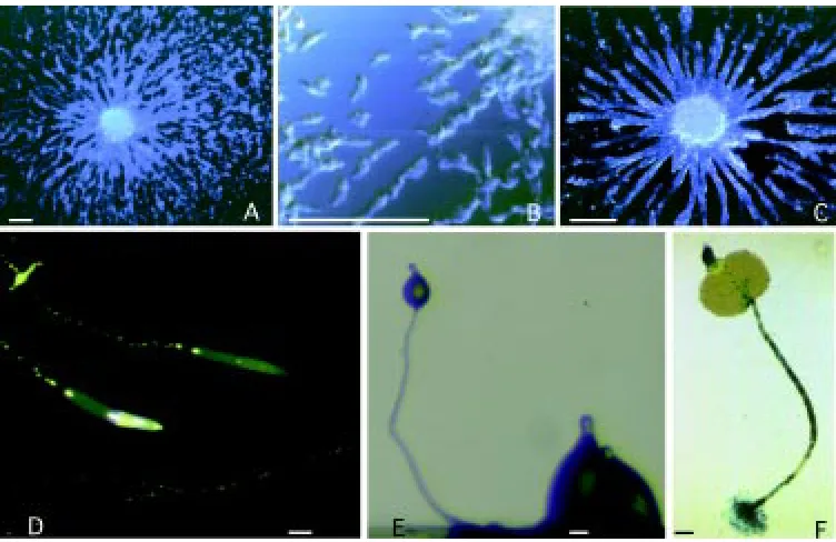
Nutrient uptake occurs in Dictyostelium by phagocytosis of bacteria (Fig. 2), though axenic strains, which grow by fluid-phase endocytosis, have been selected in the laboratory and are widely used. Dictyostelium is a very efficient phagocyte, being able to take up four bacteria/min/cell. Phagocytosis is developmentally regu-lated, and is strongly reduced after tight aggregate formation (Bozzaro, unpublished). Up to the aggregation stage, cells are able Fig. 1. Development of Dictyostelium.
Cells aggregating on a glass surface at
(A, B) an earlier and (C) a later stage. Cells move toward aggregation centers in response to chemoattractant re-leased by the cells in the aggregation center and propagated by relay. Aggre-gating cells display an elongated shape (B) and move as single cells or in streams, adhering to each other by end-to-end contacts. Slugs migrating on agar (D), or undergoing culmination
to phagocytose, but bacterial uptake leads to repression of developmentally regulated genes and reversion to the growth phase, consistent with the notion that growth and development are mutually exclusive (Gambino et al., 1992).
Particle uptake requires actin recruitment to the site of particle adhesion, such that a phagocytic cup is formed, and further actin polymerization at the distal border of the cup, in order for the particle to be totally surrounded. By expressing a chimeric GFP-actin it has been shown that GFP-actin is recruited to the membrane within seconds from particle binding, and that an actin-coated phagosome is formed in less than one minute, followed by depolymerization of the actin coat (Fig. 2) (Peracino et al., 1998). The phagocytic process can be interrupted at any stage, thus particle adhesion does not act as a trigger for a process that then proceeds irreversibly (Maniak et al., 1995; Peracino et al., 1998). Dictyostelium cells possess a heterotrimeric G protein, whose subunits have been cloned. So far eight Gα subunits have been cloned, which interact with a single βγ heterodimer. Gβ-null cells are impaired in phagocytosis, but not in fluid-phase endocytosis (Peracino et al., 1998). As a consequence their growth rate on bacteria is strongly reduced, though not totally abolished. None of the Gα-null mutants is defective in phagocytosis, suggesting that Gα subunits act redundantly in this regard. Insertion of the GFP-actin protein in Gβ-null cells has allowed correlation of the defect in phagocytosis with a reduced ability of Gβ-minus cells to reorganize their actin cytoskeleton into a functional phagocytic cup. Downstream compo-nents of the transduction pathway involve PLC and intracellular Ca2+ ions (Peracino et al., 1998}. Members of the Rho protein family, such as RacC and RacF1, have been found to be transiently localized in phagocytic cups (Seastone et al., 1998; Rivero et al., 1999), whereas they are not present in chemotactic pseudopods (Rivero et al., 1999}. They could mediate signals to the actin cytoskeleton, though no defects have been found in null mutants, possibly due to redundancy. Two actin crosslinking proteins, the gelation factor and α -actinin, could favor proper actin assembly during phagocytosis. The gelation factor is located at the phagocytic cup (Cox et al., 1996}, and α-actinin in phagosomes (Furukawa and Fechheimer, 1994). A double mutant shows a significant reduction in phagocy-tosis, compared to the single mutants and the wild type (Rivero et al., 1996b). Depolymerization of actin can also be as important for phagocytosis as correct crosslinking, as shown by the finding that mutants defective in DAip1, the Dictyostelium homologue of the yeast actin interactin protein 1, are strongly defective in phagocy-tosis {Konzok et al., 1999).
Bacterial uptake stimulates in less than 30 minutes transcription of specific genes, such as cysteine proteinases or genes of unknown function (Bozzaro and Merkl, 1985; Souza et al., 1995). Among these
genes is the multimeric V-ATPase (Bracco et al., 1997), which is present in the contractile vacuole (Heuser et al., 1993) and in endo-lysosomes (Adessi et al., 1995). Cloning the B subunit encoding vatB gene has led to generation of a GFP-VATB chimeric protein. Prelimi-nary experiments indicate that the protein is recruited very rapidly to the forming phagosome (Balbo et al., unpublished results).
Cell-cell signaling in early development
In the transition from the unicellular to the multicellular stage the profile of gene expression changes dramatically; cells express several new genes which are required for aggregation while repressing genes required for growth (Mehdy et al., 1983, Clarke and Gomer, 1995}. Among the first, are genes encoding the cAMP receptors, subunits of the heterotrimeric G proteins, downstream effectors, such as the adenylyl- and guanylyl-cyclases, the mem-brane cAMP phosphodiesterase, the extracellular phosphodieste-rase, the protein kinases A and C, or cell adhesion proteins, such as the 80 kDa glycoprotein csA.
Two signals which affect expression of these developmental genes are PSF and CMF. PSF, or prestarvation factor, is a 68 kDa protein which is secreted by growing cells, but reaches a threshold level only at high cell density. CMF, or conditioned medium factor, is an 80 kDa protein secreted by starving cells (Clarke and Gomer, 1995). Early and aggregation specific genes are induced at low level by these factors. With the production of cAMP by adenylylcyclase activity, a new signal system is build up, which becomes the major regulator of both aggregation-stage and post-aggregative genes (Reymond et al., 1995}. Cyclic AMP down-regulates growth phase specific genes (Kimmel and Carlisle, 1986; Hassanain and Kopachik, 1989), whereas up-regulates genes associated with aggregation (Darmon et al., 1975; Roos et al., 1977; Mann and Firtel, 1989}.
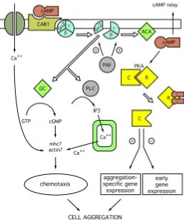
Stimulation requires the pulsatile production and secretion of cAMP, which binds to high affinity membrane receptors, and stimulates via G protein the adenylyl cyclase, leading to further cAMP production (Fig. 3). The relay system adapts after about one minute, such that adenylyl cyclase cannot be further activated, until the pathway has returned to its initial conditions. Removal of cAMP by the membrane cAMP phosphodiesterase resensitizes the cell (Theibert and Devreotes, 1986; Snaar-Jagalska and Van Haastert, 1990). The activation, desensitization and resensitization cycle results in the production of cAMP pulses with a period of about 6 minutes (Gerisch and Wick, 1975). The oscillatory stimulation by cAMP is essential for up-regulating, by a positive feedback loop, maximal transcription of the genes involved in cAMP sensing and production as well as the gene encoding the cell adhesion mol-ecule csA. When the level of cAMP rises significantly and is no Fig. 2. Phagocytosis in Dictyostelium. (Left)
more pulsatile, as it seems to occur in compact aggregates, transcription of aggregation specific genes is repressed, and post-aggregative genes are activated.
Essential role of heterotrimeric G protein in chemotaxis
and development
Both the chemotactic and hormone-like activities of cAMP are mediated by binding of the molecule to membrane receptors, which belong to the family of G protein-linked serpentine receptors with seven transmembrane domains. Of the four receptors which have been identified, and which show different pattern of expression during development, cAR1 plays the major role during aggrega-tion. Disruption of the encoding carA gene blocks development, chemotaxis and developmentally-regulated gene expression (Sun and Devreotes, 1991).
Cyclic AMP signaling through cAR1 or any of the other receptors requires coupling to the heterotrimeric G protein for intracellular transduction. Deletion mutants have been generated for 6 of the 8
α subunits identified so far, as well as the single β subunit, allowing a dissection of the role of each component. Gα2 plays an essential role in development, as it mediates the in vivo cAMP-dependent stimulation of adenylyl- and guanylyl-cyclase (Kumagai et al., 1989). Thus, Gα2-null mutants fail to aggregate, to undergo chemotaxis as well as respond to extracellular cAMP. Deletion of the remaining Gα subunits does not have dramatic effects on development, but results in subtle phenotypes (Wu and Devreotes, 1991). Deletion of the single Gβ subunit also abolishes all G protein-linked signal transduction, and thus blocks development (Wu et al., 1995). This is consistent with the view that all α subunits couple to the single β subunit, and that both the α- and the βγ− complex are required for activation of either subunit.
G protein-dependent regulation of adenylyl cyclase activity Two different adenylyl cyclases have been identified and char-acterized in Dictyostelium, ACA and ACG. ACA is maximally
expressed during aggregation and at reduced levels thereafter, while ACG is expressed in mature spores and in the growth phase (Pitt et al., 1992). ACA is stimulated by G protein-dependent activation upon cAMP binding to cAR1 (Fig. 3). Analysis of mutants defective in G protein subunits and in vitro activation studies with GTPγS have led to the conclusion that in vivo ACA is activated by interaction with the βγ subunit released from the Gα2βγ complex (Wu et al., 1995).
ACA activation requires the concomitant activity of at least four cytosolic proteins, CRAC (Insall et al., 1994), ERK2 (Segall et al., 1995), a Ras-GEF (Insall et al., 1996), and PIA (Chen et al., 1997). CRAC, or cytosolic regulator of adenylylcyclase, contains a pleckstrin homology domain, and binds to membrane PH binding sites which are generated by Gβγ, following cAMP activation (Parent and Devreotes, 1999). The MAP kinase ERK2 and RasGEF are also necessary for ACA activation, but their mechanism of action is not clear. PIA, or pianissimo, is a novel cytosolic protein without recognizable domains, which is also essential for ACA activation (Chen et al., 1997}. The synag mutant HSB1, is defective in ACA activation, fails to aggregate but is able to complete development if synergized with wild-type cells, like all synag mutants (Bozzaro et al., 1987). In this mutant, the genetic defect has been recently related to PIA (Pergolizzi et al., in preparation). Unlike other synag mutants, HSB1 is a temperature-sensitive mutant, being able to aggregate and complete development at temperatures below 17°C, but not above. Transformation of the mutant with the wild-type PIA gene completely rescues the defect. Fig. 3. Regulation of cAMP signaling. Cyclic AMP binding to cAMP
receptors (CAR) leads to dissociation of Gα from the βγ subunits, which activate adenylylcyclase (ACA). Two cytosolic proteins (CRAC and PIA) are required for ACA activation by βγ. The kinase erk2 and a RASGEF (aimless) stimulate ACA activation.. Efficient signaling requires degradation of the secreted cAMP, which is achieved by an extracellular (ePDE) and a membrane-bound (mPDE) phosphodiesterase; ePDE, but not mPDE activ-ity, is further regulated by an extracellular phosphodiesterase inhibitor (PDI) (see text for additional details).
A point-mutation in the encoding gene has been recently identified which is responsible for the temperature-dependent phenotype (Pergolizzi et al., in preparation). Interestingly, if tight aggregates formed by the mutant at permissive temperatures are shifted at the non-permissive temperature further development proceeds nor-mally, whereas a temperature shift at any time before formation of tight aggregates freezes development (Pergolizzi et al., in prepa-ration). This indicates that either PIA is required for ACA activation up to tight aggregate formation only, or that ACA itself is no more required after this stage. A G protein-independent ACB activity has been detected in slug lysates which could be responsible for cAMP production at post-aggregative stages (Kim et al., 1998; Meima and Schaap, 1999). Most, if not all, of cAMP produced by ACB remains intracellular and is rapidly degraded by the intracellular phosphodiesterase. This activity could be sufficient for intracellular PKA activation, but would not explain the persistence of cAMP oscillations in the tip of the mound and migrating slug, which apparently coordinate post-aggregative development (Siegert and Weijer, 1995). Further analysis of the HSB1 mutant could help in elucidating these discrepancies.
Platelet activating factor (PAF): a lipid component of the cAMP transduction cascade?
Dictyostelium cells produce PAF in a developmentally-regu-lated way (Bussolino et al., 1991). PAF (1-O-hexadecyl-2-O-acetyl-sn-glycero-3-phosphocholine) was initially found in plate-lets, and mediates several cellular functions in different cell types. In Dictyostelium, PAF activity undergoes oscillatory changes upon stimulation of the cells with cAMP, suggesting that its synthesis could be regulated by cAMP signaling. In turn, exogenous PAF stimulates cAMP-dependent responses, such as ACA and GC activation or Ca2+ uptake (Sordano et al., 1993). Interestingly, ACA and GC are stimulated by PAF only in concomitance with cAMP signaling, whereas Ca2+ uptake does not require addition of cAMP (Sordano et al., 1993). The latter response is strongly reduced or absent in Gβ- or Gα2-null mutants, respectively. These results suggest that PAF activity requires a functional Gα2βγ complex. PAF stimulation of Ca2+ uptake is developmentally regulated and coincides with the spike-shaped cellular oscillations, further sug-gesting a role of PAF in cAMP pulsatile signaling (Schaloske et al., 1995).
Inhibitors of the IP3-sensitive Ca2+ stores inhibit PAF-induced Ca2+ uptake (Schaloske et al., 1995). Thus, PAF seems to act upstream of the IP3-sensitive Ca2+ store, possibly at the G protein level. The effects on Ca2+ uptake and the synergistic effects on cAMP-induced ACA and GC activation are reminiscent of similar activities stimulated by PAF in mammalian cells, and suggest that
this lipid molecule may play a general role in G protein dependent signal transduction. A unifying hypothesis of the effects described is that PAF stimulates binding of PH domains, such as those of CRAC, to the membrane, thus amplifying Gβγ responses (see next section). Genetic manipulation of PAF metabolism by cloning and disrupting the homologue of the PAF acetylhydrolase, which has been recently cloned in mammalian cells, could help in unraveling the function of PAF in Dictyostelium development.
G protein linked transduction in chemotactic locomotion Oriented cell motility in response to cAMP as chemoattractant, is mediated by a transduction pathway distinct from the one leading to cAMP relay. ACA-minus cells are unable to produce cAMP, but move chemotactically toward a cAMP source. In contrast, mutants with altered cGMP metabolism are affected in chemotaxis (Liu and Newell, 1988, 1994; Kuwayama et al., 1993). GC is activated within seconds from cAMP binding to CAR1 leading to transient accumu-lation of cGMP (Fig. 4). GC is activated either by Gα2 or by Gβγ released from the Gα2βγ complex (van Haastert, 1995). Several lines of evidences suggest that cGMP could regulate oriented cell motility by regulating translocation to the membrane of myosin heavy chain (MHC) and MHK kinase. MHC phosphorylation would cause it to return to the cytosol (Liu and Newell, 1994; Kuwayama et al., 1993; Dembinsky et al., 1996). In contrast with these results, gene knock-out experiments showed that neither the conventional myosin II nor the myosins I is essential for chemotaxis (Titus et al., 1995), though they could contribute to efficient chemotaxis under stringent conditions of development (see further below).
Cyclic GMP could also act on the actomyosin cytoskeleton by regulating Ca2+ uptake (Newell et al., 1995). Cross-linking pro-teins, such as α-actinin or synexin, are regulated by Ca2+ ions (Noegel and Luna, 1995). Disruption of α-actinin, or gelation factor, affect both cell speed and orientation of the cells, though chemo-taxis is not completely blocked (Rivero et al., 1996a). A double mutation in α-actinin/34 kDa bundling factor strongly inhibits ag-gregation and fruiting body formation (Ponte et al., 2000). Chemo-tactic orientation, though albeit reduced, still occurs in the mutant, suggesting that signal transduction leading to chemotactic pseu-dopod formation must involve targets upstream of the actin crosslinking proteins. Parent and Devreotes (1999) have recently proposed that membrane binding sites for pleckstrin (PH) domains present on Gβγ or other membrane components could be respon-sible for localized recruitment of transducers, which then lead to actomyosin assembly in leading edges. CRAC and PKB, both containing PH domains, have been elegantly shown to translocate very rapidly to the membrane region closer to the source of cAMP, even in cells rounded up by treatment with actin inhibitors (Parent Fig. 5. Development of Dictyostelium
and Devreotes, 1999). Small G proteins, such as Rac, which have been shown to regulate the actin cytoskeleton, also contain PH domains, and thus their recruitment to the membrane, by exposure of specific binding sites could be the first step in the formation of a pseudopod or a phagocytic cup.
Cell-cell adhesion in Dictyostelium development
Developmental changes in cell-cell adhesion
Growth phase cells are barely cohesive: when incubated on a solid substratum, they form transient contacts only, whereas in shaken suspension they form small, loose aggregates, which are easily dissociated with 1-2 mM EDTA or EGTA. The class of adhesion molecule responsible for this form of adhesion was named contact sites B (csB) (Beug et al., 1973). A 24 kDa glycoprotein, with some homologies to cadherins, is possibly identical with csB (Knecht et al., 1987; Brar and Siu, 1993).
Aggregating cells differ from growth-phase cells by their stronger cohesion, which leads to formation of large aggregates under shaking, and their resistance to dissociation by EDTA or EGTA. This is due to the expression on the cell surface of a new class of adhesion molecules, which have been called contact sites A (csA), and are identical with a glycoprotein of 80 kDa, which undergoes homophilic interactions (Müller and Gerisch, 1978; Kamboj et al., 1989). The csA glycoprotein starts to be expressed on the cell surface after about three hours from starvation and reaches maximal expression 2 hours later, at the onset of aggregation.
Disruption of the single csA encoding gene generates null mutants, which fail to form EDTA-resistant aggregates at the aggregation stage. The mutant cells are, however, able to undergo aggregation and complete development when incubated on agar (Harloff et al., 1989). If tipped aggregates or slugs formed by the mutant are dissociated and the cells incubated under shaking in the presence of EDTA, EDTA-resistant aggregates are formed, indi-cating the existence of a third EDTA-resistant class of adhesion molecules, different from csA and active after the tip stage (Bozzaro and Ponte, 1995).
The molecular basis of the post-aggregative adhesion system is less well understood. The aggregates formed at tip or slug stage show a much more compact, tissue-like, structure than aggregates from the aggregation-stage. In addition, prestalk and prespore cells populations within the slugs display different degrees of adhesiveness (Lam et al., 1981). It is not known, whether these differences between prestalk and prespore cells result from the expression of new, cell type specific, adhesion molecules or from quantitative differences in adhesion molecules common to both cell types. A glycoprotein of 150 kDa has been involved in post-aggregative cell-cell adhesion, based on the fact that purified gp150 inhibits aggregate formation of cells dissociated from slugs (Gao et al., 1992). The glycoprotein could be identical with the product of the lagC gene, which encodes a 95 kDa polypeptide containing a putative transmembrane domain (Dynes et al., 1994, Siu et al.,1997). LagC-null cells are blocked at loose mound stage, and can be rescued when mixed with wild-type cells in a proportion that is compatible with LagC being a cell adhesion molecule (Dynes et al., 1994). If so, LagC would be a component of a heterophilic adhesion system, since constitutive expression of the protein in growth phase cells failed to induce cell clumping (Dynes et al., 1994).
The role of csA-mediated adhesion in development
The developmental role of the csA glycoprotein in Dictyostelium development has been a puzzling problem until recently. As men-tioned, csA-null mutants develop quite normally on agar, in contrast to gp24 and LagC-null mutants, which either fail to aggregate or are blocked at loose mound stage, respectively. Several lines of evi-dence complied with the finding that csA is an adhesion molecule, and that its activity is mainly restricted to the aggregation stage. CsA expression was reported to be strictly regulated: the protein is not expressed during growth, and the encoding gene starts to be transcribed shortly after starvation; transcription is strongly en-hanced by cAMP pulsatile signaling, and is rapidly suppressed upon formation of tight aggregates, though the protein disappears slowly from the cell surface due to its long half-life. The strict regulation of transcription favored the notion that csA plays a major role during aggregation. Accordingly, EDTA-resistant adhesion during the ag-gregation stage was completely abolished in csA-null mutants, thus confirming that the glycoprotein was responsible for this form of adhesion, and that no other protein compensated for it, at least at this stage. Conversely, constitutive expression of csA induced formation of EDTA-resistant aggregates already in growing cells (Faix et al., 1992). On the other hand, csA-null mutants aggregated and com-pleted development when incubated on agar or filter paper (Harloff et al., 1989). The puzzle was solved when development of wild type cells and csA-null mutant was studied under conditions closer to the natural ones. When cells are incubated on Petri dishes filled with soil particles, instead of agar, csA-null cells are strongly impaired to aggregate and form fruiting bodies (Ponte et al., 1998). When co-cultured with wild-type cells under these conditions, csA-null cells are also at clear disadvantage, failing to enter aggregates and fruiting bodies (Ponte et al., 1998). Thus, the strength of cell-cell adhesion induced by the expression of csA is crucial for normal development in the natural habitat, but is dispensable under the less stringent conditions commonly used in the laboratory (see also next section).
These results offer an explanation for the finding that cell-type specific sorting was observed to occur in co-aggregates of wild-type and csA-null cells under shaking, but not in co-cultures aggregating on agar (Ponte et al., 1998). The shear forces which cells are exposed to under shaking tend to detach the less cohesive csA-null cells from the wild-type cells expressing csA, such that cell-type specific aggregates are formed, the csA-null aggregates being much smaller.
in a region that is flanked by two of the five N-glycosylation sites present in the polypeptide backbone (Kamboj et al., 1989). N- or O-linked glycosylation are necessary for transport of the protein on the cell surface and for its stability to proteases, but not essential for mediating adhesion (Hohmann et al., 1987 a,b). The O-carbo-hydrates chains may, however, contribute to optimal adhesion, since mutant cells defective in O glycosylation undergo partial cell-type specific sorting when mixed with wild-cell-type cells under shaking (Ponte, unpublished observations).
Genetic redundancy and the detection of subtle
pheno-types
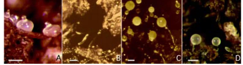
One of the advantages of Dictyostelium as an experimental system is that its entire 24-hours development can be easily followed under the microscope under a wide range of conditions. A spot of cells can undergo development on a surface of either agar or wetted filter paper, or on glass or plastic surfaces in a humidified chamber. Cells will grow and develop up to tight aggregates also in shaken liquid cultures (Beug et al., 1970); cell differentiation and sorting into pre-spore and prestalk regions occur in the aggregates, provided the flasks are oxygenated (Sternfeld and Bonner, 1977). Conditions closer to the natural ones, namely the rough-and-tumble world of forest soil, can be reproduced in the laboratory; Petri dishes containing commercially available garden soil have been introduced as an alternative substratum to study develop-ment under more stringent conditions, in order to emphasize subtle phenotypes (Ponte et al., 1998, and Fig. 5). Mutated genes which fail to reveal a defective phenotype on agar or filter paper, can thus be detected on soil plates. In particular, genes which affect cell-cell and cell-substratum adhesion or cell motility can be assumed to be particularly sensitive to environmental conditions. In this regard, aggregation is the most crucial developmental step, as cells have to move actively on the substratum in order to aggregate, and must be cohesive enough to overcome the shear forces deriving from cell-substratum adhesion. In addition, cAMP diffusion and chemo-tactic locomotion are three-dimensional on soil particles, while being two-dimensional on agar. Depending on the substratum, defects in cytoskeletal proteins or adhesion molecules may result in smaller aggregates or even in complete inhibition of aggregation. Once aggregates are formed, further development depends on the ability of slugs to migrate well on soil, where they have to bridge chasms between soil particles (Fig. 5), and on prestalk and prespore cell motility within the culminating slug, where high tension forces are generated (Dormann et al., 1996). Interactions with the substratum may affect slug migration and culmination. The availability of alternative developmental assays with different de-grees of stringency allows to detect graded phenotypic defects, and to define better to which extent a gene product is or not essential for development. As mentioned above, the adhesion glycoprotein csA has been shown to be essential when cells develop on soil, but dispensable when cells develop on agar (Ponte et al., 1998). Similar results have been recently obtained in single and double mutants defective in actin-binding proteins, such as α -actinin, gelation factor, 34-kDa actin bundling protein, synexin, and interaptin (Fig. 5D; Ponte et al., 2000). In some cases, a phenotypic defect not evident on agar emerged on soil, whereas in other cases a subtle phenotype barely detectable on agar was emphasized on soil.
These results support the notion that gene redundancy may result in many cases from the standard laboratory conditions not being as selective as the natural ones. By combining different developmental assays with single and double mutations it is also possible to establish a hierarchy between proteins belonging to a redundant network, i.e. sharing a similar activity or an overlapping function with other gene products. Thus, among the actin-crosslinking proteins, α-actinin seems to play a major role in the regulation of actin-based motility processes relevant to aggrega-tion and fruiting body formaaggrega-tion, followed in the order by gelaaggrega-tion factor and 34-kDa bundling protein.
Conclusions
Dictyostelium has emerged as an organism that is well suited to a molecular dissection of the different aspects of development, because of a unique combination of cell biological, biochemical and molecular genetic approaches that can be used in this system. Both the intracellular dynamics in living cells, or cell behavior within the multicellular organism can be easily followed by optical tools and digital image-processing. The introduction of green-fluores-cent protein technology has been exploited to monitor the shuttling of proteins in living cells in cytokinesis, phagocytosis, cell motility and chemotaxis. The small haploid genome (somewhere between yeast and Drosophila) and the high frequency of site specific recombinations make this organism ideal for gene disruption studies. Insertional mutagenesis has proven to be a powerful and effective tool for discovering new genes; saturation mutagenesis of existing mutants to generate genetic suppressors has recently been introduced (Loomis, 1996). Moreover, full scale sequencing of both cDNA libraries prepared from distinct developmental stages and genomic DNA is at an advanced stage and will open new opportunities for molecular genetic studies.
Acknowledgements
Results from the laboratory have been supported by funds of the European Community, Italian CNR and MURST.
References
ADESSI, C., CHAPEL, A., VINCON, M., RABILLOUD, T., KLEIN, G., SATRE, M. and GARIN, J. (1995). Identification of major proteins associated with Dictyostelium discoideum endocytic vesicles. J. Cell Sci. 108: 3331-3337.
AUBRY, L., KLEIN, G. and SATRE, M. (1997). Cytoskeletal dependence and modula-tion of endocytosis in Dictyostelium discoideum amoebae. In Dictyostelium - A model system for cell and developmental biology (Eds. Y. Maeda, K. Inouye, and I. Takeuchi). Universal Academy Press, Tokyo, pp. 65-74.
BEUG, H., GERISCH, G., KEMPFF, S., RIEDEL, V. and CREMER, G. (1970). Specific inhibition of cell contact formation in Dictyostelium by univalent antibodies. Exp. Cell Res. 63: 147-158.
BEUG, H., KATZ, F.E. and GERISCH, G. (1973). Dynamics in antigenic membrane sites relating to cell aggregation in Dictyostelium discoideum. J. Cell Biol. 56: 647-658.
BONNER, J.T. (1967). The cellular slime molds. Second edition. Princeton Univ. Press, Princeton, NJ.
BOZZARO, S. and PONTE, E. (1995). Cell adhesion in the life cycle of Dictyostelium. Experientia 51: 1175-1188.
BOZZARO, S. and MERKL, R. (1985). Monoclonal antibodies against Dictyostelium plasma membranes: their binding to simple sugars. Cell Differ. 17: 83-94.
BRACCO, E., PERACINO, B., NOEGEL, A.A. and BOZZARO, S. (1997). Cloning and transcriptional regulation of the gene encoding the vacuolar H+ ATPase B subunit
of Dictyostelium discoideum. FEBS Lett. 419: 37-40.
BRAR, S.K. and SIU, C.H. (1993). Characterization of the cell adhesion molecule gp24 in Dictyostelium discoideum - mediation of cell-cell adhesion via a Ca2+-dependent mechanism. J. Biol. Chem. 268: 24902-24909.
BUSSOLINO, F., SORDANO, F., BENFENATI, E. and BOZZARO, S. (1991). Dictyostelium cells produce platelet-activating factor in response to cAMP. Eur. J. Biochem. 196: 609-616.
CHEN, M.Y., LONG, Y. and DEVREOTES, P.N. (1997). A novel cytosolic regulator, pianissimo, is required for chemoattractant receptor and G protein-mediated activation of the 12 transmembrane domain adenylyl cyclase in Dictyostelium. Genes Devel. 11: 3218-3231.
CLARKE, M. and GOMER, R.H. (1995). PSF and CMF, autocrine factors that regulate gene expression during growth and early development of Dictyostelium. Experientia 51: 1124-1134.
COX, D., WESSELS, D., SOLL, D.R., HARTWIG, J. and CONDEELIS, J. (1996). Re-expression of ABP-120 rescues cytoskeletal, motility, and phagocytosis defects of ABP-120(-) Dictyostelium mutants. Mol. Biol. Cell 7: 803-823.
DARMON, M., BRACHET, P. and PEREIRA DA SILVA, L.H. (1975). Chemotactic signals induce cell differentiation in Dictyostelium discoideum. Proc. Natl. Acad. Sci. USA 72: 3163-3166.
DEMBINSKY, A., RUBIN, H. and RAVID, S. (1996). Chemoattractant-mediated increases in cGMP induce changes in Dictyostelium myosin II heavy chain-specific protein kinase C activities. J. Cell Biol. 134: 911-921.
DORMANN, D., SIEGERT, F. and WEIJER, C.J. (1996). Analysis of cell movement during the culmination phase of Dictyostelium development. Development 122: 761-769.
DYNES, J.L., CLARK, A.M., SHAULSKY, G., KUSPA, A., LOOMIS, W.F. and FIRTEL, R.A. (1994). LagC is required for cell interactions that are essential for cell-type differentiation in Dictyostelium. Genes Devel. 8: 948-958.
FAIX, J., GERISCH, G. and NOEGEL, A.A. (1992). Overexpression of the csA cell adhesion molecule under its own cAMP-regulated promoter impairs morphogen-esis in Dictyostelium. J. Cell Sci. 102: 203-214.
FISHER, P.R. (1997). Directional movement of migrating slugs. In Dictyostelium. A model system for Cell and Developmental Biology (Eds. Y. Maeda, K. Inouye, I. Takeuchi). Universal Acad. Press, Tokyo, pp. 437-452.
FURUKAWA, R. and FECHHEIMER, M. (1994). Differential localization of α-actinin and the 30 kDa actin-bundling protein in the cleavage furrow, phagocytic cup, and contractile vacuole of Dictyostelium discoideum. Cell Motil. Cytoskel. 29: 46-56.
GAMBINO, M., KAY, R.R. and BOZZARO, S. (1992). Morphogenesis and differentia-tion of Dictyostelium cells interacting with immobilized glucosides - dependence on DIF production. Differentiation 49: 133-141.
GAO, E.N., SHIER, P. and SIU, C.H. (1992). Purification and partial characterization of a cell adhesion molecule (gp150) involved in post-aggregation stage cell-cell binding in Dictyostelium discoideum. J. Biol. Chem. 267: 9409-9415.
GELTOSKY, J., WESEMAN, J., BAKKE, A. and LERNER, R. (1979). Identification of a cell surface glycoprotein involved in cell aggregation in Dictyostelium discoideum. Cell 18: 391-398.
GERISCH, G. (1987). Cyclic AMP and other signals controlling cell development and differentiation in Dictyostelium. Annu. Rev. Biochem. 56: 853-879.
GERISCH, G. and WICK, U. (1975). Intracellular oscillations and release of cyclic AMP from Dictyostelium cells. Biochem. Biophys. Res. Commun. 65: 364-370.
GERISCH, G. and WEBER, I. (2000). Cytokinesis without myosin II. Curr. Opin Cell Biol. 12: 126-132.
HADWIGER, J.A., LEE, S., and FIRTEL, R.A. (1994). The Gα subunit Gα 4 couples to pterin receptors and identifies a signaling pathway that is essential for multicel-lular development in Dictyostelium. Proc. Natl. Acad. Sci. USA 91: 10566-10570.
HARLOFF, C., GERISCH, G. and NOEGEL, A.A. (1989). Selective elimination of the contact site A protein of Dictyostelium discoideum by gene disruption. Genes Devel. 3: 2011-2019.
HARWOOD, A.J., HOPPER, N.A., SIMON, M.N., BOUZID, S., VERON, M. and WILLIAMS, J.G. (1992). Multiple roles for cAMP-dependent protein kinase during Dictyostelium development. Dev. Biol. 149: 90-99.
HARWOOD, A.J., PLYTE, S.E., WOODGETT, J., STRUTT, H. and KAY, R.R. (1995). Glycogen synthase kinase 3 regulates cell fate in Dictyostelium. Cell 80: 139-148.
HASSANAIN, H.H. and KOPACHIK, W. (1989). Regulatory signals affecting a selective loss of messenger RNA in Dictyostelium discoideum. J. Cell Sci. 94: 501-509.
HEUSER, J., ZHU, Q.L. and CLARKE, M. (1993). Proton pumps populate the contractile vacuoles of Dictyostelium amoebae. J. Cell Biol. 121: 1311-1327.
HOHMANN, H.-P., BOZZARO, S., MERKL, R., WALLRAFF, E., YOSHIDA, M., WEINHART, U. and GERISCH, G. (1987a). Post-translational glycosylation of the contact site A protein of Dictyostelium discoideum is important for stability but not for its function in cell adhesion. EMBO J. 6: 3663-3671.
HOHMANN, H.-P., BOZZARO, S., YOSHIDA, M., MERKL, R. and GERISCH, G. (1987b). Two-step glycosylation of the contact site A protein of Dictyostelium discoideum and transport of an incompletely glycosylated form to the cell surface. J. Biol. Chem. 262: 16618-15524.
INSALL, R. H., BORLEIS, J. and DEVREOTES, P. N. (1996). The aimless RasGEF is required for processing of chemotactic signals through G-protein-coupled receptors in Dictyostelium. Curr. Biol. 6: 719-729.
INSALL, R., KUSPA, A., LILLY, P.J., SHAULSKY, G., LEVIN, L.R., LOOMIS, W. F. and DEVREOTES, P. (1994). CRAC, a cytosolic protein containing a pleckstrin homology domain, is required for receptor and G protein-mediated activation of adenylyl cyclase in Dictyostelium. J. Cell Biol. 126: 1537-1545.
KAMBOJ, R.K., GARIEPY, J. and SIU, C.H. (1989). Identification of an octapeptide involved in homophilic interaction of the cell adhesion molecule gp80 of Dictyostelium discoideum. Cell 59: 615-625.
KAWATA, T., SHEVCHENKO, A., FUKUZAWA, M., JERMYN, K.A., TOTTY, N.F., ZHUKOVSKAYA, N.V., STERLING, A.E., MANN, M. and WILLIAMS, J. G. (1997). SH2 signaling in a lower eukaryote: A STAT protein that regulates stalk cell differentiation in Dictyostelium. Cell 89: 909-916.
KAY, R.R. (1997). DIF Signaling. In Dictyostelium - A model system for cell and developmental biology (Eds. Y. Maeda, K. Inouye and I. Takeuchi). Universal Academy Press, Tokyo, pp. 279-292.
KIM, H.J., CHANG, W.T., MEIMA, M., GROSS, J.D. and SCHAAP, P. (1998). A novel adenylyl cyclase detected in rapidly developing mutants of Dictyostelium. J. Biol. Chem. 273: 30859-30862.
KIMMEL, A.R. and CARLISLE, B. (1986). A gene expressed in undifferentiated vegetative Dictyostelium is repressed by developmental pulses of cAMP and reinduced during dedifferentiation. Proc. Natl. Acad. Sci. USA 83: 2506-2510.
KLEIN, P.S., SUN, T.J., SAXE III, C.L., KIMMEL, A.R., JOHNSON, R.L. and DEVREOTES, P.N. (1988). A chemoattractant receptor controls development in Dictyostelium discoideum. Science 241: 1467-1472.
KNECHT, D.A., FULLER, D.L. and LOOMIS, W.F. (1987). Surface glycoprotein, gp24, involved in early adhesion of Dictyostelium discoideum. Dev. Biol. 121: 277-283.
KONZOK, A., WEBER, I., SIMMETH, E., HACKER, U., MANIAK, M. and MÜLLER-TAUBENBERGER, A. (1999). DAip1, a Dictyostelium homologue of the yeast actin-interacting protein 1, is involved in endocytosis, cytokinesis and motility. J. Cell Biol. 146: 453-464.
KUMAGAI, A., PUPILLO, M., GUNDERSEN, R., MIAKE-LYE, R., DEVREOTES, P.N. and FIRTEL, R.A. (1989). Regulation and function of G-alpha protein subunits in Dictyostelium. Cell 57: 265-275.
KUSPA, A. and LOOMIS, W.F. (1992). Tagging developmental genes in Dictyostelium by restriction enzyme-mediated integration of plasmid DNA. Proc. Natl. Acad. Sci. USA 89: 8803-8807.
KUWAYAMA, H., ISHIDA, S. and VAN HAASTERT, P.J.M. (1993). Non-chemotactic Dictyostelium discoideum mutants with altered cGMP signal transduction. J. Cell Biol. 123: 1453-1462.
LAM, T.Y., PICKERING, G., GELTOSKY, J. and SIU, C.H. (1981). Differential cell cohesiveness expressed by prespore and prestalk cells of Dictyostelium discoideum. Differentiation 20: 22-28.
LIU, G. and NEWELL, P.C. (1988). Evidence that cyclic GMP regulates myosin interaction with the cytoskeleton during chemotaxis of Dictyostelium. J. Cell Sci. 90: 123-129.
LOOMIS, W.F. (1975). Dictyostelium discoideum. A developmental system. Aca-demic Press, New York.
LOOMIS, W.F., KUSPA, A. and SHAULSKY, G. (1994). Gene discovery in Dictyostelium. Genetic Engin. 16: 49-64.
LOOMIS, W.F. (1996). Genetic networks that regulate development in Dictyostelium cells Microbiol. Rev. 60: 135-150.
MAEDA, M., AUBRY, L., INSALL, R., GASKINS, C., DEVREOTES, P.N. and FIRTEL, R.A. (1996). Seven helix chemoattractant receptors transiently stimulate mitogen-activated protein kinase in Dictyostelium - Role of heterotrimeric G proteins. J. Biol. Chem. 271: 3351-3354.
MAEDA, Y. (1997). Cellular and molecular mechanisms of the transition from growth to differentiation in Dictyostelium cells. In Dictyostelium - A model system for cell and developmental biology (Eds. Y. Maeda, K. Inouye and I. Takeuchi). Universal Academy Press, Tokyo, pp. 207-218.
MANIAK, M., RAUCHENBERGER, R., ALBRECHT, R., MURPHY, J. and GERISCH, G. (1995). Coronin involved in phagocytosis: dynamics of particle-induced relocalization visualized by a green fluorescence protein tag. Cell 83: 915-924.
MANN, S.K.O. and FIRTEL, R.A. (1993). cAMP-dependent protein kinase differen-tially regulates prestalk and prespore differentiation during Dictyostelium develop-ment. Development 119: 135-146.
MANN, S.K. and FIRTEL, R.A. (1989). Two-phase regulatory pathway controls cAMP receptor-mediated expression of early genes in Dictyostelium. Proc. Natl. Acad. Sci. USA 86: 1924-1928.
MATSUNAGA, T. and MORI, N. (1987). The origin of the immune system. The possibility that immunoglobulin superfamily molecules and cell adhesion mol-ecules of chicken and slime mould are all related. Scand. J. Immunol. 25: 485-495.
MEHDY, M.C., RATNER, D. and FIRTEL, R.A. (1983). Induction and modulation of cell-type specific gene expression in Dictyostelium. Cell 32: 763-771.
MEIMA, M.E. and SCHAAP, P. (1999). Fingerprinting of adenylyl cyclase activities during Dictyostelium development indicates a dominant role for adenylyl cyclase B in terminal differentiation. Dev. Biol. 212: 182-190.
MÜLLER, K. and GERISCH, G. (1978). A specific glycoprotein as the target site of adhesion blocking Fab in aggregating Dictyostelium cells. Nature 274: 445-449.
NANJUNDIAH, V. (1997). Models for pattern formation in the Dictyostelid slime molds. In Dictyostelium - A model system for cell and developmental biology (Eds. Y. Maeda, K. Inouye and I. Takeuchi). Universal Academy Press, Tokyo, pp. 305-322.
NEWELL, P.C., MALCHOW, D. and GROSS, J.D. (1995). The role of calcium in aggregation and development of Dictyostelium. Experientia 51: 1155-1165.
NOEGEL, A.A. and LUNA, J.E. (1995). The Dictyostelium cytoskeleton. Experientia 51: 1135-1143.
NOEGEL, A.A., GERISCH, G., STADLER, J. and WESTPHAL, M. (1986). Complete sequence and transcript regulation of a cell adhesion protein from aggregating Dictyostelium cells. EMBO J. 5: 1473-1476.
PARENT, C.A. and DEVREOTES, P.N. (1996). Molecular genetics of signal transduc-tion in Dictyostelium. Annu. Rev. Biochem. 65: 411-440.
PARENT, C.A. and DEVREOTES, P.N. (1999). A cell’s sense of direction. Science 284: 765-770.
PERACINO, B., BORLEIS, J., JIN, T., WESTPHAL, M., SCHWARTZ, J.M., WU, L.J., BRACCO, E., GERISCH, G., DEVREOTES, P. and BOZZARO, S. (1998). G protein beta subunit-null mutants are impaired in phagocytosis and chemotaxis due to inappropriate regulation of the actin cytoskeleton. J. Cell Biol. 141: 1529-1537.
PITT, G.S., MILONA, N., BORLEIS, J., LIN, K.C., REED, R.R. and DEVREOTES, P.N. (1992). Structurally distinct and stage-specific adenylyl cyclase genes play different roles in Dictyostelium development. Cell 69: 305-315.
PONTE, E., BRACCO, E., FAIX, J. and BOZZARO, S. (1998). Detection of subtle phenotypes: The case of the cell adhesion molecule csA in Dictyostelium. Proc. Natl. Acad. Sci. USA 95: 9360-9365.
PONTE, E., RIVERO, F., FECHHEIMER, M., NOEGEL, A.A. and BOZZARO, S. (2000). Severe developmental defects in Dictyostelium null mutants for actin binding proteins. Mech. Devel. 91: 153-161.
PUPILLO, M., KUMAGAI, A., PITT, G.S., FIRTEL, R.A. and DEVREOTES, P.N. (1989). Multiple alpha subunits of guanine nucleotide-binding proteins in Dictyostelium. Proc. Natl. Acad. Sci. USA 86: 4892-4896.
REYMOND, C.D., SCHAAP, P., VERON, M. and WILLIAMS, J.G. (1995). Dual role of cAMP during Dictyostelium development. Experientia 51: 1166-1174.
RIVERO, F., ALBRECHT, R., DISLICH, H., BRACCO, E., GRACIOTTI, L., BOZZARO, S. and NOEGEL, A.A. (1999). RacF1, a novel member of the Rho protein family in Dictyostelium discoideum, associates transiently with cell contact areas, macropinosomes and phagosomes. Mol. Biol. Cell 10: 1205-1219.
RIVERO, F., FURUKAWA, R., NOEGEL, A.A. and FECHHEIMER, M. (1996a). Dictyostelium discoideum cells lacking the 34,000-Dalton actin-binding protein can grow, locomote and develop, but exhibit defects in regulation of cell structure and movement: A case of partial redundancy. J. Cell Biol. 135: 965-980.
RIVERO, F., KOEPPEL, B., PERACINO, B., BOZZARO, S., SIEGERT, F., WEIJER, C.J., SCHLEICHER, M., ALBRECHT, R. and NOEGEL, A.A. (1996b). The role of the cortical cytoskeleton: F-actin crosslinking proteins protect against osmotic stress, ensure cell size, cell shape and motility and contribute to phagocytosis and development. J. Cell Sci. 109: 2679-2691.
ROGERS, K.C., GINSBURG, G.T., MU, X., GOLLOP, R., BALINT-KURTI, P., LOUIS, J.M. and KIMMEL, A.R. (1997). The cAMP receptor gene family of Dictyostelium discoideum: expression, regulation and function. In Dictyostelium - A model system for cell and developmental biology (Eds. Y. Maeda, K. Inouye and I. Takeuchi). Universal Academy Press, Tokyo, pp. 163-172.
ROOS, W., SCHEIDEGGER, C. and GERISCH, G. (1977). Adenylate cyclase activity oscillations as signals for cell aggregation in Dictyostelium discoideum. Nature 266: 259-261.
SCHALOSKE, R., SORDANO, C., BOZZARO, S. and MALCHOW, D. (1995). Stimulation of calcium influx by platelet activating factor in Dictyostelium. J. Cell Sci. 108: 1597-1603.
SEASTONE, D.J., LEE, E., BUSH, J., KNECHT, D. and CARDELLI, J. (1998). Overexpression of a novel Rho family GTPase, RacC, induces unusual actin-based structures and positively affects phagocytosis in Dictyostelium discoideum. Mol. Biol. Cell 9: 2891-2904.
SEGALL, J.E., KUSPA, A., SHAULSKY, G., ECKE, M., MAEDA, M., GASKINS, C. and FIRTEL, R.A. (1995). A MAP kinase necessary for receptor-mediated activation of adenylyl cyclase in Dictyostelium. J. Cell Biol. 128: 405-413.
SIEGERT, F. and WEIJER, C.J. (1995). Spiral and concentric waves organize multicellular Dictyostelium mounds. Curr. Biol. 5: 937-943.
SIU, C.-H., HARRIS, T.J.C., WONG, E.F.S., YUNG, C., SESAKI, H. and WANG, J. (1977). Cell adhesion molecules in Dictyostelium. In Dictyostelium - A model system for cell and developmental biology (Eds. Y. Maeda, K. Inouye and I. Takeuchi). Universal Academy Press, Tokyo, pp. 111-144.
SNAAR-JAGALSKA, B.E. and VAN HAASTERT, P.J.M. (1990). Pertussis toxin inhibits cAMP-induced desensitization of adenylate cyclase in Dictyostelium discoideum. Mol. Cell. Biochem. 92: 177-189.
SORDANO, C., CRISTINO, E., BUSSOLINO, F., WURSTER, B. and BOZZARO, S. (1993). Platelet activating factor modulates signal transduction in Dictyostelium. J. Cell Sci. 104: 197-202.
SOUZA, G.M., HIRAI, J., MEHTA, D.P. and FREEZE, H.H. (1995). Identification of two novel Dictyostelium discoideum cysteine proteinases that carry N-acetylglucosamine-1-P modification. J. Biol. Chem. 270: 28938-28945.
STADLER, J., KEENAN, T.W., BAUER, G. and GERISCH, G. (1989). The contact site A glycoprotein of Dictyostelium discoideum carries a phospholipid anchor of a novel type. EMBO J. 8: 371-377.
STERNFELD, J. and BONNER, J.T. (1977). Cell differentiation in Dictyostelium under submerged conditions. Proc. Natl. Acad. Sci. USA 74: 268-271.
SUN, T.J. and DEVREOTES, P.N. (1991). Gene targeting of the aggregation stage cAMP receptor cAR1 in Dictyostelium. Genes Devel. 5: 572-582.
THEIBERT, A. and DEVREOTES, P. (1986). Surface receptor-mediated activation of adenylate cyclase in Dictyostelium. Regulation by guanine nucleotides in wild-type cells and aggregation deficient mutants. J. Biol. Chem. 261: 15121-15125.
TITUS, M.A., NOVAK, K.D., HANES, G.P. and URIOSTE, A.S. (1995). Molecular genetic analysis of myoF, a new Dictyostelium myosin I gene. Biophys. J. 68: S152-S157.
VAN HAASTERT, P.J.M. (1995). Transduction of the chemotactic cAMP signal across the plasma membrane of Dictyostelium cells. Experientia 51: 1144-1154.
WILLIAMS, J. (1997). Prestalk and stalk cell heterogeneity in Dictyostelium. In Dictyostelium - A model system for cell and developmental biology (Eds. Y. Maeda, K. Inouye and I. Takeuchi). Universal Academy Press, Tokyo, pp. 293-304.
WONG, E.F.S., BRAR, S.K., SESAKI, H., YANG, C.Z. and SIU, C.H. (1996). Molecular cloning and characterization of DdCAD-1, a Ca2+-dependent cell-cell adhesion
molecule, in Dictyostelium discoideum. J. Biol. Chem. 271: 16399-16408.
WU, L.J. and DEVREOTES, P.N. (1991). Dictyostelium transiently expresses eight distinct G-protein alpha-subunits during its developmental program. Biochem. Biophys. Res. Commun. 179: 1141-1147.