IGF-I, IGF-II and insulin promote differentiation of
spermatogonia to primary spermatocytes
in organ culture of newt testes
YUKI NAKAYAMA
1, TAKASHI YAMAMOTO
2and SHIN-ICHI ABÉ
1*
1Department of Materials and Life Science, Graduate School of Science and Technology and 2Department of Biological Science,
Faculty of Science, Kumamoto University, Kumamoto, Japan
ABSTRACT Recombinant human insulin-like growth factors (rhIGF-I and rhIGF-II) and human insulin promoted the differentiation of spermatogonia into primary spermatocytes in newt testes fragments cultured in a chemically defined medium. The biological potency for promoting differentiation was dose-dependent for all the ligands with the highest potency displayed by IGF-I, followed by IGF-IIGF-I, and the least by insulin. The difference in potency was larger between IGF-II and insulin than that between IGF-I and IGF-II. This order of biological potency was in good accordance with the order of affinity in binding specificity of [125I]I to the testicular membrane fractions:
IGF-II and insulin competed the binding of [125I]IGF-I only at concentrations 20-fold and 100-fold higher,
respectively, than IGF-I. Specific binding was observed in both somatic cells (mostly Sertoli cells) and germ cells (spermatogonia and primary spermatocytes), though the binding to somatic cells was about 2.7 times higher than that to germ cells. These results indicate that (1) specific binding sites for IGF-I are present in the newt testes, (2) IGF-II and insulin also bind to these receptors but to a lesser degree, and (3) IGF-II and insulin as well as IGF-I promote spermatogonial differentiation into primary spermatocytes by binding to the IGF-I receptor.
KEY WORDS: IGFs, insulin, meiosis initiation, newt testes, organ culture
0214-6282/99/$10.00
© UBC Press Printed in Spain http://www.lg.ehu.es/ijdb
*Address for reprints: Department of Materials and Life Science, Graduate School of Science and Technology, Kumamoto University, Kurokami 2-39-1, Kumamoto 860-8555, Japan. FAX: 81-96-342-3437. e-mail:abeshin@gpo.kumamoto-u.ac.jp
Introduction
Spermatogenesis commences with the proliferation of sper-matogonia which enter meiosis and differentiate into primary spermatocytes. The number of mitoses that spermatogonia un-dergo before entering meiosis is species-specific (Roosen Runge, 1977), but the exact mechanisms controlling this proliferation and entrance into meiosis requires elucidation. It is known that germ cell proliferation and differentiation are controlled mainly by Sertoli cells which remain closely attached to germ cells throughout spermatogenesis (Parvinen et al., 1986; Griswold et al., 1988; Skinner, 1991; Jégou, 1993). It is believed that Sertoli cells are stimulated by follicle-stimulating hormone (FSH) and androgens to produce paracrine or autocrine factors which in turn induce the proliferation and differentiation of germ cells (Bellvé and Zheng, 1989; Skinner, 1991; Jégou, 1993).
The subject of our current investigation focuses on insulin-like growth factor I (IGF-I), a factor thought to play autocrine and paracrine roles during spermatogenesis. On one hand, IGF-I is produced by testicular cells (Ritzén, 1983) and was purified from
Original Article
Abbreviations used in this paper: FSH, follicle-stimulating hormone; SM-C,
basal and hCG-supported cAMP and testosterone production in cultured Leydig cells (Bernier et al., 1986; Lin et al., 1986a,b; Chatelain et al., 1987; Kasson and Hsueh, 1987; Smith et al., 1987).
With respect to the action of IGF-I on germ cells, Söder et al. (1992) reported first in mammals that IGFs stimulate premitotic DNA synthesis and may maintain premeiotic DNA synthesis in cultured segments of rat seminiferous tubules. In fishes, rhIGF-I increases DNA synthesis in cultures of premeiotic dogfish spermatocysts (Dubois and Callard, 1993). Furthermore, IGF-I stimulated DNA synthesis in trout spermatogonia and primary spermatocytes in a dose-dependent manner, indicating that IGF-I acts directly as a stimulator of proliferation (Loir, 1994). In fact, rhIGF-I was shown to bind specifically to rainbow trout spermatogonia and primary sper-matocytes (Loir and Le Gac, 1994), and IGF-I mRNA and receptors are present to a greater extent in Sertoli cell-enriched populations and in spermatogonia with primary spermatocytes (Le Gac et al., 1996). Thus, the above studies implicate IGF-I as an important factor in governing the proliferation of male germ cells and support the view that Sertoli cells have both an autocrine and paracrine role as both targets and sources of growth factors.
The structure of mammalian testes is quite complex and con-sists of seminiferous tubules, each containing many generations of spermatogenic cells residing with Sertoli cells; this organization involves complex interactions among testicular cells in various stages (Callard, 1991). In contrast to mammalian testes, the structure of testes in fish and urodeles is quite simple, and consists of spermatocysts each comprising a clone of germ cells and a stage-synchronized clone of Sertoli cells (Callard, 1991). Further-more, spermatogenesis progresses unidirectionally from the cepha-lic to the caudal region of fish and urodele testes, permitting the
investigator to analyze individual zones, each containing germ cells in the same stage.Using this advantageous organiza-tion in urodeles, we previously showed in cultures of newt testes fragments that mammalian FSH stimulates spermatogo-nial proliferation and their differentiation into primary spermatocytes (Ji et al., 1992; Abé and Ji, 1994).Furthermore, we dem-onstrated that this proliferation and differ-entiation is mediated by Sertoli cells (Maekawa et al., 1995; Ito and Abé, 1999), suggesting that paracrine factors produced by Sertoli cells stimulate this proliferation and differentiation.Here we report that human recombinant IGF-I, IGF-II and in-sulin promote differentiation of newt sper-matogonia into primary spermatocytes in cultures of testes fragments.
Results
Differentiation of secondary sper-matogonia into primary spermatocytes in organ culture of testis fragments by IGF-I, IGF-II an insulin.
Testes fragments containing germ cells in the spermatogonial stage (Fig. 1A) were cultured in the absence or presence of Fig. 1. Photomicrographs of histological sections from testis fragments on 0 day (A) and after
2 weeks of culture (B-D).(A) Most of the cysts contained secondary spermatogonia. (B) In control medium, most of the germ cells remained as secondary spermatogonia. (C) In medium containing 100 ng/ml IGF-I. (D) In medium containing 100 ng/ml IGF-II. In (C) and (D), most of the germ cells are primary spermatocytes in pachytene stage. Arrows and arrowheads show pericystic cells and Sertoli cells, respectively. Bar, 50 µm.
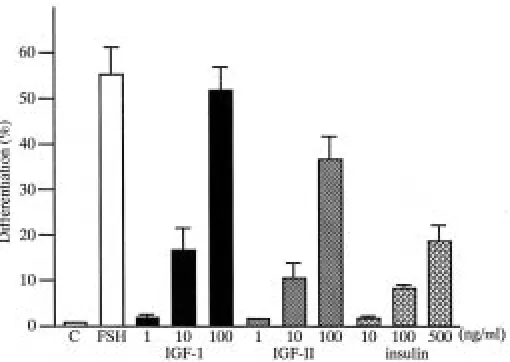
IGF-I, IGF-II or insulin. After 2 weeks most of the germ cells remained as spermatogonia in control medium (Fig. 1B). On the other hand, in the presence of IGF-I (100 ng/ml) spermatogonia differentiated into primary spermatocytes in the pachytene stage (Fig. 1C) in more than 50% of the cysts (Fig. 2). IGF-II (100 ng/ml; Fig. 1D) as well as insulin (500 ng/ml) also promoted the differen-tiation of spermatogonia into primary spermatocytes. The effects of IGF-I, IGF-II and insulin were dose-dependent (Fig. 2). The extent of differentiation with 100 ng/ml IGF-I was almost comparable to that with FSH (5 µg/ml); the most advanced stage attained in both cases was the pachytene stage. The percentage of cysts that differentiated into primary spermatocytes was the highest in 100 ng/ml IGF-I, intermediate in 100 ng/ml IGF-II and the lowest in 500 ng/ml insulin. Even 100 ng/ml insulin promoted less differentiation than 10 ng/ml IGF-I, and the value attained with 500 ng/ml insulin was almost comparable to that with 10 ng/ml IGF-I. These results indicate that IGF-I is more potent than IGF-II in promoting the differentiation of spermatogonia into primary spermatocytes, whereas insulin is the least potent, i.e. approximately 9 (6.5–11.6) and 6 (4.6–7.5) fold less than IGF-I and IGF-II, respectively.
Binding of [125I]IGF-I to testicular cells
The binding of [125I]IGF-I to membrane fractions prepared from
the testes in the spermatogonial stage was examined. Unlabeled competitors with increasing concentrations inhibited [125I]IGF-I
and insulin exert their effects on spermatogonia by binding to the IGF-I receptor.
IGF-I binding was also determined for preparations of isolated germ (spermatogonia and primary spermatocytes) and somatic cells (mostly Sertoli cells). As shown in Figure 3B, specific binding was observed not only in somatic cells but also in germ cells, though the specific binding for somatic cells was about 2.7 times higher than that for germ cells. The purity of germ cells (Gm) was 98% (spermatogonia, 62%; primary spermatocytes, 36%; somatic cells, 2%), as was the purity of somatic cells (Sm) (Sertoli cells, 71%; pericystic cells, 27%; primary spermatocytes, 2%). These results indicate that both germ and somatic cells (mainly Sertoli cells) contained binding sites for IGF-I.
Discussion
Our current study demonstrates for the first time in vertebrates that human IGF-I, IGF-II and insulin promote the differentiation of spermatogonia into primary spermatocytes in cultures of newt testes fragments. The biological potency for promoting differentia-tion was the highest for IGF-I, followed by IGF-II, and the least for insulin. The difference of potency between IGF-II and insulin is larger than that between IGF-I and IGF-II. This order of biological potency accords with the order of affinity in binding specificity of [125I]IGF-I to the testicular membrane fractions. The observed
affinity order in competition binding assays (IGF-I > IGF-II >> insulin) is similar to that described for the IGF-I receptor in mam-mals (Sara and Hall, 1990; Francis et al., 1993), Xenopus (Janicot et al., 1991) and rainbow trout (Loir and Le Gac, 1994; Le Gac et al., 1996).
With respect to receptors, IGF-IR, IGF-IIR and insulin receptor have been characterized in mammals; IGF-IR is structurally ho-mologous to the insulin receptor but binds insulin with low affinity (Chernausek et al., 1981; Massague and Czech, 1982; Ullrich et al., 1986). The biological activity of IGFs is mediated largely through their interaction with the IGF-IR (Van Wyk and Casella, 1991; Soos et al., 1992). In the case of Xenopus laevis oocytes, IGF-IR mediates the effects of insulin, IGF-I and IGF-II, even
though IGF-IIR is also present (Janicot et al., 1991). Our present data on newt testes indicate that specific binding sites for IGF-I are present, and that IGF-II and insulin promote spermatogonial differ-entiation to primary spermatocytes through the IGF-I receptor. However, since the effect of IGF-II on spermatogonial differentia-tion is comparable to that of IGF-I, IGF-II may act through both the IGF-I and IGF-II receptors. This possibility can be evaluated by developing antibodies against the newt IGF-I and IGF-II receptors, and examining the effect of IGF-I, IGF-II and insulin on sper-matogonial differentiation in the presence of their antibodies.
Previous studies of testes demonstrated the presence of IGF-IR on both somatic and germ cells, such as mammalian Leydig (Handelsman et al., 1985; Lin et al., 1986a,b; Kasson and Hsueh, 1987; Saez et al., 1988) and Sertoli cells (Borland et al., 1984; Oonk and Grootegoed, 1988), and trout Sertoli cells, spermatogo-nia and primary spermatocytes (Le Gac et al., 1996). In addition, IGF-I binding was observed on rat pachytene spermatocytes (Tres et al., 1986), and IGF-IR immunoreactivity was localized on human secondary spermatocytes and early spermatids (Vannelli et al., 1988). Similarly, our current studies detected [125I]IGF-I binding to
both spermatogonia-rich and Sertoli-rich fractions. These results pose several interpretations regarding the mechanism of IGF-I action on spermatogonial differentiation. First, IGF-I directly stimu-lates spermatogonial proliferation and/or differentiation into pri-mary spermatocytes. Indeed, Loir (1994) showed initially in verte-brates that IGF-I directly stimulates the proliferation of male germ cells, but the differentiation of spermatogonia into primary sperma-tocytes was not detected as the spermatogonia were cultured only for 3 days. Second, IGF-I stimulates the proliferation of Sertoli cells. Third, IGF-I activates Sertoli cells which in turn produce factors that stimulate spermatogonial proliferation and/or differen-tiation into primary spermatocytes. Finally, a fourth possibility is that IGF-I directly stimulates spermatogonial proliferation, whereas Sertoli cells, activated by IGF-I, secrete factor(s) required for the differentiation of spermatogonia into primary spermatocytes.
Recently we established reconstituted cultures composed of newt spermatogonia and somatic cells (mainly Sertoli cells) in which FSH stimulates germ cell proliferation and differentiation into primary spermatocytes (Ito and Abé, 1999). Comparative studies involving separate cultures of germ and somatic cells with recon-stituted cultures might reveal the role of IGF-I in the proliferation and/or differentiation of spermatogonia, the cell type expressing IGF-I, and the hormone(s) regulating the expression of IGF-I.
Materials and Methods
Animals and reagents
Adult male newts (Cynops pyrrhogaster) were purchased from a dealer (Hamamatsu Seibutsu Kyozai Ltd., Hamamatsu, Japan), kept at 22°C under 12L: 12D illumination, and fed frozen Tubifex. All chemicals were obtained from Nacalai Tesque, Inc., Kyoto, Japan, unless otherwise stated.
Organ culture of testicular fragments
All operations were carried out in sterile conditions. Testis fragments were cultured as previously described (Ji et al., 1992). In brief, the immature part of each testis that was in the spermatogonial stage was cut into 1-2 mm diameter fragments. Four fragments were placed on a nucleopore filter (pore size 0.2 µm; diameter, 25 mm; Corning, Acton, MA, USA) which was floated on culture medium in a 35 mm dish. Porcine follicle-stimulating hormone (FSH; Sigma, St. Louis, MO, USA), recombinant human insulin-like growth factor-I (rhIGF-I, Genzyme, Cambridge, MA, USA), insulin-insulin-like Fig. 2. Effects of FSH, IGF-I, IGF-II and insulin on the percentage of primary
growth factor-II (rhIGF-II, Genzyme) and insulin (Becton Dickinson, Franklin Lakes, NJ, USA) were added to the culture medium (Leibovitz's L-15) at various concentrations. The dishes were stored in a dark incubator (22°C) and the culture medium was changed once a week.
Histology and quantitative analysis of differentiation
Four fragments from each culture dish were fixed together with the filter in Bouin’s solution after 2 weeks of culture. The samples were dehydrated in an ethanol series and embedded in a block of paraffin (paraplast plus tissue embedding medium, Oxford Labware, St. Louis, MO, USA). The blocks were sectioned serially (5 µm) and stained according to the Delafield hematoxylin-eosin method.
To estimate the extent of differentiation occurring after 2 weeks of culture, one section in the central area of each fragment was photographed. The cysts within the fragments were classified into two groups; cysts consisting of spermatogonia and those consisting of primary spermato-cytes. The percentage of each group was averaged for all fragments (4 fragments/dish x 2 dishes) examined, and expressed as the extent of differentiation. Triplicate experiments were performed.
Separation of testicular cells
The spermatogenic cells were separated from the somatic cells (mostly Sertoli cells) according to Maekawa et al. (1995) with some modifications. Testis fragments rich in secondary spermatogonia and primary spermato-cytes were collected and minced in L-15 medium followed by treatment with 0.1% collagenase (type N-2, Nitta Zeratin Co., Tokyo) at 22°C for 2 h. The cell suspension (5 ml) was layered onto 5% Nycodenz (Nycomed Pharma As, Oslo, Norway) in OR-2 (10 ml); this had been underlayered with 10% (10 ml) which in turn was underlayered with 15% Nycodenz (5 ml). The suspension was centrifuged at 1,500g for 10 min at room temperature. Spermatogenic cells were recovered from the boundary between 10% and 15%, and somatic cells from the boundary between L-15 and 5%.
IGF-I binding study
Preparation of cellular membranes was performed according to Oonk and Grootegoed (1988). All steps in the preparation were carried out at 0-4°C. Newt testes containing both spermatogonia and primary spermatocytes were homogenized in 5 volumes of ice-cold 0.3M sucrose containing 25 mM Tris-HCl (pH 7.6) with 5 up-and-down strokes of a mechanically driven Teflon/glass homogenizer. The homogenate was centrifuged at 900g for 10
min to eliminate nuclei and tissue fragments. The resulting supernatant was centrifuged at 100,000g for 60 min, and the pellet was resuspended in 25 mM Tris-HCl (pH 7.6) buffer containing 0.12M NaCl, 2.5 mM KCl and 6 mM MgSO4, to a protein concentration of 4-10 mg/ml.
Two hundred micrograms of the membrane preparation (200 µl) were incubated for 16 h at 4°C in the presence of 35,000 cpm of [125I] human
IGF-I (NEN, Boston, MA, USA) and various concentrations of unlabeled rhIGF-I (Genzyme), rhIGF-II (Genzyme) or human insulin (Becton Dickinson). The reaction was stopped by the addition of 1 ml of 25 mM Tris-HCl (pH 7.6) buffer containing 0.12M NaCl, 2.5 mM KCl, 6 mM MgSO4 and 0.5% BSA; the tubes were centrifuged at 15,000g for 20 min.
The supernatant was aspirated and the pellet was washed once with ice-cold buffer. The radioactivity of the pellet was counted using an automatic gamma counter (Wallac, Turk, Finland). Nonspecific binding was deter-mined by the addition of a 2000-fold excess of unlabeled human IGF-I to the reaction mixture.
Acknowledgments
We thank Professor Marie A. DiBerardino for critical reading and editing the manuscript. This work was supported by Grants-in-Aid for Scientific Research (no.09480206) and Priority Areas (no.07283104) from the Min-istry of Education, Science, Sports and Culture of Japan, and by Special Coordination Funds for Promoting Science and Technology.
References
ABÉ, S.-I. and JI, Z.-S. (1994). Initiation and stimulation of spermatogenesis in vitro by mammalian follicle-stimulating hormone in the Japanese newt, Cynops pyrrhogaster. Int. J. Dev. Biol. 38: 201-208.
BELLVÉ, A.R. and ZHENG, W. (1989). Growth factors as autocrine and paracrine modulators of male gonadal functions. J. Reprod. Fertil. 85: 771-793.
BERNIER, M., CHATELAIN, P., MATHER, J.P. and SAEZ, J.M. (1986). Regulation of gonadotropin receptors, gonadotropin responsiveness, and cell multiplication by somatomedin-C and insulin in cultured pig Leydig cells. J. Cell. Physiol. 129: 257-263.
BORLAND, K., MITA, M., OPPENHEIMER, C.L., BLINDERMAN, L.A., MASSAGUE, J., HALL, P.F. and CZECH, M.P. (1984). The actions of insulin-like growth factors I and II on cultured Sertoli cells. Endocrinology 114: 240-246.
CAILLEAU, J., VERMEIRE, S. and VERHOEVEN, G. (1990). Independent control of the production of insulin-like growth factor I and its binding protein by cultured testicular cells. Mol. Cell. Endocrinol. 69: 79-89.
CALLARD, G.V. (1991). Spermatogenesis. In Vertebrate Endocrinology: fundamen-tals and biomedical implications (Pang, P.K.T. and Schreibman, M.T., eds.), Academic Press, New York, vol.4, part A, pp. 303-341.
CHATELAIN, P.G., NAVILLE, D. and SAEZ, J.M. (1987). Somatomedin-C/insulin-like growth factor 1-like material secreted by porcine Sertoli cells in vitro: characteriza-tion and regulacharacteriza-tion. Biochem. Biophys. Res. Commun 146: 1009-1017.
CHERNAUSEK, S.D., JACOBS, S. and VAN WYK, J.J. (1981). Structural similarities between human receptors for somatomedin C and insulin: analysis by affinity labeling. Biochemistry 20: 7345-7350.
DUBOIS, W. and CALLARD, G.V. (1993). Culture of intact Sertoli/germ cell units and isolated Sertoli cells from Squalus testis. II. Stimulatory effects of insulin and IGF-I on DNA synthesis in premeiotic stages. J. Exp. Zool. 267: 233-244.
FRANCIS, G.L., APLIN, S.E., MILNER, S.J., MCNEIL, K.A., BALLARD, F.J. and WALLACE, J.C. (1993). Insulin-like growth factor (IGF)-II binding to IGF-binding proteins and IGF receptors is modified by deletion of the N-terminal hexapeptide or substitution of arginine for glutamate-6 in IGF-II. Biochem. J. 293: 713-719.
GRISWOLD, M.D., MORALES, C. and SYLVESTER, S.R. (1988). Molecular biology of the Sertoli cell. In Oxford reviews of reproductive biology. (Ed. J.R. Clarke). Vol. 10. Oxford University Press, Oxford, pp. 125-161.
HANDELSMAN, D.J., SPALIVIERO, J.A., SCOTT, C.D. and BAXTER, R.C. (1985). Identification of insulin-like growth factor-I and its receptors in the rat testis. Acta Endocrinol. 109: 543-549.
ITO, R. and ABÉ, S.-I. (1999). FSH-initiated differentiation of newt spermatogonia to primary spermatocytes in germ-somatic cell reaggregates cultured within a collagen matrix. Int. J. Dev. Biol. 43: 111-116.
Fig. 3. IGF-I binding to testicular membrane (A) and fractionated populations of testicular cells (B). (A) Competitive binding assay be-tween [125I]IGF-I and unlabeled IGF-I (●-●), IGF-II (■-■) and insulin (▲-▲)
for binding to testicular membrane fraction. (B) Binding of [125I]IGF-I to
JAILLARD, C., CHATELAIN, P.G. and SAEZ, J.M. (1987). In vitro regulation of pig Sertoli cell growth and function: effects of fibroblast growth factor and somatome-din-C. Biol. Reprod. 37: 665-674.
JANICOT, M., FLORES-RIVEROS, J.R. and LANE, D.M. (1991). The insulin-like growth factor 1 (IGF-1) receptor is responsible for mediating the effects of insulin, IGF-1, and IGF-2 in Xenopus laevis oocytes. J. Biol. Chem. 266: 9382-9391.
JÉGOU, B. (1993). The Sertoli-germ cell communication network in mammals. Int. Rev. Cytol. 147: 25-96.
JI, Z.-S., KUBOKAWA, K., ISHII, S. and ABÉ, S.-I. (1992). Differentiation of secondary spermatogonia to primary spermatocytes by mammalian follicle stimulating hor-mone in organ culture of testes fragments from the newt, Cynops pyrrhogaster. Dev. Growth Differ. 34: 649-660.
KASSON, B.G. and HSUEH, A.J. (1987). Insulin-like growth factor-I augments gonadotropin-stimulated androgen biosynthesis by cultured rat testicular cells. Mol. Cell. Endocrinol. 52: 27-34.
LE GAC, F., LOIR, M., LE BAIL, P.-Y. and OLLITRAULT, M. (1996). Insulin-like growth factor (IGF-I) mRNA and IGF-I receptor in trout testis and in isolated spermatogenic and Sertoli cells. Mol. Reprod. Dev. 44: 23-35.
LIN, T., HASKELL, J., VINSON, N. and TERRACIO, L. (1986a). Direct stimulatory effects of insulin-like growth factor-I on Leydig cell steroidogenesis in primary culture. Biochem. Biophys. Res. Commun. 137: 950-956.
LIN, T., HASKELL, J., VINSON, N. and TERRACIO, L. (1986b). Characterization of insulin and insulin-like growth factor I receptors of purified Leydig cells and their role in steroidogenesis in primary culture: a comparative study. Endocrinology 119: 1641-1647.
LOIR, M. (1994). In vitro approach to the control of spermatogonia proliferation in the trout. Mol. Cell. Endocrinol. 102: 141-150.
LOIR, M. and LE GAC, F. (1994). Insulin-like growth factor-I and -II binding and action on DNA synthesis in rainbow trout spermatogonia and spermatocytes. Biol. Reprod. 51: 1154-1163.
MAEKAWA, K., JI, Z.-S. and ABÉ, S.-I. (1995). Proliferation of newt spermatogonia by mammalian FSH via Sertoli cells in vitro. J. Exp. Zool. 272: 363-373.
MASSAGUE, J. and CZECH, M.P. (1982). The subunit structures of two distinct receptors for insulin-like growth factors I and II and their relationship to the insulin receptor. J. Biol. Chem. 257: 5038-5045.
MITA, M., BORLAND, K., PRICE, J.M. and HALL, P.F. (1985). The influence of insulin and insulin-like growth factor-I on hexose transport by Sertoli cells. Endocrinology 116: 987-992.
OONK, R.B. and GROOTEGOED, J.A. (1988). Insulin-like growth factor I (IGF-I) receptors on Sertoli cells from immature rats and age-dependent testicular binding of IGF-I and insulin. Mol. Cell. Endocrinol. 55: 33-43.
PARVINEN, M., VIHKO, K.K. and TOPPARI, J. (1986). Cell interactions during the seminiferous epithelial cycle. Int. Rev. Cytol. 104: 115-151.
RITZÉN, E.M. (1983). Chemical messengers between Sertoli cells and neighbouring cells. J. Steroid Biochem. 19: 499-504.
ROOSEN-RUNGE, E.C. (1977). The Process of Spermatogenesis in Animals. Cambridge University Press, London.
SAEZ, J.M., CHATELAIN, P.G., PERRARD-SAPORI, M.-H., JAILLARD, C. and NAVILLE, D. (1988). Differentiating effects of somatomedin-C/insulin-like growth factor I and insulin on Leydig and Sertoli cell functions. Reprod. Nutr. Dev. 28: 989-1008.
SARA, V.R. and HALL, K. (1990). Insulin-like growth factors and their binding proteins. Physiol. Rev. 70: 591-641.
SKINNER, M.K. (1991). Cell-cell interactions in the testis. Endocr. Rev. 12: 45-77.
SKINNER, M.K. and FRITZ, I.B. (1986). Identification of a non-mitogenic paracrine factor involved in mesenchymal-epithelial cell interactions between testicular peritubular cells and Sertoli cells. Mol. Cell. Endocrinol. 44: 85-97.
SMITH, E.P., SVOBODA, M.E., VAN WYK, J.J., KIERSZENBAUM, A.L. and TRES, L.L. (1987). Partial characterization of a somatomedin-like peptide from the medium of cultured rat Sertoli cells. Endocrinology 120: 186-193.
SÖDER, L., BANG, P., WAHAB, A. and PARVINEN, M. (1992). Insulin-like growth factors selectively stimulate spermatogonial, but not meiotic, deoxyribonucleic acid synthesis during rat spermatogenesis. Endocrinology 131: 2344-2350.
SOOS, M.A., FIELD, C.E., LAMMERS, R., ULLRICH, A., ZHANG, B., ROTH, R.A., ANDERSEN, A.S., KJELDSEN, T. and SIDDLE, K. (1992). A panel of monoclonal antibodies for type I insulin-like growth factor receptor. Epitope mapping, effects on ligand binding, and biological activity. J. Biol. Chem. 267: 12955-12963.
TRES, L.L., SMITH, E.P., VAN WYK, J.J. and KIERSZENBAUM, A.L. (1986). Immunoreactive sites and accumulation of somatomedin-C in rat Sertoli-sper-matogenic cell co-cultures. Exp. Cell Res. 162: 33-50.
ULLRICH, A., GRAY, A., TAM, A.W., YANG-FENG, T., TSUBOKAWA, M., COLLINS, C., HENZEL, W., LE BON, T., KATHURIA, S., CHEN, E., JACOBS, S., FRANCKE, U., RAMACHANDRAN, J. and FUJITA-YAMAGUCHI, Y. (1986). Insulin-like growth factor I receptor primary structure: comparison with insulin receptor suggests structural determinants that define functional specificity. EMBO J. 5: 2503-2512.
VAN WYK, J.J. and CASELLA, S.J. (1991). A dual binding site hypothesis of the type 1 insulin-like growth factor receptor. In Molecular Mechanisms in Cellular Growth and Differentiation (Ed. R. Bellvé and H.J. Vogel) Academic Press, New York. pp. 3-8.
VANNELLI, B.G., BARNI, T., ORLANDO, C., NATALI, A., SERIO, M. and BALBONI, G.C. (1988). Insulin-like growth factor-I (IGF-I) and IGF-I receptor in human testis: an immunohistochemical study. Fertil. Steril. 49: 666-669.