Distribution and possible function of an Adrenomedullin-like
peptide in the developing chick limb bud
M. REZA SEGHATOLESLAMI
1, ALFREDO MARTÍNEZ
2, FRANK CUTTITTA
2and ROBERT A. KOSHER*
,11University of Connecticut Health Center, Farmington, Connecticut, USA and 2Cell and Cancer Biology Department, National Cancer Institute, National Institutes of Health, Rockville, Maryland, USA
ABSTRACT Adrenomedullin (AM) is a multifunctional peptide that exhibits discrete domains of expression during mouse embryogenesis consistent with a role in regulating growth and differ-entiation during morphogenesis. Here we report that AM immunoreactivity is present at high levels throughout the apical ectodermal ridge (AER) of the chick limb bud as the AER is directing the outgrowth and patterning of underlying limb mesoderm. Immunostaining is particularly strong along the surfaces of the contiguous cells of the AER. AM immunoreactivity attenuates as the AER regresses and is absent from the distal apical ectoderm of stage 20 limbless mutant limb buds which fail to develop an AER. To explore the possible role of AM in AER activity, we examined the effect of exogenous AM and an AM inhibitor on the in vitro morphogenesis of limb mesoderm, cultured in the presence and absence of the AER. Although exogenous AM cannot substitute for the AER in promoting outgrowth of limb mesoderm in vitro, a specific AM antagonist, AM(22-52), impairs the outgrowth and proliferation of limb mesoderm cultured in the presence of the AER. This is consistent with the possibility that inhibition of endogenous AM activity in the AER impairs the ability of the AER to promote limb morphogenesis. Taken together, these studies suggest that an AM-like molecule may function in an autocrine fashion to regulate some aspect of AER activity.
KEY WORDS:
adrenomedullin, limb development, AER, limbless mutant chick embryos, proliferation
0214-6282/2002/$25.00 © UBC Press
Printed in Spain www.ijdb.ehu.es
*Address correspondence to: Dr. Robert A. Kosher. Ph.D. Department of BioStructure and Function, MC3705, University of Connecticut Health Center, 263 Farmington Ave., Farmington, Connecticut 06030, USA. Fax: +1-860-679-2910. e-mail: [email protected]
Abbreviations used in this paper: AER, apical ectodermal ridge; AM, adrenomedullin; BrdU, 5-bromodeoxyuridine, FGF, fibroblast growth fac-tor; IGF, insulin-like growth factor.
Introduction
Adrenomedullin (AM) is a 52 amino acid multifunctional peptide that is conserved across many species, and has been implicated in regulating a variety of physiological processes (Kitamura et al., 1993a,b, 1994; Sakata et al., 1993; Martínez et al., 1995, 1996; Martínez and Cuttitta, 1998). Studies have suggested that AM can function as an autocrine or paracrine regulator of growth and proliferation (Withers et al., 1996; Miller et al., 1996; Martínez and Cuttitta, 1998). AM is highly expressed by numerous malignant cell lines, and a neutralizing anti-AM monoclonal antibody inhibits tumor cell growth (Miller et al., 1996).
It has recently been demonstrated that AM exhibits discrete spatial and temporal patterns of expression during mouse and rat embryogenesis that are consistent with its possible involvement in regulating growth and differentiation during morphogenesis (Montuenga et al., 1997, 1998). Notably, AM is localized at several sites of epithelial-mesenchymal interactions in the developing embryo, suggesting a possible role in inductive tissue interactions (Montuenga et al., 1997, 1998).
present in high amounts in the AER as the AER is promoting the outgrowth and patterning of limb mesoderm. We also provide evi-dence suggesting that an AM-like molecule may function in an autocrine fashion to regulate some aspect of AER activity.
Results
Distribution of AM Immunoreactivity during Early Develop-ment of the Chick Limb Bud and in Limbless Mutant Limb Buds
From stage 18, which is shortly after the formation of the limb bud, through stage 25, a high level of AM immunoreactivity is present throughout the AER at the distal periphery of the limb bud, which is directing the outgrowth and patterning of the underlying limb mesoderm (Fig. 1 A-E). The intensity of immunostaining in the AER is highest during the early stages of limb development (Fig. 1 A-D) and attenuates by stage 25 (Fig. 1E). Little immunostaining is detectable in the distal apical ectoderm by stage 31, by which time the AER has lost its ability to promote the outgrowth of limb mesoderm (Rubin and Saunders, 1972). At least at early stages of
development, weak immunostaining is detectable in dorsal/ventral limb ectoderm and perhaps in mesodermal cells directly subjacent to the ectoderm (Fig. 1 A-D). However, immunostaining in these regions is very modest compared to the intense AM immunoreac-tivity present in the AER. It is noteworthy that very intense immunostaining is present along the surfaces of the contiguous cells of the AER (Fig. 1F).
To explore further the association of AM with AER activity, we examined its distribution during the development of the limb buds of limbless mutant chick embryos. Limbless is an autosomal recessive mutation that directly affects the ectoderm, not the mesoderm, of limb buds (Carrington and Fallon, 1988). Limb buds of limbless mutant embryos form at the proper time in develop-ment, but fail to undergo further outgrowth and subsequently degenerate (Carrington and Fallon, 1988). The lack of outgrowth of limbless limb buds results from their failure to form an AER (Carrington and Fallon, 1988), and this is reflected in the absence of expression of a variety of AER-characteristic genes in limbless apical ectoderm (Ros et al., 1996; Grieshammer et al., 1996; Coelho et al., 1991; Robert et al., 1991).
AM immunoreactivity is present in the ectoderm during the initial formation of limbless mutant limb buds at stage 17 (Fig. 2B), which is an event that occurs prior to and independent of the formation of the AER (Carrington and Fallon, 1988; Todt and Fallon, 1984).
Fig. 1. (Left) Distribution of AM immunoreactivity during early development of the chick limb bud. Frontal (A,B) and sagittal (C-E) sections through stage 19/20 (A), stage 22 (B,C), stage 23 (D) and stage 25 (E) limb buds. Immunostaining is detectable throughout the AER (arrows). (F) Sagittal section through the AER of a stage 22 limb bud.Intense AM immunostaining is present along the surfaces of the contiguous cells of the AER.
Fig. 2. (Right) Distribution of AM immunoreactivity during the development of limbless mutant limb buds. (A,C) Frontal sections through stage 18 (A) and stage 20//21 (C) normal (non-mutant) limb buds. AM immunostaining is present throughout the AER of normal limb buds at both stages. (B,D) Frontal sections through stage 17 (B) and stage 20/21 (D) limbless mutant limb buds. AM immunostaining is present throughout the apical ectoderm of limbless limb buds at stage 17, but not at stage 20/21.
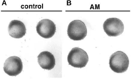
Fig. 3.Day 4 control (A) and 10-6 M AM-treated (B) posterior limb
mesoderm explants cultured in the absence of the AER and limb ectoderm. Exogenous AM does not promote the outgrowth of the explants.
A
B
C
D
A
B
A
D
B
E
C
However, as shown in Fig. 2D, the distal apical ectoderm of stage 20 limbless mutant limb buds, which have failed to develop an AER, exhibits little or no immunostaining for AM, as might be expected for a molecule involved in AER activity.
In Vitro Studies of the Possible Role of AM in AER Activity
The presence of a high level of AM immunoreactivity in the AER as it is directing the outgrowth of limb mesoderm and its absence from the apical ectoderm of stage 20 limbless mutant limb buds which fail to develop an AER suggests that an AM-like molecule may play a role in regulating or mediating some aspect of AER activity. One possibil-ity is that AM expressed in the AER may function in a paracrine fashion to mediate the effect of the AER on the outgrowth of limb mesoderm. To explore this possibility, we determined if exogenous AM would be able to promote the outgrowth of posterior limb mesoderm subjected to organ culture in the absence of the AER and limb ectoderm, as do other signaling molecules such as IGF-I and FGFs that have been implicated in mediating the outgrowth of limb mesoderm in response to the AER (Dealy and Kosher, 1995, 1996). As shown in Fig. 3, exogenous AM at concentrations ranging from 10 -8 M to 10-5 M does not promote the outgrowth of posterior limb
mesoderm explants lacking an AER.
Another possibility is that AM present in the AER may be regulat-ing in an autocrine fashion some aspect of the activity of the AER itself. Consistent with this possible autocrine mode of action is the observation that a high level of AM immunoreactivity is present along the surfaces of the contiguous cells of the AER (Fig. 1F). If AM is indeed acting in an autocrine fashion to influence AER activity, then inhibiting endogenous AM activity in the AER might impair the ability of the AER to promote the morphogenesis of limb mesoderm. Accordingly, we examined the effect of the AM antagonist AM(22-52)
on the morphogenesis of posterior limb mesoderm explants cultured in the presence of the AER. AM(22-52) contains the C-terminal 30 amino acids of AM which mediate binding of AM to its receptor, but lacks the N-terminal portion of the molecule required for signal transduction following receptor binding (Eguchi et al., 1994).
The gross morphogenesis of control and AM(22-52)-treated posterior limb mesoderm explants cultured in the presence of the AER is shown in Fig. 4. During the 5 day culture period, control explants progressively undergo polarized proximodistal outgrowth (Fig. 4 A-D). The outgrowth and contour of AM(22-52)-treated explants is comparable to controls through day 2 of culture (Fig. 4E). However, in contrast to controls, AM(22-52)-treated explants un-dergo little or no further outgrowth during the subsequent 3 days of culture (Fig. 4 E-H). The size and shape of day 3-5 AM(22-52)-treated explants is comparable to that of day 2 control and treated explants. To confirm the observations on living explants indicating that AM(22-52) impairs the outgrowth of limb mesoderm in response to the AER, we compared cell proliferation in control and AM(22-52)-treated explants by immunohistochemical analysis of BrdU incorpo-ration into nuclei engaged in DNA synthesis during a 1 hour labeling period on days 3 and 4 of culture. As shown in Fig. 5, a highly proliferating population of cells is present in the mesoderm underly-ing the AER of control explants, and there is a strikunderly-ing reduction in number of proliferating cells in the subridge mesoderm of the AM(22-52)-treated explants. The subridge mesoderm of control explants
Fig. 4.Morphogenesis of a control (A-D) and 10-6 M AM(22-52)-treated
(E-H) posterior limb mesoderm explant cultured in the presence of the AER. The control explant undergoes progressive proximodistal outgrowth during the 5 day culture period, whereas the AM(22-52)-treated explant undergoes little or no outgrowth between days 2 and 5.
Fig. 5.Cell proliferation in control (A,C,E) and 10-6 M (B) or 10-5 M (D,F)
AM(22-52)-treated posterior limb mesoderm explants cultured in the presence of the AER as assayed by the immunohistochemical analysis of BrdU incorporation into dividing cells during a 1 hour labeling period on day 3 (A,B; C,D) or day 4 (E,F) of culture. A highly proliferating population of cells is present in the mesoderm underlying the AER of control explants, and there is a striking reduction in number of proliferating cells in the subridge mesoderm of the AM(22-52)-treated explants.
A
B
C
D
E
F
A
B
C
D
TABLE 1
EFFECT OF AM(22-52) ON CELL PROLIFERATION IN THE SUBRIDGE MESODERM OF POSTERIOR LIMB MESODERM
EX-PLANTS CULTURED IN THE PRESENCE OF THE AER
Number of proliferating cellsa
Control explants AM(22-52)-treated explants 287 ± 29 (n = 6) 131 ± 21 (n = 9)
aThe number of BrdU-labeled nuclei in the subridge mesoderm of sections through the center of control and 10-5 M AM(22-52)-treated explants that had been incubated in the presence of BrdU for 1 hour on day 4 of culture.
detectable with antiserum that had been preabsorbed to purified human AM coupled to Sepharose.
Preparation of Limb Buds from Limbless Mutant Chick Embryos Windows were cut into eggs obtained from mating members of limbless heterozygous chickens maintained at the University of Connecticut, and the right forelimbs of embryos were removed and individually processed as described below. The windows in the shells of the donor embryos were sealed, and the donor embryos were reincubated until the phenotype of their remaining limb buds was clearly evident. Individual limb buds to be used for immunohistochemistry were fixed in Bouin’s solution, processed to 70% ethanol, and stored in 70% ethanol until their phenotype was established.
Preparation and Analysis of Organ Cultures
The ectoderm was removed from the posterior 2/3 of stage 20/21 (Hamburger and Hamilton, 1951) wing buds as previously described (Dealy and Kosher, 1995), and the posterior mesoderm was cultured in the presence or absence of 10-5-10-8 M of human adrenomedullin (Peninsula Laboratories; Phoenix Pharmaceuticals) on nutrient agar substrates (Ko-sher et al., 1979) containing serum-free BGJb/F12 (7/3) medium supple-mented with 0.1% bovine serum albumin (Dealy and Kosher, 1995). In other experiments, the posterior 2/3 of stage 20/21 wing buds were cultured with the AER and limb ectoderm intact in the presence or absence of 10-5 -10-6 M of the AM antagonist, AM(22-52) (Peninsula Laboratories).
The morphogenesis of living explants was evaluated daily with the dissecting microscope and living explants were photographed. Cell prolif-eration was examined as described by Dealy and Kosher (1995) by immunohistochemical analysis of the incorporation of 5-bromodeoxyuridine (BrdU) into nuclei engaged in DNA synthesis during a 1 hr labeling period with BrdU on days 3 and 4 of culture.
Acknowledgments
We thank Caroline N. Dealy for the limbless mutant limb buds. Sup-ported by NIH Grant HD22610 to R.A.K.
References
CARRINGTON, J.L. and FALLON, J.F. (1988). Initial limb budding is independent of apical ectodermal ridge activity: evidence from a limbless mutant. Development 104: 361-367.
COELHO, C.N.D., KRABBENHOFT, K., UPHOLT, W.B., FALLON, J.F. and KO-SHER, R.A. (1991). Altered expression of the chicken homeobox-containing genes Ghox-7 and Ghox-8 in the limb buds of limbless mutant chick embryos. Development 113: 1487-1493.
DEALY, C.N. and KOSHER, R.A. (1995). Studies on insulin-like growth factor-I and insulin in chick limb morphogenesis. Dev. Dynamics 202: 67-79.
DEALY, C.N. and KOSHER, R.A. (1996). IGF-I, insulin, and FGFs induce outgrowth of the limb buds of amelic mutant chick embryos. Development 122: 1323-1330.
EGUCHI, S., HIRATA, Y., IWASAKI, H., SATO, K., WATANABE, T.X., INUI, T., NAKAJIMA, K., SAKAKIBARA, S. and MARUMO, F. (1994). Structure-activity relationship of adrenomedullin, a novel vasodilatory peptide, in cultured rat vascular smooth muscle cells. Endocrinology 135: 2454-2458.
GOULD, S.E., UPHOLT, W.B. and KOSHER, R.A. (1995). Characterization of chicken syndecan-3 as a heparan sulfate proteoglycan and its expression during embryogenesis. Dev. Biol. 168: 438-451.
GRIESHAMMER, U., MINOWADA, G., PISENTI, J.M., ABBOTT, U.K and MARTIN, G.R. (1996). The chick limbless mutation causes abnormalities in limb bud dorsal-ventral patterning: implications for the mechanism of apical ridge formation. Development 122: 3851-3861.
HAMBURGER, V. and HAMILTON, H.L. (1951). A series of normal stages in the development of the chick embryo. J. Morphol. 88: 49-92.
KITAMURA, K., KANGAWA, K., KAWAMOTO, M., ICHIKI, Y., NAKAMURA, S., MATSUO, H.and ETO, T. (1993a). Adrenomedullin: a novel hypotensive peptide isolated from human pheochromocytoma. Biochem. Biophys. Res. Comm. 192: 553-560.
contains more than twice as many proliferating (BrdU-labeled) cells as AM(22-52)-treated explants (Table 1). Thus, the outgrowth and proliferation of limb mesoderm in response to the AER is clearly impaired in the presence of AM(22-52).
Discussion
AM is a multifunctional peptide that has recently been shown to exhibit discrete spatial and temporal patterns of expression during mouse and rat embryogenesis, particularly at sites of epithelial-mesenchymal tissue interactions (Montuenga et al., 1997, 1998). Here we demonstrate that AM immunoreactivity is present at high levels in the AER during the early stages of development of the chick limb bud when the AER is directing the outgrowth and patterning of limb mesoderm. AM immunoreactivity attenuates during later stages of development as the AER flattens and loses its ability to promote limb outgrowth. Furthermore, little or no AM immunoreactivity is detectable in the apical ectoderm of stage 20 limbless mutant limb buds which have failed to develop an AER. These results suggest that an AM-like molecule may play a role in regulating or mediating some aspect of the activity of the AER of the developing chick limb bud.
Our results suggest that an AM-like molecule may function in an autocrine fashion to regulate some aspect of AER activity. Intense immunostaining for AM is present along the surfaces of the cells of the AER, an observation consistent with the possibility that AM expressed by AER cells may be bound to receptors on contiguous cells. Moreover, we have found that although exogenous AM cannot substitute for the AER in promoting the outgrowth of limb mesoderm, the AM antagonist AM(22-52) impairs the outgrowth and proliferation of limb mesoderm cultured in the presence of the AER. This is consistent with the possibility that inhibition of endog-enous AM activity in the AER impairs the ability of the AER to promote the proliferation of limb mesoderm.
Materials and Methods
Immunohistochemistry
KITAMURA, K., KANGAWA, K., KOJIMA, M., ICHIKI, Y., MATSUO, H.and ETO, T. (1994). Complete amino acid sequence of porcine adrenomedullin and cloning of cDNA encoding its precursor. FEBS Lett. 338: 306-310.
KITAMURA, K., SAKATA, J., KANGAWA, K., KOJIMA, M., MATSUO, H.and ETO, T. (1993b). Cloning and characterization of cDNA encoding a precursor for human adrenomedullin. Biochem. Biophys. Res. Comm. 194: 720-725.
KOSHER, R.A., SAVAGE, M.P. and CHAN, S.-C. (1979). In vitro studies on the morphogenesis and differentiation of the mesoderm subjacent to the apical ectodermal ridge of the embryonic chick limb-bud. J. Embryol. Exp. Morph. 50: 75-97.
MARTÍNEZ, A. and CUTTITTA, F. (Eds.) (1998). Adrenomedullin. IOS Press, Amsterdam.
MARTÍNEZ, A., MILLER, M.J., UNSWORTH, E.J., SIEGFRIED, J.M. and CUTTITTA, F. (1995). Expression of adrenomedullin in normal human lung and in pulmo-nary tumors. Endocrinology 136: 4099-4105.
MARTÍNEZ, A., UNSWORTH, E.J. and CUTTITTA, F. (1996). Adrenomedullin-like immunoreactivity in the nervous system of the starfish Marthasterias glacialis. Cell Tissue Res. 283: 169-172.
MILLER, M.J., MARTÍNEZ, A., UNSWORTH, E.J., THIELE, C.J., MOODY, T.W., ELASSER, T. and CUTTITTA, F. (1996). Adrenomedullin expression in human tumor cell lines. Its potential role as an autocrine growth factor. J. Biol. Chem. 271: 23345-23351.
MONTUENGA, L.M., MARTÍNEZ, A., MILLER, M.J., UNSWORTH, E.J. and CUTTITTA, F. (1997). Expression of adrenomedullin and its receptor during embryogenesis suggests autocrine or paracrine modes of action. Endocrinol-ogy 138: 440-451.
MONTUENGA, L.M., MARIANO, J.M., PRENTICE, M.A., CUTTITTA, F. and JAKOWLEW, S.B. (1998). Coordinate expression of transforming growth factor-β1 and adrenomedullin in rodent embryogenesis. Endocrinology 139: 3946-3957.
ROBERT, B., LYONS, G., SIMANDL, B.K., KUROIWA, A. and BUCKINGHAM, M. (1991). The apical ectodermal ridge regulates Hox-7 and Hox-8 gene expres-sion in developing chick limb buds. Genes Dev. 5: 2363-2374.
ROS, M.A., LÓPEZ-MARTÍNEZ, A., SIMANDL, B.K., RODRÍGUEZ, C., IZPISUA-BELMONTE, J.C., DAHN, R. and FALLON, J.F. (1996). The limb bud mesoderm determines initial limb bud anteroposterior asymmetry and budding independent of sonic hedgehog or apical ectodermal gene expressions. Development 122: 2319-2330.
RUBIN, L. and SAUNDERS, J.W., JR. (1972). Ectodermal mesodermal interactions in the growth of limb buds in the chick embryo: constancy and temporal limits of the ectodermal induction. Dev. Biol. 28: 94-112.
SAKATA, J., SHIMOKUBO, T., KITAMURA, K., NAKAMURA, S., KANGAWA, K., MATSUO, H. and ETO, T. (1993). Molecular cloning and biological activities of rat adrenomedullin, a hypotensive peptide. Biochem. Biophys. Res. Comm. 195: 921-927.
SAUNDERS, J.W., JR. (1948). The proximo-distal sequence of origin of parts of the chick wing and the role of the ectoderm. J. Exp. Zool. 108: 363-404.
SAUNDERS, J.W., JR. (1977). The experimental analysis of chick limb development. In Vertebrate Limb and Somite Morphogenesis (Eds. Ede, D.A., Hinchliffe, J.R. and Balls, M.). Cambridge University Press, Cambridge, pp. 1-24.
SAUNDERS, J.W., JR.and GASSELING, M.T. (1968). Ectodermal-mesenchymal interactions in the origin of limb symmetry. In Epithelial-Mesenchymal Interactions (Eds. Fleischmajer, R. and Billingham, R.F.). Williams and Wilkins, Baltimore, pp. 78-97.
SOLURSH, M., SINGLEY, C.T. and REITER, R.S. (1981). The influence of epithelia on cartilage and loose connective tissue formation by limb mesenchyme cultures. Dev. Biol. 86: 471-482.
TODT, W.L. and FALLON, J.F. (1984). Development of the apical ectodermal ridge in the chick wing bud. J. Embryol. Exp. Morphol. 80: 21-41.
VOGEL, A. and TICKLE, C. (1993). FGF-4 maintains polarizing activity of posterior limb bud cells in vivo and in vitro. Development 119: 199-206.