HIGHLIGHTED ARTICLE INVESTIGATION
A Novel Ribosomopathy Caused by Dysfunction of
RPL10 Disrupts Neurodevelopment and Causes
X-Linked Microcephaly in Humans
Susan S. Brooks,*,1Alissa L. Wall,†,1Christelle Golzio,†David W. Reid,‡Amalia Kondyles,†Jason R. Willer,† Christina Botti,* Christopher V. Nicchitta,‡,§Nicholas Katsanis,†and Erica E. Davis†,2 *Department of Pediatrics, Rutgers Biomedical and Health Sciences, Robert Wood Johnson Medical School, New Brunswick, New
Jersey 08901, and†Center for Human Disease Modeling,‡Department of Biochemistry, and§Department of Cell Biology, Duke
University Medical Center, Durham, North Carolina 27710 ORCID IDs: 0000-0002-1618-7127 (S.B.); 0000-0002-2412-8397 (E.D.)
ABSTRACT Neurodevelopmental defects in humans represent a clinically heterogeneous group of disorders. Here, we report the genetic and functional dissection of a multigenerational pedigree with an X-linked syndromic disorder hallmarked by microcephaly, growth retardation, and seizures. Using an X-linked intellectual disability (XLID) next-generation sequencing diagnostic panel, we identified a novel missense mutation in the gene encoding 60S ribosomal protein L10 (RPL10), a locus associated previously with autism spectrum disorders (ASD); the p.K78E change segregated with disease under an X-linked recessive paradigm while, consistent with causality, carrier females exhibited skewed X inactivation. To examine the functional consequences of the p.K78E change, we modeled RPL10 dysfunction in zebrafish. We show that endogenousrpl10expression is augmented in anterior structures, and that suppression decreases head size in developing morphant embryos, concomitant with reduced bulk translation and increased apoptosis in the brain. Subsequently, usingin vivocomplementation, we demonstrate that p.K78E is a loss-of-function variant. Together, our findings suggest that a mutation within the conserved N-terminal end of RPL10, a protein in close proximity to the peptidyl transferase active site of the 60S ribosomal subunit, causes severe defects in brain formation and function.
N
EURODEVELOPMENTAL defects in humans representa diagnostic challenge. Displaying marked phenotypic overlap, examples include autism spectrum disorders (ASD), intellectual disability (ID), microcephaly, and seizures; in some instances, common genetic defects can underscore each of these clinical entities. For example, mutations in
the voltage-gated sodium channel Nav1.2 encoded bySCN2A
are associated with the manifestation of early infantile
epi-lepsy (Sugawara et al. 2001). However, recent exome
se-quencing studies have also identified SCN2A mutations as
rare contributors to disease in autism cohorts, thereby
ex-panding the phenotypic spectrum underscored by Nav1.2
channel dysfunction (Sanderset al.2012). A gender bias of
1.3–1.4 males to 1 female with a neurodevelopmental
dis-order has complicated further our mechanistic understanding
of such defects (Leonard and Wen 2002; Ellisonet al.2013).
One obvious explanation for an unbalanced representation of the sexes among individuals with a structural or functional brain defect is an abundance of developmentally important
genes on the X chromosome. To date,.100 genes have been
associated with ASD, ID, microcephaly, or seizures primarily in hemizygous males and, to some extent, their carrier
moth-ers (De Brouweret al.2007; Tarpeyet al. 2009; Lubset al.
2012).
Here, we report the genetic dissection of a novel form of X-linked human genetic disease characterized by microcephaly, seizures, growth retardation, and hypotonia. Combined genetic, functional, and biochemical assays suggest that a missense mutation in RPL10, a component of the 60S large ribosomal subunit, can cause syndromic central nervous system defects, likely because of defects in bulk translation and increased apoptosis in the brain.
Copyright © 2014 by the Genetics Society of America doi: 10.1534/genetics.114.168211
Manuscript received July 13, 2014; accepted for publication August 22, 2014 Supporting information is available online athttp://www.genetics.org/lookup/suppl/ doi:10.1534/genetics.114.168211/-/DC1.
1These authors contributed equally to this work.
2Corresponding author: Duke University Medical Center, Box 3709, Durham, NC
Materials and Methods
Clinical genetic screening and confirmatory testing
Nine members of the family consented for genetic testing. X chromosome inactivation status was established by analysis of DNA methylation at the human androgen receptor locus in DNA from the two mutation carrier mothers (individuals I-2 and II-4; Center for Genetic Testing, Saint Francis Health System). An X-linked intellectual disability (XLID) next-generation sequencing panel targeting 82 genes (supporting
information,Table S1) was conducted at a commercial
lab-oratory (Ambry Genetics), using a DNA sample from af-fected individual II-1. Segregation analysis of p.K78E was
carried out by Sanger sequencing of RPL10 exon 5 in all
seven additional available family members. DNA constructs and in vitro transcription
We obtained a human wild-type (WT)RPL10open reading
frame (ORF) construct [pENTR221, Ultimate ORF Collec-tion by Invitrogen (Carlsbad, CA); Life Technologies, clone IOH2895] and we generated constructs encoding missense variants p.K78E, p.L206M, p.H213Q, and p.S202N as
de-scribed (Niederriteret al.2013). Following sequence confi
r-mation of the mutation and ORF integrity using Sanger sequencing, pENTR constructs were then cloned into the pCS2+ vector, using LR clonase II-mediated recombination
(Life Technologies). Sequence-confirmed WT and mutant
RPL10constructs in the pCS2+ vector were linearized withNotI
and transcribedin vitro, using the SP6 mMessage mMachine Kit
(Ambion).
Zebrafish embryo manipulation and injections
We developed anin vivocomplementation assay as described
in Niederriter et al. (2013). Translation blocking (tb) (59
TGCGATCTGTAACGTACACAATAAC 39) and splice blocking
(sb) (59AAAATACATGGCTTACCAGGAACAC 39) morpholinos
(MOs) (Gene Tools) were diluted to appropriate concentra-tions in nuclease-free water (0.5, 0.6, and 0.7 ng/nl for the tb-MO dose response; 1, 2, and 3 ng/nl for the sb-MO dose response; 0.6 ng/nl tb-MO for rescue experiments; and 0.7 ng/nl tb-MO or 3 ng/nl sb-MO for transferase-mediated dUTP nick end labeling (TUNEL) and phospho-histone H3 antibody
staining) and injected into WT zebrafish embryos (Ekkwill3
AB F1outcross) at the one- to four-cell stage. To assess sb-MO
efficiency, endogenousrpl10 expression was determined by
extracting total RNA from 1 day postfertilization (dpf)
em-bryos with Trizol (Invitrogen) according to manufacturer’s
instructions. Oligo(dT)-primed total RNA was reverse tran-scribed using SuperScriptIII reverse transcriptase (Invitrogen) and the resulting complementary DNA (cDNA) was PCR
am-plified. To rescue morphant phenotypes, we injected tb-MO
with 50 pg capped human messenger RNA (mRNA). Embryos
were scored at 2 dpf and classified as normal or abnormal
(microcephalic) when compared to age-matched controls from the same clutch. Embryos were then dechorionated,
anesthe-tized with Tricaine solution, fixed in 4% paraformaldehyde
solution overnight, and then transferred to 13PBS prior to
quantitative phenotypic analysis. RNA in situ hybridization
We PCR amplifiedDanio rerio rpl10transcript
correspond-ing to cDNA clone MGC:56154 (GenBank: BC045950), using 1 dpf whole-embryo cDNA as template. We labeled sense and antisense RNA probes with digoxigenin and
per-formed whole-mount RNA in situ hybridization on 2 dpf
embryos as described in Thisse and Thisse (2008). Lateral images were acquired on a Nikon (Garden City, NY) AZ100 microscope, using Nikon NIS Elements Software.
Bright-field imaging and measurements
Lateral and dorsal images were acquired on a Nikon SMZ745 microscope, using Nikon NIS Elements Software
(n = 30 larvae per injection batch; investigator masked to
injection cocktail; repeated twice). We measured head size, body length, and somite angle with ImageJ software; for body length measurements (from lateral images), a polyline was drawn beginning at the anteriormost point of yolk at-tachment and terminating at the posteriormost point on the tail; for somite angle measurements (from lateral images), we measured the angle of the somite located at the midpoint between the yolk and the anus; for forebrain area measure-ments (from dorsal images), an outline was drawn begin-ning at the posteriormost point of eye and tracing around
the head to terminate at the starting point. A Student’st-test
was used to determine the statistical significance of
differ-ences between injection batches. Polysome gradients
Zebrafish larvae were anesthetized in tricaine solution at
5 dpf and decapitated with microsurgical scissors, and heads and bodies were lysed in separate pools in 200 mM KOAc,
15 mM MgCl2, 25 mM K-HEPES (pH 7.2), and 2%
dodecyl-maltoside (DDM) (n= 20 larvae per injection batch). For
each sample, 250 A260units of the tissue extracts were then
layered over a 10–50% sucrose gradient and centrifuged for
3 hr at 35,000 rpm in a SW-41 rotor (Beckman-Coulter, Pasadena, CA). Gradients were collected using a Teledyne-Isco gradient fractionator with continuous absorbance mon-itoring at 254 nm.
Whole-mount TUNEL assay, phospho-histone H3
immunostaining, andfluorescence microscopy
We utilized terminal deoxynucleotidyl TUNEL to assay
apo-ptosis, using the ApopTag rhodaminein situApoptosis
Detec-tion kit (Chemicon) as described in Golzioet al.(2012). For
whole-mount histone H3 immunostaining, we used anti-phospho-histone H3 (ser10)-R antibody (diluted 1:750;
sc-8656-R, Santa Cruz) as described in Golzio et al. (2012).
For each of TUNEL and phospho-histone H3 immunostaining,
fluorescent signals were imaged on laterally positioned larvae
and z-stacked on a Nikon AZ100 microscope, using NIS
cells (histone H3) or pixels (TUNEL) in defined regions of the head by using ImageJ software; positive cells in the eyes were
removed from cell counts for histone H3 (n = 20 embryos
imaged per injection batch; masked scoring; repeated twice).
Results and Discussion
A novel missense variant in RPL10 segregates with X-linked syndromic microcephaly
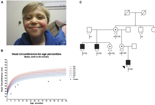
We consulted for an 11-month-old male of European-American origin who presented with a syndromic neurodevelopmental disorder of unknown etiology. Following a pregnancy that was complicated with polyhydramnios at 27 weeks gestation, the index case (III-1) was born at 35 weeks. He failed his newborn hearing screen and displayed multiple congenital abnormali-ties that included digit malformations, right cryptorchidism, sacral dimple, and dysmorphic craniofacial features (Table 1; Figure 1A). He underwent surgical procedures to correct some of his congenital abnormalities: pneumo-eustachian tube place-ment at 4 months, sacral lipoma removal at 9 months, and
a right orchiopexy at 11 months. He had chronic reflux and
growth retardation, was generally hypotonic, and was hospi-talized for several pneumonias. At birth, his head circumfer-ence was 28 cm (2.6 standard deviations below the mean); his head circumference velocity declined, and at his most recent clinical assessment at age 4.5 years measured 44.5 cm (5 stan-dard deviations below the mean, Figure 1B). Laboratory stud-ies including chromosomal microarray, plasma amino acid, acylcarnitine, urine organic acids, creatine, and guanidoacetic acid were normal. Although both of his parents were reported to be healthy, a review of his family history revealed two ma-ternal uncles who had unexplained syndromic encephalopathy disorders (Figure 1C). Both of the maternal uncles (II-1 and II-2) were evaluated and medical records were reviewed at 19 and 25 years, respectively. They shared multiple phenotypic features with that of the index case, including a history of microcephaly, seizures, growth retardation, hypotonia, genito-urinary abnormalities, and prognathism (Table 1). Addition-ally, both maternal uncles were born with cardiac valve defects, developed hearing loss, are essentially nonverbal, and are min-imally or nonambulatory (Table 1).
Suspicious of an X-linked disorder, we tested both het-erozygous carrier mothers (I-2 and II-4) for nonrandom X-inactivation patterns. Favorable skewing is a well-documented phenomenon in which cells containing an active mutation-bearing X chromosome are selected against during cell division, resulting in a predominance of cells with an active X chromosome containing the normal allele (Migeon 1998; Van Den Veyver 2001). Evaluation of DNA methylation at the human androgen receptor locus in each of I-2 and II-4 pro-duced results consistent with our hypothesis; each female showed fully skewed inactivation of their mutation-bearing X chromosome.
With no clear diagnostic criteria to assign the family to a described syndrome, we ordered an X-linked sequencing
panel covering the coding regions and intron–exon boundaries
of 82 genes implicated previously in XLID (XLID panel, Ambry
Genetics;Table S1). An estimated 42% of affected individuals
with a family history of XLID are anticipated to have a dele-terious mutation in a gene represented on the panel (De
Brouwer et al.2007), and analytic sensitivity of this test is
reported to be 83%. This approach identified a novel
single-nucleotide change (c.232A . G; p.K78E) within the gene
encoding 60S ribosomal protein L10 (RPL10), a gene on Xq28 reported previously to be an ASD candidate (Klauck et al.2006). Mutational screening of all the other 81 genes was negative; this variant was also absent from all publicly available control exomes and genomes [NHLBI Exome Var-iant Server (EVS), dbSNP, and 1000 Genomes] and had not been reported in cases of XLID.
To explain further the significance of this variant, we
conducted segregation analysis in eight available family mem-bers representative of three generations with Sanger
sequenc-ing ofRPL10exon 5. This variant segregated as expected for
an X-linked disorder: the proband and both affected maternal
uncles were hemizygous carriers of p.K78E; the proband’s
mother (II-4) and his maternal grandmother (I-2) were het-erozygous for p.K78E; and importantly, an unaffected great uncle of the index case (I-3) harbored the WT allele (Figure 1C).
Together, our mutational findings from the XLID panel,
segregation with disease in multiple generations, and fully skewed X inactivation in carrier mothers provided genetic
evidence suggesting that RPL10 p.K78E might be the
pri-mary cause of syndromic microcephaly in this family.
rpl10 suppression in zebrafish results in microcephaly
and p.K78E is pathogenic
Missense mutations in RPL10 encoding p.L206M and
p.H213Q have been reported previously to confer
suscepti-bility to ASD (Klauck et al.2006; Chiocchetti et al.2011).
Even so, individuals bearing these nonsynonymous changes were not reported to display microcephaly or the constella-tion of syndromic features present in the three hemizygous
males with RPL10p.K78E. Thus, the rarity of RPL10
varia-tion among cases (3/521 ASD pedigrees) (Klauck et al.
2006; Chiocchetti et al.2011) in the absence of replication
in other ASD cohorts (Gonget al.2009) and control cohorts
bereft of functional variation (1/2443 males and 3/4060
females harboring missenseRPL10changes; EVS) precluded
us from implicating p.K78E in severe neurodevelopmental
phenotypes. Moreover, in silico prediction programs were
conflicting; p.K78E was predicted to be benign by PolyPhen-2
(Adzhubei et al. 2010), but damaging by Mutation Taster
(Schwarz et al.2010) and SIFT (Kumaret al.2009).
There-fore, we turned to the developing zebrafish as a model both to
determine the physiological relevance ofRPL10to disease and
to test the pathogenic potential of p.K78E on RPL10 function.
Previous studies have shownD. rerioto represent a useful
surrogate to study neurodevelopmental defects in humans
(Komoikeet al.2010; Tianet al.2010; Bicknellet al.2011;
Table 1 Phe notype s o f affec ted ma les with RPL10 p. K78E Patie nt identi fi er Wk of gesta tion Bi rth we ight Hea d circu mfere nce Microcepha ly Seizure s
Severe growth
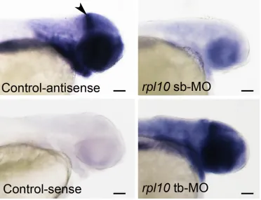
Dauberet al.2013; Schafferet al.2014). Although reported previously to be expressed ubiquitously (Thisse and Thisse
2004), in situ hybridization of rpl10 riboprobes in 2 dpf
embryos demonstrated enriched expression in the anterior
vs.posterior structures, particularly at the midbrain–hindbrain
boundary (Figure S1). Next, to determine the functional
consequences of RPL10 suppression, we employed
MO-induced knockdown of the singleD. rerio ortholog (92%
identity, 97% similarity). We designed a tb-MO targeting
the translational start site of the rpl10 transcript. We
injected one- to four-cell stage WT zebrafish embryos
with increasing doses of tb-MO (0.5, 0.6, and 0.7 ng;
n= 50 embryos per injection batch). We observed a
mi-crocephaly phenotype that was dose dependent at 2 dpf as determined by qualitative scoring masked to injection
cocktails (Figure 2A;Figure S2, A and C). To assess this defect
quantitatively, we measured the forebrain cross-sectional area
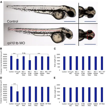
of morphants and controls and observed a significant decrease
in head size, but not body length, when evaluated at 2 dpf
(P,0.0001;n= 30; repeated twice; Figure 2, A–E;Table S2,
Table S3), suggesting that rpl10 suppression did not cause
a generalized developmental delay. Additionally, we recorded
similar somite angle measurements in morphants vs.controls
(mean somite angle of 99.8°vs.99.1°, respectively; n= 30;
repeated twice), and only a minor proportion of morphants displayed affected structures other than the head (tail
exten-sion defects; 3/60 evaluated), indicating thatrpl10suppression
affects predominantly anterior structures. To replicate these observations, we designed a sb-MO targeting the donor site ofrpl10 exon 2 (Figure S1,Figure S2, A and B); the sb-MO titration curve (1, 2, and 3 ng) resulted in a similar reduction of embryo head size at 2 dpf in a dose-dependent manner
(Figure S2D). Next, we co-injected WT human RPL10mRNA
with tb-MO. This resulted in a significant improvement of the
microcephaly phenotype according to both qualitative and
quantitative measures (qualitative, 70% vs.30% affected for
MO alonevs.WT rescue,P,0.0001; quantitative, mean
cross-sectional area 49,337mm2vs.60,203mm2for tb-MO alonevs.
WT rescue;P,0.0001; Figure 2, A–C;Table S2). Together,
these data indicate that loss ofrpl10 in developing zebrafish
Figure 1 Microcephaly in an X-linked pedigree harboringRPL10p.K78E. (A) The proband (III-1) at age 41 months. He displays microcephaly, a thin upper lip, and mandibular prognathism (see Table 1 for full phenotypic description). (B) Head circumference chart for the proband. Microcephaly was of
prenatal onset and growth continued to follow a normal curve at this reduced trajectory. (C) X-linked segregation ofRPL10c.232A.G; p.K78E in
a three-generation pedigree. Subsequent to identification of p.K78E in individual II-1 by a next-generation sequencing X-linked intellectual disability
diagnostic panel, Sanger sequencing confirmed segregation of the mutation in three affected males and three carrier females. A healthy maternal great
results in an anatomically similar neurodevelopmental pheno-type to that of the hemizygous males harboring p.K78E.
Both our group and others have shown that in vivo
com-plementation assays in zebrafish embryos are a sensitive and
specific approach to test the pathogenicity of missense
muta-tions implicated in human genetic disease (Zaghloul et al.
2010; Wan et al.2012; Niederriteret al. 2013; Daviset al.
2014). To test the effect of p.K78E on RPL10 function,
co-injection ofrpl10tb-MO with mRNA harboring p.K78E failed
to rescue the morphant phenotype and resulted in signifi
-cantly decreased forebrain cross-sectional area (43,390mm2)
when compared to the WT rescue (60,203mm2;P,0.0001;
n= 30; repeated twice); body length was not affected (P=
0.34;n= 30; Figure 2, B and C,Table S3). Comparison of the
forebrain cross-sectional area for tb-MO plus p.K78E mRNA
vs. MO-injected embryo batches was statistically
indistin-guishable (P = 0.066; n = 30; Figure 2, B and C). Next,
we tested the twoRPL10variants associated previously with
ASD and a putative benign variant (rs4909) also reported
previously (Klauck et al. 2006). Co-injection of each of
p.S202N, p.L206M, or p.H213Q encoding mRNAs with rpl10 tb-MO fully rescued the microcephaly phenotype, as Figure 2 Suppression ofrpl10in zebrafish results in reduced head size and p.K78E is a loss-of-function variant. (A) Live larval images of control (top) andrpl10tb-MO-injected embryos (bottom). Left panels show lateral views of whole larvae with similar body lengths; right panels show dorsal views
showing a reduced head size in morphants. Red lines indicate head size measurements quantified in B and D and body length measurements quantified
in C and E. Bars, 500mm. (B) Quantification of head area forrpl10MO and MO co-injected with humanRPL10mRNA. (C) Quantification of body length
for rpl10MO and MO co-injected with human RPL10mRNA. (D) Quantification of head size for embryos injected with RPL10mRNA alone. (E)
Quantification of body length for embryos injected withRPL10mRNA alone. Head area and body length measurements were carried out at 2 days
postfertilization (dpf), using embryos injected with 0.6 ng tb-MO and/or 50 pg mRNA;n= 30 for each injection batch with masked scoring were
repeated with similar results. Error bars indicate standard error of the mean (SEM).***P,0.0001 (two-tailedt-test comparisons between MO-injected
indicated by similar forebrain cross-sectional areas and body lengths in comparison to batches co-injected with a cocktail
of WT mRNA and rpl10tb-MO (Figure 2, B and C). These
data are not surprising, given that p.L206M and p.H213Q were reported as hypomorphic changes that do not disrupt the basic functions of translation, but do alter discrete cel-lular protein signatures that may result in the dysregulation
of oxidative stress response (Klaucket al.2006; Chiocchetti
et al.2014). Moreover, these C-terminally positioned muta-tions (a) can fully complement temperature-sensitive strains
of RPL10 mutant yeast (Klaucket al.2006) and (b) do not
induce skewing of X inactivation in mutation carrier females, suggesting that they may be tolerated in some cellular
con-texts (Chiocchetti et al. 2011). Therefore, the functional
capacity of RPL10 bearing each of these two variants prob-ably exceeds the cellular threshold required to rescue the
MO-induced microcephaly phenotypes in our zebrafish
mod-els, thereby resulting in a benign score.
Importantly, injection of each of the four missenseRPL10
mRNAs alone resulted in relatively similar head sizes and body lengths in comparison to WT mRNA alone, arguing against mRNA toxicity or dominant negative effects (Figure
2, D and E, Table S2andTable S3). Taken together, these
data suggest that p.K78E is a pathogenic variant and is a functional null in this assay and support the genetic
argu-ments from within our RPL10 pedigree to implicateRPL10
p.K78E as the driver of severe neurodevelopmental phenotypes.
Suppression of rpl10 results in decreased bulk
translation in the zebrafish head
RPL10 is conserved among eukaryotic taxa and, in mam-malian cells, is one of the 46 proteins that make up the 60S large ribosomal subunit in cooperation with three ribosomal
(r)RNAs (Ben-Shemet al. 2011). The 60S subunit harbors
the peptidyl transferase center (PTC) and the exit tunnel for newly synthesized polypeptides, whereas its functional part-ner, the 40S small ribosomal subunit, facilitates the
interac-tion between transfer RNA (tRNA) and mRNA (Spahnet al.
2001; Klingeet al.2011). Ribosomes are ubiquitous cellular
components responsible for the translation of all mRNAs, and perturbed ribosome biogenesis and ribosome dysfunc-tion can therefore give rise to numerous and varied
down-stream consequences (Scheperet al.2007).
Advances in protein crystallography have enabled exqui-site resolution of the structure of the 60S large ribosomal
subunit (Klinge et al.2011). RPL10 is one of six large
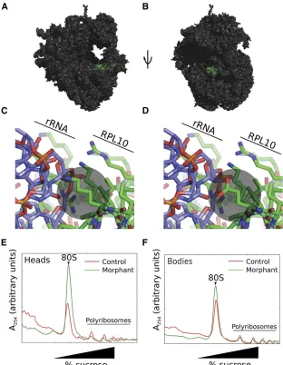
sub-unit proteins with immediate proximity to the PTC (in ad-dition to RPL3, RPL4, RPL8, RPL21, and RPL29; Figure 3, A and B); to date only RPL21 has been implicated in human genetic disease: a missense mutation at this locus has been associated with a nonsyndromic hair loss disorder,
heredi-tary hypotrichosis simplex (HHS) (Zhou et al. 2011). The
model suggests that mutation of K78, notably to an
acid-ic residue, disrupts RPL10 protein–28S rRNA interactions
(Figure 3, C and D), alters basic translational functions,
and as a consequence alters significantly protein expression
signatures to confer specific phenotypes to the central
ner-vous system.
To gain preliminary insight into the biochemical under-pinnings of the severe structural brain defects that result from altered RPL10, we asked whether reduction of RPL10 levels disrupted general protein synthesis. We injected WT
zebrafish embryos with sb-MO and allowed them to grow to
5 dpf (Figure S2E). After separating heads from bodies, we
analyzed polyribosome structure in each of the anterior and posterior portions of larvae. Morphant anterior structures displayed an increase in 80S ribosome abundance with a corresponding decrease in polyribosomes, consistent with a decrease in translation activity (Figure 3E). In contrast, polyribosome structure from bodies is relatively unchanged in morphants compared to controls (Figure 3F). These data
indicate that RPL10 is important for translation specifically
in zebrafish heads. At present, it is not known why loss of
RPL10 expression alters polyribosome structure in the
zebra-fish head but not the posterior region. Such differences may
reflect the apparently enriched expression of rpl10 in the
anterior portion of the embryo or overall translation
de-mands in specific spatiotemporal contexts.
rpl10 morphants display augmented apoptosis in the brain
Given the specific spatial reduction in bulk translation of
rpl10 morphant brains, we wondered what cellular conse-quences were induced in the absence of RPL10. We modeled our hypothesis on reports from a clinically distinct
ribosomo-pathy, Diamond–Blackfan anemia (DBA), in which
muta-tions in ribosomal proteins can result in cell cycle arrest or
induction of apoptosis (Aspesi et al. 2014). For example,
studies using patient cells harboring mutations in the most
common DBA gene,RPS19, can give rise to altered
prolifer-ation (Kuramitsuet al.2008) and/or cell death (Gazdaet al.
2006; Choesmelet al.2007). Therefore, we tested these two
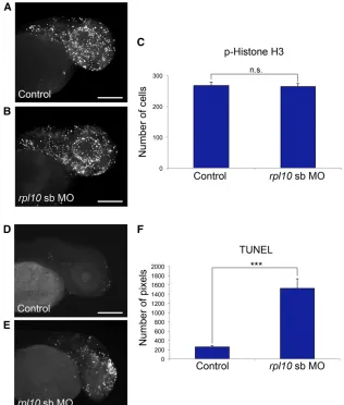
possibilities by quantifying markers of cell cycle (M-phase marker phospho-histone H3) and cell death (TUNEL) in rpl10 morphants.
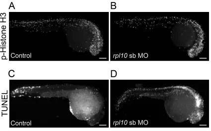
First, we injected WT embryos withrpl10sb-MO (Figure
4, A and B),fixed them at 2 dpf, and stained them with
anti-phospho-histone H3 antibody (n= 20 embryos per injection
batch). We saw no difference in cell proliferation in
mor-phants vs. controls upon quantification of stained cells in
defined areas of the zebrafish head (Figure 4, A–C; repeated
twice for the sb-MO, with similar results for the tb-MO, not shown). Moreover, we observed similar cell proliferation
signatures at 1 dpf (Figure S3, A and B), a time point that
typically precludes the observation of a head size defect in
other zebrafish models of microcephaly (Golzioet al.2012;
Beunderset al.2013).
Next, we monitored apoptosis in age-matched rpl10
rpl10 morphants in comparison to controls, determined by the number of stained pixels in laterally positioned images
(Figure 4, D–F;P,0.0001;n= 20 embryos quantified per
injection batch; repeated twice for the sb-MO, with similar results for the tb-MO, not shown). While TUNEL staining in 1 dpf morphant embryos showed increased generalized
apo-ptosis in the hindbrain and along the neural tube (Figure S3,
C and D), it was temporally distinct from the localized cell death in the forebrain at 2 dpf, suggesting that apoptosis is
likely to be a specific mechanistic driver of microcephaly in
embryos with compromised RPL10 function.
Conclusion
Here, we report an X-linked pedigree with three hemizygous males who display severe central nervous system defects, including microcephaly and seizures in combination with growth retardation and a multitude of additional congenital defects. We propose that this novel syndrome is underscored by a missense p.K78E-encoding mutation in the 60S large
ribosomal subunit component, RPL10. Ourin vivofunctional
studies using zebrafish models support our human genetics
data and highlight the power of a physiologically relevant vertebrate system to (a) establish relevance of a novel disease gene to human phenotypes; (b) determine variant pathogenic potential for private mutations; and (c) begin to elucidate the pathomechanism from biochemical evaluation of ribosomal output and cellular consequences, including cell death.
Although we are always cautious about elaboratingfi
nd-ings from a single pedigree, our combined phenotypic, ge-netic, and functional data suggest that RPL10 dysfunction causes a novel ribosomopathy. To date, most reported mu-tations in ribosomal proteins have been associated with DBA. Frequently associated with loss-of-function mutations in at least 10 ribosomal components, DBA is a rare, clinically het-erogeneous disorder hallmarked by red blood cell aplasia and incompletely penetrant defects in facio-skeletal development. Notably, affected individuals with DBA and our RPL10 family display overlapping features including syndactyly, mandibular and cleft defects, and genitourinary malformations (Vlachos Figure 3 rpl10morphants display reduced bulk trans-lation, especially in larval heads, as indicated by poly-ribosome structure. (A) Rendering of RPL10 in green on a eukaryotic ribosome in proximity to the peptidyl transferase active site. Protein Data Bank (PDB) entries 4a17, 4a19, and 2xzm were merged using a yeast ri-bosome (PDBs 2xzm and 3o58) as a guide. (B) As in A, rotated 90°. (C) Interaction of wild-type RPL10 K78 with the ribosomal (r)RNA. K78 interacts mostly with the negatively charged 28S rRNA and is indicated by a gray circle. (D) Simulation of the K78E mutation, in-dicated by a gray circle. (E) Sucrose gradient analysis of polyribosome structure in heads of 5 day
postfertiliza-tion (dpf)rpl10morphants and controls. Note the
in-crease in 80S abundance with a corresponding dein-crease
in polyribosomes for morphantsvs.controls, indicative
of a decrease in translational activity. (F) Sucrose
gradi-ent analysis of bodies ofrpl10morphants and controls.
et al.2014). However, the defining features of each ribosomal disorder described here are distinctly different; DBA is char-acterized by fully penetrant anemia and the most prominent phenotypes in our RPL10 pedigree are in the central nervous system. Moreover, the affected males with a hemizygous RPL10 mutation are not anemic, suggesting that they do not have a variant form of DBA.
Finally, our findings add to the accumulating repertoire
of ubiquitously expressed genes that give rise to tissue-specific
phenotypes. Another such example includes cleavage and polyadenylation factor I subunit 1 (CLP1), a multifunctional kinase implicated in tRNA, mRNA, and small interfering RNA (siRNA) maturation that when mutated, gives rise to
neuro-degenerative disease (Schafferet al.2014). Also, the general
pre-mRNA splicing factors, such as PRPF3, PRPF8, and
PRPF31, are substantial contributors to isolated retinitis pig-mentosa when rendered dysfunctional, yet mutation-bearing
individuals do not display syndromic features (McKie et al.
2001; Vithana et al. 2001; Chakarova et al. 2002; Liu and
Zack 2013). Althoughrpl10is expressed widely in the
zebra-fish embryo (Thisse and Thisse 2004), but with augmented
expression levels in the developing zebrafish head in
compar-ison to the posterior region, our data indicate that central
nervous system defects are likely due to altered quantitative, and potentially qualitative, translational activity restricted to the anterior structures. We do not know whether reduced bulk
translation, altered translation of certain neuronal-specific
transcripts, or both phenomena in concert result in
micro-cephaly in hemizygous males withRPL10mutations. Future
systematic polysome profiling of cells derived from either
affected individuals or model organisms corresponding to
dif-fering ribosomal components will be required to refine the
precise mechanisms governing diverse and tissue-specific
phe-notypic outcomes.
Acknowledgments
We are grateful to the family in our study for their encour-agement and support of our work. We acknowledge Dustin Dowless for technical assistance. This work was supported by funding from the Duke University Undergraduate Research
Support Office (to A.L.W.), a National Alliance for Research on
Schizophrenia and Depression (NARSAD) Young Investigator Grant from the Brain and Behavior Research Foundation (to C.G.), National Institutes of Health (NIH) grant GM101533 (to C.V.N.), and the Simons Foundation Autism Research Initiative Figure 4 rpl10morphants display normal cell prolifer-ation and increased apoptosis in the brain. (A and B) Whole-mount phospho-histone H3 staining for
prolif-erating cells (M-phase marker) in control and rpl10
morphants at 2 dpf (lateral views). (C) Quantification of phospho-histone H3-positive cells from 20 embryos
each (control embryos or embryos injected withrpl10
MO). Data are represented as the mean6SEM. n.s.,
nonsignificant (two-tailedt-test comparisons between
sb-MO-injected and rescued embryos). (D and E) TUNEL staining for apoptotic cells in control and
rpl10morphants at 2 dpf (lateral views). (F) Quantifi
-cation of TUNEL staining intensities from 20 embryos
each (control embryos or embryos injected withrpl10
sb-MO). Data are represented as the mean 6SEM.
***P,0.0001 (two-tailedt-test comparisons between
MO-injected and rescued embryos); similar results
grant 239983 and NIH grant P50MH094268 (to N.K.). N.K. is a Distinguished George W. Brumley Professor.
Literature Cited
Adzhubei, I. A., S. Schmidt, L. Peshkin, V. E. Ramensky, A. Gerasimova
et al., 2010 A method and server for predicting damaging
mis-sense mutations. Nat. Methods 7: 248–249.
Aspesi, A., E. Pavesi, E. Robotti, R. Crescitelli, I. Boria et al., 2014 Dissecting the transcriptional phenotype of ribosomal protein deficiency: implications for Diamond-Blackfan anemia. Gene 545: 282–289.
Ben-Shem, A., N. Garreau de Loubresse, S. Melnikov, L. Jenner, G. Yusupovaet al., 2011 The structure of the eukaryotic ribo-some at 3.0 A resolution. Science 334: 1524–1529.
Beunders, G., E. Voorhoeve, C. Golzio, L. M. Pardo, J. A. Rosenfeld
et al., 2013 Exonic deletions in AUTS2 cause a syndromic form
of intellectual disability and suggest a critical role for the C terminus. Am. J. Hum. Genet. 92: 210–220.
Bicknell, L. S., S. Walker, A. Klingseisen, T. Stiff, A. Leitch et al., 2011 Mutations in ORC1, encoding the largest subunit of the origin recognition complex, cause microcephalic primordial dwarfism resembling Meier-Gorlin syndrome. Nat. Genet. 43: 350–355.
Chakarova, C. F., M. M. Hims, H. Bolz, L. Abu-Safieh, R. J. Patel
et al., 2002 Mutations in HPRP3, a third member of pre-mRNA
splicing factor genes, implicated in autosomal dominant retinitis pigmentosa. Hum. Mol. Genet. 11: 87–92.
Chiocchetti, A., G. Pakalapati, E. Duketis, S. Wiemann, A. Poustka
et al., 2011 Mutation and expression analyses of the ribosomal
protein gene RPL10 in an extended German sample of patients with autism spectrum disorder. Am. J. Med. Genet. A. 155A: 1472–1475.
Chiocchetti, A. G., D. Haslinger, M. Boesch, T. Karl, S. Wiemann
et al., 2014 Protein signatures of oxidative stress response in
a patient specific cell line model for autism. Mol Autism 5: 10. Choesmel, V., D. Bacqueville, J. Rouquette, J. Noaillac-Depeyre, S. Fribourg et al., 2007 Impaired ribosome biogenesis in Diamond-Blackfan anemia. Blood 109: 1275–1283. Dauber, A., C. Golzio, C. Guenot, F. M. Jodelka, M. Kibaeket al.,
2013 SCRIB and PUF60 are primary drivers of the multisyste-mic phenotypes of the 8q24.3 copy-number variant. Am. J. Hum. Genet. 93: 798–811.
Davis, E. E., S. Frangakis, and N. Katsanis, 2014 Interpreting hu-man genetic variation with in vivo zebrafish assays. Biochim. Biophys. Acta (in press).
de Brouwer, A. P., H. G. Yntema, T. Kleefstra, D. Lugtenberg, A. R. Oudakkeret al., 2007 Mutation frequencies of X-linked mental retardation genes in families from the EuroMRX consor-tium. Hum. Mutat. 28: 207–208.
Ellison, J. W., J. A. Rosenfeld, and L. G. Shaffer, 2013 Genetic basis of intellectual disability. Annu. Rev. Med. 64: 441–450. Gazda, H. T., A. T. Kho, D. Sanoudou, J. M. Zaucha, I. S. Kohane
et al., 2006 Defective ribosomal protein gene expression alters
transcription, translation, apoptosis, and oncogenic pathways in Diamond-Blackfan anemia. Stem Cells 24: 2034–2044. Golzio, C., J. Willer, M. E. Talkowski, E. C. Oh, Y. Taniguchiet al.,
2012 KCTD13 is a major driver of mirrored neuroanatomical phenotypes of the 16p11.2 copy number variant. Nature 485: 363–367.
Gong, X., R. Delorme, F. Fauchereau, C. M. Durand, P. Chasteet al., 2009 An investigation of ribosomal protein L10 gene in autism spectrum disorders. BMC Med. Genet. 10: 7.
Klauck, S. M., B. Felder, A. Kolb-Kokocinski, C. Schuster, A. Chiocchettiet al., 2006 Mutations in the ribosomal protein
gene RPL10 suggest a novel modulating disease mechanism for autism. Mol. Psychiatry 11: 1073–1084.
Klinge, S., F. Voigts-Hoffmann, M. Leibundgut, S. Arpagaus, and N. Ban, 2011 Crystal structure of the eukaryotic 60S ribosomal subunit in complex with initiation factor 6. Science 334: 941–948. Komoike, Y., K. Shimojima, J. S. Liang, H. Fujii, Y. Maegakiet al., 2010 A functional analysis of GABARAP on 17p13.1 by knock-down zebrafish. J. Hum. Genet. 55: 155–162.
Kumar, P., S. Henikoff, and P. C. Ng, 2009 Predicting the effects of coding non-synonymous variants on protein function using the SIFT algorithm. Nat. Protoc. 4: 1073–1081.
Kuramitsu, M., I. Hamaguchi, M. Takuo, A. Masumi, H. Momose
et al., 2008 Deficient RPS19 protein production induces cell
cycle arrest in erythroid progenitor cells. Br. J. Haematol. 140: 348–359.
Leonard, H., and X. Wen, 2002 The epidemiology of mental re-tardation: challenges and opportunities in the new millennium. Ment. Retard. Dev. Disabil. Res. Rev. 8: 117–134.
Liu, M. M., and D. J. Zack, 2013 Alternative splicing and retinal degeneration. Clin. Genet. 84: 142–149.
Lubs, H. A., R. E. Stevenson, and C. E. Schwartz, 2012 Fragile X and X-linked intellectual disability: four decades of discovery. Am. J. Hum. Genet. 90: 579–590.
McKie, A. B., J. C. McHale, T. J. Keen, E. E. Tarttelin, R. Goliath
et al., 2001 Mutations in the pre-mRNA splicing factor gene
PRPC8 in autosomal dominant retinitis pigmentosa (RP13). Hum. Mol. Genet. 10: 1555–1562.
Migeon, B. R., 1998 Non-random X chromosome inactivation in mammalian cells. Cytogenet. Cell Genet. 80: 142–148. Niederriter, A. R., E. E. Davis, C. Golzio, E. C. Oh, I. C. Tsaiet al.,
2013 In vivo modeling of the morbid human genome using Danio rerio. J. Vis. Exp. 78: e50338.
Sanders, S. J., M. T. Murtha, A. R. Gupta, J. D. Murdoch, M. J. Raubeson et al., 2012 De novo mutations revealed by whole-exome sequencing are strongly associated with autism. Nature 485: 237–241.
Schaffer, A. E., V. R. Eggens, A. O. Caglayan, M. S. Reuter, E. Scott
et al., 2014 CLP1 founder mutation links tRNA splicing and
maturation to cerebellar development and neurodegeneration. Cell 157: 651–663.
Scheper, G. C., M. S. van der Knaap, and C. G. Proud, 2007 Translation matters: protein synthesis defects in inherited disease. Nat. Rev. Genet. 8: 711–723.
Schwarz, J. M., C. Rodelsperger, M. Schuelke, and D. Seelow, 2010 MutationTaster evaluates disease-causing potential of sequence alterations. Nat. Methods 7: 575–576.
Spahn, C. M., R. Beckmann, N. Eswar, P. A. Penczek, A. Saliet al., 2001 Structure of the 80S ribosome from Saccharomyces cerevisiae–tRNA-ribosome and subunit-subunit interactions. Cell 107: 373–386.
Sugawara, T., Y. Tsurubuchi, K. L. Agarwala, M. Ito, G. Fukuma
et al., 2001 A missense mutation of the Na+ channel alpha II
subunit gene Na(v)1.2 in a patient with febrile and afebrile seiz-ures causes channel dysfunction. Proc. Natl. Acad. Sci. USA 98: 6384–6389.
Tarpey, P. S., R. Smith, E. Pleasance, A. Whibley, S. Edkins et al., 2009 A systematic, large-scale resequencing screen of X-chromosome coding exons in mental retardation. Nat. Genet. 41: 535–543. Thisse, B., and C. Thisse, 2004 Fast release clones: a high
throughput expression analysis. ZFIN Direct Data Submission. Available at:http://zfin.org.
Van den Veyver, I. B., 2001 Skewed X inactivation in X-linked disorders. Semin. Reprod. Med. 19: 183–191.
Vithana, E. N., L. Abu-Safieh, M. J. Allen, A. Carey, M. Papaioannou
et al., 2001 A human homolog of yeast pre-mRNA splicing
gene, PRP31, underlies autosomal dominant retinitis pigmen-tosa on chromosome 19q13.4 (RP11). Mol. Cell 8: 375–381. Vlachos, A., L. Blanc, and J. M. Lipton, 2014 Diamond Blackfan
anemia: a model for the translational approach to understand-ing human disease. Expert Rev. Hematol. 7: 359–372.
Wan, J., M. Yourshaw, H. Mamsa, S. Rudnik-Schoneborn, M. P. Menezes
et al., 2012 Mutations in the RNA exosome component gene
EXOSC3 cause pontocerebellar hypoplasia and spinal motor neuron degeneration. Nat. Genet. 44: 704–708.
Zaghloul, N. A., Y. Liu, J. M. Gerdes, C. Gascue, E. C. Oh et al., 2010 Functional analyses of variants reveal a significant role for dominant negative and common alleles in oligogenic Bardet-Biedl syndrome. Proc. Natl. Acad. Sci. USA 107: 10602–10607. Zhou, C., D. Zang, Y. Jin, H. Wu, Z. Liuet al., 2011 Mutation in ribosomal protein L21 underlies hereditary hypotrichosis sim-plex. Hum. Mutat. 32: 710–714.
GENETICS
Supporting Information
http://www.genetics.org/lookup/suppl/doi:10.1534/genetics.114.168211/-/DC1
A Novel Ribosomopathy Caused by Dysfunction of
RPL10 Disrupts Neurodevelopment and Causes
X-Linked Microcephaly in Humans
Susan S. Brooks, Alissa L. Wall, Christelle Golzio, David W. Reid, Amalia Kondyles, Jason R. Willer, Christina Botti, Christopher V. Nicchitta, Nicholas Katsanis, and Erica E. Davis
Figure S1 Expression patterns of rpl10 in control and morphant embryos. Embryos were hybridized in situ with
digoxigenin-‐labeled rpl10 antisense and sense probes at 2 dpf. Although expression was detected throughout the
embryo, there was an enrichment of transcript detected in the anterior structures, with distinct staining at the midbrain-‐hindbrain boundary (arrowhead in upper left panel, lateral view shown). Expression is similar between
control and rpl tb-‐MO injected embryos, while sb-‐MO embryos displayed reduced expression in concordance with RT-‐
Figure S2 D. rerio rpl10 locus and characterization of morpholinos. (A) Schematic of the zebrafish rpl10 locus. Blue, exons; dashed lines, introns; white, untranslated regions; red boxes, morpholinos (MO)s; tb, translation blocker; sb, splice blocker; ATG indicates the translational start site; green arrows, RT-‐PCR primers; number indicates the targeted
exon (59 bp). (B) Agarose gel images of rpl10 RT-‐PCR products in morphants and age matched controls. rpl10 sb
results in skipping of exon 2 encoding a premature stop codon, p.C8X (C) tb-‐MO titration curve. (D) sb-‐MO titration
curve; for panels D and E, embryos were scored qualitatively as normal or abnormal at 2 dpf (n=50 embryos/injection;
masked scoring. Both MOs produced a dose-‐dependent response. (E) Live larval images of control (top) and rpl10 sb-‐
MO injected embryos (bottom) at 5 dpf; right panels show dorsal views and reduced head size in sb morphants similar
Figure S3 rpl10 morphants display normal cell proliferation and increased generalized apoptosis at 1 day post fertilization. (A, B) Whole-‐mount Phospho-‐Histone H3 staining for proliferating cells in control and rpl10 morphants
at 1 dpf (lateral views). (C, D) Whole-‐mount TUNEL assay for apoptotic cells in control and rpl10 morphant at 1 dpf
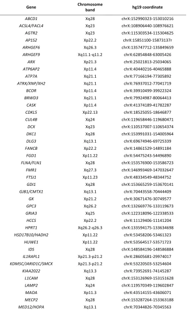
Table S1 Genes on the next-‐generation sequencing XLID panel (Ambry).
Gene Chromosome band hg19 coordinate
ABCD1 Xq28 chrX:152990323-‐153010216
ACSL4/FACL4 Xq23 chrX:108906440-‐108976621
AGTR2 Xq23 chrX:115303534-‐115304625
AP1S2 Xp22.2 chrX:15851100-‐15873137r
ARHGEF6 Xq26.3 chrX:135747712-‐135849659
ARHGEF9 Xq11.1-‐q11.2 chrX:62854848-‐63005426
ARX Xp21.3 chrX:25021813-‐25034065
ATP6AP2 Xp11.4 chrX:40440216-‐40465888
ATP7A Xq21.1 chrX:77166194-‐77305892
ATRX/XNP/XH2 Xq21.1 chrX:76937012-‐77041719
BCOR Xp11.4 chrX:39910499-‐39922324
BRWD3 Xq21.1 chrX:79924987-‐80064413
CASK Xp11.4 chrX:41374189-‐41782287
CDKL5 Xp22.13 chrX:18525055-‐18646877
CUL4B Xq24 chrX:119658446-‐119680471
DCX Xq23 chrX:110537007-‐110654374
DKC1 Xq28 chrX:153991031-‐154005964
DLG3 Xq13.1 chrX:69674946-‐69725339
FANCB Xp22.2 chrX:14861529-‐14891184
FGD1 Xp11.22 chrX:54475243-‐54496890
FLNA/FLN1 Xq28 chrX:153576900-‐153586723
FMR1 Xq27.3 chrX:146993469-‐147032647
FTSJ1 Xp11.23 chrX:48334549-‐48344752
GDI1 Xq28 chrX:153665259-‐153670141
GJB1/CMTX1 Xq13.1 chrX:70443558-‐70444409
GK Xp21.2 chrX:30671476-‐30749577
GPC3 Xq26.2 chrX:132669776-‐133119673
GRIA3 Xq25 chrX:122318096-‐122338533
HCCS Xp22.2 chrX:11129406-‐11141204
HPRT1 Xq26.2-‐q26.3 chrX:133594175-‐133634698
HSD17B10/HADH2 Xp11.22 chrX:53458206-‐53461323
HUWE1 Xp11.22 chrX:53564517-‐53571723
IDS Xq28 chrX:148584196-‐148586884
IL1RAPL1 Xp21.3-‐p21.2 chrX:28605681-‐29974017
KDM5C/JARID1C/SMCX Xp21.3-‐p21.2 chrX:53220503-‐53254604
KIAA2022 Xq13.3 chrX:73952691-‐74145287
L1CAM Xq28 chrX:153126969-‐153151628
LAMP2 Xq24 chrX:119570349-‐119602847
MAOA Xp11.3 chrX:43514155-‐43606071
MECP2 Xq28 chrX:153287264-‐153363188
MID1 Xp22.2 chrX:10422910-‐10645779
MTM1 Xq28 chrX:149737047-‐149841616
NDP Xp11.3 chrX:43808024-‐43832921
NDUFA1 Xq24 chrX:119005734-‐119010629
NHS Xp22.13 chrX:17393543-‐17754113
NLGN3 Xq13.1 chrX:70364681-‐70391051
NLGN4/NLGN4X Xp22.32-‐p22.31 chrX:5808083-‐6146706
OCRL Xq26.1 chrX:128722863-‐128726530
OFD1 Xp22.2 chrX:13752832-‐13787480
OPHN1 Xq12 chrX:67262186-‐67653299
OTC Xp11.4 chrX:38211736-‐38280703
PAK3 Xq23 chrX:110346387-‐110464173
PDHA1 Xp22.12 chrX:19362011-‐19379825
PGK1 Xq21.1 chrX:77361859-‐77382324
PHF6 Xq26.2 chrX:133507342-‐133562822
PHF8 Xp11.22 chrX:53969048-‐54069627
PLP1 Xq22.2 chrX:103031781-‐103047547
PORCN Xp11.23 chrX:48367371-‐48379202
PQBP1 Xp11.23 chrX:48755775-‐48760422
RPL10 Xq28 chrX:153626571-‐153630680
PRPS1 Xq22.3 chrX:106871654-‐10689425
RPS6KA3/RSK2 Xp22.12 chrX:20168029-‐20285523
SHROOM4/KIAA1202 Xp11.22 chrX:50376178-‐50386683
SLC9A6 Xq26.3 chrX:135067583-‐135129428
SLC16A2/MCT8 Xq13.2 chrX:73641328-‐73753764
SMC1A/SMC1L1 Xp11.22 chrX:53431731-‐53449618
SMS Xp11.23 chrX:21958691-‐22012955
SOX3 Xq27.1 chrX:139585152-‐139587225
SRPX2 Xp22.1 chrX:99899163-‐99926296
SYN1 Xp11.23 chrX:47431300-‐47479256
SYP Xp11.23 chrX:49044265-‐49056661
TIMM8A Xq22.1 chrX:100603026-‐100603957
TSPAN7/TM4SF2 Xp11.4 chrX:38420731-‐38548172
UBE2A Xq24 chrX:118714298-‐118718379
UPF3B Xq24 chrX:118967989-‐118986991
ZDHHC9 Xq26.1 chrX:128948634-‐128978124
ZNF41 Xp11.23 chrX:47305561-‐47342610
ZNF81 Xp11.23 chrX:47696301-‐47781655
ZNF674 Xp11.3-‐p11.23 chrX:46357160-‐46404892
ZNF711 Xq21.1 chrX:84520124-‐84527248
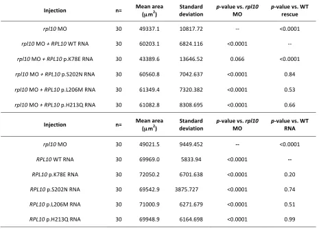
Table S2 Brain cross sectional areas, means and p-‐values in rpl10 models
Injection n= Mean area (µm2)
Standard
deviation p-‐value vs. MO rpl10 p-‐value vs. WT rescue
rpl10 MO 30 49337.1 10817.72 -‐-‐ <0.0001
rpl10 MO + RPL10 WT RNA 30 60203.1 6824.116 <0.0001 -‐-‐
rpl10 MO + RPL10 p.K78E RNA 30 43389.6 13646.52 0.066 <0.0001
rpl10 MO + RPL10 p.S202N RNA 30 60560.8 7042.637 <0.0001 0.84
rpl10 MO + RPL10 p.L206M RNA 30 61349.4 7320.382 <0.0001 0.53
rpl10 MO + RPL10 p.H213Q RNA 30 61082.8 8308.695 <0.0001 0.66
Injection n= Mean area
(µm2)
Standard deviation
p-‐value vs. rpl10
MO
p-‐value vs. WT RNA
rpl10 MO 30 49021.5 9449.452 -‐-‐ <0.0001
RPL10 WT RNA 30 69969.0 5833.94 <0.0001 -‐-‐
RPL10 p.K78E RNA 30 72050.2 6701.638 <0.0001 0.20
RPL10 p.S202N RNA 30 69542.9 3875.727 <0.0001 0.74
RPL10 p.L206M RNA 30 71000.9 6271.679 <0.0001 0.51
RPL10 p.H213Q RNA 30 69948.9 6164.698 <0.0001 0.99
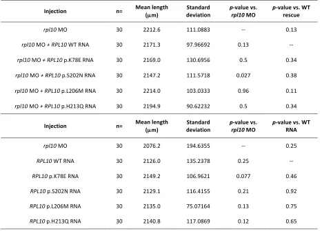
Table S3 Body length measurements, means and p-‐values in rpl10 models
Injection n= Mean length (µm)
Standard
deviation p-‐rpl10 value vs. MO p-‐value vs. WT rescue
rpl10 MO 30 2212.6 111.0883 -‐-‐ 0.13
rpl10 MO + RPL10 WT RNA 30 2171.3 97.96692 0.13 -‐-‐
rpl10 MO + RPL10 p.K78E RNA 30 2169.0 130.6956 0.5 0.34
rpl10 MO + RPL10 p.S202N RNA 30 2147.2 111.5718 0.027 0.38
rpl10 MO + RPL10 p.L206M RNA 30 2214.0 103.0333 0.96 0.11
rpl10 MO + RPL10 p.H213Q RNA 30 2194.9 90.62232 0.5 0.34
Injection n= Mean length
(µm)
Standard deviation
p-‐value vs.
rpl10 MO
p-‐value vs. WT RNA
rpl10 MO 30 2076.2 194.6355 -‐-‐ 0.25
RPL10 WT RNA 30 2126.0 135.2378 0.25 -‐-‐
RPL10 p.K78E RNA 30 2149.2 106.9621 0.077 0.46
RPL10 p.S202N RNA 30 2129.1 116.4155 0.21 0.92
RPL10 p.L206M RNA 30 2135.0 75.07164 0.13 0.75