Scholarship@Western
Scholarship@Western
Electronic Thesis and Dissertation Repository
8-22-2011 12:00 AM
Exploring Bacterial Nanowires: From Properties to Functions and
Exploring Bacterial Nanowires: From Properties to Functions and
Implications
Implications
Kar Man Leung
The University of Western Ontario
Supervisor Jun Yang
The University of Western Ontario
Graduate Program in Mechanical and Materials Engineering
A thesis submitted in partial fulfillment of the requirements for the degree in Doctor of Philosophy
© Kar Man Leung 2011
Follow this and additional works at: https://ir.lib.uwo.ca/etd
Part of the Biological and Chemical Physics Commons, Microbial Physiology Commons, and the Nanoscience and Nanotechnology Commons
Recommended Citation Recommended Citation
Leung, Kar Man, "Exploring Bacterial Nanowires: From Properties to Functions and Implications" (2011). Electronic Thesis and Dissertation Repository. 235.
https://ir.lib.uwo.ca/etd/235
This Dissertation/Thesis is brought to you for free and open access by Scholarship@Western. It has been accepted for inclusion in Electronic Thesis and Dissertation Repository by an authorized administrator of
(Spine Title: Exploring the Properties and Functions of Bacterial Nanowires)
(Thesis format: Monograph)
by
Kar Man Leung
Department of Mechanical and Materials Engineering Faculty of Engineering
A thesis submitted in partial fulfillment of the requirements for the degree of
Doctor of Philosophy
The School of Graduate and Postdoctoral Studies The University of Western Ontario
London, Ontario, Canada
ii
THE UNIVERSITY OF WESTERN ONTARIO School of Graduate and Postdoctoral Studies
CERTIFICATE OF EXAMINATION
Supervisor
______________________________ Dr. Jun Yang
Co-Supervisor
______________________________ Dr. Leo W. M. Lau
Examiners
______________________________ Dr. Xueliang Sun
______________________________ Dr. Liying Jiang
______________________________ Dr. Jayshri Sabarinathan
______________________________ Dr. P. Ravi Selvaganapathy
The thesis by
Kar Man Leung
entitled:
Exploring Bacterial Nanowires: From Properties to Functions and
Implications
is accepted in partial fulfillment of the requirements for the degree of
Doctor of Philosophy
iii
Abstract
The discovery of electrically conductive bacterial nanowires from a broad range of microbes
provides completely new insights into microbial physiology such as extracellular electron
transfer, energy distribution, and cell-to-cell communication. There have been a variety of
hypothesized functions of bacterial nanowires that raise a number of fundamental questions
on these biological nanostructures. This thesis work is carried out to provide answers to these
fundamental, but important, questions by employing advanced nanofabrication techniques
and nanoscopic characterization tools.
By limiting the availability of electron acceptors, Shewanella oneidensis strain MR-1, a
dissimilatory metal-reducing bacterium, produces extracellular bacterial nanowires
connecting neighboring cells. Characterized by a variety of microscopy techniques,
Shewanella nanowires are found to be up to tens of micrometers long, with a lateral
dimension of ~10 nm for single, non-bundled nanowires. Fluorescence microscopy reveals
that the nature of bacterial nanowires is protein. In addition, CP-AFM and STM images
suggest that bacterial nanowires are efficient electrical conductors.
Direct electrical transport measurements along Shewanella nanowires using nanofabricated
electrodes and CP-AFM consistently reveal a measured nanowire resistivity on the order of 1
Ω·cm along micro-meter length scales. With electron transport rates up to 109/s at 100 mV,
bacterial nanowires can serve as a viable microbial strategy for extracellular electron
transport. By modulating the nanowire conductance in NW-FET configurations and CP-AFM
measurements on nanowires supported on different electrode materials, it has been found that
iv
5.3 eV. This finding further suggests that Shewanella nanowires may serve as electronic
cables for efficient energy distribution and electronic signal exchange within microbial
communities.
Two separate AFM experiments, which are real-time elastic modulus mapping using
torsional harmonic cantilevers and conventional AFM nanoindentation, mutually confirm
that the elastic modulus of Shewanella nanowires is on the order of 1 GPa, with no
significant variations in local elasticity along individual nanowires. With electrical properties
comparable to those of moderately doped inorganic semiconductors and elasticity close to
polymeric materials, bacterial nanowires may represent a new class of functional
bionanomaterials that will potentially be building blocks for bionanoelectronics and flexible
nanoelectronics.
Keywords
v
Acknowledgments
This thesis is the result of not only my efforts but also many others who have supported me
in different ways throughout my Ph.D. study.
First of all, I would like to express my deepest gratitude to my supervisors, Prof. Jun Yang
and Prof. Leo Lau. Prof. Yang has always been friendly, supportive, and motivating me to
think outside the box to get new insights into each research topic. His valuable comments and
suggestions have led to significant achievements in this thesis work. Prof. Lau’s guidance,
deep knowledge, and brilliant advice gave me the confidence to carry out this research work
and face all its challenges. Also, I enjoyed the parties in his house very much every
Christmas and on other important days.
I would like to thank Prof. Gordon Southam, who taught me a lot about microbiology.
Taking his Geomicrobiology course and working in his lab are very enjoyable experiences. I
must also thank my collaborator, Dr. Greg Wanger, who is also my microbiology teacher.
From them, I learned the subject of microbiology, from knowledge to hand-on skills, with a
lot of fun. Moreover, I am so grateful to Prof. Yuri Gorby and Prof. Mohamed Y. El-Naggar
for their helpful comments and suggestions. Our collaboration has been very productive and
successful.
Technical support from the staff at the Western Nanofabrication Facility, including Dr. Todd
Simpson, Dr. Rick Glew, and Mr. Tim Goldhawk, are highly appreciated. I would also like to
appreciate assistance from the following colleagues: Dr. Yu Liu, Mr. Tomas Trebicky, Dr.
Frank Zhao, Mr. Qiuquan Guo, Dr. Thomas Lei, Mr. Tingjie Li, and Dr. Mei Liu. Special
vi
Suzanne Hicks for assistance in purchasing and logistics, and Mr. Ross Davidson, Mr. Brad
Kobe and Mr. Gary Good for their technical assistance.
Finally, I would like to express my highest gratefulness to my wife, Ka Po, for her patience
and understanding, for going through all ups and downs with me, and for her faith in me. I
am forever grateful to my parents, Richard and Betty, and my brother, Eric, for their
vii
Table of Contents
CERTIFICATE OF EXAMINATION... ii
Abstract... iii
Acknowledgments... v
Table of Contents... vii
List of Tables ... x
List of Figures ... xi
List of Abbreviations ... xvi
Chapter 1... 1
1 Introduction... 1
1.1 Brief Background... 1
1.2 Research Aim and Outline of this Thesis ... 2
Chapter 2... 5
2 Literature Review... 5
2.1 Generation of Energy for Metabolism by Microbes ... 5
2.2 Bacterial Strategies for Extracellular Electron Transfer... 8
2.3 Extracellular Electron Transfer Toward Practical Applications in Environmental Biotechnology and Bio-energy Generation ... 12
2.3.1 Bioremediation... 12
2.3.2 Microbial Fuel Cells... 15
2.4 Long-range Electron Transfer Facilitated by Extracellular Nanowires... 17
2.4.1 Bacterial Nanowires from Geobacter sulfurreducens... 17
2.4.2 Bacterial Nanowires Produced by Shewanella oneidensis Strain MR-1 ... 20
viii
Chapter 3... 24
3 Cultivation of Shewanella oneidensis MR-1 for Bacterial Nanowire Production and Characterization Techniques... 24
3.1 Cultivation in Continuous Flow Chemostat Bioreactors ... 24
3.2 Sample Preparation and Characterizations of Bacterial Nanowires ... 29
3.2.1 Scanning Electron Microscopy ... 30
3.2.2 Transmission Electron Microscopy... 35
3.2.3 Fluorescence Microscopy... 39
3.2.4 Atomic Force Microscopy... 41
3.2.5 Scanning Tunneling Microscopy ... 46
3.3 Concluding Remarks... 49
Chapter 4... 50
4 Electrical Transport Measurements along Bacterial Nanowires from Shewanella oneidensis MR-1 ... 50
4.1 Introduction... 50
4.2 Experimental Methods... 50
4.2.1 Cell Cultivation ... 51
4.2.2 Nanofabricated Devices for Electrical Measurements... 52
4.2.3 Conducting-probe Atomic Force Microscopy Measurements ... 57
4.3 Direct Electrical Measurements Using Nanofabricated Electrodes... 59
4.4 Measurements with Conducting Atomic Force Microscopy Probes ... 63
4.5 Discussion... 68
4.6 Conclusions... 69
Chapter 5... 70
5 Semiconducting Behavior of Shewanella Bacterial Nanowires ... 70
5.1 Introduction... 70
ix
5.3 Electronic Transport Chacteristics of Shewanella Nanowires Coupled to
Electrodes... 74
5.4 Modulating the Conductance of Shewanella Nanowires Configured in Nanowire Field-Effect Transistor Structures... 78
5.5 Discussion... 81
5.6 Concluding Remarks... 82
Chapter 6... 84
6 Mechanical Properties of Shewanella Nanowires Studied by Atomic Force Microscopic Techniques ... 84
6.1 Introduction... 84
6.2 Experimental Methods... 87
6.3 Quantitative Nanomechanical Property Mapping of Shewanella Nanowires by Torsional Harmonic AFM Probes... 89
6.4 Conventional AFM Nanoindentation on Shewanella Nanowires... 94
6.5 Discussions ... 96
6.6 Concluding Remarks... 97
Chapter 7... 99
7 Thesis Summary and Future Work ... 99
7.1 Summary ... 99
7.2 Thesis Contributions ... 102
7.3 Future Work... 103
References... 104
x
List of Tables
xi
List of Figures
Figure 2.1: Simplified diagram of aerobic respiration of bacteria by oxidizing glucose and reducing oxygen.. ... 7
Figure 2.2: Pathway of extracellular Fe(III) reduction in S. oneidensis with outer-membrane
c-type cytochromes OmcA and MtrC. Black solid circles represent heme groups.... ... 10
Figure 2.3: Two bacterial strategies for indirect extracellular electron transfer to iron oxide. (a) A chelator (yellow oval) brings soluble iron to the cell for reduction. (b) An electron shuttle (blue closed oval is in the oxidized state, open oval in the reduced state) catalyzes electron transfer from the cell to iron oxide at a distance from the cell... 11
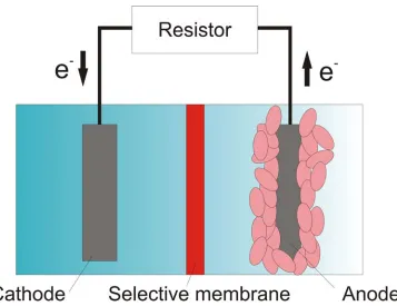
Figure 2.4: A schematic drawing illustrating the basic structure of a microbial fuel cell... ... 16
Figure 2.5: A conceptual bio-geobattery model describing generation of electric field by transferring electrons from the anode (reduced zone) to the cathode (oxidized zone) through bacterial nanowires... 21
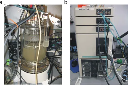
Figure 3.1: (a) Continuous flow bioreactor employed for cultivation of S. oneidensis MR-1 (wild-type). The bioreactor is integrated with (b) a controlling unit for gas flow control, agitation control, pH and dissolved oxygen concentration monitoring, fresh medium supply, and old medium withdrawal... ... 25
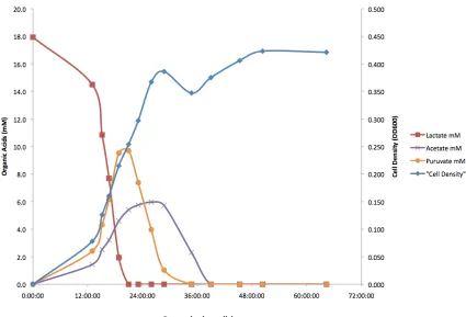
Figure 3.2: Organic acid concentration (mM) and cell density (OD600) versus growth time (h) for S. oneidensis MR-1 (wild-type) cultivated in batch mode... 27
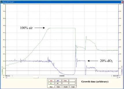
Figure 3.3: A screen view captured from the controller display showing the dissolved oxygen concentration of air and the percentage air versus nitrogen supplied to the bioreactor as a function of growth time... 29
xii
express a high amount of nanofilaments connecting neighboring cells. This sample was chemically fixed, dehydrated through a graduated series of ethanol, critical-point dried, and coated with Pt prior to SEM imaging.... ... 33
Figure 3.5: An SEM image showing cells of S. oneidensis MR-1 harvested in a continuous flow chemostat bioreactor operating under electron-acceptor-limited conditions. This sample was only chemically fixed and Pt coated prior to SEM imaging. Without going through ethanol dehydration processes and critical-point drying, the cell structures were severely deformed and collapsed, especially when subjected to high-vacuum environment in SEM, although nanofilamentous structures could still be observed.... ... 34
Figure 3.6: An SEM image showing cells of S. oneidensis MR-1 harvested in batch culture. In addition to nanofilaments, extracellular polymeric substances (EPS) are observed in the sample, which can challenge microscopic analysis and electrical measurements on bacterial nanowires.... ... 35
Figure 3.7: (a) TEM micrographs showing a negatively stained S. oneidensis MR-1 bacterium with nanofilaments (indicated by arrows in (b) and (c)) emanating from the cell body with a lateral dimension of ~10 nm measured from the TEM images... 37
Figure 3.8: (a) TEM micrographs showing a negatively stained S. oneidensis MR-1 bacterium with a flagellum (indicated by the arrow in (b)) emanating from the pole of the cell and a branched nanofilament (indicated by the arrow in (c)) from the side of the cell... 38
Figure 3.9: An optical micrograph showing cells of S. oneidensis MR-1. Nanofilaments such as nanowires and flagella cannot be observed owing to the limitation of resolution, which is typically 200 nm for visible light illumination.... ... 40
xiii
Figure 3.11:Tapping-mode (a) topography and (b) amplitude AFM images of S. oneidensis
MR-1 cells. The scale bar is 2 µm... 43
Figure 3.12:(a) Topography image and (b) simultaneous conductivity map of a bacterial nanowire from S. oneidensis MR-1 collected in batch culture accompanied with some non-conducting substances, possibly EPS. (c) The height and (d) current profiles (indicated by black lines) across the nanowire reveal that the radial dimension of the nanowire is ~10 nm and the nanowire is electrically conductive... 44
Figure 3.13:Simple model explaining the broadening effect due to AFM tip geometry. Comparing with the actual width of the object (upper), the apparent width in the topographic image (lower) is over estimated but the height can be measured very accurately... 45
Figure 3.14:(a) STM image and (b) height profile confirm that bacterial nanowires from S. oneidensis MR-1 are highly conductive and can be composed of bundles of individual nanofilaments... ... 48
Figure 4.1: Optical micrograph of a microfabricated structure on a SiO2/Si substrate with Au
microelectrodes and contact pads for electrical measurements... 53
Figure 4.2: Schematics of microfabrication procedures of devices used as platforms for electrical measurements of bacterial nanowires... ... 54
Figure 4.3: SEM images of (a) close-circuit and (b) open-circuit controls. (c) I-V curves of (a) and (b) are indicated by squares and circles, respectively... ... 56
Figure 4.4: AFM topography image of prefabricated Au microgrids on a SiO2/Si substrate...
... 58
xiv
Figure 4.6: (a) SEM image showing a single bacterial nanowire emanating from a wild-type
S. oneidensis MR-1 cell contacted with two nanofabricated Pt electrodes. (b) Current-voltage curve of the bacterial nanowire in (a). (c) SEM image of another bacterial nanowire with the measured I-V curve shown in (d)... ... 61
Figure 4.7: (a) SEM image showing a single bacterial nanowire from the S. oneidensis
flagellin-deficient mutant contacted with two nanofabricated Pt electrodes. (b) Current-voltage curve of the bacterial nanowire... ... 62
Figure 4.8: (a and b) SEM images showing two individual extracellular appendages from the
ΔmtrC/omcA mutants that are morphologically consistent with wild-type nanowires but are electrically non-conductive as confirmed by electrical measurements... ... 63
Figure 4.9: Topographic AFM image showing air-dried S. oneidensis MR-1 cells and extracellular appendages deposited randomly on a SiO2/Si substrate patterned
with Au microgrids... 64
Figure 4.10:Schematic diagram illustrating electronic transport along a single bacterial nanowire in contact with Au microgrids measured by a Pt-coated AFM tip... .. 65
Figure 4.11: (a) Contact mode AFM image showing a bacterial nanowire reaching out from a
S. oneidensis MR-1 cell to the Au electrode. (b) An I-V curve obtained by probing the nanowire at a length of 600 nm away from the Au electrode (at the position marked by the black dot in (a)). (Inset) The I-V curves obtained on bare Au and SiO2, respectively... 66
Figure 4.12: A plot of total resistance as a function of distance between AFM tip and Au electrode. The contact resistance is estimated by extrapolating the linear curve to zero length... ... 67
xv
Figure 5.2: (a and b) Current-voltage curves collected on two individual Shewanella bacterial nanowires when sandwiched between a Pt-coated AFM tip and HOPG substrate... 76
Figure 5.3: Structure of a NW-FET device with a single bacterial nanowire contacted by source (S) and drain (D) electrodes. The topography shown is an amplitude AFM image of the device.... ... 79
Figure 5.4: (a) IDS-VDS characteristics of a Shewanella nanowire at different gate voltages. (b)
Transfer characteristic of the NW-FET at VDS = –1 V... 80
Figure 6.1: SEM image of a torsional harmonic cantilever with a T-shaped geometry and an off-axis tip.... ... 87
Figure 6.2: A tip-sample force waveform captured when operating under HarmoniX mode AFM.... ... 89
Figure 6.3: (a) Topography of a bacterial nanowire supported on HOPG and (b) the corresponding elastic modulus map simultaneously generated. Numerical values across the sections indicated by the white lines are plotted below the images....
... 91
Figure 6.4: Topographic (a and b) and corresponding elastic modulus (c and d) micrographs of bacterial nanowires supported on PMMA and borosilicate glass substrates, respectively. Numerical values across the sections indicated by the white lines are plotted below the images.... ... 92
xvi
List of Abbreviations
ADP Adenosine diphosphate
AFM Atomic force microscopy
ATP Adenosine triphosphate
CP-AFM Conducting-probe atomic force microscopy D Drain
DMRB Dissimilatory metal-reducing bacteria DOS Density of states
EBID Electron-beam induced deposition
EPS Extracellular polymeric substances
FET Field-effect transistor
GIS Gas injection system
HOPG Highly oriented pyrolytic graphite I-V Current-voltage
MFC Microbial fuel cell
MOSFET Metal-oxide-semiconductor field-effect transistor
NTA Nitro-triacetic acid
NW Nanowire
PBS Phosphate buffered saline
PEM Proton exchange membrane
PMMA Polymethylmethacrylate S Source
SEM Scanning electron microscopy STM Scanning tunneling microscopy TEM Transmission electron microscopy THC Torsional harmonic cantilever
Chapter 1
1
Introduction
The background of this thesis work is briefly introduced here in the first chapter. In
addition, the research aim and outline of this thesis are presented.
1.1 Brief
Background
In microbial world, microbes can generate energy using diverse strategies for cell growth
and/or maintenance. Such energy generating processes can be simplified as a chain of
electron-transfer reactions, from extracting electrons from an electron donor (organic
foodstuffs) to dumping electrons to a terminal electron acceptor, involving a series of
redox reactions within the whole process (more details are discussed in Chapter 2).
Although most bacteria use soluble electron acceptors such as oxygen or carbon dioxide,
some bacteria are capable of extracellular electron transfer in harsh environments where
the only available electron acceptors are insoluble or poorly soluble, for instance,
metal-oxide minerals. In recent years, increasing attention has been paid to microbial
metabolisms that involve electron transfer between cells and extracellular substrates. The
recent discovery of electrically conductive bacterial nanowires produced by a variety of
microbes suggests that electron transfer via nanowires may be widespread in nature, and
inspires this thesis work in further exploring the properties of bacterial nanowires, testing
and communication, and looking for their potential applications in nanobiotechnology
and nanobioelectronics.
1.2 Research Aim and Outline of this Thesis
It is the aim of this thesis work to explore the functions and properties of bacterial
nanowires from a dissimilatory metal-reducing bacterium, Shewanella oneidensis strain
MR-1, which have immense implications in microbial physiology, ecology,
electromicrobiology, biotechnology, and nanotechnology. To achieve this aim, the
electrical, electronic and mechanical properties of Shewanella nanowires are studied
using techniques including microfabricated chips, nanofabricated devices, and
AFM-based electrical and mechanical measurements.
Chapter 2 presents a review of the currently known microbial strategies for electron
transfer to extracellular electron acceptors and related applications in environmental
biotechnology as well as in bio-energy production. It is also discussed that bacterial
nanowires may be a more general, but previously unknown, microbial strategy for
effective electron transfer to distantly located electron acceptors, including solid-phase
metal-oxide minerals, electrodes and possibly neighboring cells.
In Chapter 3, detailed cultivation procedures and the optimal growth conditions to induce
bacterial nanowire production by Shewanella oneidensis MR-1 are described.
Characterizations of bacterial nanowires by a variety of microscopy techniques including
characterizations of bacterial nanowires using scanning probe microscopy (CP-AFM and
STM) are demonstrated.
Chapter 4 demonstrates direct electrical transport measurements along bacterial
nanowires from S. oneidensis MR-1 by two independent techniques: (i) nanofabricated
electrodes patterned on top of individual nanowires, and (ii) conducting AFM probing at
various points along a single nanowire bridging a prefabricated metallic microelectrode
and the conductive AFM tip. This is the first study to prove that bacterial nanowires are
able to conduct electrons along micron length scales.
Chapter 5 presents electronic transport studies across junctions between bacterial
nanowires and electrodes using CP-AFM, and modulation of bacterial nanowire
conductance under NW-FET device structures. This work reveals the mechanism of
electrical transport in these biological nanostrucutres, and suggests that Shewanella
nanowires may not only serve as conduits to transfer electrons throughout the
extracellular matrix, but also as cables for efficient energy distribution and electronic
information exchange such as for cell-cell communication within microbial communities.
Chapter 6 presents studies on the mechanical properties of Shewanella bacterial
nanowires using two separate AFM techniques: (i) real-time elastic modulus mapping
using torsional harmonic cantilevers with a T-shaped geometry and an offset tip, and (ii)
conventional AFM nanoindentation with subsequent curve fitting based on the classical
Hertz model. This work inspires us with new applications of bacterial nanowires: with
semiconductors and elasticity similar to polymeric materials, bacterial nanowires may
function as building blocks for bionanoelectronics and flexible nanoelectronics.
Most of the work presented in this thesis has been published in peer-reviewed journals or
Chapter 2
2
Literature Review
In this chapter, different reported strategies of how microbes generate energy are
reviewed. The basic principle of biological energy generation is based on oxidation and
reduction processes involving electron transfer within cells and/or outside of the cells to
terminal electron acceptors. In the case of electron transfer outside of the cells known as
extracellular respiration or extracellular electron transfer, a number of mechanisms have
been proposed although not many of them are completely understood. Some of the
mechanisms have been experimentally proved to be in good agreement with the proposed
theories, but there are definitely still knowledge gaps in each model yet to be filled and
perhaps unknown mechanisms to be explored.
2.1 Generation of Energy for Metabolism by Microbes
Microbes can generate energy using diverse strategies. The fundamental principle of
energy generation for metabolism is based on transfer of electrons from electron donors
with lower (or more negative) reduction potentials (also known as reduction/oxidation or
redox potentials), through an electron transport chain, to terminal electron acceptors with
higher (or more positive) reduction potentials. The electron transport chain couples the
electron transfer to the transfer of protons across a cell membrane, producing an
electrochemical proton gradient as a result of a series of redox reactions, releasing
currency used for intracellular energy transfer. ATP provides biological energy for a
variety of cellular functions such as motility, synthesis of macromolecules (DNA,
proteins, etc), and cell division.
Organic substances are the most common electron donors, whereas some prokaryotes
(bacteria and archaea) can use inorganic substances as their energy sources. As
mentioned earlier, the electron transport chain consists of a series of redox reactions in
which electrons are transferred from a donor to an acceptor. The driving force of such
reactions is based on the Gibbs free energy of the reactants and products. Whether a
reaction can proceed depends on whether the reaction can decrease the Gibbs free energy
of the system. In general when the electron acceptor is a freely diffusible gas (e.g.
oxygen) in the liquid environment, electrons are extracted from oxidization of organic
nutrients and transferred through multiple redox reactions, carried out by a series of
protein complexes in the cell membranes, to oxygen, which is reduced to water. This
process, which drives the production of ATP, is called oxidative phosphorylation. A
simplified diagram of aerobic respiration of bacteria using glucose as the electron donor
and oxygen as the electron acceptor is illustrated in Figure 2.1. An equation for the
overall reaction is shown below:
C6H12O6 + 6 O2→ 6 CO2 + 6 H2O
In this respiration process, electrons are transported from enzymes to enzymes, which are
embedded in the cell membrane, while protons (H+) are transferred across the membrane,
creating a proton gradient. Powered by this transmembrane gradient, one of the most
ATP by coupling adenosine diphosphate (ADP) and inorganic phosphate (Pi) when the
protons are transferring back through the channel in ATP synthase. The overall reaction
is:
ADP + Pi→ ATP
Oxygen generates the highest Gibbs free energy change on hydrolysis than any other
electron acceptors in the electron transport chain. Facultative anaerobic bacteria use
oxygen as the terminal electron acceptor, but they can also survive by “breathing” other
electron acceptors if oxygen is not present. Some bacteria such as strict anaerobes even
die in presence of oxygen. Therefore, these types of bacteria use alternative electron
acceptors for cellular respiration. Some of these acceptors are soluble such as nitrate,
sulfate, and carbonate, while some are insoluble minerals, such as iron and manganese
oxides. The latter case involves electron transfer outside of cells to solid interfaces. This
process is called extracellular electron transfer or extracellular respiration.
2.2 Bacterial Strategies for Extracellular Electron Transfer
When electron acceptors are not freely soluble or readily dissolved, bacteria seek other
strategies to facilitate interactions between the cellular electron transport chain and the
extracellular substrates. Extracellular electron transfer is a promising research field
including but not limited to cell biology, genetics, and biochemistry [1]. Although our
understanding on how bacteria transfer electrons outside of cells is incomplete, a variety
of mechanisms have been proposed and observed. The mechanism of bacterial
extracellular electron transfer can be mainly divided into two categories: direct and
indirect interactions.
For direct interactions, bacteria express proteins on the cell surfaces that are able to
transfer electrons to the solid-phase electron acceptors when the proteins and the
the most abundant form of Fe(III) in sedimentary environments, has been observed in
dissimilatory metal-reducing bacteria (DMRB) for genera such as Shewanella [2, 3],
Geobacter [3, 4], Pelobacter [5], Geovibrio [6], Ferrimonas [7], Desulfuromusa [8], and
Desulfuromonas [9]. Commonly, the proteins that are involved in electron transfer to
minerals such as iron/manganese (hydr)oxides are localized to the outer membrane of
Gram-negative bacteria. In Gram-negative bacteria such as Shewanella oneidensis MR-1
and Geobacter sulfurreducens, it has been suggested that outer-membrane redox-active
proteins called cytochromes play critical roles in dissimilatory reduction of solid metal
(hydr)oxides by facilitating electron transport across the cell envelope [3]. Lower et al.
showed that specific interaction forces are induced between Shewanella oneidensis and
goethite (α-FeOOH) under anaerobic conditions in which extracellular electron transfer is
expected [10]. Xiong et al. observed high-affinity binding and direct electron transfer to
hematite by the Shewanella oneidensis MR-1 outer-membrane c-type cytochrome OmcA
[11]. Parikh and Chorover employed attenuated total reflection (ATR) Fourier transform
infrared (FTIR) spectroscopy and revealed significant short-range bonding interactions
during adhesion to iron oxide (α-Fe2O3) by Shewanella oneidensis and other
Gram-positive bacteria [12]. To date, the mechanism of how electrons are transported from
these outer-membrane cytochromes is not fully understood, but studies shown above
provided supporting evidences on bacterial extracellular electron transfer through direct
contact. A model of Fe(III) reduction pathway by S. oneidensis is illustrated in Figure
2.2. In this model, electrons are extracted from lactate as an electron donor and
transferred to a series of multi-heme cytochromes, which are located in the inner
Figure 2.2: Pathway of extracellular Fe(III) reduction in S. oneidensis with outer-membrane c-type cytochromes OmcA and MtrC. Black solid circles represent heme
groups. Figure copied from Ref [13].
For indirect extracellular electron transfer, bacteria are not or may not able to be in direct
contact with the terminal electron acceptors. They adopt another strategy by producing
some small molecules that traverse the space between the cells and the extracellular
substrates. In indirect electron transfer to poorly soluble minerals, these small molecules
act either to chelate metals and deliver them to an intracellular metal oxidoreductase or
by themselves serving as electron shuttles [1]. In the case of metal chelators, ligands such
as citrate and nitro-triacetic acid (NTA) are capable of delivering soluble iron to the cell
for reduction, either on the outside or on the inside [1]. An electron shuttle is a mobile
molecule that serves as the terminal electron acceptor and is able to undergo redox
cycling for thousands of times [14]. At its reduced state, the electron shuttle can transfer
serving as the terminal electron acceptor. Some organic molecules that can play this role
are humic substances such as quinones, phenazines, and thiol-containing molecules like
cysteine [14, 15]. A schematic diagram is presented in Figure 2.3 demonstrating how a
bacterium uses a chelator or electron shuttle for extracellular respiration.
Figure 2.3: Two bacterial strategies for indirect extracellular electron transfer to iron oxide. (a) A chelator (yellow oval) brings soluble iron to the cell for reduction.
(b) An electron shuttle (blue closed oval is in the oxidized state, open oval in the reduced state) catalyzes electron transfer from the cell to iron oxide at a distance
2.3 Extracellular Electron Transfer Toward Practical
Applications in Environmental Biotechnology and
Bio-energy Generation
Microorganisms that are able to sustain metabolism in anoxic conditions by carry out
extracellular electron transfer have been of great interest in biotechnological applications
such as bioremediation and electricity generation. In order to realize practical
applications in these areas, research is continuously ongoing to investigate the
mechanism of extracellular electron transfer, how electrons are transferred to electron
acceptors, and what factors control the rate and extent of the process [16]. In this
subsection, the basic concepts of how extracellular electron transfer plays an important
role in these applications will be outlined along with some examples.
2.3.1 Bioremediation
The goal of bioremediation is to restore contaminated environments by means of
microbial metabolism in an inexpensive and environmentally friendly way. The
restoration can be aided by utilizing proper microorganisms to oxidize, bind, immobilize,
volatilize, or transform the contaminants [16]. Contaminants in these environments
include organic pollutants (aromatic hydrocarbons and pesticides), chlorinated
compounds, and metals. Aerobic degradation of toxic organic contaminants to non-toxic
products can be achieved by utilizing aerobic microorganisms, for instance,
Pseudomonas species, which are able to oxidize (extract electrons from) these
contaminants and transfer electrons to oxygen as the terminal electron acceptor [17].
soils, are anoxic, lack of accessibilities and on large scales. Therefore, bioremediation in
these subsurface environments are of great research interest.
The respiratory flexibility of some bacteria makes them good agents for bioremediation
in subsurface, anaerobic environments. For example, Geobacter species are capable of
oxidizing organic (aromatic) contaminants coupled to reduction of Fe(III) oxides [18]. In
addition to choosing proper bacterial species, the efficiency of bioremediation is also
dependent on the availability of suitable electron acceptors for the chosen species in the
contaminated areas. In subsurface environments, Fe(III) is usually the most abundant
electron acceptors for the oxidation of organic compounds [19]. Thus, further enhancing
the availability of Fe(III) in the sites can stimulate anaerobic degradation of organic
contaminants, enriching the species and increasing the bioremediation efficiency [20].
The concentration of sulfate in seawater is high so sulfate is an important electron
acceptor for biodegradation of organic contaminants in marine environments. In such
environments, sulfate-reducing bacteria, such as Desulfobacula and Desulfobacterium
species, are good candidates for the oxidization of hydrocarbons with sulfate as the
electron acceptor [21]. Halogenated compounds are one of the most significant types of
toxic pollutants. Dehalogenating microorganisms are able to use these pollutants as
electron acceptors [22]. This process is called reductive dehalogenation, which is mainly
known to occur under anaerobic conditions [23].
Bioremediation of metals and radionuclides such as chromium (Cr), technetium (Tc), and
uranium (U), is an important process in controlling the fate and transport of these
hazardous substances in anoxic subsurface environments [24]. Because toxic metals
their spread and make the collection or removal processes possible. As the reduced forms
of some metals and radionuclides are insoluble or have very low solubility, they can be
precipitated as immobile forms stimulated by microbial reduction. For example,
bioremediation of uranium-contaminated waters and soils has been suggested by reducing
soluble uranyl ion (UO22+) to highly insoluble uraninite (UO2) [25, 26]. When uranium is
reduced from the U(VI) oxidation state to U(IV), the solubility decreases resulting in
immobilization. A variety of bacterial species are known to be capable of reducing U(VI)
to U(IV) such as Shewanella [27, 28], Geobacter [29], Desulfosporosinus [30],
Desulfovibrio [31, 32], and Clostridium [33]. The biomolecular mechanisms of uranium
reduction are not well understood yet. However, it was reported for Shewanella
oneidensis MR-1 that c-type cytochromes are essential for the reduction of U(VI) and
formation of extracellular UO2 nanoparticles [34], implying a direct electron transfer
mechanism from cytochromes located in the outer membrane to extracellular U(VI).
Uraninite particles produced by microbial reduction are less than 3 nm in diameter [35].
Owing to the small size and high surface-to-volume ratio, it would be expected that the
UO2 nanoparticles are still relatively mobile in liquid environments and can be
re-oxidized easily. Further procedures after microbial reduction to uraninite, such as
flocculation and adsorption onto colloidal particles, have been suggested in order to
reduce the transport potential [35]. To realize practical uranium bioremediation, the
following research direction should be on looking for an effective way to facilitate
2.3.2 Microbial
Fuel
Cells
Other than metal oxide minerals that can serve as extracellular terminal electron
acceptors, electrodes represent another possibly more convenient electron acceptor for
some bacteria [36-38]. One of the most important potential applications utilizing
microbial extracellular electron transfer to electrodes is electricity generation called
microbial fuel cells (MFCs). In MFCs, the fuel source can be any microbially degradable
organic matter. Although MFCs are not yet commercialized owing to challenges such as
low power densities and high production costs [39], MFCs have a number of advantages
over typical abiotic fuel cells that are powered by hydrogen and alcohol. These abiotic
fuel cells require precious, expensive metal catalysts [40] to promote oxidation of the fuel
source, in contrast to MFCs in which the catalysts are naturally occurring bacteria that are
able to oxidize a variety of “costless” fuels such as organic waste and renewable biomass
in soils and sediments. In addition, the catalysts used in some abiotic fuel cells can be
poisoned by carbon monoxide. Some commercial fuel cells that are less susceptible to
poisoning usually need to operate at high temperatures [41], whereas MFCs operates at
room temperature or at temperatures that microbial metabolism can be sustained.
As shown in Figure 2.4, a MFC consists of an anode, which accepts electrons from the
bacteria, and a cathode, which donates electrons to an electron acceptor. The anode
compartment should be maintained under a condition that is free from available electron
acceptors for the bacteria so that the only means for respiration is to transfer electrons to
the anode. The cathode can be suspended in aerobic media or exposed to air [42].
Electrons flow from the anode to the cathode, as a result of electrochemical potential
resistor or other electrical components. The anode and cathode is typically separated by a
proton exchange membrane (PEM), which restricts oxygen to diffuse from the cathode to
the anode compartment and allows protons that are released from the oxidation of the
organic fuel source to pass through the membrane from the anode to the cathode. The
number of electrons that transfer from the anode to the cathode must matched by an equal
number of protons moving between the electrodes in order to preserve the
electroneutrality. It has also been assumed that both anode and cathode can be
biologically catalyzed by placing proper species of bacteria in each electrode
compartment such that the cathode can serve as the electron donor to the bacteria [43].
2.4 Long-range Electron Transfer Facilitated by
Extracellular Nanowires
A novel, previously unrecognized, extracellular electron transfer pathway was reported in
2005 by Reguera et al. [44] that the DMRB Geobacter sulfurreducens can produce
extracellular appendages termed as microbial nanowires, which are electrically
conductive pili, to aid in establishing contacts with and transferring electrons to Fe(III)
oxides. A similar phenomenon was discovered by Gorby et al. [45] in a variety of
bacteria such as the DMRB Shewanella oneidensis MR-1, the oxygenic photosynthetic
cyanobacterium Synechocystis PCC6803, and the thermophilic, fermentative bacterium
Pelotomaculum thermopropioncium that can also produce electrically conductive
appendages, namely bacterial nanowires, when grown under specific conditions. It should
be noted that the terms “microbial nanowires” and “bacterial nanowires” are used
interchangeably in the literature and are both referred to electrically conductive
appendages with nanowire architectures produced by microbes. In this section, the
discoveries and assessments of electrically conductive bacterial nanowires produced by
G. sulfurreducens and S. oneidensis MR-1 are discussed.
2.4.1 Bacterial
Nanowires
from
Geobacter sulfurreducens
Geobacter sulfurreducens, which are strictly anaerobic bacteria and the predominant
Fe(III) reducers, must be in direct contact with Fe(III) oxides to reduce them. Reguera et
al. [44] observed that G. sulfurreducens produced monolateral pili that were proposed to
aid in establishing contact with Fe(III) oxides. Mutants with the gene, PilA, deleted did
crystalline Fe(III) and Mn(IV) oxides, suggesting that G. sulfurreducens required the
assembly of functional pili in order to reduce insoluble electron acceptors. To evaluate
the role of these pili in electron transfer to Fe(III) oxides, the authors measured the
electrical conductivity through the pili with an atomic force microscope (AFM) equipped
with a conductive tip. It was shown that there was a strong current response along the
pilus filament when a voltage was applied to the tip, indicating that the pili were highly
conductive and might function as the electrical connection between the cell and the
surface of the Fe(III) oxides. However, it should be noted that the electrical
measurements performed in such settings were “across-the-thickness” but not
“along-the-length” measurements. Therefore, whether electrons can actually flow along Geobacter
nanowires remains uncertain. In addition, another study showed that there are several
proteins other than PilA can yield pilin-like filaments in G. sulfurreducens [46]. Whether
any of these filaments are also conductive and which protein(s) is (are) the essential
component(s) for electrical conduction require further investigations.
G. sulfurreducens are also electricigens that are capable of donating electrons to the
anode of MFCs. In typical MFCs, the requirement for electricigens to establish contact
with the fuel cell anode in order to produce electrical current can potentially be one of the
limiting factors in power production. Therefore, it is generally thought that increasing the
anode surface areas would be necessary to increase the power output of MFCs. Reguera
et al. [47] developed MFCs employing G. sulfurreducens to convert acetate to electricity
and observed a direct linear increase in the amount of biomass on the anodes as the
current increased and biofilms developed. It was shown that the output current increased
These findings suggested that the electrically conductive nanowires of G. sulfurreducens
allowed increased stacking of cells on the anode surface and promoted long-range
electron transfer across the multilayer biofilms on anodes. This study has important
implications for the design of MFCs because it proves that it is possible to increase
electricity production not only by increasing the surface area of the anode in contact with
cells but also increasing the number of active cells donating electrons on a given surface.
For G. sulfurreducens,there are a number of c-type cytochromes that have been proposed
to be on the outer cell surface and are essential for extracellular electron transfer [48]. For
instance, OmcZ, primarily localized in the extracellular matrix, was shown to aid in
electron conduction from G. sulfurreducens biofilms to the anodes of MFCs [49]. OmcS,
another abundant outer-surface c-type cytochromes in G. sulfurreducens that is essential
for the reduction of Fe(III) oxides, was revealed to be localized along Geobacter pili by
immunogold labeling [50]. Although OmcS was associated with the pili, the large
spacing (>2 nm) observed between individual OmcS molecules suggested that OmcS
only facilitated electron transfer from pili to Fe(III) oxides in vitro rather than promoting
electron conduction along the length of the pili. Therefore, it is more likely that electrons
are conducted along Geobacter nanowires as a result of the intrinsic conductive
properties of the pili, possibly due to the amino acid sequence of the type IV pilin
2.4.2
Bacterial Nanowires Produced by
Shewanella oneidensis
Strain MR-1
Shewanella oneidensis is a facultative bacterium that is capable of surviving and
proliferating in both aerobic and anaerobic environments. Similar to G. sulfurreducens, S.
oneidensis is able to reduce Fe(III) and Mn(IV) mineral oxides as alternative terminal
electron acceptors in anoxic environments. Bacterial nanowires produced by Shewanella
oneidensis MR-1 were first discovered by Gorby et al. [45], also in 2005 while the report
published in 2006, when the bacterial cells were grown under oxygen-limited conditions.
Not only Shewanella nanowires, the authors also reported production of electrically
conductive nanowires from the oxygenic phototropic cyanobacterium Synechocystis
PCC6803 and the thermophilic, fermentative bacterium Pelotomaculum
thermopropionicum, suggesting that electrically conductive nanowires are not exclusive
to DMRB and nanowires may represent a common bacterial strategy for efficient electron
transfer and energy distribution. In this subsection, only characterizations of Shewanella
nanowires are discussed.
To assess the ability of the discovered Shewanella appendages to conduct an electrical
current, the authors employed a scanning tunneling microscope (STM) to image isolated
appendages on conductive graphite surfaces under ambient conditions in air. Operating in
constant-current mode, STM images revealed that the widths of the appendages ranged
from 50 to 150 nm with the apparent heights of ~10 nm. The STM images also revealed a
ridged structure of the appendages running along their lengths. The authors suggested
that the ridges, with diameters of ~3 to 5 nm, appeared as individual electrically
height should be close to the actual height of the sample, whereas the apparent height of
non-conductive samples should be zero. Therefore, the STM results indicated that the
investigated Shewanella appendages (nanowires) were electrically conductive.
Figure 2.5: A conceptual bio-geobattery model describing generation of electric field by transferring electrons from the anode (reduced zone) to the cathode (oxidized
zone) through bacterial nanowires. Figure copied from Ref [51].
Ntarlagiannis et al. [51] used quartzitic sand columns saturated with nutrient medium to
associated with electrical potentials. The authors measured the self potentials (SP) at
different locations along the columns relative to a reference electrode located at the
bottom of the column. The SP measurements revealed that microbially induced charge
transfer developed in the Shewanella oneidensis MR-1 column after a lag time of ~220
hours. In contrast, no significant voltages were recorded in the column using mutants
lacking genes (OmcA and MtrC) that are suggested to be necessary to produce
conductive nanowires. The authors associated the large SP gradients developed in the
Shewanella oneidensis MR-1 column to the ability to transfer electrons through the
nanowires, and proposed a conceptual bio-geobattery model (Figure 2.5) associated with
the charge transfer mechanism in the column system performing similar to a MFC but
without the use of direct electrical wiring between cathode and anode. In this model,
electron donating (oxidation of lactate) and accepting (reduction of oxygen) processes
serve as the anode and cathode reactions, respectively. The presence of nanowires serve
as electrical connections linking the oxidizing and reducing zones, thus permitting
electron transfer and producing an electric field.
El-Naggar et al. [52] reported quantitative electronic transport measurements across
bacterial nanowires produced by Shewanella oneidensis MR-1 by resting a conductive
AFM tip over individual bacterial nanowires supported on a conductive highly oriented
pyrolytic graphite (HOPG) substrate, and measuring the current response as the voltage
across each nanowire was swept. By converting the experimental data into normalized
differential conductance spectra, which are (dI/dV)(I/V)-1, the voltage dependence of the
conductance revealed peaks indicating discrete energy levels with higher electronic
structure of the molecules present on the bacterial nanowires that was suggested to
mediate electron transport along Shewanella nanowires. Whether electrons are
transported by hopping between localized states or through delocalized energy states
across the nanowire remains unclear and requires further investigations.
2.5 Summary
A number of microbial strategies for electron transfer to extracellular electron acceptors
have been reviewed. In addition to electron transfer through outer-membrane
cytochromes and biosynthesized organic molecules, bacterial nanowires appear to be a
common, but previously unrecognized, bacterial strategy for electron transfer to
long-distance electron acceptors and possibly efficient energy distribution within microbial
communities. Although the electrically conductive behavior of bacterial nanowires,
particularly those of G. sulfurreducens and S. oneidensis MR-1, has been preliminarily
confirmed, their specific conduction mechanisms as well as other physical properties are
Chapter 3
3
Cultivation of
Shewanella oneidensis
MR-1 for Bacterial
Nanowire Production and Characterization Techniques
It has been remarked in the previous chapter that the DMRB S. oneidensis MR-1
produces electrically conductive bacterial nanowires under specific growth environments,
which are electron-acceptor-limited conditions. In this chapter, experimental details of
the bacterial cell cultivation method and growth parameters for inducing bacterial
nanowire production by S. oneidensis MR-1 are presented. In addition, techniques
employed in characterizing the Shewanella bacterial nanowires as well as procedures for
preparing the biological samples for characterizations are discussed.
3.1 Cultivation in Continuous Flow Chemostat Bioreactors
Cells of S. oneidensis strain MR-1 (wild-type) were cultured in continuous flow
bioreactors (New Brunswick Scientific BioFlo 110, Figure 3.1) operating in chemostat
mode with a dilution rate of 0.05 h-1 and an operating volume of 1 liter. Agitation was
maintained at 50 rpm, to minimize mechanical shear forces, and pH was continuously
monitored and maintained at 7.0 by using 2 M HCl. A chemically defined medium was
used, containing the following: sodium lactate, 18 mmol/liter; PIPES buffer, 3
mmol/liter; ammonium chloride, 28 mmol/liter; NaH2PO4, 4.35 mmol/liter; ferric
nitrilotriacetic acid (NTA), 0.1 mmol/liter; and sodium selenate, 0.001 mmol/liter. 10 ml
supplied from stock solutions. The 10× Wolfe’s vitamin solution (per liter of deionized
water) contained 2.0 mg of biotin, 2.0 mg of folic acid, 10.0 mg of pyridoxine HCl, 5.0
mg of riboflavin, 5.0 mg of thiamine, 5.0 mg of nicotinic acid, 5.0 mg of pantothenic
acid, 0.1 mg of cyanocobalamin, 5.0 mg of p-aminobenzoic acid, and 5.0 mg of thioctic
acid. The 10× Wolfe’s mineral solution (per liter of deionized water) contained 2.14 g of
NTA, 0.1 g of MnCl2 • 4H2O, 0.3 g of FeSO4 • 7H2O, 0.17 g of CoCl2 • H2O, 0.2 g of
ZnSO4 • 7H2O, 0.03 g of CuCl2 • 2H2O, 5 mg of KAl(SO4)2 • 12H2O, 5 mg of H3BO4,
0.09 g of Na2MoO4, 0.11 g of NiSO4 • 6H2O, and 0.02 g of Na2WO4 • 2H2O.
Figure 3.1: (a) Continuous flow bioreactor employed for cultivation of S. oneidensis MR-1 (wild-type). The bioreactor is integrated with (b) a controlling unit for gas flow control, agitation control, pH and dissolved oxygen concentration monitoring,
The bioreactor was first operated in batch mode, which is a closed loop system. In batch
culture, the growth conditions undergo continual changes as no additional nutrients are
added to supplement that depleted. Cell growth in batch culture can be divided into four
distinct phases: lag, exponential or logarithmic phase, stationary phase, and dead phase.
Figure 3.2 displays the organic acid concentration profile and cell density versus time for
S. oneidensis MR-1 grown in batch culture. The organic acid concentrations were
measured using high-performance liquid chromatography (HPLC), whereas the cell
densities were obtained at 600 nm on a UV/Vis spectrophotometer. Lactate was the sole
electron donor applied to the system at an initial concentration of 18 mM. As cells were
grown aerobically, oxygen served as the sole terminal electron acceptor. In the first 12 h,
cells were growing in their lag phase. In this phase, lactate was being consumed and the
cell density started to increase moderately. Within this period, puruvate and acetate were
detected because lactate was not completely oxidized to carbon dioxide (CO2) but to
puruvate. Puruvate could serve as electron donor for the cells and they were subsequently
oxidized to acetate, which could also serve as electron donor. In the exponential or
logarithmic phase (between 12 and 30 h of growth), the cell density increased rapidly
with rapid consumption of lactate and production of puruvate and acetate. Approaching
the stationary phase (~30 h of growth), all lactate and puruvate were consumed and only
acetate was remaining to provide electrons to the cells. After 38 h of growth, all of the
remaining acetate was depleted. In this phase, the cell density became stabilized as no
nutrients (electron donors) were available in the system. This growth environment is
called electron-donor-limited condition. At this stage, the bioreactor would be ready for
Figure 3.2: Organic acid concentration (mM) and cell density (OD600) versus growth time (h) for S. oneidensis MR-1 (wild-type) cultivated in batch mode.
Throughout cultivation in both batch and continuous modes, the dissolved oxygen (O2)
tension (DOT) was monitored using a polarographic O2 probe and meter and was
maintained at desired values by using a control loop and switching valves that
automatically adjusted the air-nitrogen (N2) ratio of the influent gas stream. Figure 3.3
shows a captured screen view of the dissolved O2 concentration (blue line) and the
amount of air (green line) supplied to the bioreactor versus growth time. The system was
set up with two gas lines (air and pure N2). As the cells were growing and consuming the
oxygen dissolved in the media, the controller had to put in a higher percentage of air
versus N2 into the bioreactor, which was observed by the increase in the green line at the
initial growth stage. After certain time the cells were so active and their density was so
high that the controller could not keep up, resulting in saturation at 100% (green line),
meaning that 100% of the gas going into the bioreactor was air. At this point the active
cell density was so high that all of the oxygen in the system was being consumed,
therefore the dissolved O2 concentration (blue line) dropped to the minimum level. A big
spike in dissolved O2 was suddenly observed at the point when the cells had consumed all
of the electron donors (lactate, puruvate, and acetate) in the system. As the electron
donors ran out, the cells stopped respiring. Since the controller was still supplying 100%
air to the bioreactor, the dissolved O2 began to rise as the cells had no more “food”. At
this point, the bioreactor was switched to continuous mode and fresh media were fed at a
slow rate. The dissolved O2 was stepped down through the controller from 20% of air, to
10% and 5%. The cells were now poised to produce nanowires as the supply of electron
acceptor was limited. By setting the dissolved O2 to 0% (or below the detection of the
polarographic O2 electrode) (not shown in Figure 3.3), bacterial nanowires were
produced in response to electron acceptor (O2) limitation. It should be noted here that the
0% dissolved O2 was not equivalent to an anaerobic condition but it was a balance where
a small amount of air was supplied that was consumed immediately by the cells. The
bioreactor was allowed to run in this steady state for 24 h or more before nanowires were
Figure 3.3: A screen view captured from the controller display showing the dissolved oxygen concentration of air and the percentage air versus nitrogen
supplied to the bioreactor as a function of growth time.
3.2 Sample Preparation and Characterizations of Bacterial
Nanowires
After the bacteria and nanowire samples were harvested from the bioreactor, they were
examined by a variety of microscopic methods (electron, optical and scanning probe
methods) and their electrical conductivity characterized by scanning probe techniques.
For biological specimens, special preparation procedures and treatments are required for
specific imaging methods in order to preserve the delicate samples, allow them to be
imaged under certain working environments, and enhance the imaging quality. All Gram-100% air
20% dO2
negative bacteria present pili, flagella or other extracellular appendages that play specific
biological roles such as biofilm formation [53-56] or DNA transfer between cells [57-59].
Without recognizing the specific functions of these appendages, a more general term
“nanofilament” can be used, considering their similarity in size, to describe appendages
attached to the bacteria [60]. Instead, the term “nanowire” is used to describe a specific
function of the nanofilament, i.e. transport of electrons, but not a particular structure. In
the following subsection, different techniques employed in this thesis work for
characterizing bacterial nanofilaments and nanowires are discussed, and details of the
sample preparation procedures are presented.
3.2.1 Scanning
Electron
Microscopy
For many years, scanning electron microscopy (SEM) has been used to image bacterial
cells as well as their ultrafine structures such as bacterial nanofilaments [61-63]. SEM is
one type of electron microscopy technique that images a specimen by scanning it with a
beam of high-energy electrons (on the order of keV) in a raster scan pattern. The
electrons interact with the surface atoms of the specimen, generating a variety of signals
that contain information about the specimen’s topography, composition, and other
properties. Conventional SEM imaging requires specimens to be electrically conductive
and electrically grounded to prevent the accumulation of electrostatic charge at the
surface. Non-conductive specimens can be coated with a thin layer of conductive material
such as gold, platinum, osmium, carbon, etc, on the surface to prevent the charging effect.
Since the working chamber of typical SEM is at high vacuum (on the order of 10-5 to 10-7
samples usually require chemical fixation and dehydration treatment to preserve and
stabilize their structures.
After samples were harvested from the bioreactor, SEM was one of the first
characterization tools used for assessing whether bacterial nanowires were produced by
the cells. Harvested cells (with potential nanowires) were first chemically fixed with 2%
glutaraldehyde. Fixation is usually the first step to preserve biological samples (tissue and
cells) as close to their natural states as possible for electron microscopy or other analysis.
Chemical fixatives such as aldehydes are commonly used for preserving bacterial cells by
killing the cells and thus preventing post-mortem decay that can cause autolysis and
putrefaction. After chemical fixation, the bacterial samples were pushed through a
membrane filter with pore size of 0.2 µm using a syringe setting such that only the liquid
media were removed while the cells remained on the filter as they could not pass through
the pores. The filter with bacterial samples on it was then gently washed successively
with pH 7 phosphate buffer solution (PBS), diluted PBS (1:1 with distilled water) and
deionized water. The samples were then dehydrated through a graduated series of ethanol
with increasing concentrations (25, 50, 75, 90, and 100%). After three final changes in
100% ethanol, the samples were subjected to critical-point drying. Air (evaporative)
drying of specimens can cause severe deformation and collapse of structure. Such sample
damage is primarily caused by the effects of surface tension as the common specimen
medium is water which has a high surface tension to air. Critical-point drying is a drying
technique that achieves a phase change of CO2 from liquid to dry gas without the effects
critical-point drying, samples were coated with a thin layer of Pt in a sputter-coater and
examined on a LEO 1540XB field emission SEM.
Figure 3.4 shows an SEM image of cells of S. oneidensis MR-1 cultivated in a continuous
flow chemostat bioreactor under electron-acceptor-limited conditions. It can be seen that
a high amount of nanofilaments connecting neighboring cells are present in the bacterial
colony. The observed length of the nanofilaments ranges from tens of nanometers to tens
of micrometers, however the diameter of the nanofilaments cannot be accurately
estimated from the SEM images. As the sample had gone through standard biological
sample preparation procedures for SEM, i.e. chemical fixation, ethanol dehydration
followed by critical-point drying, the cells structures were preserved and remained intact
throughout the analysis. In contrast, if the bacterial cells were not dehydrated and
critical-point dried prior to imaging, the cell structures appeared to be deflated, severely
deformed and even collapsed as shown in Figure 3.5.
Bacteria produce extracellular polymeric substances (EPS) to facilitate cell attachment on
surfaces, aggregation, flocculation and biofilm formation. EPS are composed of
polysaccharides, proteins, lipids, nucleic acids and other biological macromolecules. As
EPS would challenge microscopic analysis of the physical character of bacterial
nanowires and electrical/electronic measurements, cultivating S. oneidensis MR-1 in
continuous culture is referred over batch culture to eliminate EPS in the samples. Figure
3.6 shows cells collected in batch culture, which express EPS in addition to
Figure 3.4: An SEM image showing cells of S. oneidensis MR-1 cultivated in a continuous flow chemostat bioreactor under electron-acceptor-limited conditions. The cells express a high amount of nanofilaments connecting neighboring cells. This
Figure 3.5: An SEM image showing cells of S. oneidensis MR-1 harvested in a continuous flow chemostat bioreactor operating under electron-acceptor-limited
conditions. This sample was only chemically fixed and Pt coated prior to SEM imaging. Without going through ethanol dehydration processes and critical-point drying, the cell structures were severely deformed and collapsed, especially when
Figure 3.6: An SEM image showing cells of S. oneidensis MR-1 harvested in batch culture. In addition to nanofilaments, extracellular polymeric substances (EPS) are
observed in the sample, which can challenge microscopic analysis and electrical measurements on bacterial nanowires.
3.2.2
Transmission Electron Microscopy
Transmission electron microscopy (TEM) has been used since two decades ago to image
bacteria as well as their pili and flagella [64-67]. The magnification and
high-resolution imaging capability of TEM allows it to provide much more accurate
estimation, particularly on the lateral dimensions of nanofilaments which are usually less
than 15 nm, when compared with SEM. Typically, TEM uses an electron beam with
relatively high energy (~100 keV or higher) that is transmitted through the specimen