An Approach to Detect Bone Tumor Using
Comparative Analysis of Segmentation
Technique
Krupali D. Mistry, Bijal J. TalatiM.E Student, Dept. of Computer Engineering, Sardar Vallabhbhai Patel Institute of Technology, Vasad, Gujarat, India
H.O.D, Dept. of Computer Engineering, Sardar Vallabhbhai Patel Institute of Technology, Vasad, Gujarat, India
ABSTRACT:A tumor is an abnormal growth of new tissues that can occur in any of the body organs. In recent years, there are many kinds of tumors in human body like brain tumor, bone tumor, lung tumor, etc., which are detected manually by doctors but its time consuming. Medical Imaging technique is used to create human body images for medical and research work. MRI is the best technique to acquire images as it has higher resolution. This paper proposes an approach to detect bone tumor in MRI images. A proposed approach integrates pre-processing technique and to extract tumor region we have used four image segmentation techniques, viz., Thresholding and Morphological operation, K-Means Clustering, Fuzzy C-Means Clustering and Rough Fuzzy Clustering. An experimental result presents the comparison of segmentation techniques for detection of bone tumor.
KEYWORDS: Medical Imaging; Bone Tumor Segmentation; Thresholding; Morphological Operations; Clustering
I. INTRODUCTION
Medical image processing is an important field of research as its outcomes are used for the betterment of health issues. A tumor is an abnormal growth of tissues [6]. As the tumor grows, the abnormal tissue displaces healthy tissue. Bone tumors develop when cells within a bone divide uncontrollably, forming a lump or mass of abnormal tissue [6]. There is a large class of bone tumor types which have different characteristics. There are two types of bone tumors, Noncancerous (Benign) and Cancerous (Malignant) [6]. The benign tumor grows very large and press on nearby tissues, once removed by surgery, they don’t usually reoccur. Malignant tumor has a larger nucleus that looks different from a normal cell’s nucleus and can also reoccur after they are removed [8] [10].
There are different image modalities like X-ray, MRI and CT scans. The MR imaging technique is the best because it has a higher resolution. Magnetic resonance imaging (MRI) is a non-invasive medical system used to show 2D images of the body [3]. This technique is based on a process that uses highly charged magnetic fields and radio waves to make images of the inside the body [1]. It is a unharmed method of obtaining images of the human body. Its data are most relevant and it helps in early detection of tumors and precise estimation of tumor boundaries. Magnetic resonance (MR) sequences such as T1-weighted, T2-weighted, contrast-enhanced T1W and T2W, STIR (Short T1 inversion recovery), PD-Weighted series provide different information about tumors. On T1W images bone tumor appears darker than brain tissue and on T2W they appear brighter than brain tissue. In this paper we are using T1W, T2W, STIR and PDW contrast enhanced sagittal MRI sequence of images.
Accurate bone tumor detection procedure is very important in many medical imaging applications. It helps in planning for early treatment, evaluation of therapy, etc. Because sometimes doctors cannot identify the diseases quickly, which can create problems that make the human life very hazardous and patients suffer from many difficulties. Therefore, radiologist needs great accuracy in the diagnosis of bone tumor from imaging investigations. Accurate analysis may help radiologists solve the problems.
So bone tumor segmentation and detection are highly required in order to generate quickly satisfactory result. This will be very useful for fresher doctor to detect tumor easily.
This paper is organized as follows. In section 2, related work of different authors on this concept is explained. In section 3, the bone tumor part is extracted from the images using one of the four techniques of image segmentation such as thresholding and morphological operation, K-Means, Fuzzy C-Means, and Rough Fuzzy C-Means Clustering. Section 4, presents the proposed approach for detection of bone tumor. Section 5,shows the experimental results and comparison of four segmentation techniques. Finally, conclusions and future work are drawn.
II. RELATED WORK
In [1] author detected bone cancer from MRI scan imagery using K-Means Clustering approach for segmentation and clustering and for detection he used Man Pixel Intensity method in which he defined a range for cancer. And those cancers which fall outside this range are non-cancer part and he got 95% accuracy with less computational time. In [2] author showed comparative study of three segmentation algorithms for brain tumor detection. They are Histogram Thresholding, K-Means and Fuzzy C-Means and he did different combination of these three algorithms. In [3] a bone tumor name Enchondroma is detected using Thresholding technique and after that he applied morphological operation for précised segmentation. In [4] brain tumor is detected using three segmentation techniques such as K-Means clustering, Fuzzy Means clustering and Region Growing from MRI images. In [5] author explained the fuzzy C-Means clustering and Rough Fuzzy set theory so he combined Rough set with Fuzzy set and modified it and finally he implemented Modified Rough fuzzy C-means clustering in MRI images. In [6] K-Means clustering is applied and two traditional morphological operation erosion and dilation are applied for precise segmentation.
III.IMAGE SEGMENTATION
Image segmentation means the partitioning, image into multiple regions. Segmentation aims to extract useful information from images in medical imaging applications as well. Image segmentation algorithms are based on one of the two fundamental properties of image intensity values: discontinuity and similarity [13]. In the formal category, the segmentation approach is based on partitioning the processed image based on changes in intensity [13]. It includes methods such as edge detection which segments an image which have varied in intensity between the dissimilar regions. The second one is based on partitioning an image into regions that are similar due to a set of predefined criteria [13]. It includes region based segmentation and clustering techniques as it has some predefined criteria. Bone tumor segmentation means segregating tumor from nontumors tissues.
Image segmentation is very challenging as it is difficult to select appropriate technique for a particular kind of image. Thus, there is no universally accepted method for image segmentation. Each technique has its own advantages as well its drawbacks, so it depends on the user which technique he uses to solve his problem to the best extent. In this paper, we will study four segmentation techniques – Thresholding segmentation and morphological operation, K-Means clustering, Fuzzy C-K-Means clustering and Rough Fuzzy clustering.
A. Thresholding and Morphological Operations:
Thresholding technique in segmentation is easy and effective. It converts the grayscale image into binary image which contains two values 0 and 1. So, accordingly the threshold values (min and max) are decided to detect tumors. It is very effective with the images which have high levels of contrast. So, we will take high contrast bone tumor images. The threshold value based on maximum pixel value in the image is as follows [3]:
Algorithm:
1.) Take maximum vector from the image. 2.) If max>0.8 then
T=max*0.7 3.) Else if max<0.6 %
This means this image tends to be in dark range, so it is not related T=0.6
4.) Else
5.) If bw(x,y) is the threshold version of I(x,y) then, 1 if I(x,y)> T
bw(x,y)
0 otherwise
Thresholding technique alone is not enough for accurate segmentation as in most cases the images have artifacts or false segmentation. So, two elementary morphological operations such as erosion and dilation are applied to the output image of thresholding. Erosion operation is used to shrink or eliminate small objects and command used in MATLAB is imerode. Dilation operation is used to expand regions and edges and the command used in MATLAB is imdilate. We have used here disk as structuring element for both morphological operations.
Erode [6]:
(f b )(x )= in f f(x + y) - b (y)
yB
(1)[6]
where, f(x) is the image , b(x) is the structuring element, B is the space defined for b(x) and ‘inf’ means infimum i.e. greatest lower bound or minimum intensity value of the image[6].
Dilate [6]:
(f b )(x )= s u p f(y) + b (x -y)
yE
(2)[6]
where, f(x) is the image , b(x) is the structuring element, E is the Euclidean space defined for b(x) and ‘sup’ means supremum i.e. least upper bound or maximum intensity value of the image[6].
The drawback of Thresholding technique is that it did not take into account the spatial characteristics. The threshold image contains only two values either black or white i.e.0 or 1. But the bitmap image contains 0 to 255 gray scale values. This resulted in some of the tumor cells being wrongly interpreted or falsely segmented.
B. Clustering Technique
Clustering is an approach in which pixels are grouped to form a cluster, which is closest among all clusters [1]. A cluster is a collection of objects which are “similar” between clusters and are “dissimilar” to the objects belonging to other clusters. Pixels having homogenous characteristics belong to the same cluster and pixels must follow the homogeneity criteria in the same cluster [1]. Clustering techniques provide better results for exact shapes, range and area of tumors or any sort of abnormal growth [2]. The segmentation techniques used in this paper are discussed below.
1) K-Means Clustering
K-Means clustering is a type of hard clustering algorithm. It belongs to unsupervised cluster analysis algorithm and achieves partitional clustering method [2]. It is also used as a preprocessing step for other algorithms. It is a key technique in pixel-based methods, where pixel-based methods based on K-means clustering are simple and the computational complexity is relatively low compared with other region-based or edge-based methods, the application is more practicable. In the K-means algorithm initially we define the number of clusters k. Then k-cluster center are chosen randomly. After that the distance between the each pixel to each cluster centers are calculated. The distance is the simplified Euclidean function. A single pixel is compared to all cluster centers using the distance formula. The pixel is moved to that particular cluster, which has the shortest distance amongst all. Then the center is recalculated. Again, each pixel is compared to all centers. The process continues until the center converges [2].
Mathematical Representation Calculation of cluster means M [2],
. ( )
M = , k=1,...,K i
k
i c i k x
N
(3)[2]
Calculate the distance between cluster center and each pixel [2]:
2
( )
arg min
i-
k, i=1,...,N
D i
x
M
(4)[2] Repeat the above two steps until mean value converges [2]. Algorithm [2]:
3. Calculate mean or center of the cluster
4. Calculate the distance b/w each pixel to each cluster center 5. If the distance is near to the center then move to that cluster. 6. Otherwise move to next cluster.
7. Re-estimate the center.
8. Repeat the process until the center doesn't move.
This algorithm minimizes the total distance of data points to the cluster center, of the cluster they are assigned to it. K-means has the great advantage of being faster and easier to implement, but here the clusters need to be predefined.
The quality of the final clustering depends on random selection of initial center [15].If it is randomly chosen, it will get different results for different initial centers. So it should be carefully chosen to get our desire segmentation [15].
2) Fuzzy C-Means Clustering
Fuzzy C-Means is a method of clustering which allows one pixel to belong to two or more clusters [2]. It is a soft clustering technique and unsupervised clustering algorithm. Fuzzy logic is a form of probabilistic logic which contains only approximate values. The fuzzy logic is a way to process the data by giving a partial membership value to each pixel in the image [2]. A membership function (MF) defines how each point in the input space is mapped to a membership value (or degree of membership) and it lies between 0 and 1. The sum of all membership degrees for any given data point is equal to 1. The membership function defines the fuzziness of an image and also to define the information contained in the image [2]. The full membership function contains value 1. The non-membership function contains 0.The intermediate or partial membership with value between 0 and 1 [2].
Mathematical Representation, Objective Function [2]:
2 1 , 2 , ...,
1 1 1
( , ) m
n i ij ij
c c n
i i j
J U c c c J u d
(5)[2]
ij
u
is between 0 and 1.i
c
is the centroid of cluster i.ij
d
is the Euclidian distance between thi
centroid (c
i ) and thj
data point.
1,
m
is a weighting exponent.
Update of membership function is given by [2],
2 ( 1 )
1 1 i j i j k j c m k u d d
(6)[2]Clusters centers given by [2],
1 1 m ij j i ij n j n m j u x c u
(7)[2]Algorithm [2]:
1.) Initialize U = [
u
ij] matrix,
U
(0)1 1 m ij j i ij n j n m j
u x
c
u
3.) Update the membership matrix u for the
k
thstep and(
1)
nthk
.2 ( 1) 1
1
ij ij kj c m ku
d
d
Where,
d
ij
x
j
c
i4.) If
(k 1 ) (k)
U U
then STOP, otherwise return to step 2 [2].
Here,
is a termination criterion between 0 and 1.Fuzzy C-means (FCM) algorithm is efficient than other algorithms of clustering. But the major drawback of the FCM algorithm is takes more computational time required.
3) Rough Fuzzy C-Means Clustering
Rough set theory offers a novel approach to manage uncertainty that has been used for the discovery of data dependencies, importance of features, patterns in sample data, feature space dimensionality reduction, and the classification of objects. With the help of Rough set we can find out the elements which completely belong to a particular cluster as well as the set of elements which probably or partially belongs to that cluster. Thus applying the concept of Fuzzy and Rough set, objective function of this hybrid algorithm can be improved, which can give better segmented results than the traditional algorithm[5].
Incorporating both fuzzy and rough sets, rough-fuzzy c-means (RFCM) is implemented by Jun-Hao Zhang, Ming-Hu Ha, Jing Wu [5]. The RFCM algorithm adds the concept of fuzzy membership of fuzzy sets, and lower and upper approximations of rough sets into c-means algorithm. While the membership of fuzzy sets enables efficient handling of overlapping partitions, the rough sets deal with uncertainty, vagueness, and incompleteness in class definition.
The RFCM partitions a set of n objects into c clusters by minimizing the objective function
2
1 1, ( )
J ( , )
j i
n k
m
m ij ij
j i x upper w
U V
d
[5](8)
Constraint conditions are:
1 1, ( )
[0,1], 0
,
1
j i
n k
ij ij ij ij j i
j i x upper w
N
and d
x
v
We also get membership formula of rough fuzzy c-means algorithm as follows
1
2 1
2
1, ( )
1
j i ij m k ijl x upper w ij
1
1
n m
ij j
j
i n
m ij j
x
v
[5](10) Algorithm [5]:
1.Determine the class number k(2 ≤ c ≤ n) , parameter m, initial matrix of member function, an appropriate number
ε>0 and s = 0.
2.According to equation (10), we can calculate centroids {vi(s)}.
3.If xj≠upper(wi) , then µij =0 . Otherwise, according to equation (9), we can update µij(s).
4. If ||U(s) −U(s+1) ||<ε , then stop, else s = s +1, goto the step (2).
IV.PROPOSED ALGORITHM
The proposed system consists of pre-processing of images, segmentation using one of the four methods we have discussed, i.e. Thresholding and morphological technique, K-Means Clustering, Fuzzy C-Means clustering and Rough Fuzzy C-Means clustering and finally tumor detection is performed.
Fig.1. Proposed System Flow
A. Dataset:
Datasets that has been used was taken from Radiopedia.org. We chose T1W, T2W, STIR, PD contrast enhanced images which give tumor in bright range. The reason behind the selection of these images is that our techniques can be applied on images to the tumor of higher intensity rather than other tissues or regions in bone. A set of MR image containing two tumor types, namely Enchondroma, Giant Cell Tumor are taken for experimental results.
B. Pre-processing:
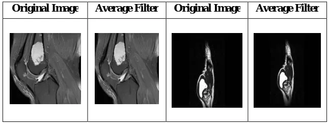
The experimentation proposed is initiated by preprocessing bone tumor MRI image. The main motive of preprocessing is to improve the quality of images to make segmentation precise. As generally the captured image is of poor quality, so filters are used for removing noise, sharpening and preserving edges and smoothing defective images created by MR imaging system.
surrounding pixels. So, the average filter gives effective results for smoothing the images. Here Table 1 shows results of average filter on the original image MRI image of knee and finger of enchondroma bone tumor.
Table 1. Results of Average Filter
Original Image Average Filter Original Image Average Filter
C. Segmentation
Segmentation phase mainly concentrates on tumor detection. An image segmentation technique such as thresholding and morphological operation, K-Means Clustering, Fuzzy C-Means Clustering and Rough Fuzzy C-Means clustering is used in this paper and the results are compared. In detection of tumor each segmentation technique is applied to the image obtained [4].
D. Tumor Detection
The clusters with high intensity pixels are isolated from the MRI image which forms the tumor image. Some of the features like as area, centroid and perimeter can be calculated from the tumor image.
V. EXPERIMENTAL RESULTS AND COMPARISONS
The four techniques of segmentation are applied on each sequence of MRI images and the results are shown below. 40 MRI images are taken from Radiopedia.org of Enchondroma, Giant cell tumor. It is implemented in MATLAB 2014a.Table 2 shows comparison of segmentation techniques such as Thresholding and morphological operation, K-Means, Fuzzy C-Means and Rough Fuzzy C-Means on average filtered image.From the results, the traditional segmentation method thresholding and morphological operation gives good result as per the location of the tumor, but false segmentation occurs as in MRI images the tumor and other cartilage have same intensity, which cannot be removed by morphological operation as it is big. In K-Means Clustering segmentation technique, it gives good results as per location and shape, but false segmentation occurs in this also as it depends on the numbers of clusters initialized.In Fuzzy C-Means Clustering segmentation technique we get the better results as per location, but the shape is inefficient, it takes more computation time and contains small artifacts. Lastly in Rough Fuzzy C-Means gives best segmentation results amongst all and it takes less computation time compared to Fuzzy C-Means.
Table 2.Comparisions of various segmentation techniques
Average Filtered Image
Thresholding and Morphological
Operation
K-Means Fuzzy C-Means Rough Fuzzy
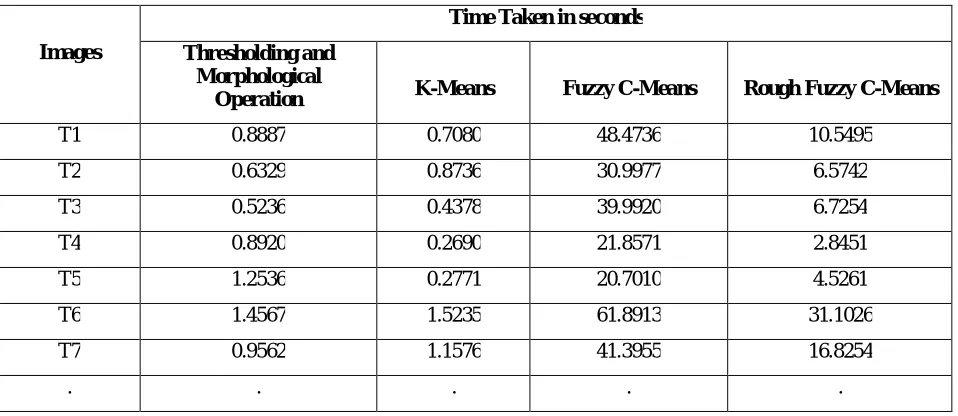
The time comparison between Thresholding and morphological operation, K-Means, Fuzzy C-Means and Rough Fuzzy C-Means is shown in Table 3.Here the time is calculated in seconds. We observed that K-Means and Thresholding technique takes less computational time comparatively to Fuzzy C-Means and Rough Fuzzy C-Means.
Table 3.Computational time taken by various segmentation techniques
Images
Time Taken in seconds
Thresholding and Morphological
Operation K-Means Fuzzy C-Means Rough Fuzzy C-Means
T1 0.8887 0.7080 48.4736 10.5495
T2 0.6329 0.8736 30.9977 6.5742
T3 0.5236 0.4378 39.9920 6.7254
T4 0.8920 0.2690 21.8571 2.8451
T5 1.2536 0.2771 20.7010 4.5261
T6 1.4567 1.5235 61.8913 31.1026
T7 0.9562 1.1576 41.3955 16.8254
. . . . .
VI.CONCLUSION AND FUTURE WORK
After analyzing the experimental results, we conclude that Rough Fuzzy C-Means gives more accurate results and takes less computation time. Fuzzy C-Means takes more time computation compared to all the segmentation methods. K-Means and Thresholding and morphological operation, both take equivalent time which is less than Fuzzy methods but gives false segmentation. Thus, Rough Fuzzy C-Means can be used further for Medical Imaging field and it predicts the tumor cells accurately in less computation time.There is much scope for further research work and the results can be improved by combining the algorithms. The features such as area, location, shape, stage can be calculated.
REFERENCES
1. Avula, Madhuri, Narasimha Prasad Lakkakula, and Murali Prasad Raja. "Bone Cancer Detection from MRI Scan Imagery Using Mean Pixel Intensity." In Modelling Symposium (AMS), 2014 8th Asia, pp. 141-146. IEEE, 2014.
2. Afshan, Nailah, Shaima Qureshi, and Syed Mujtiba Hussain. "Comparative study of tumor detection algorithms." In Medical Imaging, m-Health and Emerging Communication Systems (MedCom), 2014 International Conference on, pp. 251-256. IEEE, 2014.
4. Hooda, Heena, Om PrakashVerma, and TriptiSinghal. "Brain tumor segmentation: A performance analysis using k-means, fuzzy c-means and region growing algorithm." In Advanced Communication Control and Computing Technologies (ICACCCT), 2014 International Conference on, pp. 1621-1626. IEEE, 2014..
5. Bhattacharya, Avik, and K. S. Patnaik. "Modified Rough Fuzzy C Means Algorithm for MR Image Segmentation." In Machine Intelligence and Research Advancement (ICMIRA), 2013 International Conference on, pp. 407-411. IEEE, 2013.
6. Vijay, J., and J. Subhashini. "An efficient brain tumor detection methodology using K-means clustering algoriftnn." In Communications and Signal Processing (ICCSP), 2013 International Conference on, pp. 653-657. IEEE, 2013.
7. Benign and Malignant Bone Tumors:Radiological Diagnosis and Imaging Features Katharina Grünberg, M.D.; Christoph Rehnitz, M.D.; Marc-André Weber, M.D., M.Sc.
8. Diagnostic Imaging -Imaging of Bone Tumors and Tumor-Like Lesions Techniques and Applications. 9. Maheshwari, J. “ORTHOPADICS BOOK “ Mehta Publishers. ISBN : 81-88039-32-2.
10. Patel, Jay, and Kaushal Doshi. "A study of segmentation methods for detection of tumor in brain MRI." Advance in Electronic and Electric Engineering 4, no. 3 (2014): (279-284), 2014.
11. Case courtesy, Radiopaedia.org, orthoinfo.aaos.org 12. Dr. Viral ,Radiology Department, New Civil Hospital, Surat.
13. Abdel-Maksoud, Eman, Mohammed Elmogy, and Rashid Al-Awadi. "Brain tumor segmentation based on a hybrid clustering technique." Egyptian Informatics Journal 16, no. 1 (2015): (71-81), 2015.
14. Sneha Dhurkunde, Shailaja Patil.” Segmentation of Brain Tumor in Magnetic Resonance Images using Various Techniques.”
International Journal of Innovative Research in Science, Engineering and Technology,Vol. 5, Issue 1, Januray 2016.
15. Dhanachandra, Nameirakpam, Khumanthem Manglem, and Yambem Jina Chanu. "Image Segmentation Using K-means Clustering Algorithm and Subtractive Clustering Algorithm." Procedia Computer Science 54 (2015): (764-771), 2015.
16. www.orthoinfo.aaos.org/bonetumors
BIOGRAPHY