Article
1
Carotenoid production by Dunaliella salina in red
2
light
3
Yanan Xu and Patricia J. Harvey *
4
University of Greenwich, Faculty of Engineering and Science, Central Avenue, Chatham Maritime, Kent, ME4
5
4TB, UK
6
* Correspondence: p.j.harvey@greenwich.ac.uk; Tel.: +44-20-8331-9972
7
8
Received: date; Accepted: date; Published: date
9
10
Abstract: The halotolerant photoautotrophic marine microalga Dunaliella salina is one of the
11
richest sources of natural carotenoids. Here we investigated the effects of high intensity blue, red
12
and white light from light emitting diodes (LED) on the production of carotenoids by strains of D.
13
salina under nutrient sufficiency and strict temperature control favouring growth.
14
Growth in high intensity red light was associated with carotenoid accumulation and a high rate
15
of oxygen uptake. On transfer to blue light, a massive drop in carotenoid content was recorded along
16
with very high rates of photo-oxidation. In high intensity blue light, growth was maintained at the
17
same rate as in red or white light, but without carotenoid accumulation; transfer to red light
18
stimulated a small increase in carotenoid content. The data support chlorophyll absorption of red
19
light photons to reduce plastoquinone in photosystem II, coupled to phytoene desaturation by
20
plastoquinol:oxygen oxidoreductase, with oxygen as electron acceptor. Partitioning of electrons
21
between photosynthesis and carotenoid biosynthesis would depend on both red photon flux
22
intensity and phytoene synthase upregulation by the red light photoreceptor, phytochrome. Red
23
light control of carotenoid biosynthesis and accumulation reduces the rate of formation of reactive
24
oxygen species (ROS) as well as increases the pool size of anti-oxidant.
25
26
Keywords: Dunaliella salina; microalgae; red LED; blue LED; growth; carotenoids;
27
plastoquinol:oxygen oxidoreductase; photosynthesis.
28
29
1. Introduction
30
Carotenoids are orange, yellow or red pigments which are synthesized by all photosynthetic
31
organisms for light-harvesting and for photo-protection, and for stabilising the pigment-protein
32
light-harvesting complexes and photosynthetic reaction centres in the thylakoid membrane. They
33
may also be accumulated by some non-photosynthetic archaea, bacteria, fungi and animals for
34
pigmentation [1-3]. Carotenoids are also the precursors of a range apocarotenoids of biological and
35
commercial importance, such as the phytohormone abscisic acid, the visual and signalling molecules
36
retinal and retinoic acid, and the aromatic volatile beta-ionone [4]. Increasingly sought after as
37
natural colorants, there is accumulating evidence that carotenoids protect humans against ageing
38
and diseases that are caused by harmful free radicals and may also reduce the risks of cataract,
39
macular degeneration, neurodegeneration and some cancers [5,6]. They have also been implicated as
40
the actives for treating diseases associated with retinoids [4].
41
In most plants and algae containing chlorophyll a (max ~ 680 nm) and b (max ~ 660 nm), photons
42
with a wavelength of 660-680 nm yield the highest quantum efficiencies. However the solar
43
spectrum at the surface of the Earth is at its maximum intensity in the blue and green regions of the
44
visible spectrum (400-550 nm), which is where carotenoids have strong absorption. In
45
photosynthetic organisms in the light, carotenoids drive photosynthesis by transferring absorbed
46
excitation energy to chlorophylls, which have poor absorption in this range. Carotenoids are also
47
able to protect photosynthetic organisms from the harmful effects of excess exposure to light by
48
permitting triplet–triplet energy transfer from chlorophyll to carotenoid and by quenching reactive
49
oxygen species (ROS) [2].
50
Dunaliella salina, a halotolerant chlorophyte, is one of the richest sources of natural carotenoids
51
and, similar to various members of the Chlorophyceae, accumulates a high content (up to 10 % of the
52
dry biomass) of carotenoids under conditions that are sub-optimal for growth i.e. high light
53
intensity, sub-optimal temperatures, nutrient limitation and high salt concentrations. In D. salina, the
54
major accumulated carotenoid is β-carotene, which is stored in globules of lipid and proline-rich,
55
carotene globule protein in the inter-thylakoid spaces of the chloroplast (βC-plastoglobuli) [7-10].
56
The pathway for β-carotene synthesis and accumulation in D. salina has been partly mapped out
57
[11,12], but the physiological role and signals triggering its accumulation are not well-established. In
58
other members of the Chlorophyceae, such as Haematococcus pluvialis and Chlorella zofingiensis, high
59
levels of oxygen-rich, secondary ketocarotenoids, astaxanthin and canthaxanthin, also accumulate
60
under high light stress or nutrient stress, often in lipid bodies located outside the chloroplast in the
61
cytoplasm. Accumulation of these may also be accompanied by cell encystment. Lemoine and
62
Schoefs [13] proposed that these carotenoids accumulate as a metabolic means of lowering ROS
63
levels by lowering cellular oxygen concentration, as well as serving as a convenient way to store
64
energy and carbon for further synthesis under less stressful conditions [13,14]. Chemically generated
65
ROS will trigger astaxanthin accumulation [15] and recently Sharma et al. [16] showed that a small
66
dose (up to 50 mJ cm2) of UV-C light (200-280 nm) in cultures of either D. salina or H. pluvialis
67
massively increased carotenoids accumulation as well as detached the flagellae to increase cell
68
settling, 24h after exposure: UV- light exposure is typically accompanied by ROS formation.
69
However in D. salina there may be additional mechanisms leading to carotene accumulation.
70
Jahnke [17] for example found that whilst supplements to visible radiation of UV-A radiation
71
(320-400 nm) specifically increased carotenoid levels and the ratio of carotenoids to chlorophylls in
72
the closely related D. bardawil, neither blue light nor UV-B light (290-320 nm) supplements were
73
similarly effective.In blue light, Loeblich [8] found that green cells of D. salina with a low carotenoid
74
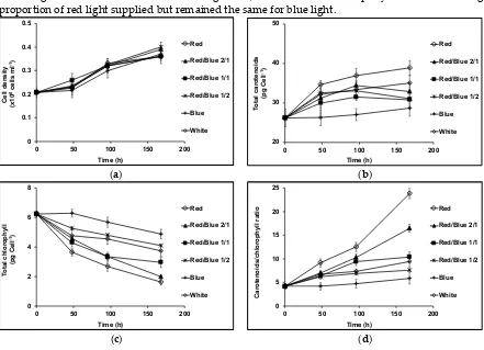
to chlorophyll ratio had a relatively depressed photosynthetic activity, which was even more
75
exaggerated in red cells with a high carotenoid to chlorophyll ratio. They proposed that blue light,
76
which was absorbed by the accumulated β-carotene, was not available for photosynthetic oxygen
77
evolution. Amotz et al. [18] on the other hand found a marked photo-inhibition for both red and
78
green cells under high intensity red light, which is absorbed by chlorophylls, but red cells, when
79
transferred to high intensity blue light were seemingly photoprotected. Since the accumulated
80
carotenoids were physically distant from chlorophylls located in thylakoid membranes, Amotz et al.
81
[18] proposed that in high intensity red light, the carotenoids were unable to provide
82
photoprotection against chlorophyll-generated ROS or quench chlorophyll excited states,
83
supporting the argument that carotene globules may function as a screen against high irradiation in
84
blue light to protect photosynthetic reaction centres in D. salina. Fu et al. [19] examined the effects of
85
different light intensities of red LED light on carotenoid production in D. salina, and showed that the
86
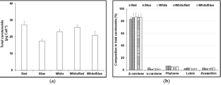
major carotenoids changed in parallel to the chlorophyll b content and that both carotenoids and
87
chlorophyll b decreased with increasing red light intensity and increased with nitrogen starvation.
88
Light-emitting diodes (LEDs) can be applied to adjust the biochemical composition of the
89
biomass produced by microalgae via single wavelengths at different light intensities [20-22]. In this
90
paper we explore the effects of red, blue and white LEDs on the growth and content of carotenoids
91
and chlorophyll in 4 different D. salina strains under nutrient-sufficient conditions using a
92
temperature-controlled PBR favouring growth. We show that in this system, cultivation using red
93
LED was particularly effective in supporting a high rate of carotenoid productivity. We suggest that
94
in strains of Dunaliella salina, accumulating carotenoids may be synthesised principally as a
95
mechanism for maintaining cellular homeostasis under conditions which might otherwise lead to
96
over-reduction of electron transport chains, formation ROS and of a hyperoxidant state and
97
ultimately lead to cell death.
2. Materials and Methods
99
2.1 Strains and cultivation
100
Strains D. salina rubeus CCAP 19/41 and D. salina salina PLY DF17 were isolated from a salt pond
101
in Israel. D. salina CCAP 19/40 was isolated from a salt pond in Monzon, Spain. Strain UTEX 2538 (D.
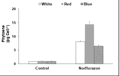
102
salina bardawil) was purchased from CCAP.
103
Algae were cultured in 500 ml Modified Johnsons Medium [14] containing 1.5 M NaCl, in
104
Erlenmeyer flasks in an ALGEM Environmental Modelling Labscale Photobioreactor (Algenuity,
105
UK) at 25 °C. Cells were grown under 12/12 LD with 200 μmol photons m-2 s-1 supplied by white
106
LED light to exponential growth phase and then dark-adapted for 36 hours. After dark adaption,
107
cultures were exposed continuously to blue, red or white LED light at light intensities of 200, 500, or
108
1000 μmol photons m-2 s-1. Cultures acclimated to white, red or blue LED light for 24 hours were
109
used to monitor the changes in cellular carotenoids after further growth for 24 hours in white, red or
110
blue LED light. Cell density of the cultures was determined by counting the cell number of cultures
111
using a haemocytometer after fixing with 2 % formalin.
112
113
2.2 Pigment analysis
114
The composition of pigments was analysed by High-Performance Liquid Chromatography
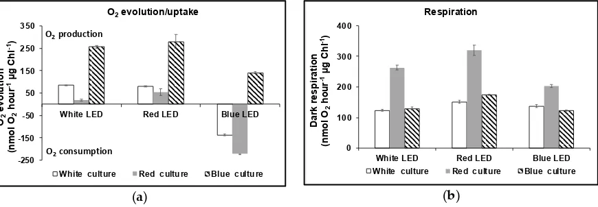
115
with Diode-Array Detection (HPLC-DAD), using a YMC30 250 X 4.9 mm I.D S- 5μ HPLC column at
116
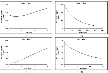
25 ºC with an isocratic solvent system of 80 % methanol: 20 % MTBE and flow rate of 1 mL min-1 at a
117
pressure of 78 bar. Carotenoid standards of -carotene, -carotene, lutein, zeaxanthin and phytoene
118
were obtained from Sigma-Aldrich (UK) and dissolved in methanol or acetone to generate standard
119
curves and DAD scans analysed at wavelengths of 280 nm (phytoene), 355 nm (phytofluene), 450 nm
120
(β-carotene, -carotene, lutein and zeaxanthin), and 663 nm (chlorophylls). Pigments were extracted
121
from the biomass of 15 mL samples of culture. Samples were harvested by centrifugation at 3,000 g
122
for 10 min and pigments extracted after sonication for 20 s with 10 mL MTBE-MeOH (20:80).
123
Samples were clarified at the centrifuge then filtered (0.45 μm filter) into amber HPLC vials before
124
analysis.
125
Total carotenoids and total chlorophyll in the cultures were measured using a UV/Vis
126
spectrophotometer. Pigments were extracted from the harvested algal biomass of 1 mL culture using
127
1 mL of 80 % (v/v) acetone, then clarified at 10,000 g. The content of total carotenoids was
128
calculated from absorbance values at 480 nm according to Strickland & Parsons [15]. Chlorophyll a,
129
b and total chlorophyll content was measured at 664 nm and 647 nm according to Porra et al. [16].
130
131
2.3 Oxygen evolution and dark respiration
132
Samples of cultures exposed to white, red or blue LED light were collected and the rates of O2
133
evolution and dark respiration were measured as described by Brindley at al. [23] at 25 C using a
134
Clark-type electrode (Chlorolab 2, Hansatech, UK). O2 evolution/uptake was induced by white, red,
135
or blue LED light supplied by the manufacturer at a light intensity of 1000 μmol m-2 s-1. After an
136
initial period of 30 min of dark adaption of 1.5 mL of each culture, the rate of O2 evolution/uptake
137
was measured for 20 min followed by dark respiration for 20 min. The average rate of
138
photosynthesis was determined from the linear rate of oxygen evolution during 5-15 min of the light
139
period. Dark respiration was determined by following the same procedure, except that the rate was
140
calculated using the data from the last 15 min of the measurement. Air saturated water and nitrogen
141
were used to calibrate the electrode.
142
3. Results
143
3.1. Cell growth and carotenoids production in acclimated cultures
144
Figure 1a shows that in high intensity blue, white or red LED light, the growth rate recorded as cell
145
density for D. salina CCAP 19/41 was the same. There was no significant difference in cell size.
146
However the contents of total carotenoids and total chlorophyll depended on the relative
147
proportions of blue or red light supplied. The initial phase of growth in all high intensity light
conditions, apart from blue, caused an initial sharp drop in chlorophyll content; the drop was
149
greatest in high intensity red LED light but decreased depending on the relative proportions of red:
150
blue light supplied. On the other hand, cultures maintained in red LED accumulated carotenoids at
151
the highest rate; in blue, the content declined depending again on the relative proportions of red:
152
blue light supplied (Figure 1b, 1c). The carotenoids/chlorophyll ratio is often used to evaluate
153
carotenogenesis in D. salina. As shown in Figure 1d, the ratio increased rapidly with the increasing
154
proportion of red light supplied but remained the same for blue light.
155
(a) (b)
(c) (d)
Figure 1. (a) Cell growth; (b) Cellular content of total carotenoids; (c) cellular content of total
156
chlorophyll; (d) Carotenoids/Chlorophyll ratio in D. salina CCAP 19/41 grown under different ratios
157
of red and blue light (Red/Blue 1/0, 2/1, 1/1, 1/2, 0/1) or white light with a total light intensity of 1000
158
μmol photons m-2 s-1 after dark-acclimation. Each culture condition was set up at least in triplicate.
159
Different Dunaliella strains responded differently to cultivation in high intensity blue or white
160
LED (see Figure 2). All showed a decline in chlorophyll content in white LED compared to
161
cultivation in high intensity blue but only strains CCAP 19/41 and UTEX 2538 showed a significant
162
increase in carotenoid content in white compared to blue light.
163
(a) (b)
0 0.1 0.2 0.3 0.4 0.5
0 50 100 150 200
C e ll d e n s it y (x 1 0 6c e ll s m l -1) Time (h) Red Red/Blue 2/1 Red/Blue 1/1 Red/Blue 1/2 Blue White 20 30 40 50
0 50 100 150 200
T o ta l c a ro te n o id s (p g C e ll -1) Time (h) Red Red/Blue 2/1 Red/Blue 1/1 Red/Blue 1/2 Blue White 0 2 4 6 8
0 50 100 150 200
T o ta l c h lo ro p h y ll (p g C e ll -1) Time (h) Red Red/Blue 2/1 Red/Blue 1/1 Red/Blue 1/2 Blue White 0 5 10 15 20 25
0 50 100 150 200
C a ro te n o id s /c h lo ro p h y ll r a ti o Time (h) Red Red/Blue 2/1 Red/Blue 1/1 Red/Blue 1/2 Blue White 0 4 8 12 16
CCAP 19/41 PLY DF17 DF40 UTEX 2538
P ig m e n t c o n te n t (p g C e ll -1) White light Chlorophyll Carotenoids 0 4 8 12 16
CCAP 19/41 PLY DF17 DF40 UTEX 2538
Figure 2. Cellular content of total carotenoids and of total chlorophyll for different D. salina strains
164
cultivated each to the mid log phase under either white (a) or blue (b) light with a total light intensity
165
of 1000 μmol photons m-2 s-1 after dark-acclimation. Each culture condition was set up at least in
166
triplicate.
167
Carotenoids in D. salina CCAP 19/41 cultures exposed to different light conditions: white,
168
red or blue LED light at 1000 μmol photons m-2 s-1, a mixture of white and red (1:1) or a mixture
169
of white and blue (1:1) with a total intensity of 1000 μmol photons m-2 s-1 for 48 hours were
170
extracted and the major carotenoids were identified and quantified by HPLC. Cultures exposed
171
to continuous red LED light had the highest contents of all the identified carotenoids, while
172
cultures maintained under blue LED light showed the lowest content. The difference was
173
mainly due to variation in β-carotene content between treatments: there was no significant
174
difference in relative content of all other carotenoids (Figure 3).
175
(a) (b)
Figure 3. (a) Cellular content of total carotenoids; (b) Relative composition of major carotenoids
176
characterised by HPLC in total carotenoids in D. salina CCAP19/41 cells exposed to continuous LED
177
of different wavelength distribution (red, blue, white, white/red 1:1, and white/blue 1:1) for 48 hours.
178
The total light intensity for all conditions was the same at 1000 μmol photons m-2 s-1.
179
180
The total carotenoids and total chlorophyll contents after 48 h exposure to red or blue LED at
181
different light intensities are shown in Figure 4. Carotenoids accumulated with increasing blue LED
182
intensity between 200 μmol m-2 s-1 and 1000 μmol m-2 s-1. In red LED light, cultures contained high
183
amounts of carotenoids even under low light intensity and the content increased with increasing red
184
LED intensity up to 500 μmol m-2 s-1. With further increase in light intensity to 1000 μmol m-2 s-1 ,
185
carotenoids declined slightly (<10 % of the value recorded at 500 μmol m-2 s-1), but chlorophylls
186
declined 34 %. The carotenoids/chlorophyll ratio increased with the increase of light intensity both
187
red and blue LED light, however, under red LED light a much higher carotenoids/chlorophyll ratio
188
was recorded than under blue. Cellular content of β-carotene and phytoene showed a similar trend
189
to that of total carotenoids, except that the highest β-carotene content under red light was achieved
190
at 500 μmol m-2 s-1, while the highest phytoene content under red light was achieved at 1000 μmol
191
m-2 s-1.
192
193
0 10 20 30 40
Red Blue White White/Red White/Blue
T
o
ta
l
c
a
ro
te
n
o
id
s
(p
g
C
e
ll
-1)
0 20 40 60 80 100
β-carotene α-carotene Phytoene Lutein Zeaxanthin
C
o
m
p
o
s
it
io
n
i
n
t
o
ta
l
c
a
ro
te
n
o
id
s
(
%
)
(a) (b)
Figure 4. Cellular content of total carotenoids and total chlorophyll (a) and carotenoids/chlorophyll
194
ratio (b) in D. salina CCAP19/41 grown under continuous red or blue LED light at three different light
195
intensities of 200, 500 and 1000 μmol m-2 s-1 for 48 hours. Each culture condition was set up at least in
196
triplicate.
197
A phytoene desaturase inhibitor norflurazon known to cause accumulation of phytoene was
198
used to treat D. salina cultures maintained under red, blue or white LED light. Figure 5 shows that
199
under these conditions the cellular content of phytoene increased, as expected, but cultures
200
maintained under red LED accumulated a significantly higher amount of phytoene compared to
201
cultures maintained under white or blue light.
202
Figure 5. Cellular content of phytoene in cultures treated with no inhibitors (control) or with 5 μM
203
norflurazon. Cultures were maintained under red, blue and white LED light at 200 μmol m-2 s-1 for 48
204
hours.
205
206
3.2. Acclimation and carotenoids production in response to wavelength switching
207
Dark-adapted cultures of D. salina CCAP19/41 were cultivated in red, white or blue LED light
208
for 24 hours (T0), and then cultivated for a further 24 hours in red, blue, or a mixture of red and
209
blue LED light (1:1), or the dark. Blue-acclimated cells produced slightly more carotenoids (14 %
210
greater content) when transferred to red LED but chlorophyll content declined from that at the start
211
of the experiment to an amount only 62 % of that in continuous blue (Figure 6a). On the other hand
212
the chlorophyll content increased when red-acclimated cells were exposed to blue light, but the
213
total carotenoids content declined sharply, approximately in proportion to the amount of blue LED
214
supplied (see Figure 6b). Red LED cultures maintained for a further 24 h in red LED accumulated
215
24.5 ± 1.3 pg carotenoid cell-1, but after 24 h in blue LED instead of red, the carotenoid content was
216
50 % lower (11.4 ± 0.4 pg carotenoid cell-1) and less than if they had been transferred to the dark
217
(12.3 ± 0.5 pg carotenoid cell-1).
218
0 5 10 15 20 25 30
R200 R500 R1000 B200 B500 B1000
C
e
ll
u
la
r
c
o
n
te
n
t
(p
g
C
e
ll
-1)
Chlorophyll Carotenoids
0 2 4 6 8
R200 R500 R1000 B200 B500 B1000
C
a
ro
te
n
o
id
s
/C
h
lo
ro
p
h
y
ll
r
a
ti
o
0 5 10 15 20
Control Norflurazon
P
h
y
to
e
n
e
(p
g
C
e
ll
-1)
(a) (b)
Figure 6. Cellular content of total carotenoids and chlorophyll under continuous blue (a) or red (b)
219
LED light at 1000 μmol m-2 s-1 for 24 hours followed by 24 h growth under either red light, a mix of
220
1:1 red and blue light, blue light at the same light intensity of 1000 μmol m-2 s-1 or dark. Each culture
221
condition was set up at least in triplicate.
222
Dark-adapted cultures were cultivated in red, blue or white light at 1000 μmol m-2 s-1 for 48
223
hours before measuring the rates of oxygen evolution/uptake over a 20 minute period with
224
illumination supplied once more by white, red or blue LED lights at 1000 μmol m-2 s-1. The rate
225
profiles of oxygen evolution are shown in Figure 7a. Dark respiration was also recorded (Figure 7b).
226
Red LED supported net oxygen evolution (55 ± 15 nmol O2 hour-1 g chlorophyll-1) but on transfer
227
to blue light in the Clark-type electrode, photo-oxidation massively exceeded the rate of oxygen
228
evolution and oxygen was consumed at an exponentially increasing rate (Figure 7a; 222 ± 32 nmol O2
229
hour-1 g chlorophyll-1). Significantly cultures grown in red LED also supported the highest rate of
230
dark respiration (320 ± 17 nmol O2 hour-1 g chlorophyll-1, ~ 2.6-fold greater than that for cultures
231
maintained in either blue or white LED light), but this also declined when cultures were transferred
232
to blue light in the Clark-type electrode. By contrast, cultures maintained in blue LED supported
233
~3-fold higher rate of net oxygen evolution (141 nmol O2 hour-1 g chlorophyll-1) in the Clark-type
234
electrode in blue light, compared to those in maintained in red LED. On transfer of blue light
235
cultures to red light in the Clark-type electrode, the rate of oxygen evolution doubled to 280 nmol O2
236
hour-1 g chlorophyll-1 (Figure 7b), and was maintained at a linear rate during the period of
237
measurement. The rate of dark respiration also increased slightly from 123 ± 2.7 nmol O2 hour-1 g
238
chlorophyll-1 to 175 ± 5.0 nmol O2 hour-1 g chlorophyll-1.
239
(a) (b)
Figure 7. Oxygen evolution/uptake by D. salina cultures in different white, red or blue LED light
240
sources (a) and in the dark (b). Cultures were grown under continuous red, blue or white light at 1000
241
μmol m-2 s-1 for 48 hours before measurement.
242
243
0 5 10 15 20 25 30T0 Red Mix Blue Dark
C e ll u la r c o n te n t (p g C e ll -1) Chlorophyll Carotenoids 0 5 10 15 20 25 30
T0 Red Mix Blue Dark
C e ll u la r c o n te n t (p g C e ll -1) Chlorophyll Carotenoids -250 -150 -50 50 150 250 350
White LED Red LED Blue LED
O2 e v o lu ti o n (n m o l O2 h o u r -1 µ g C h l -1)
O2evolution/uptake
White culture Red culture Blue culture
O2 production
O2 consumption 0
100 200 300 400
White LED Red LED Blue LED
D a rk r e s p ir a ti o n (n m o l O2 h o u r -1 µ g C h l -1) Respiration
Figure 8. Rate profiles for oxygen uptake/evolution measured with different wavelengths of
244
LED lights measured using a Clark-type electrode for 1.5 mL cultures of D. salina CCAP19/41
245
maintained at 25 °C. (a) Red light acclimated cultures measured under blue light. (b) Blue light
246
acclimated cultures measured under red light; (c) White light acclimated cultures measured under
247
red light; (d) White light acclimated cultures measured under blue light.
248
249
4. Discussion
250
LEDs with different wavelengths have been increasingly used to study the wavelength effects
251
on the growth and productivity of photoautotrophic microalgae, and much effort is being invested
252
to understand the most energy-efficient way to incorporate their use for large-scale algal cultivation
253
[20-22,24,25]. In the present work we explored the effects of using red, white, blue and mixtures of
254
red and blue LEDs at different intensities to evaluate the basis for carotenoid accumulation in strains
255
of Dunaliella salina.
256
The emission spectrum of the red LED used in the present work (625-680 nm) emits photons
257
with the exact range required by molecules of chlorophyll a and chlorophyll b to initiate
258
photosynthesis [26]. In D. salina, action spectra of O2 evolution rates show maximum photosynthetic
259
activity within the red absorption bands of the chlorophylls [8]. Photosystems I and II (PSI, PSII),
260
which both contain chlorophyll a, work together in a series of more than 40 steps that proceed with
261
the efficiency of nearly 100 % to transfer electrons from water to NADP [2]. Consequently the
262
wavelength range of the red LED should be the most efficient emission required for photosynthesis
263
in this alga and deliver the highest specific growth rate. However this also depends on the rate at
264
which the absorbed light energy from any given applied photon flux density is converted to
265
chemical energy: with increasing photon flux density, photosynthesis eventually achieves a
light-266
saturated maximum rate that is limited by the rate of carbon fixation in the Calvin cycle. Spirulina
267
platensis for example exhibited the highest specific growth rate using high intensity red LED [24]. C.
268
reinhardtii however, showed unstable growth in high intensity orange-red and deep red LED, which
269
ceased completely after a few days and was accompanied by cell agglomeration [21]: agglomeration
270
is typical of oxidant stress and formation of a hyperoxidant state [27].
271
(a) (b)
In the present work, we found that in high intensity red light in conditions of nutrient
272
sufficiency, D. salina strains maintained a growth rate at least equal to that in white light or blue LED
273
light, seemingly in contrast with the work of others [8,18,19]. However we also found that some but
274
not all strains accumulated carotenoids rapidly, within 48 hours of exposure. Carotenoids are
275
known antioxidants that are synthesized by many microalgae as part of the battery of
276
photoprotective mechanisms necessary to prevent photoinhibition caused by photo-oxidation of
277
photosynthetic reaction centres [2, 28, 29]. Photo-oxidation may occur in photon flux density levels
278
that result in absorption of more light than is required to saturate photosynthesis. At the molecular
279
level, when a photon is absorbed by a chlorophyll molecule, it enters a short-lived singlet excited
280
state (1Chl*): the longer the excitation of 1Chl* lasts, which increases under saturating light
281
conditions, the greater the chance that the molecule will enter the triplet excited state (3Chl*) via
282
intersystem crossing. 3Chl* has a longer excitation lifetime and can transfer energy to the ground
283
state of O2 to form singlet oxygen, 1O2, predominantly at the reaction centre of PSII and, to a lesser
284
extent, in the light-harvesting complexes. Photooxidative damage occurs to the photosynthetic
285
apparatus when species such as 1O2 react with fatty acids form lipid peroxides, setting up a chain of
286
oxygen activation events that may eventually lead to a hyperoxidant state and cell death.
287
Carotenoids may protect the photosystems by reacting with lipid peroxidation products to terminate
288
these chain reactions; by scavenging 1O2 and dissipating the energy as heat; by reacting with 3Chl* to
289
prevent formation of 1O2 or by dissipation of excess excitation energy through the xanthophyll
290
cycle. It is tempting therefore to suppose that the differences observed by different workers simply
291
reflects differences in carotenoids content between different strains, but this does not explain what
292
triggered the differences in carotenoids content.
293
In the D. salina strain CCAP 19/41, accumulation of carotenoids was accompanied by the highest
294
rate of O2 consumption and a low rate of net O2 evolution, which might imply 1O2 formation and
295
ROS accumulation. In the non-photosynthetic, astaxanthin-accumulating yeast, Phaffia rhodozyma,
296
artificially generated 1O2 was proposed to degraded astaxanthin to relieve feedback inhibition of
297
carotenoid biosynthesis and also to induce carotenoid synthesis by gene activation [30]. However
298
these authors also found that carotenoid biosynthesis was linked to O2 consumption by a
299
cyanide-insensitive alternative oxidase, serving to consume oxygen without chemiosmotic synthesis
300
of ATP. In C. reinhardtii, a specific thylakoid-associated, terminal plastoquinol:oxygen
301
oxidoreductase has been identified with homology to the mitochondrial alternative oxidase [31]. The
302
smaller rate of oxygen uptake compared to mitochondrial respiration suggested a function in
303
directly coupling oxygen uptake and the exergonic reaction of plastoquinol oxidation with
304
plastoquinone reduction by a phytoene/phytoene desaturase couple, to permit endergonic carotene
305
desaturation without ATP involvement [31]. In D. bardawil, a decrease in oxygen consumption rate
306
coupled to phytoene accumulation caused by norflurazon inhibition of phytoene desaturase also
307
suggests a connection between direct desaturation of phytoene and chloroplastic oxygen dissipation
308
[32].
309
In the present work, high intensity red light in conditions of nutrient sufficiency maintained
310
growth at the same rate as in blue or white light, and red light also led to carotenoid accumulation
311
albeit to different extents in different strains. These data support involvement of a
312
plastoquinol:oxygen oxidoreductase as originally proposed for C. reinhardtii [31], but controlled by
313
red photon flux intensity (see Scheme 1). In this scheme, chlorophyll absorption of red light photons
314
is coupled to plastoquinone reduction in photosystem II, and oxygen reduction is coupled to
315
phytoene desaturation by plastoquinol:oxygen oxidoreductase leading to carotenoid accumulation.
316
Partitioning of electrons between photosynthesis and carotenoid biosynthesis would depend on
317
both red photon flux intensity as well as upregulation of phytoene synthase. The observed increase
318
in O2 consumption coupled to accumulation of carotenoids via the carotenoid biosynthetic pathway
319
would reduce the tendency for 1O2 formation under high photon flux and maintain cytosolic redox
320
potential.
321
The coupling of reduction of the plastoquinone pool to carotenoid synthesis driven by
322
chlorophyll absorption of red light may involve the red light photoreceptor phytochrome.
Photosynthetic organisms are known to perceive red light signals via phytochrome. The synthesis of
324
phytoene by phytoene synthase is under phytochrome regulation [33-35] and is upregulated by
325
both red and far-red light [33]. Red light also lowers the concentration of the transcription factor
326
PIF1, a repressor of carotenoid biosynthesis [36]. In those strains which do not accumulate
327
carotenoids, alternative mechanisms may serve to consume energy e.g. via NAD(P)H reduction of
328
dihydroxyacetone phosphate to form glycerol [37].
329
330
331
Scheme 1. Partitioning electron flux between photosynthesis and carotenoid biosynthesis.
332
Red photon flux intensity controls the partitioning of electrons either for carotenoid biosynthesis or
333
for photosynthesis, via energy absorption by chlorophyll and the PQ pool. Red photon flux density
334
also controls phytochrome regulation of phytoene synthase.
335
336
In support of this model, transfer from high intensity red light to blue with higher energy
337
content caused a massive drop in the accumulated carotenoid content, very high rates of
338
photo-oxidation and low respiratory rates. Carotenoids in both the accumulated pool and in the
339
light harvesting antenna, but not chlorophyll, absorb photons in the range 400-550 nm, exactly
340
overlapping the emission spectrum of the blue LED (440-500 nm). Failure of chlorophyll molecules
341
to use the absorbed energy to reduce the plastoquinone pool would be expected to reduce the rate of
342
electron flux through the plastoquinol:oxygen oxidoreductase as well as uncouple carotenoid
343
synthesis and consequently increase cellular O2 concentration. This would lead in turn to increased
344
ROS formation by reaction of O2 with reduced electron transport chains, initiating further oxygen
345
radical chain reactions, and carotenoid oxidation. Furthermore, in continuous high intensity blue
346
LED, growth was maintained without carotenoid accumulation, but transfer to high intensity red
347
LED light stimulated a small increase in carotenoid content, once again putting red light absorption
348
by chlorophylls and transfer of absorbed energy to the plastoquinone pool at the centre of carotenoid
349
biosynthesis. Transfer from blue to red light in the Clark-type electrode would cause absorption of
350
more light than was required to saturate photosynthesis; if upregulation of the carotenoid
351
biosynthetic pathway via phytochrome perception was required before coupling with O2 uptake via
352
the plastoquinol:oxygen oxidoreductase, this would result in initial increase in the rate of
353
photoinhibition and O2 uptake in the Clark-type electrode, and consequent loss in chlorophyll
354
content, as was observed.
355
β-carotene accumulation in βC-plastoglobuli has parallels with that for astaxanthin
356
accumulation, serving both as a carbon sink and end-product of an alternative oxygen-consuming
357
biosynthetic pathway that on the one hand, controls over-reduction of photosynthetic (and
358
respiratory) electron transport chains at the same time as removes oxygen from the plastid to limit
359
formation of ROS. It is also able to quench any ROS that form. In blue light it may serve as a screen
360
to absorb excess irradiation [7,18] but clearly offers photoprotection in red light as well. These
functions are seen as distinct from its role as an accessory pigment in light-harvesting antennae
362
systems.
363
Recently Davidi et al. [10] showed that the formation of cytoplasmic TAG under N deprivation
364
preceded that of βC-plastoglobuli, reaching a maximum after 48h of N deprivation and then
365
decreasing. They suggested that βC-plastoglobuli are made in part from hydrolysis of chloroplast
366
membrane lipids and in part by a continual transfer of TAG or of fatty acids derived from
367
cytoplasmic lipid droplets. TAG synthesis represents a pathway for restricting over-reduction of
368
electron transport chains [38] and its recruitment in formation of βC-plastoglobuli is entirely
369
consistent with steps to dissipate excessive energy absorbed by chlorophyll in high intensity red
370
light.
371
Overall, cultivation with red light may hold potential to enhance carotenoids production in
372
carotenoid-accumulating strains of D. salina. Red light treatment has also been reported as an
373
effective way to accelerate ripening of tomato fruit and increase the content of carotenoids [39].
374
Compared to other commonly used approaches to induce carotenogenesis, such as high light stress,
375
high salt stress and addition of hydrogen peroxide or sodium hypochlorite, the use of red light
376
provides a clean, convenient and economic alternative to promote carotenoids production from D.
377
salina in a short time.
378
5. Conclusions
379
This study shows light wavelength plays an important role in regulating the production of
380
carotenoids in carotenoid-accumulating strains of D. salina. Red light enhanced the production of
381
carotenoids, mostly β-carotene, by upregulating the entire biosynthetic pathway of carotenoids.
382
The data support a model of flexible co-operation between photosynthesis and carotenoid
383
production via the plastoquinone pool. Chlorophyll absorption of red light photons and
384
plastoquinone reduction in photosystem II is coupled to oxygen reduction and phytoene
385
desaturation by plastoquinol:oxygen oxidoreductase. Partitioning of electrons between
386
photosynthesis and carotenoid biosynthesis depends on photon flux intensity as well as
387
upregulation of phytoene synthase by the red light photoreceptor phytochrome. Red light control of
388
carotenoid biosynthesis and accumulation reduces the rate of formation of ROS as well as increases
389
the pool size of anti-oxidant.
390
Red light may have industrial value as an energy-efficient light source for carotenoid
391
production by D. salina.
392
393
Author Contributions: conceptualization, Y.X., P.H; methodology, Y.X.; formal analysis, Y.X., P.H.; data
394
curation, Y.X.; writing—original draft preparation, Y.X., P.H; writing—review and editing, Y.X., P.H.;
395
supervision, P.H.; project management, P.H.
396
Funding: This research received funding from EU KBBE.2013.3.2-02 programme (D-Factory: 368 613870) and from
397
the Interreg 2 Seas programme 2014-2020 co-funded by the European Regional Development Fund under subsidy
398
contract No ValgOrize 2S05017
399
Conflicts of Interest: The authors declare no conflict of interest.
400
References
401
1. Tanaka, Y.; Sasaki, N.; Ohmiya, A. Biosynthesis of plant pigments: anthocyanins, betalains and
402
carotenoids. Plant J. 2008, 54, 733–749.
403
2. Hashimoto, H.; Sugai, Y.; Uragami, C.; Gardiner, A. T.; Cogdell, R. J. Natural and artificial
404
light-harvesting systems utilizing the functions of carotenoids. J Photochem Photobiol C: Photochem Rev
405
2015, 25, 46–70.
406
3. Rodriguez-Concepcion, M.; Avalos, J.; Bonet, M. L.; Boronat, A.; Gomez-Gomez, L.; Hornero-Mendez, D.;
407
Limon, M. C.; Meléndez-Martínez, A. J.; Olmedilla-Alonso, B.; Palou, A.; Ribot, J.; Rodrigo, M. J.; Zacarias,
L.; Zhu, C. A global perspective on carotenoids: Metabolism, biotechnology, and benefits for nutrition and
409
health. Progress in Lipid Research 2018, 70, 62–93.
410
4. Auldridge, M. E.; McCarty, D. R.; Klee, H. J. Plant carotenoid cleavage oxygenases and their
411
apocarotenoid products. Curr. Opin. Plant Biol. 2006, 9, 315–321.
412
5. Zhang, J.; Sun, Z.; Sun, P.; Chen, T.; Chen, F. Microalgal carotenoids: beneficial effects and potential in
413
human health. Food Funct 2014, 5, 413–425.
414
6. Gong, M.; Bassi, A. Carotenoids from microalgae: A review of recent developments. Biotechnol. Adv. 2016,
415
34, 1396–1412.
416
7. Ben-Amotz, A.; Katz, A.; Avron, M. Accumulation of β-Carotene in Halotolerant Algae: Purification and
417
Characterization of β-Carotene-Rich Globules From Dunaliella Bardawil (Chlorophyceae). J Phycol 1982, 18,
418
529–537.
419
8. Loeblich, L. A. Photosynthesis and pigments influenced by light intensity and salinity in the halophile
420
Dunaliella Salina (Chlorophyta). J. Mar. Biol. Assoc. U. K. 1982, 62, 493–508.
421
9. Lamers, P. P.; Janssen, M.; De Vos, R. C. H.; Bino, R. J.; Wijffels, R. H. Exploring and exploiting
422
carotenoid accumulation in Dunaliella salina for cell-factory applications. Trends Biotechnol 2008, 26, 631–
423
638.
424
10. Davidi, L.; Shimoni, E.; Khozin-Goldberg, I.; Zamir, A.; Pick U. Origin of β-carotene-rich plastoglobuli
425
in Dunaliella bardawil. Plant Physiol. 2014, 164, 2139–2156.
426
11. Lers, A.; Biener, Y.; Zamir, A. Photoinduction of massive beta-carotene accumulation by the alga
427
Dunaliella bardawil: kinetics and dependence on gene activation. Plant Physiol. 1990, 93, 389–395.
428
12. Jin, E.; Polle, J. Carotenoid biosynthesis in Dunaliella (Chlorophyta). In The Alga Dunaliella Biodiversity,
429
Physiology, Genomics and Biotechnology.6, 147-171; Ben-Amotz, A.; Polle, E. W.; Subba Rao, D. V., Eds.;
430
2009.
431
13. Lemoine, Y.; Schoefs, B. Secondary ketocarotenoid astaxanthin biosynthesis in algae: a multifunctional
432
response to stress. Photosynth Res 2010, 106, 155–177.
433
14. Takaichi, S. Carotenoids in algae: distributions, biosyntheses and functions. Mar Drugs 2011, 9, 1101–
434
1118.
435
15. Ip, P.-F.; Chen, F. Employment of reactive oxygen species to enhance astaxanthin formation in Chlorella
436
zofingiensis in heterotrophic culture. Process Biochem. 2005, 40, 3491–3496.
437
16. Sharma, K. K.; Ahmed, F.; Schenk, P. M.; Li, Y. UV-C mediated rapid carotenoid induction and settling
438
performance of Dunaliella salina and Haematococcus pluvialis. Biotechnol. Bioeng 2015, 112, 106–114.
439
17. Jahnke, L. S. Massive carotenoid accumulation in Dunaliella bardawil induced by ultraviolet-A radiation.
440
J. Photochem. Photobiol. B, Biol. 1999, 48, 68–74.
441
18. Ben-Amotz, A.; Shaish, A.; Avron, M. Mode of action of the massively accumulated β-carotene of
442
Dunaliella bardawil in protecting the alga against damage by excess irradiation. Plant Physiol. 1989, 91,
443
1040–1043.
444
19. Fu, W.; Guðmundsson, O.; Paglia, G.; Herjólfsson, G.; Andrésson, OS.; Palsson, BO.; Brynjólfsson, S.
445
Enhancement of carotenoid biosynthesis in the green microalga Dunaliella salina with light-emitting diodes
446
and adaptive laboratory evolution. Appl. Microbiol. Biotechnol. 2013, 97, 2395–2403.
447
20. Schulze, P. S. C.; Barreira, L. A.; Pereira, H. G. C.; Perales, J. A.; Varela, J. C. S. Light emitting diodes
448
(LEDs) applied to microalgal production. Trends Biotechnol 2014, 32, 422–430.
449
21. de Mooij, T.; de Vries, G.; Latsos, C.; Wijffels, R. H.; Janssen, M. Impact of light color on
450
photobioreactor productivity. Algal Research 2016, 15, 32–42.
22. Nwoba, E. G.; Parlevliet, D. A.; Laird, D. W.; Alameh, K.; Moheimani, N. R. Light management
452
technologies for increasing algal photobioreactor efficiency. Algal Research 2019, 39, 101433.
453
23. Brindley, C.; Acién, F. G.; Sevilla, J. M. F. The oxygen evolution methodology affects photosynthetic rate
454
measurements of microalgae in well‐defined light regimes. Biotechnol. Bioeng 2010, 106, 228–237.
455
24. Wang, C.-Y.; Fu, C.-C.; Liu, Y.-C. Effects of using light-emitting diodes on the cultivation of Spirulina
456
platensis. Biochem Eng J. 2007, 37, 21–25.
457
25. Gordon, J. M.; Polle, J. E. W. Ultrahigh bioproductivity from algae. Appl. Microbiol. Biotechnol. 2007, 76,
458
969–975.
459
26. Milne, B. F.; Toker, Y.; Rubio, A.; Nielsen, S. B. Unraveling the intrinsic color of chlorophyll. Angew.
460
Chem. Int. Ed. Engl. 2015, 54, 2170–2173.
461
27. Hansberg, W.; Aguirre, J. Hyperoxidant states cause microbial cell differentiation by cell isolation from
462
dioxygen. J Theor Biol. 1990, 142, 201–221.
463
28. Erickson, E.; Wakao, S.; Niyogi, K. K. Light stress and photoprotection in Chlamydomonas reinhardtii.
464
Plant J. 2015, 82, 449–465.
465
29. Yokthongwattana, K.; Jin, E.; Melis, A. Chloroplast acclimation, photodamage and repair reactions of
466
Photosystem-II in the model green alga, Dunaliella salina. In The Alga Dunaliella Biodiversity, Physiology,
467
Genomics and Biotechnology. 11, 273-299. Ben-Amotz, A.; Polle, E. W.; Subba Rao, D. V., Eds.; 2009.
468
30. Schroeder, W. A.; Johnson, E. A. Singlet oxygen and peroxyl radicals regulate carotenoid biosynthesis in
469
Phaffia rhodozyma. J. Biol. Chem. 1995, 270, 18374–18379.
470
31. Bennoun, P. Chlororespiration and the process of carotenoid biosynthesis. Biochim Biophys Acta Bioenerg.
471
2001, 1506, 133–142.
472
32. Salguero, A.; la Morena, de, B.; Vigara, J.; Vega, J. M.; Vilchez, C.; León, R. Carotenoids as protective
473
response against oxidative damage in Dunaliella bardawil. Biomol Eng 2003, 20, 249–253.
474
33. Bonk, M.; Batschauer, A. Light‐dependent regulation of carotenoid biosynthesis occurs at the level of
475
phytoene synthase expression and is mediated by phytochrome in Sinapis alba and Arabidopsis thaliana
476
seedlings. Plant J. 1997, 12, 625–634.
477
34. Welsch, R.; Beyer, P.; Hugueney, P.; Kleinig, H.; Lintig, von, J. Regulation and activation of phytoene
478
synthase, a key enzyme in carotenoid biosynthesis, during photomorphogenesis. Planta 2000, 211, 846–854.
479
35. Welsch, R.; st, F. W.; r, C. B.; Al-Babili, S.; Beyer, P. A Third Phytoene Synthase Is Devoted to Abiotic
480
Stress-Induced Abscisic Acid Formation in Rice and Defines Functional Diversification of Phytoene Synthase
481
Genes. Plant Physiol. 2008, 147, 367–380.
482
36. Llorente, B.; Martinez-Garcia, J. F.; Stange, C.; Rodriguez-Concepcion, M. Illuminating colors: regulation
483
of carotenoid biosynthesis and accumulation by light. Curr. Opin. Plant Biol. 2017, 37, 49–55.
484
37. Xu, Y.; Ibrahim, I. M.; Wosu, C. I.; Ben-Amotz, A.; Harvey, P. J. Potential of New Isolates of Dunaliella
485
Salina for Natural β-Carotene Production. Biology 2018, 7, 14.
486
38. Johnson, X.; Alric, J. Central carbon metabolism and electron transport in Chlamydomonas reinhardtii:
487
metabolic constraints for carbon partitioning between oil and starch. Eukaryot Cell 2013, 12, 776–793.
488
39. Panjai, L.; Noga, G.; Fiebig, A.; Hunsche, M. Effects of continuous red light and short daily UV exposure
489
during postharvest on carotenoid concentration and antioxidant capacity in stored tomatoes. Sci Hort 2017,
490
226, 97–103.