Cancer Management and Research
Dove
press
R e v i e w
open access to scientific and medical research
Open Access Full Text Article
Personalized treatment for advanced colorectal
cancer: KRAS and beyond
Gargi Surendra Patel1
Christos S Karapetis1,2
1Department of Medical Oncology, Flinders Medical Centre, 2Flinders Centre for innovation in Cancer, Flinders University, Bedford Park, Adelaide, SA, Australia
Correspondence: Christos S Karapetis Flinders Centre for innovation in Cancer, Flinders University, Bedford Park, Adelaide, SA 5042, Australia email c.karapetis@flinders.edu.au
Abstract: Targeted therapies have improved the survival of patients with advanced colorectal cancer (CRC). However, further improvements in patient outcomes may be gained by the development of predictive biomarkers in order to select individuals who are most likely to benefit from treatment, thus personalizing treatment. Using the epidermal growth-factor receptor (EGFR) pathway, we discuss the existing and potential predictive biomarkers in clinical development for use with EGFR-targeted agents in metastatic CRC. The data and technological issues surrounding such biomarkers as expression of EGFR or its family members or ligands, KRAS-, NRAS-, and
BRAF-mutation status, PI3K/PTEN expression, and imaging and clinical biomarkers, such as
rash and hypomagnesemia, are summarized. Although the discovery of KRAS mutations has improved patient selection for EGFR-targeted treatments, further biomarkers are required, especially for those patients who exhibit KRAS mutations rather than the wild-type gene.
Keywords: EGFR, colorectal cancer, predictive biomarker, KRAS
Introduction
The advent of targeted therapies for colorectal cancer (CRC) has brought the potential to prescribe therapy for the specific abnormalities within an individual tumor and hence personalize treatment. Although discoveries such as KRAS gene mutations have made inroads into this field, we have not yet realized the full potential of targeted therapies. Several factors are required for the personalization of treatment, including identifica-tion of the aberrant pathway/s involved and the development of drugs to target these specific pathways, in order to select the right drug for the right patient. Furthermore, methods of monitoring “on-target” drug effects are required in order to monitor the development of resistance and effect an early change in therapies for patients not responding to treatment. This review will discuss the existing and upcoming predictive biomarkers available for the use of epidermal growth-factor receptor (EGFR)-targeted agents in metastatic CRC.
A predictive biomarker indicates the likelihood of response to a particular therapy, whereas a prognostic biomarker provides information on the outcome irrespective of the treatments used.1 Biomarkers may be both prognostic and predictive, as is the case for the human epidermal growth-factor receptor 2 (HER2) in breast cancer. In this review, we will focus on predictive biomarkers as opposed to prognostic biomarkers, as these hold the greatest potential in selecting the most appropriate targeted treatments for the individual, potentially reducing toxicity and expense, whilst improving survival rates. The ideal predictive biomarker must possess several characteristics, including detection of specific pathogenic changes both at the anatomical and physiological
Cancer Management and Research downloaded from https://www.dovepress.com/ by 118.70.13.36 on 20-Aug-2020
For personal use only.
Number of times this article has been viewed
This article was published in the following Dove Press journal: Cancer Management and Research
Dovepress
Patel and Karapetis
level, ie, including the activated state of molecular targets, high sensitivity and specificity, detection of on-target drug effects whilst the patient is on treatment, and a validated, standardized methodology for use. Moreover, the predictive biomarker measurement should be relatively easy to perform and the procedure needs to be demonstrably cost-effective.
Over the last few decades, research into CRC genomics and epigenetics has significantly advanced our knowledge of CRC pathogenesis and highlighted potential new targets for treatment. Three distinct pathways involving different genetic or epigenetic abnormalities have been described for the development of CRC: chromosomal instability (CIN), microsatellite instability (MSI), and CpG-island methylator phenotype (CIMP). The canonical pathway for the development of CRC is the CIN pathway leading to the “adenoma–carcinoma” sequence. The initial reports of this transformation described inactivation of the APC tumor-suppressor gene first, followed by the development of activating mutations in KRAS that promote tumor pro-gression only in the presence of APC mutations.2 Recent studies have found that many other genes may be involved. CIN is defined as an accelerated rate of gains or losses of whole or large chromosomes, resulting in an imbalance in chromosome number (aneuploidy) and a high frequency of loss of heterozygosity, which may be seen in 65%–70% of sporadic CRC. In hereditary cancers, an alternative pathway involving MSI is thought to play a role. Germ-line mutations in the DNA mismatch-repair genes such as MLH1, leading to a failure to repair errors in repeated sequences, causes the distinctive mutational signature of MSI, which may be found in 15% of all CRC and 90% of hereditary nonpolyposis colorectal carcinomas.3 Alterna-tively, hypermethylation of islands of regulatory genes rich in C–G sequences, called CpG sites, are involved in the CIMP pathway. Sequential hypermethylation of CpG sites in tumor-suppressor genes may lead to progressive gene silencing and the evolution of CRC.4 This phenotype commonly involves genes such as PTEN, RUNX3, and
UNC5C.5–7 Global hypomethylation is described in a group of CRCs that have a unique methylation pattern and a better prognosis, possibly involving an alternative pathway for CRC development.8
These discoveries have paved the way to understand-ing the pathogenetic steps involved in the development of CRC, but have not yet helped tailor treatments to the specific pathways involved. Research in molecular biology has identified one of the major aberrant pathways in CRC – the EGFR pathway – which can be targeted for
treatment, and downstream mutations that may lead to resistance to EGFR-targeted therapies, to be discussed further. However, in order to profile real-time changes in tumor biology, novel molecular imaging technologies will be required to map tumor resistance and to highlight sites for biopsy in order to uncover resistant pathways and to select the next appropriate drug to target these pathways. Furthermore, continued drug development is essential to ensure the availability of drugs to combat resistance. In this review, we describe the existing and potential technologies available to fulfill these criteria and personalize treatments for CRC patients. The major potential biomarkers derived from clinical trials thus far are summarized in Table 1.
Current treatment paradigms
for advanced CRC
Chemotherapy has been the mainstay of treatment for advanced CRC with single agents until recently. Infusional 5-fluorouracil (5-FU) with leucovorin was first used to provide a 2- to 6-month improvement in overall survival (OS) compared with best supportive care.9,10 Combination chemotherapy, with FOLFOX (oxaliplatin with infusional 5-FU and leucovorin) or FOLFIRI (irinotecan, 5-FU, leuco-vorin) further improved OS up to 20 months.11 The addition of drugs targeted against two critical pathways in CRC – the EGFR pathway and angiogenesis – led to further improve-ments in OS.12,13 However, response rates are only 10% in the unselected population. Biomarkers such as hypertension and circulating levels of vascular endothelial growth factor (VEGF),14,15 are being investigated for prediction of response to antiangiogenics, but none has yet been validated for clini-cal use. We focus on the EGFR pathway, as the predictive biomarkers for anti-EGFR targeted therapy have provided the greatest clinical benefit in the selection of patients who may respond to, for example, cetuximab or panitumumab.
Until recently, clinicians routinely relied upon extent of disease, prior treatment, type and severity of symptoms, patient performance status, and patient preference to choose the most appropriate therapy for the individual patient.16 Emerging stud-ies have demonstrated that additional clinical factors may not only be useful but also cost-effective. These biomarkers will be discussed further (Clinical biomarkers section). We first focus on biomarkers of response to EGFR-targeted treatments.
EGFR in colorectal cancer
The epidermal growth factors are a family of transmembrane receptor tyrosine kinases consisting of EGFR or HER1,
Cancer Management and Research downloaded from https://www.dovepress.com/ by 118.70.13.36 on 20-Aug-2020
Dovepress Personalized treatment for advanced colorectal cancer
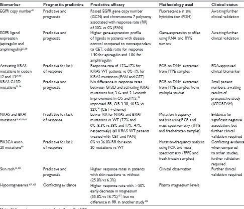
Table 1 Synopsis of major biomarkers derived from clinical studies for use with eGFR-targeted therapies in CRC
Biomarker Prognostic/predictive Predictive efficacy Methodology used Clinical status
eGFR copy number23 Predictive and prognostic
Raised eGFR gene copy number (GCN) and chromosome 7 polysomy associated with response rate (RR) of 30% vs 0% (PAN)
Fluorescence in situ hybridization (FiSH)
Awaiting further clinical validation
eGFR ligand expression (epiregulin and amphiregulin)27,28
Predictive and prognostic
Higher gene-expression profile of ligands in patients with disease control compared to nonresponders to CeT; odds ratio for response 1.90 for epiregulin and 1.86 for amphiregulin
Gene-expression profiles using RNA and FFPe tumors
Awaiting further clinical validation
Activating KRAS mutations in codon 12 and 1339,40
Predictive for lack of response
Response rate of 12%–17% for KRAS wT patients vs 0%–1% for KRAS mutations (PAN and CeT)
PCR on DNA extracted from FFPe samples
FDA-approved clinical biomarker
KRAS G13D mutations55,56
Predictive and prognostic
No difference in response rates between G13D and activating KRAS mutations but, 3.6- and 2.1-month improvement in OS and PFS,55 improved RR, OR 3.38, 40.5% vs 22%56 (CeT + chemo)
PCR on DNA extracted from FFPe samples from multiple studies
Small patient numbers; awaiting results of prospective study (iCeCReAM)
NRAS and BRAF mutations18,42,60,61
Predictive for lack of response
Lower RR for NRAS and BRAF mutations vs wT (7.7% and 0%–8.3% vs 38% and 17%–47%, respectively) (all KRAS wT patients treated with CeT and PAN)
Mutation-frequency analysis using PCR and mass spectrometry (FFPe and fresh-frozen samples)
evidence for significant negative association, but further clinical validation required PiK3CA exon
20 mutations60
Predictive for lack of response
0% vs 36.8% RR for exon 20 mutations vs wT
Mutation-frequency analysis using PCR and mass spectrometry (FFPe and fresh-frozen samples)
Conflicting evidence when compared to other studies, further validation required Skin rash13,100 Predictive and
prognostic
Higher response rates in patients with skin reactions vs without (25.8% vs 6.3%)
Clinical observation Further clinical validation required
Hypomagnesemia107,108 Conflicting evidence Higher response rate with .50% early decrease in magnesium (55.8% vs 16.7%)107; but no difference in RR in another study108
Plasma magnesium levels
Note: All figures shown are statistically significant (P , 0.05).
Abbreviations: PAN, panitumumab; CeT, cetuximab; CRC, colorectal cancer chemo, chemotherapy; eGFR, epidermal growth-factor receptor; GCN, gene copy number; RR, response rate; RNA, ribonucleic acid; FFPE, formalin-fixed paraffin-embedded; WT, wild type; OS, overall survival; PFS, progression-free survival; PCR, polymerase chain reaction; vs, versus.
HER2, HER3, and HER4. Ligand binding to the extracellular domain leads to allosteric activation via receptor dimeriza-tion and tyrosine kinase transphosphoryladimeriza-tion, thus activating the Ras/mitogen-activated protein kinase (MAPK) pathway (RAS-RAF-MAPK) and the phosphoinositide 3-kianse (PI3K) pathway (PI3K-phosphatase and tensin homologue [PTEN]-Akt).17 These downstream signaling pathways are involved in cell proliferation, differentiation, apoptosis, and cell invasion. EGFR overexpression, constitutive acti-vation, ligand overexpression, and activating mutations of downstream effectors or loss of tumor-suppressor genes, eg,
PTEN, may all lead to activation of this pathway.
EGFR has been identified as an oncogene in a variety of tumors, including CRC, non-small-cell lung cancer
(NSCLC), and head and neck cancers, leading to the use of EGFR-targeted agents in these tumor types. Cetuximab and panitumumab are monoclonal antibodies immunoglobulin [Ig]-G1 and anti-IgG2, respectively) which bind to extracellular ligand-binding sites of EGFR, thus inhibiting EGFR phosphorylation and activation of down-stream intracellular signaling pathways. Panitumumab is a fully humanized antibody, as opposed to cetuximab, which is a chimeric monoclonal antibody, which may also be able to elicit an antibody-dependent cell-mediated cytotoxicity. These antibodies are associated with a clinical improvement in progression-free survival (PFS), specifically in patients with KRAS wild-type (WT) tumors.13,18,19 Although EGFR tyrosine-kinase inhibitors such as erlotinib and gefitinib have
Cancer Management and Research downloaded from https://www.dovepress.com/ by 118.70.13.36 on 20-Aug-2020
Dovepress
Patel and Karapetis
been efficacious in NSCLC, these benefits have not yet been demonstrated for CRC.
Biomarkers related
to EGFR expression
Contrary to expectation, EGFR expression does not correlate with response to treatment, even in KRAS WT cases. Tumors that do not exhibit EGFR overexpression by immunohis-tochemistry (IHC) may respond to cetuximab.20 A number of possible explanations have been proposed, including constitutive activation of EGFR receptors and inaccurate methodologies for assessment of EGFR expression, such as unreliable antibodies that are difficult to standardize and score. However, increased copy numbers of EGFR, which may be present without a significant increase in receptor expression, have been shown to be associated with response to EGFR-targeted therapies in various retrospective analyses.21,22 Sartore-Bianchi et al demonstrated a 30% response rate for patients with increased EGFR copy number, treated with panitumumab, compared with 0% for patients without the amplification.23 Lack of correlation between EGFR copy number and protein expression alludes to a qualitative effect of gene copy number. The available technologies for assess-ment of EGFR copy number, eg, fluorescence or chromogenic in situ hybridization (FISH or CISH), are easier to quantify compared with IHC, but the cutoff levels for significance are variable and copy numbers are often heterogeneous in metastatic disease, thus complicating clinical interpretation. Furthermore, significant inter-laboratory variability has been demonstrated in the measurement of EGFR copy number by FISH in experienced laboratories.24
Mutation of the KRAS gene is the only validated bio-marker to predict for resistance to EGFR-targeted therapies, as discussed below. Although EGFR copy number is not con-sidered a reliable biomarker for EGFR-targeted treatments, use of this biomarker in tumors with wild-type KRAS may improve the positive predictive value.25
Autocrine ligand production of epiregulin and amphiregu-lin leads to EGFR activation and tumor growth. Evaluation of ligand expression by messenger RNA (mRNA) may provide both prognostic and predictive information. Low expression of epiregulin mRNA has been shown to be prognostic of improved OS for KRAS WT patients who did not receive EGFR-targeted therapy.26 Assessment of tumor mRNA for these ligands using a gene signature derived from liver metastases from patients receiving cetuximab monotherapy has been proposed as a biomarker for response to EGFR- targeted therapies.27 High levels of epiregulin
and amphiregulin have been demonstrated in 50%–60% of patients with metastatic CRC, and are associated with ben-efit from cetuximab as monotherapy or in combination with chemotherapy.28–30 Within a KRAS WT group, high versus low epiregulin gene expression was found to be able to dif-ferentiate responders to cetuximab.High ligand expression was associated with response to cetuximab, as expected due to attenuation of ligand-based EGFR activation. This study demonstrates that assessment of epiregulin expression could be used as a positive biomarker for EGFR-targeted therapies to narrow down further the target population within the KRAS WT group that may benefit from treatment. However, the available methodologies for quantification for ligand expression require validation with establishment of cutoff levels. Of the EGFR-related biomarkers, none has been approved for clinical use, in part due to the lack of standard-ized methodologies to quantify these markers.
KRAS and downstream effectors
KRAS
Three human RAS genes have been identified: KRAS, NRAS, and HRAS. The K-Ras protein, a small-cell-membrane guanosine triphosphatase, is one of the most important down-stream effectors coupling EGFR to intracellular signaling cascades, mediated by RAF kinase and mitogen-activated extracellular signal-regulated kinases (ERK) leading to cell growth, division, motility, and inhibition of apoptosis.31 Single-nucleotide point mutations in the KRAS gene, in codons 12 and 13 of exon 2, lead to constitutive activation of the MAPK pathway, and are found in approximately 40% of patients with metastatic CRC.32,33 High concordance is reported between KRAS mutations from primary tumors and metastases,34–36 alluding to KRAS mutation early in the adenoma–carcinoma cascade. Although these mutations are not prognostic, they are established biomarkers for lack of response to anti-EGFR monoclonal antibodies in patients with metastatic CRC.19,27,37–41 The discovery of this mutation as a biomarker has been a major step in the personalization of EGFR-targeted treatments for CRC.
The hypothesis behind the evaluation of KRAS mutation status in the context of EGFR-targeted therapies was that constitutive activation of the intracellular signaling pathway downstream of EGFR would attenuate the effects of EGFR-targeted monoclonal antibodies. The best evidence to support this hypothesis comes from the National Cancer Institute of Canada CO.17 study and the PIVOTAL (Study of Prostate and Pelvis Versus Prostate Alone Treatment for Locally Advanced Prostate Cancer) trial, which examined the effect of KRAS
Cancer Management and Research downloaded from https://www.dovepress.com/ by 118.70.13.36 on 20-Aug-2020
Dovepress Personalized treatment for advanced colorectal cancer
mutation in patients treated with cetuximab or panitumumab, respectively, versus best supportive care after multiple lines of chemotherapy.39,40 Response rates were 12%–17% for KRAS WT patients treated with the antibodies compared to 0%–1% for those with mutant KRAS, and an OS benefit of 4.7 months was noted for KRAS WT patients treated with cetuximab. The panitumumab study (PIVOTAL) did not demonstrate an OS benefit for patients with KRAS WT tumors, though a significant increase in PFS and response rate were shown. OS in this study may have been confounded by patients on the control arm receiving panitumumab on progression as the study design allowed crossover.
Studies including a chemotherapy backbone alongside cetuximab provide further support for this hypothesis.42,43 A meta-analysis of the CRYSTAL (Cetuximab Combined with Irinotecan in First-Line Therapy for Metastatic Colorectal Cancer) and OPUS (Oxaliplatin and Cetuximab in First-Line Treatment of Metastatic Colorectal Cancer) studies demon-strated an overall survival benefit of 4 months (23.5 versus 19.5 months) for patients with KRAS WT tumors treated with cetuximab and standard chemotherapy (FOLFOX or FOLFIRI) in the first-line setting over chemotherapy alone.44 However, both the Medical Research Council (MRC) COIN (Combination Chemotherapy and Cetuximab as First-Line Therapy in Treating Patients with Advanced and/or Metastatic Colorectal Cancer) study and the NORDIC (Randomized Phase III study of 5-Fluorouracil/Folinate/Oxaliplatin Given Continuously or Intermittently with or Without Cetuximab, as First-Line Treatment of Metastatic Colorectal Cancer) trial found that KRAS status was not predictive of benefit with the addition of cetuximab in the first-line setting.45,46 The MRC COIN study did demonstrate a survival benefit in the FOLFOX-plus-cetuximab arm but not in the arm con-taining capecitabine as part of the standard chemotherapy regime, highlighting a potential negative interaction between capecitabine and cetuximab. In contrast, the NORDIC study, comparing continuous or bolus 5-FU plus oxaliplatin plus or minus cetuximab, showed no improvement in OS in the unselected population and a worse PFS for patients with KRAS WT tumors receiving cetuximab. Further analyses are awaited, but these results suggest that the benefit of cetuximab in the first-line setting may be restricted to combination use with infusional 5-FU. With respect to panitumumab, the PRIME (Panitumumab Randomized trial In combination with chemotherapy for Metastatic colorectal cancer to determine Efficacy) study demonstrated an improvement in PFS and response rate with the addition of panitumumab to FOL-FOX, with a trend to improved OS in patients with KRAS
WT tumors in the first-line setting.37 However, patients with mutant KRAS tumors exhibited shorter PFS, suggesting a negative interaction between panitumumab and oxaliplatin, in line with a previous study.47 A further negative interaction may exist between bevacizumab and cetuximab or panitu-mumab, as demonstrated by the lack of survival benefit in patients with KRAS WT tumors in the CAIRO-2 (Cetuximab, Capecitabine, Oxaliplatin and Bevacizumab in Advanced Colorectal Cancer) and PACCE (Panitumumab Advanced Colorectal Cancer Evaluation) studies.48,49
Studies in the second-line setting for cetuximab have not stratified outcomes by KRAS status, and an attempt to do so has essentially failed due to a low rate of tissue collec-tion.50,51 For instance, the Erbitux Plus Irinotecan for meta-static colorectal Cancer (EPIC) trial originally recruited and randomized patients irrespective of KRAS status. Although a retrospective analysis of outcome data by KRAS status did not demonstrate a benefit of addition of cetuximab to irinotecan for KRAS WT patients, tissue samples from only 23% of patients were available for analysis.51 The evidence for panitumumab in this setting is stronger than that for cetuximab, with an improvement in PFS and response rate for KRAS WT tumors.38
The evidence described herein supports the use of KRAS status as a specific biomarker to select patients who may be resistant to cetuximab and panitumumab, with the greatest support in the third-line setting when examining the magni-tude of survival benefit. Based upon this evidence, the US Food and Drug Administration and the European Medicines Agency have approved the restriction of cetuximab treatment to patients with KRAS WT tumors.41 However, extensive cali-bration and validation of KRAS mutation testing is required in order to ensure that practical and reliable resources are available for the implementation of this biomarker in clinical practice, as recommended in the American Society of Clinical Oncology guidelines.41,52 Methodological improvements in
KRAS testing by, for example, using mutant-enriched
poly-merase chain reaction53 may improve the predictive capacity of this biomarker.
Recent data have suggested that tumors with specific
KRAS mutations, especially the glycine-to-aspartate
muta-tion in codon 13 (G13D) mutamuta-tion, may be sensitive to cetuximab or panitumumab. In vitro data has shown that cancer cell lines with the G13D mutation have a lower transforming potential and attenuated proliferation in the presence of cetuximab, compared to other KRAS muta-tions.54 In a combined analysis of four data sets, De Roock et al demonstrated an improvement in OS (7.6 versus
Cancer Management and Research downloaded from https://www.dovepress.com/ by 118.70.13.36 on 20-Aug-2020
Dovepress
Patel and Karapetis
5.7 months, hazard ratio [HR] 0.50, 95% confidence interval [CI] 0.31–0.81) and PFS (4 versus 1.9 months, HR 0.51, CI 0.32–0.81) for patients with G13D mutations compared with other KRAS mutations treated with cetuximab.55 Tejpar et al showed a similar benefit, although only for PFS.56 However these studies were small, with only 32 and 83 patients, respectively, with the G13D mutation. On the other hand, a number of studies have demonstrated no benefit for either cetuximab or panitumumab by specific KRAS mutations.57 Although the preclinical work delineating different KRAS mutations is promising, prospective, multicenter clinical studies are required to recruit the numbers of patients nec-essary for the validation of G13D and other mutations in
KRAS as positive biomarkers of response to EGFR-targeted
therapies. Furthermore, 40%–60% of patients with KRAS WT do not respond to EGFR-targeted therapies.58,59 Clearly, the development of further biomarkers is necessary to select patients who will respond to these treatments.
NRAS and BRAF
The NRAS gene codes for a protein, N-Ras, which is an alternate effector to K-Ras. Mutations within this gene are found in 3%–5% of mCRC patients, and are mutually exclu-sive of KRAS mutations. Although a retrospective study and the PICCOLO (Panitumumab, Irinotecan and Ciclosporin in Colorectal Cancer Therapy) trial have demonstrated a reduced response to cetuximab and panitumumab for patients with
NRAS mutations,60,61 further work is required to demonstrate the predictive capacity of these mutations.
The serine/threonine-protein kinase B-Raf is an effector in the MAPK signaling pathway, downstream of K-Ras. Mutations in the proto-oncogene BRAF are present in 5%–10% of the metastatic CRC population and are also mutually exclusive of KRAS mutations.62 BRAF V600E is the most common of all BRAF mutations (present in 90% of cases), and is enriched in a subset of patients who are female, greater than 70 years of age, with KRAS WT right-sided colon cancer.63 This mutation leads to constitu-tive activation of B-Raf by mimicking a tyrosine-kinase phosphorylation.64 Emerging evidence from the CRYSTAL, OPUS, and PICCOLO trials supports the use of BRAF mutations as negative predictors of response to such EGFR inhibitors as cetuximab and panitumumab.18,42,61 Objective response rates are significantly higher in the WT group, from 17% to 47% compared to 0%–8% in the BRAF mutant group. However, a proportion of patients with mutant BRAF tumors still derived benefit from cetuximab treatment in the meta-analysis of the CRYSTAL and OPUS data, with a median PFS
of 7.1 versus 3.7 months for patients treated with cetuximab and chemotherapy compared to chemotherapy alone. In this analysis, BRAF had a prognostic rather than a predictive impact. Further work is required to unravel the predictive significance of BRAF mutations, including the significance of various BRAF mutations, as per KRAS.
There is currently insufficient evidence to support the routine use of BRAF or NRAS mutations as negative pre-dictive biomarkers for EGFR-targeted therapies. However,
BRAF mutations may yet be used to personalize treatment by
highlighting novel targets for therapy. Further knowledge of the molecular biology and functional consequences of these mutations is required prior to integration into clinical care. For instance, sorafenib, a multikinase inhibitor against WT BRAF, BRAF V600E, CRAF, and VEGF, has demonstrated preclinical activity in CRC cell lines carrying the BRAF V600E mutation.65 However, clinical trials did not demon-strate a significant benefit.66 The selective BRAF inhibitor, vemurafenib, demonstrated a modest benefit in a phase I study, with a 26% response rate,67 although not to the same extent as seen in melanoma.68 Tyrosine-kinase inhibitors targeted against RAF are in development and early clinical trials.
Pi3K/PTeN pathway
Either EGFR or KRAS activation may lead to phosphoryla-tion of phosphoinositide 3-kinase (PI3K), contributing to cross talk and pathway redundancy within the EGFR network. Mutations in the gene encoding the PI3K catalytic subunit (PI3KCA) are found in 15%–20% of metastatic CRC patients, leading to downstream activation of the PI3K pathway. The loss of expression of PTEN is present in 20%–40% of meta-static CRC patients, leading to the loss of the sole tumor-suppressor gene in the EGFR pathway. Although both these events may predict for resistance to EGFR-targeted therapies, supporting evidence is variable.60,69–72 The largest retrospec-tive series demonstrated that mutations of the PI3KCA gene in exon 20, but not exon 9, which were more common, were associated with resistance to cetuximab.60 However, other studies have not shown a correlation between PI3K status and response to cetuximab.73 The evidence pertaining to loss of PTEN is also variable, with a high discordance between PTEN expression in primary versus metastatic sites.72 Furthermore, assessment of loss of PTEN by IHC is unreliable with significant interreporter variation. Muta-tions in PI3K and loss of PTEN may coexist with KRAS and
BRAF mutations, presenting potential targets for single or
combination therapies.
Cancer Management and Research downloaded from https://www.dovepress.com/ by 118.70.13.36 on 20-Aug-2020
Dovepress Personalized treatment for advanced colorectal cancer
Rational treatment combinations
(KRAS mutant tumors)
Molecular profiling of the members of downstream signal-ing cascades may help rationalize drug development and personalization of therapy for patients with KRAS mutant tumors. KRAS is a key “node” in the activation of receptor tyrosine kinase signaling, but has proven difficult to target. Farnesyltransferase inhibitors were designed for RAS inhi-bition, but both preclinical and clinical studies have been disappointing, with no correlation between RAS mutation and response.74,75 Drug development has focused on targets downstream of KRAS, such as BRAF, MEK, PI3K, Akt, and mTOR, which are currently in early clinical trials. Knowledge of tumor genomic aberrations may aid drug selection. For instance, inhibition of MEK, a target down-stream of BRAF, has been very successful for melanoma patients with BRAF mutations.76 However, this effect has not translated to CRC. Cross talk within the EGFR network leads to activation of negative feedback loops involving the PI3K pathway and resistance to MEK inhibition in preclinical CRC models.77 It is likely that a combination of drugs selected to target the aberrant activated pathway and potential resistance pathways may be more effective than single agents. Dual inhibition of MEK and PI3K has been shown to be more efficacious than Mek inhibition alone in a cancer cell line.78
Translation of knowledge of molecular events to clini-cal practice is key to personalizing targeted therapy. For instance, evidence to support BRAF mutation as a marker of resistance to EGFR-targeted therapy has been described previously. However, CRC cell-line data has demonstrated that treatment with vemurafenib for BRAF V600E mutations leads to a powerful feedback activation of EGFR, lead-ing to continued proliferation.79 Combined treatment with EGFR-targeted treatment and vemurafenib was synergistic, both in vitro and in vivo. These and supporting experiments provide an explanation for the poor efficacy of vemurafenib in patients with BRAF V600E mutations and a rationale for design of further clinical studies combining EGFR and BRAF inhibitors. Elucidation of the specific genomic aberrations in individual tumors may aid the selection of appropriate drugs for the patient, but only if we understand the molecular effects of these drugs.
Targets upstream of KRAS
The genetic aberrations discussed thus far may account for up to 60% of CRCs that are likely to exhibit primary resis-tance to EGFR-targeted therapies. However, only 10%–15%
of the unselected population respond to anti-EGFR mono-therapy, indicating an alternate mechanism of resistance or activation of a different pathway in the remaining 25% of patients. EGFR is one of several membrane-bound recep-tors at the apex of a hierarchy of a variety of intracellular signaling cascades. Cross talk between other members of the EGFR family, such as HER2 and HER3, and the insulin-like growth-factor 1 receptor (IGF-1R) may lead to resistance to anti-EGFR therapies. Molecular profiling of these receptors may aid in selection of treatments specific to the activated pathways. Cetuximab-resistant cell lines exhibit HER2 gene amplification and to increase in HER2 phosphorylation, whereby HER2 knockdown restores cetuximab sensitivity.80 Furthermore, the resistant cell lines exhibit overexpression of heregulin, a HER3 and HER4 ligand, and increased HER2/HER3 dimerization and signaling. Although HER2 amplification only occurs in 2% of unselected metastatic CRC cases, enrichment is evident in patients with KRAS WT who do not benefit from anti-EGFR therapy.81 Both HER2 expression and increased heregulin expression cor-related with shorter OS in patients with KRAS WT tumor treated with cetuximab.82 These findings not only highlight HER2 and heregulin expression as markers of cetuximab resistance but also as potential novel targets in CRC, with established HER2-targeted treatments such as lapatinib and pertuzumab in these patients as shown in vitro.80,81 Although HER2 amplification and protein expression are routinely measured in clinical practice for breast cancer, heregulin-expression levels are variable, and the technology for assessment has not yet been standardized. Furthermore, HER2 activation may occur in the absence of protein over-expression, leading to difficulties in identification of the population requiring treatment. Further validation of these biomarkers is essential.
Preclinical work has demonstrated that overexpression of HER3 may also predict resistance to EGFR-targeted therapies.83 Overexpression of HER3 is present in 30%–80% of CRC patients, and correlates with a poorer outcome in patients treated with cetuximab and irinotecan.84 HER3 overexpression may be used as an additional biomarker to those related to EGFR, in order to select patients who may benefit from the addition of specific anti-HER3 monoclonal antibodies, such as AMG 88 and MM-121, which are cur-rently in clinical trials.
IGF-1R may stimulate EGFR via release of one of its ligands – transforming growth factor-α85 – thus highlighting an alternate receptor upstream of KRAS that may determine response to EGFR-targeted therapies. Overexpression of
Cancer Management and Research downloaded from https://www.dovepress.com/ by 118.70.13.36 on 20-Aug-2020
Dovepress
Patel and Karapetis
IGF-1R has been associated with resistance to cetuximab in patients with KRAS WT CRC, providing modest clinical support for the use of IGF-1R expression as another nega-tive biomarker for response to EGFR-targeted therapies.85,86 However, addition of anti-IGF-1R monoclonal antibodies to anti-EGFR-targeted treatments has not been successful in clinical trials. The failure to demonstrate improvement in response rate and survival may stem from the lack of predic-tive biomarkers for anti-IGF-1R antibodies.
An alternative receptor, MET, may hold promise both as a predictive biomarker and a target for treatment. Significant cross talk between the MET, EGFR, and HER3 leads to cross-activation and potentially resistance to EGFR-targeted treatments.87,88 As high expression of MET and its ligand, hepatocyte growth factor (HGF), correlates with advanced stage and poor survival,89 targeted inhibitors are in develop-ment, with some success. For instance, a combination of rilotumumab, an antibody raised against HGF, with pani-tumumab for patients with KRAS WT CRC demonstrated an improved response rate (31% versus 21%). Further trials of MET inhibitors are underway. As MET amplification is uncommon in CRC, standardization of techniques assessing MET and HGF expression is essential prior to their use as biomarkers for selection of therapy.
Imaging biomarkers
The assessment of on-target drug effects and the timely detection of the development of resistance are key compo-nents in the personalization of treatment, in order to make expedient, appropriate changes in treatment regimes. Several studies have demonstrated a correlation between early tumor shrinkage in chemorefractory metastatic CRC patients with OS and PFS.90,91 However, retrospective, exploratory analyses of data from the first-line setting did not corroborate these results.92 Recently, analysis of data from the CRYSTAL study demonstrated that a .20% change in tumor dimensions after 8 weeks of cetuximab treatment was predictive of OS and PFS,93 hence providing an early measure of treatment efficacy.
Although standard imaging may be used, molecular and functional imaging modalities represent one of the key technologies in development to profile drug effects within the individual.94 For instance, the morpholino-[124I]-IPQA probe, which binds to the activated EGFR kinase adenosine triphosphate-binding site, but not to the inactive form, has been developed in order to image active forms of EGFR in tumor cell lines and mouse xenografts.95 Magnetic reso-nance imaging has also been used to delineate constitutively
activated EGFR using an IgG antibody targeted against a truncated constitutively active form of EGFR, conjugated to iron oxide nanoparticles and imaged in murine models.96 Furthermore, targeted delivery of antibody using this method was also shown to be therapeutic. Such assays could be used to delineate the on-target effects of EGFR-directed monoclonal antibodies, such as cetuximab, as well improve treatment efficacy.
A further challenge in personalizing treatment is the identification of the cause for development of resistance, which may impact upon treatment selection. For instance, treatment with cetuximab has been shown to resensitize patients who demonstrate primary resistance to oxaliplatin,97 alluding to the presence of cancer stem cells that are in constant flux. Molecular imaging is an alternative to “blind” tumor biopsies that may not be feasible, in order to better select sites for biopsy and the treatments required for control of tumor burden.
Clinical biomarkers
Clinical biomarkers that may be more readily measured and are less invasive are being investigated.
Skin rash
Skin rash represents the most frequently encountered tox-icity associated with EGFR monoclonal antibodies, with incidence ranging from approximately 65% to 85%.13,98–100 The appearance of an acneiform rash has been associated with response to cetuximab. The severity of skin toxicity, as graded by Common Terminology Criteria for Adverse Events (CTCAE) criteria, has been correlated with improvement in OS. Patients receiving cetuximab monotherapy with a grade 2 or worse skin toxicity exhibited the greatest improvements in OS from approximately 2 months for patients with no rash, to 9.5 months for patients with a grade 2 or worse rash.13,99 These benefits were restricted to patients with KRAS WT tumors, as expected.100 The pathological mechanism behind this effect is unknown. As EGFR receptors are present in the skin as well as the gastrointestinal tract, skin toxicity may represent receptor saturation. The EVEREST (Intrapatient Cetuximab Dose Escalation in Metastatic Colorectal Cancer According to the Grade of Early Skin Reactions) study was carried out to test prospectively whether dose escalation of cetuximab in patients with a grade 0–1 rash was feasible and if it improved clinical outcome.101 Patients who demonstrated a grade 0 or 1 rash either remained on the standard treatment arm (cetuximab plus irinotecan) or were treated with a higher dose of cetuximab (500 mg/m2 per week as opposed to
Cancer Management and Research downloaded from https://www.dovepress.com/ by 118.70.13.36 on 20-Aug-2020
Dovepress Personalized treatment for advanced colorectal cancer
250 mg/m2). Patients on the higher dose were more likely to experience grade 2 or greater skin toxicity (59% versus 35% on the standard arm) and a higher response rate (30% versus 16%), but no improvement in OS was noted. However, skin toxicity may be more prognostic of survival than predictive of response to treatment. A recent study demonstrated a sig-nificant difference in survival by degree of skin toxicity for patients with KRAS mutations in codon 12 only. This patient group does not classically respond to anti-EGFR antibodies, as described further. Although skin rash would be an attractive clinical predictive biomarker of response to EGFR-targeted treatments in CRC, further trials are required.
Obesity
Obesity, as measured by body mass index (BMI) has been proposed as a potential prognostic and predictive marker. Patients with a BMI of greater than 35 are reported to be at risk of cancer recurrence after adjuvant treatment in some studies,102 but not in others.103,104
Further studies are assessing the role of diabetes, smoking and markers of chronic inflammatory disease as prognostic and predictive biomarkers.105,106
Hypomagnesemia
The development of cetuximab-induced hypomagnesemia is also being investigated as a potential surrogate marker of response, but the evidence is conflicting. Initial reports demonstrated an association between a .50% reduction in magnesium levels with improved response rates and improved OS (11 versus 8.1 months).107 Analysis of data from CO.17 did not support this observation, demonstrating that a greater degree of hypomagnesemia correlated with poor OS in patients receiving cetuximab monotherapy, irrespec-tive of KRAS status and after adjustment for development of skin toxicity.108 Further prospective studies are required to clarify the predictive value of cetuximab-induced hypo-magnesemia as a noninvasive, cost-effective biomarker of cetuximab efficacy.
Circulating tumor DNA
Measurement of circulating tumor DNA (ctDNA) could be an attractive noninvasive biomarker of tumor response to treatment. Circulating DNA may be derived from one of three sources: normal healthy cells, tumor stromal cells, and tumor cells, and there is some overlap between the types of circulating DNA in healthy patients and those with tumors.109 Although the presence of ctDNA has been shown to be prog-nostic for poorer outcomes in CRC, its predictive capacity
requires much further work.110,111 A small fraction of ctDNA has been shown to harbor the same point mutations as those that characterize the primary tumor, such as APC, KRAS, or BRAF, which have been shown to be predictive of out-come for patients undergoing surgery or chemotherapy.112,113 However, the fraction of such ctDNA may represent less than 0.01% of total ctDNA, and the development of a reliable assay has been challenging due to technological issues.110,112 Although the detection of ctDNA harboring mutations lead-ing to resistance to EGFR inhibitors represents a promislead-ing technology for less invasive methods of tailoring targeted therapies, it has not yet been validated for CRC.
Other biomarkers in development
We have outlined a variety of tumor-related characteristics that may predict response to targeted therapies, potentially allowing personalization of treatment for the individual. As described thus far, not all patients with KRAS WT tumors respond to EGFR-targeted therapies. KRAS posttranslational modifications are novel areas of interest for the identifica-tion of KRAS WT tumors that may not respond to EGFR-targeted therapies. The cytosolic protein KRAS requires a cascade of posttranslational modifications initiated by a CAAX motif, catalyzed by farnesyltransferase (FTase) in order to localize to the cell surface for normal function. FTase inhibitors have been designed in order to inhibit this process, but with little success in CRC. This phenomenon may be due to alternative prenylation by an alternative enzyme, geranylgeranyltransferase I.114,115 MicroRNAs (miRNAs), single-stranded small noncoding RNA molecules that may regulate gene expression by translational inhibition or mRNA degradation, may be a more promising target.116 MiRNAs may act as tumor-suppressor genes, as in the case of the let-7 family of miRNAs,117 or as oncogenes, as in the case of downregulation of miR-18a and miR-143, which attenuates KRAS suppression.118 A recent retrospective study demonstrated that increased expression of miR-200b and decreased expression of miR-143 were associated with improved PFS for patients with KRAS mutant tumors, but not WT.119 Further work is required to improve the validity and reliability of these assays when performed on formalin-fixed paraffin-embedded tumor tissues.120
In addition, biomarkers for the host response have recently been investigated, in order to fully assess treatment effect. The Fc region of anti-EGFR antibodies is vital to initiation of the host immune response via Fc gamma receptors (FcγRs). Single-nucleotide polymorphisms of FcγRIIIa are predic-tive of resistance to anti-EGFR antibodies and highlight a
Cancer Management and Research downloaded from https://www.dovepress.com/ by 118.70.13.36 on 20-Aug-2020
Dovepress
Patel and Karapetis
group of tumors that may be resistant to these drugs, irrespec-tive of KRAS status.121
In contrast to the specific genomic mutations outlined herein, high-throughput technologies, including microar-rays and single-nucleotide polymorphism microarmicroar-rays, aim to identify genome-wide changes in tumor DNA in order to predict outcome and response to treatment.122 However, thus far no genomic signature has been validated as a pre-dictive marker for use in metastatic CRC, in part due to the retrospective, heterogeneous, and/or small nature of studies attempting to derive these signatures. Within the field of pharmacogenetics, an interesting tool in development is the drug-metabolizing enzymes and transporters (DMET) microarray. Drug-metabolism enzymes may affect drug phar-macokinetics and pharmacodynamics, thus altering levels of the active drug or metabolite. The DMET microarray profiles over 200 genes that may be functionally involved with such enzymes. If the drugs used in metastatic CRC can be vali-dated on this platform, interpatient variations in active drug levels may be predicted and appropriate doses prescribed, potentially improving drug efficacy. These technologies are expensive and require significant validation, due to the enormous amount of data generated from microarray and high-throughput genome analysis.
Chemotherapy-related predictive factors are in devel-opment, but few are ready for routine use. Biomarkers predicting response to chemotherapy may be related to pharmaocodynamic and pharmacokinetic factors, or due to unrelated genomic mutations, such as MSI. Mutations leading to malfunction in the DNA mismatch-repair mechanism lead to MSI in 15% of colorectal tumors.123 High-MSI tumors are associated with early stage CRC (stage II) and resistance to 5-FU adjuvant treatment, as opposed to low-MSI tumors, which may have a worse prognosis but are sensitive to 5-FU treatment in the adjuvant setting. Although MSI level may be a useful adjunct to current methods of prognostication, such as stage of disease at presentation, to direct the use of adjuvant chemotherapy for stage II CRC,124 further validation is being carried out in the Eastern Cooperative Oncology Group 5202 clinical trial. This trial randomizes patients with stage II CRC, who have had curative surgery, to observa-tion only for patients who are deemed to be at low risk for MSI, versus adjuvant chemotherapy with or without beva-cizumab for patients at high risk for MSI. Knowledge of the metabolism as well as the mechanisms of action of a drug may provide insights into novel pharmacodynamic and phar-macokinetic biomarkers. For instance, capecitabine, an oral prodrug of 5-FU, undergoes catabolism to fluorodeoxyuridine
monophosphate, which inhibits thymidylate synthase, the major mechanism of action of 5-FU. Dihydropyrimidine dehydrogenase (DPD) is the rate-limiting enzyme for this step. A low level of DPD expression is associated with better outcome with capecitabine and irinotecan, and conversely high gene expression of DPD has been associated with resistance to capecitabine in the metastatic setting.125,126 This biomarker is used in clinical practice for patients who demonstrate severe 5-FU-induced toxicities.
Conclusion
Although the discovery of KRAS mutations has paved the way for personalized treatment for patients with metastatic CRC, the type of KRAS mutation and aberrations in related proteins, such as BRAF, PTEN, and PIK3Ca are likely to be important in refining patient selection and improving outcomes. Mutations within these genes highlight a group of patients who may be resistant to anti-EGFR antibodies, but the best course of treatment for these patients is currently unclear. Conversely, the data pertaining to EGFR copy number and expression of EGFR ligands delineate a group who may be sensitive to anti-EGFR antibodies. Therefore, these positive biomarkers may be more clinically useful in selecting appropriate treatments for individual patients. However, the methodologies for these and many of the bio-markers described herein require further standardization and validation. Functional and/or molecular imaging is expected to have an important role in the noninvasive, real-time assess-ment of patient response and developassess-ment of resistance, thus helping to tailor treatment appropriately. However, the technologies required for this are not widely available, and imaging biomarkers also require validation.
Many different proteins, including those relating to the host response, are likely to be involved in tumor dynam-ics. It is imperative that we identify key “nodes” within the receptor tyrosine-kinase network, such as RAS, in order to develop combinations of drugs with the best potential for control of tumor burden. In vitro characterization of protein–protein interactions has been integrated to build signal networks to model carcinogenic pathways or response to drug treatment, for example for EGFR.127 Nodes within these networks define key pathways that are integral for carcinogenesis or as a target for therapy. These networks may be used to generate novel predictive markers and direct novel drug development. Translational studies must be carried out in parallel to drug development to ensure that biomarkers assessing the functional status of these nodes within individual patients are available, in order to select
Cancer Management and Research downloaded from https://www.dovepress.com/ by 118.70.13.36 on 20-Aug-2020
Dovepress Personalized treatment for advanced colorectal cancer
the correct combination of drugs. However, toxicities may be synergistic and render the drugs intolerable, as demon-strated when combining EGFR and VEGF inhibitors.48 In the future, the best results are likely to be achieved through a combined application of the current genomic biomark-ers with novel predictive molecular and genomic markbiomark-ers and potentially functional/molecular imaging in order to personalize therapy in real time.
Disclosure
Associate Professor Christos S Karapetis: advisory board for Amgen, Roche, and Merck Serono. The authors have no other conflicts of interest to report.
References
1. Dienstmann R, Vilar E, Tabernero J. Molecular predictors of response to chemotherapy in colorectal cancer. Cancer J. 2011;17(2):114–126. 2. Fearon ER, Vogelstein B. A genetic model for colorectal tumorigenesis.
Cell. 1990;61(5):759–767.
3. Boland CR, Goel A. Microsatellite instability in colorectal cancer. Gastroenterology. 2010;138(6):2073–2087. e3.
4. Toyota M, Ahuja N, Ohe-Toyota M, Herman JG, Baylin SB, Issa JP. CpG island methylator phenotype in colorectal cancer. Proc Natl Acad Sci U S A. 1999;96(15):8681–8686.
5. Goel A, Arnold CN, Niedzwiecki D, et al. Frequent inactivation of PTEN by promoter hypermethylation in microsatellite instability-high sporadic colorectal cancers. Cancer Res. 2004;64(9):3014–3021. 6. Goel A, Arnold CN, Tassone P, et al. Epigenetic inactivation of
RUNX3 in microsatellite unstable sporadic colon cancers. Int J Cancer. 2004;112(5):754–759.
7. Shin SK, Nagasaka T, Jung BH, et al. Epigenetic and genetic alterations in Netrin-1 receptors UNC5C and DCC in human colon cancer. Gastroenterology. 2007;133(6):1849–1857.
8. Silver A, Sengupta N, Propper D, et al. A distinct DNA methylation profile associated with microsatellite and chromosomal stable sporadic colorectal cancers. Int J Cancer. 2012;130(5):1082–1092.
9. Nordic Gastrointestinal Tumor Adjuvant Therapy Group. Expectancy or primary chemotherapy in patients with advanced asymptomatic colorectal cancer: a randomized trial. J Clin Oncol. 1992;10(6): 904–911.
10. Scheithauer W, Rosen H, Kornek GV, Sebesta C, Depisch D. Randomised comparison of combination chemotherapy plus supportive care with supportive care alone in patients with metastatic colorectal cancer. BMJ. 1993;306(6880):752–755.
11. Tournigand C, Andre T, Achille E, et al. FOLFIRI followed by FOLFOX6 or the reverse sequence in advanced colorectal cancer: a randomized GERCOR study. J Clin Oncol. 2004;22(2):229–237. 12. Hurwitz H, Fehrenbacher L, Novotny W, et al. Bevacizumab plus
irinotecan, fluorouracil, and leucovorin for metastatic colorectal cancer. N Engl J Med. 2004;350(23):2335–2342.
13. Cunningham D, Humblet Y, Siena S, et al. Cetuximab monotherapy and cetuximab plus irinotecan in irinotecan-refractory metastatic colorectal cancer. N Engl J Med. 2004;351(4):337–345.
14. Weickhardt AJ, Williams D, Lee C, et al. Vascular endothelial growth factors (VEGF) and VEGF receptor expression as predictive biomarkers for benefit with bevacizumab in metastatic colorectal cancer (mCRC): analysis of the phase III MAX study. J Clin Oncol. 2011; 29 Suppl:3531.
15. Syrigos KN, Karapanagiotou E, Boura P, Manegold C, Harrington K. Bevacizumab-induced hypertension: pathogenesis and management. BioDrugs. 2011;25(3):159–169.
16. Committee ACNCCGR. Guidelines for the prevention, early detection and management of colorectal cancer. In: Network TCCAaAC, ed. Sydney: 2005.
17. Yarden Y, Sliwkowski MX. Untangling the ErbB signalling network. Nat Rev Mol Cell Biol. 2001;2(2):127–137.
18. Van Cutsem E, Kohne CH, Hitre E, et al. Cetuximab and chemotherapy as initial treatment for metastatic colorectal cancer. N Engl J Med. 2009;360(14):1408–1417.
19. Van Cutsem E, Peeters M, Siena S, et al. Open-label phase III trial of panitumumab plus best supportive care compared with best supportive care alone in patients with chemotherapy-refractory metastatic colorectal cancer. J Clin Oncol. 2007;25(13):1658–1664.
20. Chung KY, Shia J, Kemeny NE, et al. Cetuximab shows activity in colorectal cancer patients with tumors that do not express the epidermal growth factor receptor by immunohistochemistry. J Clin Oncol. 2005;23(9):1803–1810.
21. Moroni M, Veronese S, Benvenuti S, et al. Gene copy number for epidermal growth factor receptor (EGFR) and clinical response to antiEGFR treatment in colorectal cancer: a cohort study. Lancet Oncol. 2005;6(5):279–286.
22. Cappuzzo F, Finocchiaro G, Rossi E, et al. EGFR FISH assay predicts for response to cetuximab in chemotherapy refractory colorectal cancer patients. Ann Oncol. 2008;19(4):717–723.
23. Sartore-Bianchi A, Moroni M, Veronese S, et al. Epidermal growth factor receptor gene copy number and clinical outcome of metastatic colorectal cancer treated with panitumumab. J Clin Oncol. 2007;25(22): 3238–3245.
24. Sartore-Bianchi A, Fieuws S, Veronese S, et al. Standardisation of EGFR FISH in colorectal cancer: results of an international interlabora-tory reproducibility ring study. J Clin Pathol. 2012;65(3):218–223. 25. Scartozzi M, Bearzi I, Mandolesi A, et al. Epidermal growth factor
receptor (EGFR) gene copy number (GCN) correlates with clinical activity of irinotecan-cetuximab in K-RAS wild-type colorectal cancer: a fluorescence in situ (FISH) and chromogenic in situ hybridization (CISH) analysis. BMC Cancer. 2009;9:303.
26. Kuramochi H, Nakajima G, Kaneko Y, et al. Amphiregulin and epiregulin mRNA expression in primary colorectal cancer and corresponding liver metastases. BMC Cancer. 2012;12:88.
27. Khambata-Ford S, Garrett CR, Meropol NJ, et al. Expression of epiregulin and amphiregulin and K-ras mutation status predict disease control in metastatic colorectal cancer patients treated with cetuximab. J Clin Oncol. 2007;25(22):3230–3237.
28. Jacobs B, De Roock W, Piessevaux H, et al. Amphiregulin and epiregulin mRNA expression in primary tumors predicts outcome in metastatic colorectal cancer treated with cetuximab. J Clin Oncol. 2009;27(30):5068–5074.
29. Tabernero J, Cervantes A, Rivera F, et al. Pharmacogenomic and pharmacoproteomic studies of cetuximab in metastatic colorectal cancer: biomarker analysis of a phase I dose-escalation study. J Clin Oncol. 2010;28(7):1181–1189.
30. Jonker DJ, Karapetis CS, Harbison CT, et al. High epiregulin (EREG) gene expression plus K-ras wild-type (WT) status as predictors of cetux-imab benefit in the treatment of advanced colorectal cancer (ACRC): results from the NCIC CTG CO.17 – a phase III trial of cetuximab versus best supportive care (BSC). J Clin Oncol. 2009;27 Suppl 15;4016. 31. Schubbert S, Shannon K, Bollag G. Hyperactive Ras in developmental
disorders and cancer. Nat Rev Cancer. 2007;7(4):295–308.
32. Bos JL, Fearon ER, Hamilton SR, et al. Prevalence of ras gene mutations in human colorectal cancers. Nature. 1987;327(6120):293–297. 33. Bos JL. Ras Oncogenes in human cancer: a review. Cancer Res.
1989;49(17):4682–4689.
34. Knijn N, Mekenkamp LJ, Klomp M, et al. KRAS mutation analysis: a comparison between primary tumours and matched liver metastases in 305 colorectal cancer patients. Br J Cancer. 2011;104(6):1020–1026. 35. Santini D, Loupakis F, Vincenzi B, et al. High concordance of KRAS sta-tus between primary colorectal tumors and related metastatic sites: impli-cations for clinical practice. Oncologist. 2008;13(12): 1270–1275.
Cancer Management and Research downloaded from https://www.dovepress.com/ by 118.70.13.36 on 20-Aug-2020
Dovepress
Patel and Karapetis
36. Artale S, Sartore-Bianchi A, Veronese SM, et al. Mutations of KRAS and BRAF in primary and matched metastatic sites of colorectal cancer. J Clin Oncol. 2008;26(25):4217–4219.
37. Douillard JY, Siena S, Cassidy J, et al. Randomized, phase III trial of panitumumab with infusional fluorouracil, leucovorin, and oxaliplatin (FOLFOX4) versus FOLFOX4 alone as first-line treatment in patients with previously untreated metastatic colorectal cancer: the PRIME study. J Clin Oncol. 2010;28(31):4697–4705.
38. Peeters M, Price TJ, Cervantes A, et al. Randomized phase III study of panitumumab with fluorouracil, leucovorin, and irinotecan (FOLFIRI) compared with FOLFIRI alone as second-line treatment in patients with metastatic colorectal cancer. J Clin Oncol. 2010;28(31):4706–4713. 39. Amado RG, Wolf M, Peeters M, et al. Wild-type KRAS is required for
panitumumab efficacy in patients with metastatic colorectal cancer. J Clin Oncol. 2008;26(10):1626–1634.
40. Karapetis CS, Khambata-Ford S, Jonker DJ, et al. K-ras mutations and benefit from cetuximab in advanced colorectal cancer. N Engl J Med. 2008;359(17):1757–1765.
41. Allegra CJ, Jessup JM, Somerfield MR, et al. American Society of Clinical Oncology provisional clinical opinion: testing for KRAS gene mutations in patients with metastatic colorectal carcinoma to predict response to anti-epidermal growth factor receptor monoclonal antibody therapy. J Clin Oncol. 2009;27(12):2091–2096.
42. Bokemeyer C, Bondarenko I, Hartmann JT, et al. Efficacy according to biomarker status of cetuximab plus FOLFOX-4 as first-line treatment for metastatic colorectal cancer: the OPUS study. Ann Oncol. 2011;22(7): 1535–1546.
43. Van Cutsem E, Kohne CH, Lang I, et al. Cetuximab plus irinotecan, fluorouracil, and leucovorin as first-line treatment for metastatic colorec-tal cancer: updated analysis of overall survival according to tumor KRAS and BRAF mutation status. J Clin Oncol. 2011;29(15):2011–2019. 44. Bokemeyer C, Van Cutsem E, Rougier P, et al. Addition of cetuximab to
chemotherapy as first-line treatment for KRAS wild-type metastatic col-orectal cancer: pooled analysis of the CRYSTAL and OPUS randomised clinical trials. Eur J Cancer. 2012;48(10):1466–1475.
45. Maughan TS, Adams RA, Smith CG, et al. Addition of cetuximab to oxaliplatin-based first-line combination chemotherapy for treatment of advanced colorectal cancer: results of the randomised phase 3 MRC COIN trial. Lancet. 2010;377(9783):2103–2114.
46. Tveit K, Guren T, Glimelius B, et al. Randomized phase III study of 5-fluorouracil/folinate/oxaliplatin given continuously or intermittently with or without cetuximab, as first-line treatment of metastatic colorectal cancer: The NORDIC VII study (NCT00145314), by the Nordic Colorec-tal Cancer Biomodulation Group. Ann Oncol. 2010;21 Suppl 8:viii9. 47. Bokemeyer C, Bondarenko I, Makhson A, et al. Fluorouracil, leucovorin,
and oxaliplatin with and without cetuximab in the first-line treatment of metastatic colorectal cancer. J Clin Oncol. 2009;27(5):663–671. 48. Tol J, Koopman M, Cats A, et al. Chemotherapy, bevacizumab, and
cetuximab in metastatic colorectal cancer. N Engl J Med. 2009;360(6): 563–572.
49. Hecht JR, Mitchell E, Chidiac T, et al. A randomized phase IIIB trial of chemotherapy, bevacizumab, and panitumumab compared with chemotherapy and bevacizumab alone for metastatic colorectal cancer. J Clin Oncol. 2009;27(5):672–680.
50. Sobrero AF, Maurel J, Fehrenbacher L, et al. EPIC: phase III trial of cetuximab plus irinotecan after fluoropyrimidine and oxaliplatin failure in patients with metastatic colorectal cancer. J Clin Oncol. 2008;26(14): 2311–2319.
51. Langer C, Kopit J, Awad M, et al. Analysis of KRAS mutations in patients with metastatic colorectal cancer receiving cetuximab in combination with irinotecan: results from the EPIC trial. Ann Oncol. 2008;19 Suppl 8:viii133.
52. Jimeno A, Messersmith WA, Hirsch FR, Franklin WA, Eckhardt SG. KRAS mutations and sensitivity to epidermal growth factor receptor inhibitors in colorectal cancer: practical application of patient selection. J Clin Oncol. 2009;27(7):1130–1136.
53. Molinari F, Felicioni L, Buscarino M, et al. Increased detection sen-sitivity for KRAS mutations enhances the prediction of anti-EGFR monoclonal antibody resistance in metastatic colorectal cancer. Clin Cancer Res. 2011;17(14):4901–4914.
54. Guerrero S, Casanova I, Farre L, Mazo A, Capella G, Mangues R. K-ras codon 12 mutation induces higher level of resistance to apoptosis and predisposition to anchorage-independent growth than codon 13 muta-tion or proto-oncogene overexpression. Cancer Res. 2000;60(23): 6750–6756.
55. De Roock W, Jonker DJ, Di Nicolantonio F, et al. Association of KRAS p.G13D mutation with outcome in patients with chemotherapy-refractory metastatic colorectal cancer treated with cetuximab. JAMA. 2010;304(16):1812–1820.
56. Tejpar S, Celik I, Schlichting M, Sartorius U, Bokemeyer C, Van Cutsem E. Association of KRAS G13D tumor mutations with outcome in patients with metastatic colorectal cancer treated with first-line chemotherapy with or without cetuximab. J Clin Oncol. 2012;30(29): 3570–3577.
57. Peeters M, Douillard JY, Van Cutsem E, et al. Mutant KRAS codon 12 and 13 alleles in patients with metastatic colorectal cancer: assessment as prognostic and predictive biomarkers of response to panitumumab. J Clin Oncol. 20, 2013;31(6):759–765.
58. Bardelli A, Siena S. Molecular mechanisms of resistance to cetuximab and panitumumab in colorectal cancer. J Clin Oncol. 2010;28(7): 1254–1261.
59. Linardou H, Dahabreh IJ, Kanaloupiti D, et al. Assessment of somatic k-RAS mutations as a mechanism associated with resistance to EGFR-targeted agents: a systematic review and meta-analysis of studies in advanced non-small-cell lung cancer and metastatic colorectal cancer. Lancet Oncol. 2008;9(10):962–972.
60. De Roock W, Claes B, Bernasconi D, et al. Effects of KRAS, BRAF, NRAS, and PIK3CA mutations on the efficacy of cetuximab plus chemotherapy in chemotherapy-refractory metastatic colorectal cancer: a retrospective consortium analysis. Lancet Oncol. 2010;11(8): 753–762.
61. Seymour MT, Brown SR, Richman S, Middleton GW, Maughan T, Olivier C. Addition of panitumumab to irinotecan: results of PICCOLO, a randomized controlled trial in advanced colorectal cancer (aCRC). J Clin Oncol. 2011;29 Suppl:3523.
62. De Mattos-Arruda L, Dienstmann R, Tabernero J. Development of molecular biomarkers in individualized treatment of colorectal cancer. Clin Colorectal Cancer. 2011;10(4):279–289.
63. Tie J, Gibbs P, Lipton L, et al. Optimizing targeted therapeutic development: analysis of a colorectal cancer patient population with the BRAF (V600E) mutation. Int J Cancer. 2011;128(9):2075–2084. 64. Davies H, Bignell GR, Cox C, et al. Mutations of the BRAF gene in
human cancer. Nature. 2002;417(6892):949–954.
65. Di Nicolantonio F, Martini M, Molinari F, et al. BRAF V600E confers resistance to cetuximab or panitumumab in metastatic colorectal cancer. Eur J Cancer Suppl. 2008;6(12):81.
66. Tabernero J, Konne C, O’Dwyer PJ, et al. A phase IIB double-blind, randomized study evaluating the efficacy and safety of sorafenib (SOR) compared with placebo (PBO) when administered in combination with chemotherapy (modified FOLFOX6) for first-line treatment (tx) of patients (pts) with metastatic colorectal cancer (mCRC). The RESPECT Trial. Eur J Cancer. 2011;47 Suppl 2:11.
67. Kopetz S, Desai J, Chan E, et al. PLX4032 in metastatic colorectal cancer patients with mutant BRAF tumours. J Clin Oncol. 2010; 28 Suppl 15:3534.
68. Chapman P, Puzanov I, Sosman J. Early efficacy signal demonstrated in advanced melanoma in a phase I trial of the oncogenic BRAF-selective inhibitor PLX4032. Eur J Cancer Suppl. 2009;7(3):5.
69. Sartore-Bianchi A, Martini M, Molinari F, et al. PIK3CA muta-tions in colorectal cancer are associated with clinical resistance to EGFR-targeted monoclonal antibodies. Cancer Res. 2009;69(5): 1851–1857.
Cancer Management and Research downloaded from https://www.dovepress.com/ by 118.70.13.36 on 20-Aug-2020