Breast Cancer: Targets and Therapy
Dovepress
O r i g i n A L r e s e A r C h open access to scientific and medical research
Open Access Full Text Article
estrogen receptor (er) and progesterone
receptor (Pgr) in breast cancer of indian women
Amit V Patil1
rahul s Bhamre2
rajeev singhai3
Mukund B Tayade4
Vinayak W Patil3
1Department of general surgery,
government Medical College, Miraj, Maharashtra, india; 2Department of
general surgery, DY Patil hospital and research Centre, nerul, navi Mumbai, india; 3Department of
Biochemistry, grant Medical College and sir JJ group of hospitals Mumbai, india; 4Department of general
surgery, grant Medical College and sir JJ group of hospitals Mumbai, india
Correspondence: rajeev singhai C-505, Beach Classic Chs Ltd, near gorai Pumping station, Chikoowadi, Borivali (W), Mumbai 400092, india Tel +919699856615
email dr.rajeevj@gmail.com
Objective: To determine the expressions and relationship between estrogen receptors (ERs) and progesterone receptors (PgRs) in breast cancer in Indian women.
Participants: Surgically removed breast cancer tissues were collected from Grant Medical College and Sir JJ Group of Hospitals, Mumbai, India, taking (n = 300) cases of infiltrating duct cancer of Indian women after radical mastectomy and lumpectomy; the age- and menopausal-related subgroups satisfied this requirement.
Measurements: Statistical significance was calculated by the likelihood ratio test; relative risk served to check for significant differences. Relapse-free interval probabilities were calcu-lated according to Kaplan and Meier, with Cox–Mantel test comparing survival functions and
P values.
Results: We observed that only in middle-aged postmenopausal patients bearing pT2 tumors were ER and PgR receptors shown to have a prognostic significance with the lowest tested cutoff value being 5 fmol/mg.
Conclusion: Immunohistochemistry analysis has been shown to be a prognostic factor for patients with breast cancer; the major aim of determining the ER receptor status is to assess predictive response to hormonal therapy.
Keywords: prognostic cancer tissue biomarkers, immunohistochemistry, hormone receptors, steroid receptors
Introduction
Immunohistochemistry (IHC) analysis of estrogen receptor (ER) has been shown to be a prognostic factor for patients with breast cancer in India. The major aim of determining the ER receptor status is to assess predictive response to hormonal therapy. Aromatase inhibitors have been found consistently to be more effective agents than tamoxifen in delaying recurrence when administered as adjuvant treatment to patients with ER- and progesterone receptor (PgR)-positive early breast cancer in Indian women. Prognostic factor is indicative of the inherent histopathological aggressiveness of a tumor, reflect-ing natural history of the disease after local therapy. The inherent histopathological aggressiveness of a tumor is indicative of prognosis, reflecting natural history of the disease after local therapy. It is, therefore, most accurately assessed in systematically untreated patients. For this purpose, we attempted to evaluate the prognostic signifi-cance of ER and PgR receptors in node-negative breast signifi-cancer patients in terms of relapse-free interval, treated with loco-regional therapy only. Biological behavior of breast cancer supports the assumption that patients without spreading of malignant cells to axillary lymph nodes (ANNs) can be classified into several subgroups in
RETRACTED
Breast Cancer: Targets and Therapy downloaded from https://www.dovepress.com/ by 118.70.13.36 on 20-Aug-2020
For personal use only.
Number of times this article has been viewed
This article was published in the following Dove Press journal: Breast Cancer: Targets and Therapy
Dovepress
Patil et al
keeping with the different aggressiveness.1 According to the
Consensus statement of Gooldhirsch A et al2 a large group
of node-negative breast cancer patients are treated with no expected benefit. Therefore, it is important to determine the subgroup of patients who are at high risk for recurrence and should, with no doubt, receive more aggressive adjuvant therapy than subgroups of patients who are at intermediate low risk.
In around 60% of all breast cancer cases, tumor cells carry ER and PgR. Around 20% of cases manifest no receptors for the hormones. The cases demonstrating expression of both receptors are known to be the ones to respond most frequently by remission to treatment with tamoxifen. The presence of ER by itself has been accepted as an independent prognostic and predictive factor. As compared with ER, the significance of PgR expression has proved to be much less unequivocal.
The measurement of ER concentrations in breast cancer tissue is an established method of predicting the response of a tumor to endocrine therapy, either using the traditional radioligand binding assay, or the more recent IHC techniques. Response to endocrine therapy clearly correlates with recep-tor positivity. The richer the tumor in ER, the better is the prognosis for the patient. Almost 70% of breast cancer patients have ER-positive tumors, and of these, around 60% are found to respond to endocrine therapy. The prediction of the prognosis of breast cancer patients is expected to be achieved by a subgrouping of ER-positive patients, based on the physiology of estrogen signaling. Identification of a poor-prognosis population among ER-positive breast cancer patients can be achieved by the use of selected estrogen-regulated genes (ERGs). Only low levels of response to treatment are achieved in those patients with ER-negative tumors. PgRs, which reflect the functional estrogenic stimu-lus, have also been investigated, and are believed to be at least as significant a prognostic and predictive factor as the ER status.
Materials and methods
human breast tumor tissue
collection and fixation protocol
All surgically removed breast cancer tissues were collected from Grant Medical College and Sir JJ Group of Hospitals, Mumbai, India, taking (n = 300) cases of female patients with operable primary breast cancer (infiltrating duct cancer), identified between May 2007 to July 2010. The age- and menopausal-related subgroups satisfied this requirement. The group of patients of age 44 and younger (n = 54) was mostly premenopausal. Patients aged over 59 (n = 75) were
postmenopausal except one with premenopausal status. Those aged between 45 and 59, ie, middle aged (n = 168), and (n = 3) not reporting, were premenopausal, perimenopausal, and postmenopausal, with nearly same frequency tissue sam-ples of breast cancer of Indian patients. Expressions of ER and PgR IHC breast cancer tissue biomarkers were analyzed in specimens of invasive duct breast cancer tissue of Indian women after radical mastectomy and lumpectomy.
immunohistochemistry
Tissue samples were fixed in fixative 10% neutral buffered formalin (fixative) for 12–24 hours. Tissue samples were processed in an autoprocesser, and then embedded with paraffin wax on embedding station. Paraffin blocks were cut into microtome 4 µ thickness sections and dried overnight at 37°C. Prior to antibody staining, the slides were pre-treated with microwave irradiation to unmask binding epitopes. After blocking endogenous peroxide activity with a 3% solution of hydrogen peroxide in methanol for 30 minutes, slides were immersed in 200 mL of 10 mM citric acid (pH 6.0) for 5 minutes at 100 W powers, followed by 4 cycles of 5 min-utes each at 50 W power. After topping up of the buffer with distilled water, this step was repeated. The slides were then left to stand for 10 minutes in buffer at room temperature before being washed thoroughly in tap water.
After three washes in tris-buffered saline (TBS), the slides were incubated with a 1:25 dilution of mouse anti-ER
α monoclonal primary antibody (Clone: 1D5; M7047; DakoCytomation, Denmark), 1:25 dilution of mouse anti-PgR monoclonal primary antibody (Clone: anti-PgR 636; M3569; DakoCytomation, Denmark) in TBS for 1 hour at room temperature. After three more washes in TBS, secondary anti-body (K0355; DakoCytomation, Denmark) biotinylated goat antibody (LINK) to mouse/rabbit immunoglobulin, diluted antibody (1:100) in TBS was applied for 1 hour at room temperature. After an additional three washes, streptavidin – biotin/HRP; horse radish peroxidase complex (enzyme label), (K0355; DakoCytomation, Denmark) diluted antibody (1:50) in TBS was applied for 1 hour at room temperature. After an additional three washes, the staining was visualized by add-ing diaminobenzidine (DAB kit; K3467; DakoCytomation, Denmark) for 5 minutes at room temperature. The slides were washed well in tap water and counterstained with Harris’s hematoxylin for 10 seconds to 1 minute and then dehydrated, cleared, and mounted in distrene plasticizer xylene (DPX). Positive and negative controls were performed with each batch of slides. Surgical specimens from the same patient were stained on the same run.
RETRACTED
Breast Cancer: Targets and Therapy downloaded from https://www.dovepress.com/ by 118.70.13.36 on 20-Aug-2020
Dovepress er and Pgr in breast cancer of indian women
The entire stained slide was scanned for immunostaining evaluation by light microscope. The image collection and microphotographs were taken by the Axio Imager. M1 Microscope with AxioVision software (Carl Zeiss Microscopy, Germany). Slides were checked under 10× objective to confirm that the cells were still attached to the slide, and then finally viewed under ×400 objective magnification. All images were taken under ×400 objec-tive magnification without oil immersion lens. All images were processed with AxioVision software.
scoring methods
ER and PgR receptors are steroid receptors localizing to the nucleus. ER and PgR status of a tumor impacts on disease-free survival interval in lymph node-positive groups of patients, as well as predicating the response to endocrine therapy more specifically, to the anti-estrogenic tamoxifen or in patient selection for alternative first-line treatment. ER and PgR positivity is denoted by nuclear staining brown of both the invasive and in-situ component of the breast cancer. Positive ER and PgR results are further qualified using a rapid semiquantitative H score ranging from 0 to 8 that takes into account both the intensity of staining and proportion of tumor cells staining positive for ER and PgR receptors with appro-priate cutoff values for treatment of advanced disease.
• Score for proportion staining: 0 score = denote no nuclear staining, 1 score 1%, 2 score = 1%–10%, 3 score = 11%–33%, 4 score = 34%–66%, 5 score = 67%– 100% nuclei staining.
• Score for staining intensity: 0 intensity = denote no nuclear staining, 1 intensity = weak staining, 2 intensity = moderate staining, 3 intensity = strong staining.
• Score for proportion staining multiplied by score for staining intensity is equal to score: score 0 indicates endo-crine treatments or tamoxifen will definitely not work and such patients should receive an alternative first-line treatment; score 2–3 indicates a 20% chance of response to endocrine treatment; score 4–6 indicates a 50% chance of response to endocrine treatment; score 7–8 indicates a good (75%) chance of response to endocrine treatment.
statistical analysis
IHC results estimation was performed using the log-rank test. Cox’s proportional hazards model was used for mul-tivariate analyses of prognostic values. Mean, χ2-test, and
P-value were calculated. The computing was carried out
using the SPSS-16 procedure (SPSS-16 Analytical Software Inc, Chicago, IL).
Statistical significance was calculated by the likelihood ratio test; relative risk served to check for significant differences. Relapse-free interval probabilities were calcu-lated according to Kaplan and Meier, with Cox–Mantel test for comparing survival functions and P values.
Results
er and Pgr ihC results
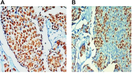
ER and PgR IHC staining of (n = 300) infiltrating duct cancer of the breast in Indian women: the entire IHC slide was scanned for immunostaining evaluation by light micro-scope. Positive nuclear immunostaining for ER is shown in Figure 1A. Positive nuclear immunostaining for PgR is shown in Figure 1B.
Quantitative prognostic values of ER and PgR in Indian breast cancer women retrospective study shows short-term outcome of ANN Indian breast cancer patients. Median follow-up time was 45 months; the aim of which was to define the patients at high risk of recurrence using first-line generation clinico-histopathological parameters.
Considering steroid receptor content, measured by the biochemical dextran-coated charcoal method recommended by the European Organization for Research and Treatment of Cancer (EORTC).3 The middle-aged postmenopausal Indian
breast cancer patient subset bearing pT2 tumors, was found to have the lowest tested cutoff value at 5 fmol/mg for ER and PgR showing prognostic significance as seen in Figure 2.
In the breast cancer patients with steroid receptor content lower than 5 fmol/mg (high risk-related subgroup), relapse-free interval probabilities were calculated according to Kaplan and Meier with Cox–Mantel test for comparing survival functions, and P values 0.05 were considered significant.
Subgroups were formed using clinico-histopathological variables within 5-year increments of patient’s age as well
A B
Figure 1 Immunostaining of infiltrating duct breast cancer of Indian women, (×400 objective magnification): dark brown nuclear positive staining for estrogen receptor (A) and dark brown nuclear positive staining for progesterone receptor (B).
RETRACTED
Breast Cancer: Targets and Therapy downloaded from https://www.dovepress.com/ by 118.70.13.36 on 20-Aug-2020
Dovepress
Patil et al
as prognostic power of the particular variable. Our first goal in this study was to form patient subgroups based on the pronounced mutual relationship between conventional parameters.
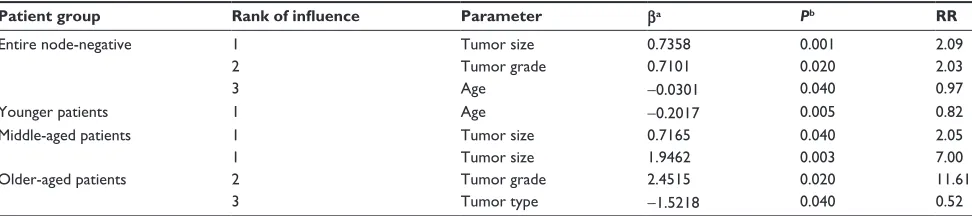
After forming the age- and menopausal-related sub-groups, the group of patients of age up to 44 years (younger group) (n = 54) was mostly premenopausal. Indian breast cancer patients aged over 59 years (older group) (n = 75) were postmenopausal, except one with premenopausal status. The group aged between 45 and 59 years (middle-aged group) (n = 168), and (n = 3) not reporting, were premenopausal, perimenopausal, and postmenopausal women with nearly same frequency. Results of multivariate analysis for all node-negative breast cancer patients, as well as for younger (up to 45 years), middle-aged (45–59 years), and older (above 59 years) patients, are shown in Table 1 (stepwise methods using the Cox regression model on a computer using BMDP-2 L Statistical Software).
ER and PgR, considered as continuous variables did not reach statistical significance to become independent variables, ie, they did not add any further information about
prognosis in the analyzed group of ANN in Indian breast cancer patients. The tumor size, grade, age, and histopatho-logical type showed different predictors of relapse across the age- and menopausal-related patient subgroups. It is well known that results of multivariate analysis obscure differ-ences among the biologically different subgroups.
ER and PgR status has a well defined small subset of patients with different prognosis in middle-aged postmeno-pausal patients bearing pT2 tumors, has overcome the bias cited above and is therefore a clinically useful model. Also, it is possible that prognostic information concerning impor-tance of ER and PgR expression could have been overlooked, ie, underestimated when considering the entire node-negative breast cancer population in India.
In that context, it is understandable why the results of prognostic values of ER and PgR in the natural course of node-negative breast cancer were controversial.4,5 It is
unreal-istic and overly simplunreal-istic to expect that any individual breast cancer biomarker alone will be prognostically powerful enough to be clinically useful.
Qualitative prognostic values of ER and PgR in Indian women with breast cancer could be determined during the histopathology course of breast cancer as related to their phenotypes, ie, using pre-determined cutoff value for ER, PgR content. Steroid hormone receptors like ER and PgR are regulated proteins.6 Provided that ER is present; PgR is
synthesized in tissue, implying a functioning ER pathway.7
Consequently, three histopathological types of ER- and PgR-related cancer should appear as: breast cancer with functional ER, ER-positive, and PgR-positive or without functional ER, ER-positive, and PgR-negative and breast tumors without both receptors ER- and PgR-negative. Although breast tumors lacking ER but containing PgR should not exist, they existed with an incidence of up to 5%. Histopathological significance of these types of breast cancer is not clear. Distribution of ER and PgR phenotypes with
0 0 0.1 0.2 0.3 0.4 0.5 0.6 0.7 0.8 0.9 1
12 24 36 48
Months
P
60 72 84 ER/PgR < 5 ER/PgR> = 5
96
Figure 2 steroid hormone receptor er and Pgr status-related disease-free probability (p) of node-negative postmenopausal middle-aged breast cancer patients with pT2 tumors.
Abbreviations: er, estrogen receptor; Pgr, progesterone receptor.
Table 1 Multivariate analysis of the entire node-negative indian breast cancer patients
Patient group Rank of influence Parameter βa Pb RR
entire node-negative 1 Tumor size 0.7358 0.001 2.09
2 Tumor grade 0.7101 0.020 2.03
3 Age -0.0301 0.040 0.97
Younger patients 1 Age -0.2017 0.005 0.82
Middle-aged patients 1 Tumor size 0.7165 0.040 2.05
1 Tumor size 1.9462 0.003 7.00
Older-aged patients 2 Tumor grade 2.4515 0.020 11.61
3 Tumor type -1.5218 0.040 0.52
Notes:aRegression coefficient for independent variable; bLikelihood ratio test.
Abbreviation: rr, relative risk.
RETRACTED
Breast Cancer: Targets and Therapy downloaded from https://www.dovepress.com/ by 118.70.13.36 on 20-Aug-2020
Dovepress er and Pgr in breast cancer of indian women
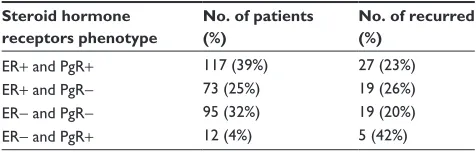
cutoff values of 10 fmol/mg and 20 fmol/mg, respectively, and observed recurrence events within each phenotype are shown in Table 2.
Twelve (4%) breast cancers displayed ER-negative but PgR-positive status. Approximately twice as high recurrence rate in the ER-negative and PgR-positive subgroup of patients was noted, compared with the other steroid hormone receptor phenotype subgroups. Relapse-free probability for each of the four receptor phenotype subgroups is shown in Figure 3.
There was a significantly lower disease-free probability of survival for patients with ER-negative and PgR-positive breast cancer than any of the other steroid hormone receptor phenotypes z0, 05 . 1.64. There was no difference in relapse-free probability between any couple of ER + positive and PgR + positive, ER + positive and PgR- negative, ER- negative and PgR- negative subgroups z0, 05 , 1.64. Considering the distribution of obtained independent prognostic parameters of entire node-negative Indian breast cancer patients, within subgroups of patients in relation to ER and PgR phenotypes shown in Table 2 and Figure 4, we did not find statistical differ-ence in tumor size and grade (χ2-test) or in age (Mann–Whitney
U-test) between ER-negative and PgR-positive subgroup and
any other receptor subgroups (P . 0.05).
Homogeneity was striking in the distribution of indepen-dent prognostic parameters such as the age of patients as well as tumor size and grade among the examined groups. Whether the worse outcome in ER- negative and PgR + positive is due to intrinsic histopathological aggressiveness needs to be determined.8 Existence of breast cancer that lacks ER but
contains PgR may be due to ER being present but masked in the binding assay because of endogenously bound estrogens.9
ER assay in these cases would be false-negative. This expla-nation has not been widely accepted so far.10 Abnormal ER
may be present, which does not bind estrogen but can bind to DNA and activate transcription.11 The presence of a PgR
gene with abnormal regulation can function in a constitutive manner if ER is truly absent and PgR is not dependent upon estrogen for regulation.
Discussion
Response to endocrine therapy clearly correlates with ER and PgR positivity, ie, the richer the tumor.12,13 Genomic- or
proteomic-based approaches has enabled understanding of the molecular picture of breast cancers, which in turn, allows cancer tissue biomarkers of response and prognosis to be identified and characterized more accurately than before. In the future, to maximize the therapeutic benefit of the patients, treatment according to the molecular portrait of their cancer tissue biomarker expression may be possible.14
Evaluations of these cancer tissue biomarkers are most valuable in predicting response to targeted therapy for these proteins. It has been reported that 85% of tumors with double-positive phenotype respond to hormonal manipula-tion, whereas less than 10% of those with double-negative phenotype respond.15,16
A total of 39% of breast cancer patients have ER-positive tumors, and of these, around 60% are found to respond to endocrine therapy. Prediction of the prognosis of breast cancer patients is expected to be achieved by a subgroup-ing of ER-positive patients, based on the physiology of ER signaling. Identification of a poor-prognosis population among ER-positive breast cancer patients can be achieved by the use of selected ER-regulated genes. Only low levels of response to treatment are achieved in those patients with ER-negative tumors.
PgR reflects the functional estrogenic stimulus and is a significant prognostic and predictive factor of the ER status. Patients, whose tumors were ER-positive and PgR-positive, have demonstrated good levels of response to endocrine treatment. ER-positive and PgR-positive breast cancer patients have lower risks of mortality after their diagnosis, compared with women with ER- and PgR-negative disease.
Table 2 Observed breast cancer in indian women recurrence relative to er and Pgr phenotypes
Steroid hormone receptors phenotype
No. of patients (%)
No. of recurred (%)
er+ and Pgr+ 117 (39%) 27 (23%)
er+ and Pgr- 73 (25%) 19 (26%)
er- and Pgr- 95 (32%) 19 (20%)
er- and Pgr+ 12 (4%) 5 (42%)
Abbreviations: er, estrogen receptor; Pgr, progesterone receptor.
0 0.00 0.10 0.20 0.30 0.40 0.50 0.60 0.70 0.80 0.90 1.00
12 24 36 48
t (Months)
Relapse-free probability
60 72 84
ER−PgR− ER+PgR− ER+PgR+
ER−PgR+
96
Figure 3 relapse-free probability of survival in node-negative breast cancer patients according to the steroid hormone receptor phenotypes.
Abbreviations: er, estrogen receptor; Pgr, progesterone receptor; t, time.
RETRACTED
Breast Cancer: Targets and Therapy downloaded from https://www.dovepress.com/ by 118.70.13.36 on 20-Aug-2020
Dovepress
Patil et al
The potential benefits of hormonal therapy can be pre-dicted by the presence or absence of ER and PgR as valuable prognostic factors. ER is the prominent breast cell mitogen, and inhibition of ER activation is an important prevention and treatment strategy. A selective ER modulator, Tamoxifen, can be used to target ER- and PgR-positive metastatic breast tumors, and these patients tend to have a greater chance of effective tumor response and longer overall survival than patients with ER- and PgR-negative tumors. Risk of recur-rence and death following adjuvant hormonal therapy are much reduced in patients with ER- and PgR-positive early breast cancer, whereas patients with ER- and PgR-negative disease are minimally benefited from these treatments.
In ER-negative tumors, which appear to gain greater benefit from chemotherapy in the metastatic and adjuvant settings, the value of ER status as a predictive cancer tissue biomarker extends to potential benefit from chemotherapy. However, treatment decisions on the basis of ER have not yet been evaluated prospectively. Tamoxifen has been the standard treatment for hormone receptor-positive breast cancer, resulting in a significant improvement in disease-free survival, regardless of nodal status.
For Indian breast cancer women with early breast cancer, ie, ER-positive, standard adjuvant treatment is with anti-ER tamoxifen for 5 years, which reduces risk of recurrence by 47% and risk of death by 26% over the next 10 years. After the initiation of therapy, resistance to tamoxifen therapy in
early breast cancer may occur as early as 12–18 months. Tamoxifen can stimulate breast cancer cell growth in some patients with resistant disease. Therefore, for early breast cancer, the role of more effective, less toxic agents, such as third generation aromatase inhibitors, has been evaluated in adjuvant therapy.
ER and PgR cause effects in the cell nuclei. The IHC detection of these two receptor groups is possible in normal breast tissue, displaying heterogeneity not just in correlation to the menstruation cycle. The induction of PgR receptors is one of the effects of ER. Malignant tumors of the breast, in most cases carcinoma of the breast, show different patterns of hormone receptors like ER and PgR expression, which are not only of great therapeutic significance but are also important regarding significantly longer relapse-free intervals.
Conclusion
IHC analysis has been shown to be a prognostic factor for patients with breast cancer in India; the major aim of determining the ER receptor status is to assess predictive response to hormonal therapy. Comprehensive studies of PgR by IHC lagged behind that of ER. First, many clinicians depend on ER status alone to select patients for hormonal therapy.
PgR alone was found to be a weaker prognostic and predictive factor as compared with ER in studies using ligand-binding assay. In addition, until relatively recently,
0
ER+, PgR+ 39%
23%
25% 26%
4% 42%
32%
20%
ER+, PgR−
Steroid Hormone receptors phonotypes
ER−, PgR− ER−, PgR+
No. of patients %
No. % recurred
5 10 15 20 25 30 35 40 45
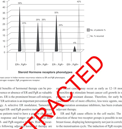
Figure 4 Observed breast cancer in indian women recurrence relative to er and Pgr phenotypes.
Abbreviations: er, estrogen receptor; Pgr, progesterone receptor.
RETRACTED
Breast Cancer: Targets and Therapy downloaded from https://www.dovepress.com/ by 118.70.13.36 on 20-Aug-2020
Breast Cancer: Targets and Therapy
Publish your work in this journal
Submit your manuscript here: http://www.dovepress.com/breast-cancer---targets-and-therapy-journal Breast Cancer: Targets and Therapy is an international, peer- reviewed open access journal focusing on breast cancer research, identification of therapeutic targets and the optimal use of preven-tative and integrated treatment interventions to achieve improved outcomes, enhanced survival and quality of life for the cancer patient.
View the full aims and scopes of this journal here. The manuscript management system is completely online and includes a very quick and fair peer-review system, which is all easy to use. Visit http:// www.dovepress.com/testimonials.php to read real quotes from published authors.
Dovepress
Dovepress
er and Pgr in breast cancer of indian women
there were only a limited number of good antibodies avail-able for PgR that worked on archival tissue. Despite these limitations, there have been several studies of assessing PgR by IHC in various settings in breast cancer. For example, three studies assessed patients receiving adjuvant hormonal therapy alone, and all three showed a significant relationship between PgR positivity and improved outcome.
Acknowledgments
Thanks to all members of the histopathology section of Grant Medical College and Sir JJ Group of Hospitals, Mumbai, India for providing surgical specimen tissue samples.
Disclosure
The authors report no conflicts of interest in this work.
References
1. Sears HF, Janus C, Kevy W, et al. Breast cancer without axillary metastases. Are there high-risk biologic subpopulations? Cancer. 1982; 50:1820–1827.
2. Gooldhirsch A, Glick JH, Gelber RD, et al. Meeting highlights: inter-national consensus panel on the treatment of primary breast cancer. J Clin Oncol. 2001;19(18):3817–3827.
3. EORTC Breast Cancer Cooperative Group. Revision of the standards for the assessment of receptors in human breast cancer. Eur J Cancer. 1980;16: 1513–1515.
4. Fisher B, Redmond C, Fisher ER, et al. National Surgical and Bowel Project Investigators: Relative worth of estrogen and progesterone recep-tors in node negative breast carcinoma patients: finding from National Surgical Adjuvant Breast and Bowel Project Protocol B-016. J Clin Oncol. 1988;6:1076–1087.
5. Thorpe S, Rose C, Rassmussen BB, Mourisden HT, Bayer T, Keiding N; for the Danish Breast Cancer Cooperative Group. Prognostic analysis of systematically untreated patients with node negative primary breast cancer. Cancer Res. 1987;7:6126–6133.
6. Muldoon TG. Regulation of steroid hormone receptor activity. Endocr Rev. 1980;1:339–364.
7. Horwitz KB, McGuire WL. Predicting response of endocrine therapy in human breast cancer: a hypothesis. Science. 1975; 189(4204):726–727.
8. Nikoliæ-Vukosavljeviæ D, Kanjer K, et al. Estrogen receptor-negative, progesterone receptor-positive: natural course of breast cancer. J Biol Markers. 2002. In press.
9. Wittliff JL. Steroid-hormone receptors in breast cancer. Cancer. 1984;53(3 Suppl):630–643.
10. Wilking N, Rutquist LE, Nordenskjold B, Skoog L, et al. Steroid recep-tor levels in breast cancer. Relationship with age and menopausal status. Acta Oncol. 1989;28:807–810.
11. Clarke R, Lippman ME. Antiestrogen resistance: mechanism and reversal. In: Teicher BA, editor. Drug Resistance in Oncology. New York: Marcel Dekker Inc; 1992:501–536.
12. Dowsett M, Allred C, Knox J, et al. Relationship between quantitative estrogen and progesterone receptor expression and human epider-mal growth factor receptor 2 (HER-2) status with recurrence in the Arimidex, Tamoxifen, Alone or in Combination trial. J Clin Oncol. 2008;26:1059–1065.
13. Varghese Christa, et al. The significance of estrogen and proges-terone receptors in breast cancer. J Clin Diagnostic Res. 2007; ISSN – 0973-709X.
14. Fuqua SA, Cui Y. Estrogen and progesterone receptor isoforms: clinical significance in breast cancer. Breast Cancer Res Treat. 2004; 87 Suppl 1:S3–S10.
15. Kumar V, Abbas AK, Fausto N, et al. The Breast Robbins and Cotran Pathologic Basis of Disease. Philadelphia, PA: Elsevier; 2004:1147. 16. Dowsett M, Houghton J, Iden C, et al. Benefit from adjuvant tamoxifen
therapy in primary breast cancer patients according estrogen receptor, progesterone receptor, EGF receptor and HER-2/neu status. Ann Oncol. 2006;17:818–826.
RETRACTED
Breast Cancer: Targets and Therapy downloaded from https://www.dovepress.com/ by 118.70.13.36 on 20-Aug-2020