Abstract
DNAzymes have been extensively studied as biosensors because of their unique functionality of cleaving substrate in the presence of metal ion cofactors. However, there are only a few reports on visual detection using gold nanoparticles. Here, we synthesized the DNAzyme microparticle (DzMP) (~1μm) via rolling circle amplification for detection of Pb2+without the help of other materials. Then, the substrate strands were labeled with two different fluorophores (6-carboxyfluorescein and Cy5) to visualize the DzMPs and to monitor the separation of substrate strands. Because of their large size, the decline in the number of fluorescent particles in the presence of Pb2+could be successfully demonstrated by a fluorescence microscopy, presenting a new platform for heavy metal detection.
Keywords:DNAzyme, Heavy metal detection, Biosensor
Background
Nucleic acids have attracted a great attention due to their versatile properties. Especially, various DNA enzymes (termed DNAzyme) have recently been developed by the systematic evolution of ligands by exponential enrichment (SELEX) since Breaker and Joyce presented the in vitro selection of catalytic DNA in 1994 [1]. These DNAzymes have unique properties that are capable of catalyzing bioorganic chemical reactions. For example, DNAzymes catalyzing RNA cleavage [1–3], ligation [4, 5], or modifica-tion of nucleic acids [6, 7] have been reported. They have been applied in various fields such as biochemistry, medi-cine, and analytical chemistry.
Particularly, DNAzymes cleaving DNA or RNA sub-strates in the presence of specific metal cofactor such as Pb2+, Mg2+, Cu2+, and UO22+ have been widely
studied [1, 8–10]. Also, Na+ [11, 12] or lanthanide [13, 14] -dependent DNAzymes have been recently reported. Because the toxicity of heavy metals is well known, these DNAzymes have often been used as a sensor to detect heavy metals. To be specific, fluor-escent techniques [15, 16], polymerase chain reaction [17], electrochemical approaches [18], and surface-enhanced Raman scattering (SERS) [19, 20] have been applied for DNAzyme-based sensors. Also, the
colorimetric sensors using gold nanoparticles were introduced [21]. However, there have been few studies on visual detection techniques and most of the conventional colorimetric detection methods need the immobilization of DNAzyme on gold nanoparticles [21–23].
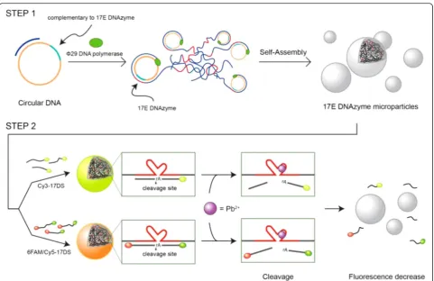
Previously, the synthesis of DNA hydrogel via ligase-mediated reaction or rolling circle amplification (RCA) has been reported [24, 25]. Based on these enzymatic ap-proaches, here we synthesized the giant catalytic DNA particles (~1 μm) without other inorganic core particles and successfully visualized the detection of Pb2+utilizing a fluorescence microscope as illustrated schematically in Fig. 1. First, the DNAzyme microparticles (DzMPs) com-prised of 17E DNAzyme for sensitive Pb2+ detection were produced by rolling circle amplification (RCA). Then, the fluorescently labeled 17DS substrate strands were hybridized with the generated particles. In the absence of Pb2+, the 17DS/E DzMPs emit bright fluores-cence. However, in the presence of Pb2+, the particles turn off their light since the cleaved substrates are falling apart from the 17E DzMPs. This giant DNAzyme particle-based strategy enables detection of Pb2+ by fluorescence microscopy. In addition, DzMPs have numerous catalytic sites, and their sequences can be flexibly changed to have diverse functionalities. Therefore, this DzMP-based ap-proach offers a simple detection method as well as a promise of a potent therapeutic tool for clinical applications.
* Correspondence:[email protected]
Department of Chemical Engineering, University of Seoul, 163 Seoulsiripdaero, Dongdaemun-gu, Seoul 130-743, South Korea
Methods
Preparation of Circular DNA
All the oligonucleotides used in the study were pur-chased from the Integrated DNA Technologies (IDT, USA) (Table 1). To prepare the circular DNA, phosphor-ylated linear single-stranded DNA (ssDNA) that has complementary sequence to the 17E DNAzyme was de-signed and the primer ssDNA was dede-signed to contain two complementary regions to each end of the linear ssDNA. For hybridization, equal concentration (3 μM) of the linear ssDNA and primer ssDNA were mixed in nuclease-free water. Next, the mixed solution was heated to 95 °C for 2 min and cooled gradually to 25 °C for 1 h. To connect the nick in the circular DNA, the
solution was incubated overnight at room temperature with T4 DNA ligase (0.03 U/μL, Promega, USA) and ligase buffer (300 mM Tris-HCl (pH 7.8), 100 mM MgCl2, 100 mM dithiothreitol, and 10 mM adenosine
triphosphate).
Synthesis of 17E DNAzyme Microparticles
To synthesize the 17E DNAzyme microparticle (17E DzMP), the prepared circular DNA was mixed withΦ29 DNA polymerase (1 U/μL, Lucigen, USA), deoxyribonu-cleotide triphosphate (2 mM), and reaction buffer (40 mM Tris-HCl (pH 7.5), 50 mM KCl, 10 mM MgCl2,
5 mM (NH4)2SO4, and 4 mM dithiothreitol). For the
RCA process, the mixed solution was incubated at 30 °C
[image:2.595.58.540.85.397.2]Fig. 1Schematic illustration of the construction of the 17DS/E DNAzyme microparticles and their catalytic activity. The DNA microparticles containing a large amount of 17E DNAzyme sites were synthesized via rolling circle amplification (RCA) (step 1). The 17DS substrate strands containing a cleavage site were added to the particle solution and then hybridized particles could be formed by denaturation and annealing. The substrate strands are cleaved when Pb2+is added and the cleaved strands are separated from the particles (step 2)
Table 1DNA sequences for the fabrication of DNAzyme microparticles
Linear DNA 5′- /Phosphate/CAA CTG TAG TGT GTT CAC GGT GCT GTA CTC ACT ATT TCG ACC GGC TCG GAG AAG AGA TGC ACT GAC AAG ACG TCA TAT CAA GTG TAT GGC AA - 3′
Primer DNA 5′- CAC TAC AGT TGT TGC CAT ACA C - 3′
Dual-labeled 17DS substrate DNA 5′- /6-FAM/ACT CAC TATrA*GGA AGA GAT GCA CTG A/Cy5/ - 3′ rArepresents the ribonucleotide adenosine
Cy3-labeled 17DS substrate DNA 5′- /Cy3/ACT CAC TATrA*GGA AGA GAT G - 3′ rArepresents the ribonucleotide adenosine
[image:2.595.59.539.650.724.2](Cy3-17DS). To determine the optimum ratio between 17E DzMP and Cy3-17DS, the different concentrations (200 nM, 1 μM, 5 μM each) of Cy3-labeled substrate strands was added into the 17E DzMP solutions. The de-naturation and annealing processes were performed to produce Cy3-labeled 17DS/17E DNAzyme microparti-cles (Cy3-17DS/E DzMPs). The solution was heated to 95 °C for 2 min and cooled gradually to 25 °C for 1 h. The substrate residues were removed by washing several times. The fluorescence intensity of the 17DS/E DzMPs were measured using Nucleo Counter (Chemometec, Denmark, NC-3000). The fluorescent images of the par-ticles were obtained by fluorescence microscopy (Nikon, Japan, Eclipse Ti).
Gel Electrophoresis
To confirm the cleavage reaction, the various concentra-tions of Pb2+solution (100 nM to 1 mM) was added to Cy3-17DS/E DzMP solutions. Then, to avoid the settling of DzMPs, the mixed solution was incubated using rotat-ing oven at room temperature for 1 h. The catalytic activity of the DNAzyme was confirmed using DNA polyacrylamide gel electrophoresis (DNA-PAGE). Gel electrophoresis was carried out on a 10 % DNA-PAGE gel at 95 V at room temperature in Tris-borate-EDTA (TBE) buffer (0.89 M Tris-borate and 0.02 M EDTA, pH 8.3) for 40 min. First, the gel electrophoresis result was analyzed without DNA-specific dyes to monitor the Cy3-17DS substrate strands. Then, the resulting DNA-zyme particles were analyzed after staining with GelRed (10−4dilution of the stock solution, Biotium, USA).
Detection of Pb2+Using a Fluorescence Microscope
To observe DzMPs using a fluorescence microscope, dual-labeled substrate was used. The substrate strand which was labeled with a FAM fluorophore (6-carboxy-fluorescein) at the 5′-end and a Cy5 at the 3′-end (6FAM/Cy5-17DS). To verify the optimal condition, dif-ferent concentrations of 6FAM/Cy5-17DS (10 nM, 100 nM, 1 μM each) were mixed with 17E DzMPs and the same denaturation and annealing processes as above were performed. The fluorescence intensity of the 6FAM/Cy5-17DS/E DzMP was measured using Nucleo
prepared from the designed circular template to generate 17E DNAzyme by RCA process. To observe the morph-ology and structure of the particles, scanning electron microscopy (SEM) was utilized. As seen in Fig. 2a, SEM image showed that the enzymatic particles have flower-like globular shape with uniform size (~1 μm). Because of the large surface area of the porous structure, numer-ous catalytic sites of the particles could participate in the enzymatic reaction. In addition, Cy3-labeled 17DS sub-strate strands (Cy3-17DS) were added to prepare 17E DzMPs to produce Cy3-17DS/E DzMPs. To determine the optimal ratio between substrates and DzMPs, three different concentrations (200 nM, 1μM, and 5μM each) of substrate solutions were tested. The hybridization between Cy3-17DS and 17E DzMPs was verified by util-izing image cytometry. As shown in Fig. 2b, fluorescence intensities of the particles increased as the concentration increased from 200 nM to 5 μM. Especially, there was a significant increase in intensity when the 5 μM of Cy3-17DS was used, compared to that of control 17E DzMPs. In addition, further study was performed using fluorescence microscopy, and the 17E particles which were mixed with 5 μM of Cy3-17DS showed bright fluorescence (Fig. 2c). These results indicated that the 17DS substrate strands were hybridized enough with the 17E particles which could catalyze the cleavage reaction. Also, the DzMPs still main-tain their structure after hybridization with the 17DS sub-strate (see Additional file 1: Figure S1).
Confirmation of Catalytic Activity of DzMPs
with 1 mM Pb2+(lane 7) and the cleaved substrate band was also observed (see Additional file 1: Figure S2). In addition, the previous gel was stained with GelRed to confirm the particles. Since the DzMPs have a size larger than the pore size, the particles were found within the wells (Fig. 3b). The intensities of the bands from lane 2 to lane 6 reveal little difference. On the other hand, in lane 7, the intensity of the band significantly decreased. This is probably due to the fact that the bulk of cleaved substrate strands were separated from the 17E DzMPs because of the catalytic activity of DNAzyme in the pres-ence of Pb2+. For a quantitative study, the normalized in-tensities have shown in Fig. 3c. Under the concentration of 1μM Pb2+, the differences between the intensities of untreated and Pb2+-treated samples were not substantial. However, the intensities were reduced up to about 25 % in the case of the concentration of 10 and 100 μM. In addition, the 1 mM treated particles showed an appre-ciable decrease of 64 % in the gel electrophoresis band intensity. Also, a gradual decrease in intensity of the
DzMPs was demonstrated by additional experiment between 20 and 200 μM Pb2+ (see Additional file 1: Figure S3, Tables S1 and S2). These results suggest that the detection of Pb2+ was successfully achieved in the presence of Pb2+.
Simple Detection of Pb2+Using a Fluorescence Microscope
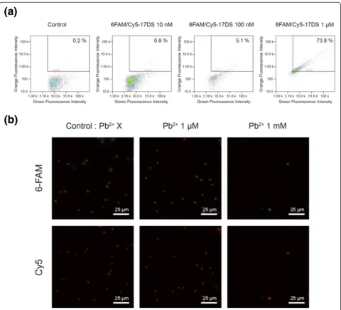
In order to develop a more convenient detection system, we designed the dual-labeled 17DS substrate. The substrate strand was labeled with a 6-carboxyfluorescein fluorophore (6-FAM) at the 5′-end and a Cy5 at the 3′-end (6FAM/ Cy5-17DS). Therefore, more precise observations are avail-able since the particles emit two different colors at once. To determine the proper concentration, different concen-trations (10 nM, 100 nM, 1μM) of 6FAM/Cy5-17DS were hybridized with the 17E DzMPs. The fluorescence in-tensities of the 6FAM/Cy5-17DS/E DzMPs were also measured by image cytometry. The results show that the number of the particles in the right upper quadrant
Fig. 2aSEM images of 17E DzMPs.bHistogram showing the fluorescence intensity of the Cy3-17DS/E DzMPs for various concentrations of the Cy3-17DSs.cFluorescence microscopy image of the 17E-17DS particles which are hybridized with the 5μM of Cy3-17DS
[image:4.595.65.538.89.220.2] [image:4.595.57.539.518.682.2]when the 1 mM Pb2+ ions were treated, the number of fluorescent particles remarkably decreased. This means
In conclusion, the DNAzyme microparticle having nu-merous DNAzyme sites was successfully synthesized.
[image:5.595.58.539.259.695.2]DNAzyme activity of the particles was confirmed by gel electrophoresis and quantification analysis and as low as 20 μM Pb2+ could be detected. Furthermore, dual-labeled substrate was designed for the simple detection platform. By utilizing the DNAzyme micro-particles, the detection of lead ions was demonstrated based on the Pb2+-dependent DNAzyme chemistry. This strategy offers a simple and intuitive detection method with a detection limit of 1 mM Pb2+ and it could be applied in environmental applications [26, 27]. Although the sensitivity of this platform should be im-proved, the DNAzyme microparticles have a lot of ad-vantages such as a large surface area, a high density of catalytic sites, and stability against nuclease [28]. More-over, the functionality of DzMPs can be modified to contain diverse DNAzymes against a target in cancer cells by controlling sequences. Therefore, this catalytic DNA microparticles may well be useful not only as a detector but as a therapeutic medicine.
Additional file
Additional file 1:Figure S1.SEM images of 17DS/E DzMPs (left) and high magnification image (right). The 17E DzMPs were annealed with the 5μM of Cy3-17DS.Figure S2.gel image showing cleavage activity of DNAzyme microparticles with different concentrations of Pb2+after 1 h reaction. Lanes 1 and 2 indicate 17DS substrate strand and DzMPs with-out Pb2+treatment. Lanes 3–7 correspond to 100 nM, 1μM, 10μM, 100μM, and 1 mM Pb2+, respectively. Gel electrophoresis was carried out on a 3 % agarose gel at 95 V at 4 °C in 1× TAE buffer (40 mM Tris, 1 mM EDTA, 40 mM acetic acid) for 80 min. The gel image was obtained with-out DNA-specific dyes.Figure S3.fluorescence intensities of the Pb2 +
-treated DzMPs. Note: the slopes of the fitted linear plots were given as 1.Table S1.the slope and intercept of the linear fitting graphs.Table S2.the slope and intercept of the linear fitting graphs. (PDF 1278 kb)
Competing interests
The authors declare that they have no competing interests.
Authors’contributions
JK and JBL designed and analyzed the data. JK performed the experiments. JK and JBL wrote the manuscript. Both authors read and approved the final manuscript.
Acknowledgements
This research was supported by Basic Science Research Program through the National Research Foundation of Korea (NRF) funded by the Ministry of Science, ICT and Future Planning (NRF-2015R1A1A1A05001174).
Received: 18 February 2016 Accepted: 3 May 2016
References
1. Breaker RR, Joyce GF (1994) A DNA enzyme that cleaves RNA. Chem Biol 1(4):223–229
2. Schubert S, GuÈl DC, Grunert HP, Zeichhardt H, Erdmann VA, Kurreck J (2003) RNA cleaving‘10-23’DNAzymes with enhanced stability and activity. Nucleic Acids Res 31(20):5982–5992
3. Silverman SK (2005) In vitro selection, characterization, and application of deoxyribozymes that cleave RNA. Nucleic Acids Res 33(19):6151–6163 4. Cuenoud B, Szostak JW (1995) A DNA metalloenzyme with DNA ligase
activity. Nature 375(6532):611–614
5. Flynn-Charlebois A, Wang Y, Prior TK, Rashid I, Hoadley KA, Coppins RL et al (2003) Deoxyribozymes with 2′-5′RNA ligase activity. J Am Chem Soc 125(9): 2444–2454
6. Li Y, Breaker RR (1999) Phosphorylating DNA with DNA. Proc Natl Acad Sci 96(6):2746–2751
7. Sheppard TL, Ordoukhanian P, Joyce GF (2000) A DNA enzyme with N-glycosylase activity. Proc Natl Acad Sci 97(14):7802–7807 8. Breaker RR, Joyce GF (1995) A DNA enzyme with Mg2+-dependent RNA
phosphoesterase activity. Chem Biol 2(10):655–660
9. Carmi N, Breaker RR (2001) Characterization of a DNA-cleaving deoxyribozyme. Bioorgan Med Chem 9(10):2589–2600
10. Moshe M, Elbaz J, Willner I (2009) Sensing of UO22+and design of logic gates by the application of supramolecular constructs of ion-dependent DNAzymes. Nano Lett 9(3):1196–1200
11. Torabi S-F, Wu P, McGhee CE, Chen L, Hwang K, Zheng N et al (2015) In vitro selection of a sodium-specific DNAzyme and its application in intracellular sensing. Proc Natl Acad Sci 112(19):5903–5908
12. Zhou W, Saran R, Chen Q, Ding J, Liu J (2016) A new Na+-dependent RNA-cleaving DNAzyme with over 1000-fold rate acceleration by ethanol. ChemBioChem 17(2):159–163
13. Huang P-JJ, Lin J, Cao J, Vazin M, Liu J (2014) Ultrasensitive DNAzyme beacon for lanthanides and metal speciation. Anal Chem 86(3):1816–1821 14. Huang P-JJ, Vazin M, MatuszekŻ, Liu J (2014) A new heavy
lanthanide-dependent DNAzyme displaying strong metal cooperativity and unrescuable phosphorothioate effect. Nucleic Acids Res. doi:10.1093/nar/gku1296 15. Zhang X-B, Wang Z, Xing H, Xiang Y, Lu Y (2010) Catalytic and molecular
beacons for amplified detection of metal ions and organic molecules with high sensitivity. Anal Chem 82(12):5005–5011
16. Wu P, Hwang K, Lan T, Lu Y (2013) A DNAzyme-gold nanoparticle probe for uranyl ion in living cells. J Am Chem Soc 135(14):5254–5257
17. Wang F, Wu Z, Lu Y, Wang J, Jiang J-H, Yu R-Q (2010) A label-free DNAzyme sensor for lead (II) detection by quantitative polymerase chain reaction. Anal Biochem 405(2):168–173
18. Shen L, Chen Z, Li Y, He S, Xie S, Xu X et al (2008) Electrochemical DNAzyme sensor for lead based on amplification of DNA—Au Bio-Bar codes. Anal Chem 80(16):6323–6328
19. Wang Y, Irudayaraj J (2011) A SERS DNAzyme biosensor for lead ion detection. Chem Commun 47(15):4394–4396
20. Ye S, Guo Y, Xiao J, Zhang S (2013) A sensitive SERS assay of L-histidine via a DNAzyme-activated target recycling cascade amplification strategy. Chem Commun 49(35):3643–3645
21. Liu J, Lu Y (2004) Accelerated color change of gold nanoparticles assembled by DNAzymes for simple and fast colorimetric Pb2+detection. J Am Chem Soc 126(39):12298–12305
22. Liu J, Lu Y (2004) Colorimetric biosensors based on DNAzyme-assembled gold nanoparticles. J Fluoresc 14(4):343–354
23. Zhang H, Lin L, Zeng X, Ruan Y, Wu Y, Lin M et al (2016) Magnetic beads-based DNAzyme recognition and AuNPs-based enzymatic catalysis amplification for visual detection of trace uranyl ion in aqueous environment. Biosens Bioelectron 78:73–79
24. Um SH, Lee JB, Park N, Kwon SY, Umbach CC, Luo D (2006) Enzyme-catalysed assembly of DNA hydrogel. Nat Mater 5(10):797–801
25. Lee JB, Peng S, Yang D, Roh YH, Funabashi H, Park N et al (2012) A mechanical metamaterial made from a DNA hydrogel. Nat Nanotechnol 7(12):816–820 26. Gichner T,Žnidar I, Száková J (2008) Evaluation of DNA damage and
mutagenicity induced by lead in tobacco plants. Mutat Res Genet Toxicol Environ Mutagen 652(2):186–190
27. Prasad M, Strzalka K (2002) Physiology and biochemistry of metal toxicity and tolerance in plants. 1st ed. Dordrecht: Kluwer Academic