INTRODUCTION
Lung transplantation has remained the only treatment option for chronic progressive lung disease since the first successful unilateral lung transplantation procedure in pulmonary fibro-sis patients in 1983.1 However, median survival thereafter is rel-atively low (5.9 years), compared with other solid organ
trans-plantation approaches. Notably, however, median survival increases to 8.1 years for patients who survive for the first year or more.2,3 Previous studies have revealed several factors relat-ed to mortality, which could be usrelat-ed to prrelat-edict prognosis.4-9 However, quality of life and performance differ according to the degree of recovery of pulmonary function, even if patients sur-vive after lung transplantation.
Lung function recovery is an important predictor of prog-nosis in lung transplant recipients and is used as a diagnostic parameter for bronchiolitis obliterans syndrome (BOS).10 BOS is defined as a sustained decline in forced expiratory volume in 1 second (FEV1).11 While inflammation, destruction of small airway, and fibrosis are putative mechanisms of BOS, this con-dition is not easy to diagnose through biopsy. Therefore, lung function deterioration on pulmonary function test (PFT) has been suggested as a diagnostic criterion for BOS.11 Pulmonary function is known to be influenced by acute rejection, infection, recurrence of primary disease, and complication at the
anasto-Factors Associated with Lung Function Recovery
at the First Year after Lung Transplantation
Bo Ra Yoon
1, Ji Eun Park
1, Chi Young Kim
1, Moo Suk Park
1, Young Sam Kim
1, Kyung Soo Chung
1,
Joo Han Song
1, Hyo-Chae Paik
2, Jin Gu Lee
2, and Song Yee Kim
11Division of Pulmonology, Department of Internal Medicine, Severance Hospital, Institute of Chest Diseases, Yonsei University
College of Medicine, Seoul;
2Department of Thoracic and Cardiovascular Surgery, Severance Hospital, Yonsei University College of Medicine, Seoul, Korea.
Purpose: Post-operative pulmonary function is an important prognostic factor for lung transplantation. The purpose of this study was to identify factors affecting recovery of forced expiratory volume in 1 second (FEV1) at the first year after lung transplantation. Materials and Methods: We retrospectively reviewed the medical records of lung transplantation patients between October 2012 and June 2016. Patients who survived for longer than one year and who underwent pulmonary function test at the first year of lung transplantation were enrolled. Patients were divided into two groups according to whether they recovered to a normal range of FEV1 (FEV1 ≥80% of predicted value vs. <80%). We compared the two groups and analyzed factors associated with lung function recovery.
Results: Fifty-eight patients were enrolled in this study: 28 patients (48%) recovered to a FEV1 ≥80% of the predicted value, where-as 30 patients (52%) did not. Younger recipients [odds ratio (OR), 0.92; 95% confidence interval (CI), 0.87–0.98; p=0.010], longer duration of mechanical ventilator use after surgery (OR, 1.14; 95% CI, 1.03–1.26; p=0.015), and high-grade primary graft dysfunc-tion (OR, 8.08; 95% CI, 1.67–39.18; p=0.009) were identified as independent risk factors associated with a lack of full recovery of lung function at 1 year after lung transplantation.
Conclusion: Immediate postoperative status may be associated with recovery of lung function after lung transplantation. Key Words: Lung transplantation, forced expiratory volume in 1 second, primary graft dysfunction
pISSN: 0513-5796 · eISSN: 1976-2437
Received: July 4, 2018 Revised: September 6, 2018 Accepted: September 6, 2018
Corresponding author: Song Yee Kim, MD, PhD, Division of Pulmonology, Depart-ment of Internal Medicine, Severance Hospital, Institute of Chest Diseases, Yonsei University College of Medicine, 50-1 Yonsei-ro, Seodaemun-gu, Seoul 03722, Korea. Tel: 82-2-2228-1940, Fax: 82-2-393-6884, E-mail: dobie@yuhs.ac
•The authors have no financial conflicts of interest.
© Copyright: Yonsei University College of Medicine 2018
This is an Open Access article distributed under the terms of the Creative Com-mons Attribution Non-Commercial License (https://creativecomCom-mons.org/licenses/ by-nc/4.0) which permits unrestricted non-commercial use, distribution, and repro-duction in any medium, provided the original work is properly cited.
mosis site; therefore, early prediction of pulmonary function deterioration is an important factor for the survival of recipi-ents.12-14 While increasing the 1-year survival rate to improve long-term survival rate is important, although lung function re-covery also serves as an important prognostic factor for surviv-ing patients.4 Notwithstanding, there has been no definitive study of factors affecting lung function recovery at the first year of lung transplantation. Thus, the purpose of this study was to identify factors affecting the recovery of pulmonary function in patients who survive for >1 year after lung transplantation.
MATERIALS AND METHODS
Study design and patient population
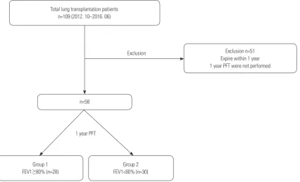
We retrospectively reviewed the electronic medical records of lung transplantation patients in a single tertiary medical insti-tution in South Korea for the period between October 2012 and June 2016. As described in Fig. 1, 109 patients underwent lung transplantation during the study period; 58 patients were in-cluded in the study. Patients who died within one year after lung transplantation (n=47, 43.1%) or who did not complete PFTs (n=4, 3.7%) were excluded. A follow-up PFT was sched-uled every 3 months after transplantation. The patients were divided into two groups according to whether they recovered to a normal range of FEV1 (FEV1 ≥80% of predicted value vs. <80%). The study protocol was approved by the Institutional Review Board (IRB) of Severance Hospital (IRB No. 4-2013-0770).
Variables and definitions
We analyzed various clinical characteristics, including recipi-ent, perioperative, postoperative, and donor factors. The re-cipient factors included age, gender, body mass index (BMI), smoking history, transplant type, primary or underlying dis-ease, and preoperative prognostic nutritional index (PNI). PNI was calculated using the following equation: [10×albumin (g/ dL) + 0.005×total lymphocyte count (per mm3)].15 Perioperative and postoperative factors, such as mechanical ventilator (MV) use or extracorporeal membrane oxygenation (ECMO) use before and after transplantation, intensive care unit (ICU) du-ration, and hospitalization dudu-ration, were analyzed. Primary graft dysfunction (PGD) and acute rejection requiring steroid pulse therapy were also reviewed. PGD was scored based on the International Society of Heart and Lung Transplantation (ISHLT) criteria; grade 2–3 was classified as high-grade PGD.16 Factors related to the operation included operation time, isch-emic time, and size mismatch between donor and recipient. Donor information, such as age, gender, BMI, smoking history, total lung capacity (TLC), size match, arterial partial pressure of oxygen (PaO2), fraction of inspired oxygen (FiO2), and ventila-tion time, was collected through the Korean Network for Organ Sharing.
Statistical analysis
All statistical analyses were performed using SPSS Statistics version 23 (IBM Corp., Armonk, NY, USA). Unadjusted vari-ables of study groups were analyzed by chi-squared test or Mann-Whitney test, then described as numbers (percentages)
1 year PFT
Exclusion Total lung transplantation patients
n=109 (2012. 10–2016. 06)
Group 1 FEV1≥80% (n=28)
Group 2 FEV1<80% (n=30) n=58
[image:2.595.95.522.456.715.2]Exclusion n=51 Expire within 1 year 1 year PFT were not performed
or medians [ranges or interquartile ranges (IQRs)]. Relation-ships between variables and lung function recovery were as-sessed by logistic regression models for multivariate analysis.
p values ≤0.05 were regarded as significant.
RESULTS
Characteristics of the patients
A total of 109 patients underwent lung transplantation during the study period; 58 patients who survived more than one year and who underwent PFTs at the first year after transplantation were enrolled. The patients were divided into two groups ac-cording to their FEV1 values at 1 year after lung transplanta-tion: 28 patients (48%) were classified as FEV1 ≥80% of the predicted value group (FEV1 ≥80% group); the remaining pa-tients (52%), whose FEV1 was less than 80%, were classified into
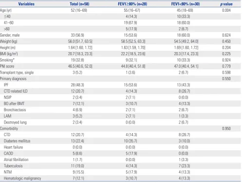
FEV1 <80% of the predicted value group (FEV1 <80% group). Baseline characteristics and variables of recipients are present-ed in Table 1. Mpresent-edian age was significantly younger in the FEV1 <80% group (55 years vs. 45 years, p=0.004). Gender, BMI, smoking history, and PNI were not significantly different be-tween the two groups. Most operations were bilateral lung transplantations, except one case (3.6%) in the FEV1 ≥80% group and two cases (6.7%) in the FEV1 <80% group. Idiopathic pulmonary fibrosis (IPF) was the most common cause of lung transplantation in both groups [15 patients (53.6%) in the FEV1 ≥80% group vs. 13 patients (43.3%) in the FEV1 <80% group,
p=0.550]. The proportions of primary pulmonary diagnoses or
comorbidities were not statistically different.
Recovery of pulmonary function after transplantation
The postoperative recovery of pulmonary function was assessed by PFT. The results are presented in Table 2 and Fig. 2. In pre-Table 1. Baseline Characteristics of the Recipients
Variables Total (n=58) FEV1≥80% (n=28) FEV1<80% (n=30) p value
Age (yr) 52 (16–69) 55 (16–67) 45 (18–69) 0.004
≤40 4 (14.3) 10 (33.3)
41–60 19 (67.9) 18 (60.0)
>60 5 (17.9) 2 (6.7)
Gender, male 33 (56.9) 15 (53.6) 18 (60.0) 0.624
Weight (kg) 56.0 [51.7, 63.5] 58.5 [52.5, 63.3] 54.5 [49.2, 64.0] 0.450 Height (m) 1.64 [1.60, 1.72] 1.63 [1.59, 1.70] 1.69 [1.60, 1.72] 0.204 BMI (kg/m2) 20.7 [18.3, 23.3] 22.2 [18.5, 23.6] 20.3 [17.4, 23.3] 0.225
Smoking* 19 (32.8) 9 (32.1) 10 (33.3) 0.924
PNI score 46.5 [40.6, 52.0] 44.8 [40.4, 51.8] 47.0 [40.4, 54.1] 0.779
Transplant type, single 3 (5.2) 1 (3.6) 2 (6.7) 0.598
Primary diagnosis 0.550
IPF 28 (48.3) 15 (53.6) 13 (43.3)
CTD related ILD 12 (20.7) 4 (14.3) 8 (26.7)
NSIP 2 (3.4) 2 (7.1) 0 (0.0)
BO after BMT 7 (12.1) 3 (10.7) 4 (13.3)
Bronchiectasis 4 (6.9) 2 (7.1) 2 (6.7)
LAM 3 (5.2) 2 (7.1) 1 (3.3)
Destroyed lung 2 (3.4) 0 (0.0) 2 (6.7)
Comorbidity 0.950
CTD 12 (20.7) 4 (14.3) 8 (26.7)
Diabetes mellitus 13 (22.4) 10 (35.7) 3 (10.0)
Heart failure 0 (0.0) 0 (0.0) 0 (0.0)
CAOD 5 (8.6) 5 (17.9) 0 (0.0)
Atrial fibrillation 1 (1.7) 0 (0.0) 1 (3.3)
Tuberculosis 11 (19.0) 4 (14.3) 7 (23.3)
NTM 9 (15.5) 5 (17.9) 4 (13.3)
Hematologic malignancy 7 (12.1) 3 (10.7) 4 (13.3)
FEV1, forced expiratory volume in 1 second; BMI, body mass index; PNI, prognostic nutritional index; IQR, interquartile range; IPF, idiopathic pulmonary fibrosis; CTD related ILD, connective tissue disease related interstitial lung disease; NSIP, nonspecific interstitial pneumonia; BO, bronchiolitis obliterans; BMT, bone mar-row transplantation; LAM, lymphangioleiomyomatosis; CTD, connective tissue disease; CAOD, coronary artery occlusive disease; NTM, nontuberculosis myco-bacterium.
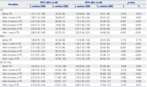
[image:3.595.43.541.305.680.2]operative PFTs, the forced vital capacity (FVC) of the FEV1 < 80% group was lower than that of the FEV1 ≥80% group (45% vs. 33%, p=0.019), while FEV1 and forced expiratory flow 25– 75% (FEF 25–75%) were not significantly different before op-eration between the two groups. After transplantation, PFTs were performed at 1, 3, 6, 9, and 12 months after transplanta-tion. All median values of FVC, FEV1, and FEF 25–75% showed better recovery in the FEV1 ≥80% group (FVC: 82% vs. 54%, p< 0.001; FEV1: 91% vs. 60%, p<0.001; FEF 25–75%: 113% vs. 77%,
p=0.017).
Perioperative factors associated with lung function recovery
Perioperative variables associated with lung function recovery are presented in Table 3. Echocardiographic variables and 6- minute walk test (6MWT) results were not significantly differ-ent between the two groups. There was no difference in pre-operative MV or prepre-operative ECMO use between the two groups. However, the median duration of postoperative MV and postoperative ECMO was longer in the FEV1 <80% group than in the FEV1 ≥80% group [MV duration; 3 (range, 1–16) vs. 9 (1–53) days, p=0.017; ECMO duration: 2 (range 1–6) vs. 2 (1– 25) days, p=0.032]. The proportion of tracheostomies was larg-er in the FEV1 <80% group (17.9% vs. 43.3%, p=0.038). Furthlarg-er- Further-more, FEV1 <80% group patients were hospitalized for longer periods and received longer ICU care than patients in the FEV1 ≥80% group (hospitalization: 30 days vs. 46 days, p=0.003; ICU
duration: 7 days vs. 13 days, p=0.009). During the postopera-tive period, four of the FEV1 <80% group patients needed renal replacement therapy, while none of the FEV1 ≥80% group re-quired this therapy [FEV1 ≥80% vs. FEV1 <80%: 0 patients (0.0%) vs. 4 patients (13.3%), p=0.047]. At 72 hours after trans-plantation, compared with the FEV1 ≥80% group, patients with FEV1 <80% had more high-grade PGD [FEV1 ≥80% vs. FEV1 < 80%: 3 patients (10.7%) vs. 16 patients (53.3%), p=0.001], while graft rejection and acute rejection requiring steroid pulse treatment within 1 year were not significantly different.
Intraoperative variables are presented in Table 4. The medi-an time of operation was longer in the FEV1 <80% group thmedi-an in the FEV1 ≥80% group (368 min vs. 415 min, p=0.034), while ischemic time, size mismatch, proportion of donor lung wedge resection, and re-operation after transplantation were not sig-nificantly different. There was no difference between the two groups in total fluid control, except for the quantity of red blood cell transfusion (FEV1 ≥80% vs. FEV1 <80%: 5 packs vs. 7 packs,
p=0.020).
Characteristics of the donors
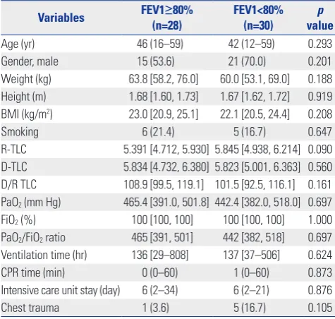
[image:4.595.57.555.84.377.2]Regarding donor variables, there was no significant difference between the two groups in regards to age, gender, BMI, smoking history, D/R TLC, PaO2/FiO2 ratio, ventilation time, CPR time, duration of ICU stay, or chest trauma history. The results are shown in Table 5.
Table 2. Univariate Analysis of Pulmonary Function Tests
Variables FEV1≥80% (n=28) FEV1<80% (n=30) p value
L, median (IQR) %, median (IQR) L, median (IQR) %, median (IQR) L %
FVC
Before TPL 1.51 (1.18, 1.85) 45 (32, 55) 1.30 (0.84, 1.88) 33 (21, 46) 0.225 0.019 After 1 month of TPL 1.99 (1.73, 2.64) 56 (50, 67) 1.65 (1.36, 2.44) 45 (37, 57) 0.058 0.001 After 3 months of TPL 2.52 (1.95, 2.83) 68 (58, 73) 1.72 (1.48, 2.21) 45 (36, 53) <0.001 <0.001 After 6 months of TPL 2.44 (2.13, 3.20) 74 (62, 83) 2.03 (1.56, 2.70) 55 (41, 64) 0.003 <0.001 After 9 months of TPL 2.85 (2.26, 3.19) 75 (70, 83) 2.07 (1.53, 2.84) 53 (40, 66) 0.002 <0.001 After 1 year of TPL 2.88 (2.40, 3.49) 82 (75, 91) 2.03 (1.64, 3.01) 54 (40, 65) <0.001 <0.001 FEV1
Before TPL 1.06 (0.79, 1.55) 42 (29, 60) 1.13 (0.66, 1.45) 33 (23, 50) 0.715 0.119 After 1 month of TPL 1.82 (1.54, 2.27) 68 (61, 78) 1.60 (1.25, 1.96) 53 (43, 63) 0.072 <0.001 After 3 months of TPL 2.21 (1.83, 2.57) 81 (74, 88) 1.50 (1.34, 1.99) 53 (42, 65) <0.001 <0.001 After 6 months of TPL 2.14 (1.85, 2.67) 86 (75, 92) 1.67 (1.43, 2.18) 62 (48, 67) 0.001 <0.001 After 9 months of TPL 2.31 (2.01, 2.81) 86 (78, 95) 1.64 (1.38, 2.33) 57 (46, 71) 0.001 <0.001 After 1 year of TPL 2.52 (2.02, 2.98) 91 (85, 102) 1.74 (1.36, 2.28) 60 (50, 74) <0.001 <0.001 FEF 25–75%
Multivariate analysis of the variables
Multivariate analysis revealed that younger recipients [odds ra-tio (OR), 0.92; 95% confidential interval (CI), 0.87–0.98; p=0.010], longer duration of MV use after surgery (OR, 1.14; 95% CI, 1.03–1.26; p=0.015), and high-grade PGD (OR, 8.08; 95% CI, 1.67–39.18; p=0.009) were independent risk factors associated with a lack of full recovery of lung function at 1 year after lung
transplantation (Table 6).
DISCUSSION
Lung transplants are increasing worldwide. According to the ISHLT registry, a total of 60107 lung transplants had been
per-100
80
60
40
20
0
120
100
80
60
40
20
0
180
160 140
120 100
80 60 40 20
0 5
4
3
2
1
0
5
4
3
2
1
0
5
4
3
2
1
0
FVC (%)
FEV1 (%)
FEF 25–75% (%)
FVC (L)
FEV1 (L)
FEF 25–75% (L)
Pre 1 3 6 9 12
Pre 1 3 6 9 12
Pre 1 3 6 9 12
Pre 1 3 6 9 12
Pre 1 3 6 9 12
Pre 1 3 6 9 12
Months after transplantation Months after transplantation Months after transplantation
Months after transplantation Months after transplantation Months after transplantation
* *
* *
* *
*
* *
*
*
* * *
*
* * *
*
* *
* *
*
* FEV1≥80%
FEV1<80%
FEV1≥80% FEV1<80%
FEV1≥80% FEV1<80%
FEV1≥80% FEV1<80%
FEV1≥80% FEV1<80%
FEV1≥80% FEV1<80% *p<0.05
*p<0.05
*p<0.05
*p<0.05
*p<0.05
*p<0.05 A
C
E
B
D
[image:5.595.41.547.69.605.2]F
formed by June 2016, although with a median survival of 6.0 years. While the median survival of lung transplant patients im-proves slightly (up to 8.1 years) in those surviving more than one year after surgery, it remains lower than the survival rate of other solid organ transplants.2 Therefore, most previous studies have involved finding survival-related risk factors to improve the over-all survival of lung transplant recipients.4-9 Meanwhile, a com-parative study of the recovery of pulmonary function in surviv-ing patients has not been a research priory. While there are some
patients whose lung function recovers well, there are some who fail to fully recover lung function. The purpose of this study was to investigate factors affecting pulmonary function recovery in patients surviving at least 1 year after lung transplantation.
[image:6.595.56.295.87.523.2]In our study, younger recipients, a longer duration of MV use after surgery, and high-grade PGD were independent risk fac-tors associated with a lack of full recovery of lung function at 1 Table 3. Univariate Analysis of Perioperative Variables
Variables FEV1(n=28)≥80% FEV1<80% (n=30) valuep
MV apply before TPL 5 (17.9) 8 (26.7) 0.425 ECMO apply before TPL 2 (7.1) 7 (23.3) 0.092 MV duration after TPL (day) 3 (1–16) 9 (1–53) 0.017 ECMO duration after TPL (day) 2 (1–6) 2 (1–25) 0.032 ECMO weaning within 24 hours 6 (21.4) 11 (36.7) 0.207 Tracheostomy 5 (17.9) 13 (43.3) 0.038 Intensive care unit stay (day) 7 (3–37) 13 (3–54) 0.009 Hospitalization days 30 (15–78) 46 (18–198) 0.003 HD usage after TPL 0 4 (13.3) 0.047 PaO2/FiO2 ratio after 48 hours 386 [313, 433] 333 [238, 404] 0.032 PaO2/FiO2 ratio after 72 hours 383 [319, 484] 294 [231, 422] 0.009 Primary graft dysfunction
Grade 0–1 25 (89.3) 14 (46.7)
Grade 2–3 3 (10.7) 16 (53.3) 0.001 Steroid pulse treatment 1 (3.6) 4 (13.3) 0.189 Echocardiography, before TPL
EF (%) 62 [59, 71] 65 [61, 68] 0.681 E/E' 8 [7, 10] 9 [7, 11] 0.678 TAPSE (cm) 1.48 [1.16, 1.60] 1.50 [1.40, 1.85] 0.615 RVSP (mm Hg) 51 [40, 70] 45 [30, 67] 0.459 RWMA 8 (28.6) 3 (10.0) 0.074 Echocardiography, after TPL
EF after TPL (%) 64 [59, 68] 65 [58, 75] 0.445 E/E' 9 [6, 10] 10 [8, 11] 0.438 RVSP (mm Hg) 31 [24, 36] 26 [22, 33] 0.229 RWMA 3 (10.7) 0 (0.0) 0.068 6 minute walk test (m)
Before TPL 160 [105, 327] 295 [150, 375] 0.281 After 1 month of TPL 337 [284, 415] 343 [200, 460] 0.789 After 3 months of TPL 390 [360, 510] 400 [250, 480] 0.291 After 6 months of TPL 445 [400, 497] 445 [400, 490] 0.638 After 1 year of TPL 500 [432, 552] 460 [335, 542] 0.096 FEV1, forced expiratory volume in 1 second; MV, mechanical ventilation; TPL, transplantation; ECMO, extracorporeal membrane oxygenation; HD, hemodi-alysis; PaO2, arterial partial pressure of oxygen; FiO2, fraction of inspired oxy-gen; EF, ejection fraction; E/E', the ratio of mitral peak velocity of early filling (E) to early diastolic mitral annular velocity (E'); TAPSE, tricuspid annular plane systolic excursion; RVSP, right ventricular systolic pressure; RWMA, regional wall motion abnormalities.
Data are presented as numbers (percentage), median (range), or median [inter-quartile range].
Table 4. Univariate Analysis of Variables Related with Transplantation Surgery
Variables FEV1(n=28)≥80% FEV1<80% (n=30) valuep
Ischemic time (min) 229 [183, 292] 237 [189, 318] 0.720 Operation time (min) 368 [352, 439] 415 [374, 481] 0.034 Size mismatch 4 (14.3) 2 (6.7) 0.620 Donor lung wedge resection 6 (21.4) 11 (36.7) 0.207 Re-operation after TPL 5 (17.9) 9 (30.0) 0.284 Input fluid (mL) 6950 [5385, 8625] 7425 [6485, 9675] 0.246 Input blood (mL) 2190 [1491, 2812] 2762 [1250, 3904] 0.392 Output urine (mL) 1485 [815, 1997] 1105 [792, 1955] 0.423 Output blood (mL) 1510 [912, 2875] 2000 [1475, 3300] 0.105 Total input/output (mL) 5771 [3537, 7563] 7290 [4582, 8231] 0.222 Transfusion (packs)
Red blood cell 5 [3, 7] 7 [4, 12] 0.020 Fresh frozen plasma 3 [2, 4] 1 [0, 5] 0.109 Platelet 6 [0, 11] 4 [0, 12] 0.921 TPL, transplantation; FEV1, forced expiratory volume in 1 second; IQR, inter-quartile range.
[image:6.595.315.553.95.297.2]Data are presented as numbers (percentage) or median [interquartile range].
Table 5. Univariate Analysis of Variables Related with Donors
Variables FEV1(n=28)≥80% FEV1<80% (n=30) valuep
Age (yr) 46 (16–59) 42 (12–59) 0.293 Gender, male 15 (53.6) 21 (70.0) 0.201 Weight (kg) 63.8 [58.2, 76.0] 60.0 [53.1, 69.0] 0.188 Height (m) 1.68 [1.60, 1.73] 1.67 [1.62, 1.72] 0.919 BMI (kg/m2) 23.0 [20.9, 25.1] 22.1 [20.5, 24.4] 0.208 Smoking 6 (21.4) 5 (16.7) 0.647 R-TLC 5.391 [4.712, 5.930] 5.845 [4.938, 6.214] 0.090 D-TLC 5.834 [4.732, 6.380] 5.823 [5.001, 6.363] 0.560 D/R TLC 108.9 [99.5, 119.1] 101.5 [92.5, 116.1] 0.161 PaO2 (mm Hg) 465.4 [391.0, 501.8] 442.4 [382.0, 518.0] 0.697 FiO2 (%) 100 [100, 100] 100 [100, 100] 1.000 PaO2/FiO2 ratio 465 [391, 501] 442 [382, 518] 0.697 Ventilation time (hr) 136 [29–808] 137 [37–506] 0.624 CPR time (min) 0 (0–60) 1 (0–60) 0.873 Intensive care unit stay (day) 6 (2–34) 6 (2–21) 0.876 Chest trauma 1 (3.6) 5 (16.7) 0.105 FEV1, forced expiratory volume in 1 second; BMI, body mass Index; TLC, total lung capacity; PaO2, arterial partial pressure of oxygen; FiO2, fraction of inspired oxygen.
[image:6.595.314.552.357.583.2]year after lung transplantation. According to the 2017 ISHLT registry report, an older recipient age is associated with a high-er rate of mortality in the first year.2 The current study applied an analysis of only surviving patients; we excluded those who expired within the first year of transplantation. Comparing the baseline characteristics of the patients excluded from and in-cluded within the study, we noted that the median age of the excluded patients was significantly higher (included patients vs. excluded patients: 52 years, range 16–69 years vs. 61 years, range 16–75 years; p<0.001). Therefore, it is difficult to inter-pret age as a significant risk factor for the lack of full recovery of lung function among survivors.
Comparing reasons for lung transplantation according to age, we found that IPF was the most common cause in patients over 40 years of age [age ≤40 vs. age >40: 3 patients (21.4%) vs. 25 patients (56.8%), p=0.022]. However, in individuals under 40 years of age, transplantation was most frequently performed because of bronchiolitis obliterans after bone marrow trans-plantation [age ≤40 vs. age >40: 5 patients (35.7%) vs. 2 patients (4.5%), p=0.002]. According to the ISHLT registry, infection is the most common cause of death within 1 year after lung trans-plantation.2 In patients with Graft-Versus-Host disease, mucosal barriers are also affected, and susceptibility to infections is in-creased, affecting lung function recovery.17
One study has described a relationship between PGD at 72 hours after transplantation and 6MWT performance.18 In our study, high-grade PGD was more common in the FEV1 <80% group. Although there was no significant difference in 6MWT after 1, 3, 6, and 12 months of transplantation in our study, we did exclude patients who did not undergo PFT, which may ac-count for the discrepancy between the results of this and the aforementioned study. We suspect that PGD may have a neg-ative effect on functional status after lung transplantation.
Interestingly, we noted no relationship between MV or ECMO
application before transplantation and recovery of pulmonary function after transplantation. However, there was a signifi-cant difference therein during the postoperative period. In the FEV1 <80% group, the duration of MV or ECMO usage was lon-ger and tracheostomies were performed more frequently dur-ing the postoperative period. Additionally, univariate analysis revealed significant differences in the use of renal replacement therapy, ICU stay, and total hospitalization days. These find-ings suggested that immediate postoperative graft function re-covery is an important factor for prognosis. In multivariate pre-dictive models of overall mortality recorded in the 2017 ISHLT registry, only allograft ischemic time was identified as an oper-ative variable for prediction models. Although we did not ana-lyze mortality, our results indicate that the degree of lung func-tion recovery can influence overall survival, consistent with previous studies.2,9
This study has limitations in that it was performed on a small number of subjects from a single institution. Age can be con-sidered an important factor; however, older adult patients were generally excluded from this study. Thus, we could not clarify the correlation between age and pulmonary function recovery. Among the components that were used to grade the degree of PGD, chest radiographs were difficult to interpret because they included mixed infiltration by pulmonary edema, infection, and changes due to vascular complications or postoperative chang-es. Further research including additional patients from multiple centers, as well as more clarified clinical factors, is needed. Nev-ertheless, our study has strengths. To date, many studies have been conducted to investigate mortality after transplantation. However, depending on lung function recovery, survivors may have a very different quality of life. Thus, in this study, we evalu-ated factors affecting pulmonary function recovery in patients who survived more than one year. This study lays the ground-work for further study.
In conclusion, postoperative MV duration and graft dysfunc-tion at 72 hours were identified as important factors affecting lung function recovery after the first year of lung transplantation. Therefore, immediate postoperative status may be associated with recovery of lung function after lung transplantation. Clini-cians should carefully follow the degree of PFT in lung trans-plant patients who experience postoperative complications.
ORCID
Song Yee Kim https://orcid.org/0000-0001-8627-486X
REFERENCES
1. Toronto Lung Transplant Group. Unilateral lung transplantation for pulmonary fibrosis. N Engl J Med 1986;314:1140-5.
[image:7.595.41.281.96.250.2]2. Chambers DC, Yusen RD, Cherikh WS, Goldfarb SB, Kucheryavaya AY, Khusch K, et al. The registry of the International Society for Heart and Lung Transplantation: thirty-fourth adult lung and heart-lung transplantation report-2017; focus theme: allograft ischemic time. J
Table 6. Multivariable Analysis of Factors Affecting Pulmonary Function Recovery
Variables OR 95% CI p value
Age 0.921 0.865–0.981 0.010 Gender 0.335 0.069–1.623 0.174 BMI 1.055 0.840–1.325 0.644 MV duration after TPL 1.138 1.025–1.264 0.015 ECMO duration after TPL 0.737 0.480–1.132 0.163 Tracheostomy 1.892 0.174–20.604 0.601 Intensive care unit stay 0.972 0.837–1.129 0.712 Hospitalization days 1.016 0.974–1.060 0.466 PGD* 8.081 1.667–39.176 0.009 RBC transfusion 1.068 0.924–1.235 0.370 Operation time 0.992 0.977–1.008 0.356 BMI, body mass index; MV, mechanical ventilator; TPL, transplantation; ECMO, extracorporeal membrane oxygenation; RBC, red blood cell; PGD, pri-mary graft dysfunction; OR, odds ratio; CI, confidence interval.
Heart Lung Transplant 2017;36:1047-59.
3. Yusen RD, Edwards LB, Kucheryavaya AY, Benden C, Dipchand AI, Dobbels F, et al. The registry of the International Society for Heart and Lung Transplantation: thirty-first adult lung and heart-lung transplant report--2014; focus theme: retransplantation. J Heart Lung Transplant 2014;33:1009-24.
4. Laporta Hernández R, Lázaro Carrasco MT, Varela de Ugarte A, Ussetti Gil P. Long-term follow-up of the lung transplant patient. Arch Bronconeumol 2014;50:67-72.
5. Pêgo-Fernandes PM, Abrão FC, Fernandes FL, Caramori ML, Sa-mano MN, Jatene FB. Spirometric assessment of lung transplant patients: one year follow-up. Clinics (Sao Paulo) 2009;64:519-25. 6. Peghin M, Hirsch HH, Len Ó, Codina G, Berastegui C, Sáez B, et
al. Epidemiology and immediate indirect effects of respiratory vi-ruses in lung transplant recipients: a 5-year prospective study. Am J Transplant 2017;17:1304-12.
7. Bozso SJ, Nagendran J, Gill RS, Freed DH, Nagendran J. Impact of obesity on heart and lung transplantation: does pre-transplant obesity affect outcomes? Transplant Proc 2017;49:344-7. 8. Moon S, Park MS, Lee JG, Jung JY, Kang YA, Kim YS, et al. Risk
fac-tors and outcome of primary graft dysfunction after lung trans-plantation in Korea. J Thorac Dis 2016;8:3275-82.
9. Osho AA, Castleberry AW, Yerokun BA, Mulvihill MS, Rucker J, Snyder LD, et al. Clinical predictors and outcome implications of early readmission in lung transplant recipients. J Heart Lung Trans-plant 2017;36:546-53.
10. Valentine VG, Robbins RC, Berry GJ, Patel HR, Reichenspurner H, Reitz BA, et al. Actuarial survival of heart-lung and bilateral se-quential lung transplant recipients with obliterative bronchiolitis. J Heart Lung Transplant 1996;15:371-83.
11. Meyer KC, Raghu G, Verleden GM, Corris PA, Aurora P, Wilson KC, et al. An international ISHLT/ATS/ERS clinical practice guideline: diagnosis and management of bronchiolitis obliterans syndrome. Eur Respir J 2014;44:1479-503.
12. Estenne M, Maurer JR, Boehler A, Egan JJ, Frost A, Hertz M, et al. Bronchiolitis obliterans syndrome 2001: an update of the diagnos-tic criteria. J Heart Lung Transplant 2002;21:297-310.
13. Davis WA, Finlen Copeland CA, Todd JL, Snyder LD, Martissa JA, Palmer SM. Spirometrically significant acute rejection increases the risk for BOS and death after lung transplantation. Am J Trans-plant 2012;12:745-52.
14. Van Muylem A, Mélot C, Antoine M, Knoop C, Estenne M. Role of pulmonary function in the detection of allograft dysfunction after heart-lung transplantation. Thorax 1997;52:643-7.
15. Onodera T, Goseki N, Kosaki G. [Prognostic nutritional index in gastrointestinal surgery of malnourished cancer patients]. Nihon Geka Gakkai Zasshi 1984;85:1001-5.
16. Christie JD, Carby M, Bag R, Corris P, Hertz M, Weill D; ISHLT Working Group on Primary Lung Graft Dysfunction. Report of the ISHLT Working Group on Primary Lung Graft Dysfunction part II: definition. A consensus statement of the International Society for Heart and Lung Transplantation. J Heart Lung Transplant 2005;24: 1454-9.
17. Holm AM, Riise GC, Hansson L, Brinch L, Bjørtuft O, Iversen M, et al. Lung transplantation for bronchiolitis obliterans syndrome after allo-SCT. Bone Marrow Transplant 2013;48:703-7.