http://dx.doi.org/10.4236/ojrad.2015.52014
How to cite this paper: Schulz, B., Eichler, K., Al-Butmeh, F., Frellesen, C., Czerny, C., Vogl, T. and Zangos, S. (2015) Com-puted Tomography Guided Percutaneous Liver Biopsy Using a Robotic Assistance Device—A Corpse Study. Open Journal of Radiology, 5, 84-91. http://dx.doi.org/10.4236/ojrad.2015.52014
Computed Tomography Guided
Percutaneous Liver Biopsy Using a Robotic
Assistance Device—A Corpse Study
Boris Schulz1*, Katrin Eichler1, Firas Al-Butmeh1, Claudia Frellesen1, Christoph Czerny2,
Thomas Vogl1, Stephan Zangos1
1Clinic of the Goethe University, Department of Diagnostic and Interventional Radiology, Theodor-Stern-Kai 7,
Frankfurt, Germany
2Clinic of the Goethe University, Department of Trauma Surgery, Theodor-Stern-Kai 7, Frankfurt, Germany
Email: *boris.schell@googlemail.com, katrin.s.eichler@googlemail.com, firas.albutmeh@googlemail.com,
c.frellesen@googlemail.com, christoph.czerny@kgu.de, T.Vogl@em.uni-frankfurt.de,
Zangos@em.uni-frankfurt.de
Received 14 March 2015; accepted 8 June 2015; published 11 June 2015 Copyright © 2015 by authors and Scientific Research Publishing Inc.
This work is licensed under the Creative Commons Attribution International License (CC BY).
http://creativecommons.org/licenses/by/4.0/
Abstract
Purpose: To investigate a robot assistance device for CT-guided percutan liver biopsy. Materials and Methods: The liver of a corpse was equipped with target dummies. Four radiologists used a 16 G needle to perform biopsy of the target region in standard free hand technique and then used a robot system which allowed planning and aligning the trajectory path. Accuracy in terms of needle tip deviation, and time efficiency and radiation exposure in terms of effective dose for the radiolo-gists were measured. Results: For in plane procedures, there was no significant benefit in accuracy when using the robot versus standard technique (4 mm vs. 5.6 mm, p = 0.11); timely effort was worse (443 sec vs. 405 sec, p = 0.64). For angulated punctures, a needle tip of 3.7 mm was meas-ured by using the robotic device (vs. 10.8 mm, p < 0.01); mean biopsy duration was 490 sec (vs. 900 sec, p < 0.01). Mean radiation exposures in freehand technique were 2.4 µSv (in plane proce-dures) and 10.8 µSv (oblique proceproce-dures); the robotic assisted procedures were performed with-out additional image guidance. Conclusion: The proposed robotic assistance device may be supe-rior for angulated interventions regarding accuracy and timely effort. Furthermore, the zero radi-ation exposure is a significant benefit for the interventional radiologist.
Keywords
Interventional Radiology, Robotic Assistance, Needle Biopsy, Radiation Dose
B. Schulz et al.
1. Introduction
Percutaneous Computed Tomography (CT)-guided liver biopsies are highly efficient due to the high accuracy and minimally invasive character of the procedure [1]. Excellent differentiations of the target region and biopsy needle, independent of their location, are the most significant advantages that help keep a low risk profile when extracting a tissue sample. For intraprocedural needle guidance, however, only thin sliced images with a thick-ness of 5 mm or less are usually acquired in transverse direction. Therefore, radiologists favor in plane access routes which allow a simultaneous view of the biopsy instrument and the target tissue. However, if a vital or bony structure obstructs the direct access of the instrument, angulated and thus more difficult approaches in sin-gle- or even double-oblique techniques are essential for safe and successful needle placement.
Furthermore, repetitive monitoring of the correct trajectory of the biopsy instrument during the procedure causes the interventional radiologist to be exposed to a significant amount of scattered X-rays [2]. Obviously, the num-ber of necessary images and therefore radiation dose increase with the complexity of the procedure and the length of the access path. Commonly used aprons only cover the body trunk and thyroid gland as these are radi-osensitive anatomical areas. However, certain other anatomical areas such as the operator’s hands and head are still exposed to the radiation regularly if not shielded in addition.
Recently, various robotic devices have been demonstrated to help to perform accurate CT-based percutaneous procedures, mainly by holding the needle device for the interventionalist [3]-[6]. In this corpse study, we tested a new and commercially available robotic device that allowed planning and guidance of in plane as well as an-gulated needle interventions using a CT dataset. Since the device integrates both planning and guidance of ne- edle-based procedures, punctures can be performed without control scans, and thus without any additional radia-tion exposure for the patient and operator. This paper analyzes the accuracy, time efficiency, overall handling and limitations of the device when performing liver biopsy.
2. Materials and Methods
2.1. Robotic Assistance DeviceThe device used in this study (MAXIO, Perfint Healthcare Pvt. Ltd. Chennai, India) is a guidance robot for per-cutaneous needle interventions that aligns itself to trajectory paths that were previously planned by the physician.
Prior to performing interventional procedures, the robotic device has to be docked to a special floor-mounted plate besides the CT table for spatial orientation. After moving the robot on the floor manually, the device then registers and docks itself semi-automatically. The complete startup procedure takes about 8 minutes and has to be repeated if the robot is moved away in between interventional procedures.
Via a data cable, the robotic device imports the dataset of a previously conducted CT scan of the relevant anatomic area. With the use of an integrated planning computer targeting, a lesion is targeted in plane or in an angulated fashion on multiplanar reconstructed images. After planning the skin access point and target, the in-terventionalist can verify the needle path and modify if necessary. The software displays further relevant infor-mation such as the CT table position required and minimal length of the biopsy device. Then, the robot positions its needle-holding arm to the planned trajectory at the correct distance from the skin surface, considering the in-dividual needle lengths up to 20 cm (Figure 1andFigure 2). With the combination of aligned access path and defined needle length, it will then be possible to reach the predefined target region without the need of any fur-ther image guidance.
2.2. Experimental Set-Up
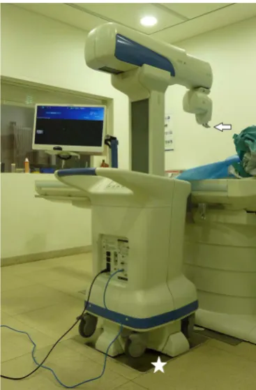
Figure 1. The experimental setup. The robotic device is mounted onto a docking plate (*) next to the CT table for spatial orientation. With the inte-grated planning computer access paths can be visualized by using a graphical user interface that supports multiplanar view angles. The white arrow marks the needle holding arm, which is then aligned to the planned trajectory. At the tip instruments from 11 to 24 gauge can be inserted through dedicated clamped adapters. The data cable, which is linked to the CT workstation and the power cord can be seen at the bottom of the image.
Figure 2. The final result of the needle tip next to the target dummy (arrow).
Needle tip is at the level of the clip, the measured deviation to the center of the clip was 2 mm in this case.
Germany) and then repeated the procedure with the help of the robotic device. The radiologists were free to choose the approach path to the target, whether in or off plane view.
[image:3.595.173.457.466.575.2]B. Schulz et al.
the achieved dosage and number of images taken for guidance were noted for each procedure and interventio-nalist. Furthermore, the time taken for planning (planning time) and executing the procedure (intervention time) was documented for each case. A post-procedural scan was performed to measure the total length of the access path and the needle point to the target distance.
In a second approach, the radiologists used the robotic device to plan and perform the needle interventions. For this approach, no image guidance was used during the needle advance, as the robotic arm was expected to position itself correctly regarding the target’s position and needle length. Again, the length of access path, accu-racy and timely effort were documented for each procedure.
2.3. Data Analysis
Average time taken for planning and performing the procedures, total access path length and accuracy (needle tip to target distance) were compared between manual and robot assistance interventions. Radiation exposure of the radiologists and the number of images required for the freehand technique was assessed, as was the patient radiation dosage for both techniques. Where applicable, Student’s t-Test was used to analyze significant differ-ences while assuming a confidence interval of 95% (α ≤ 0.05).
3. Results
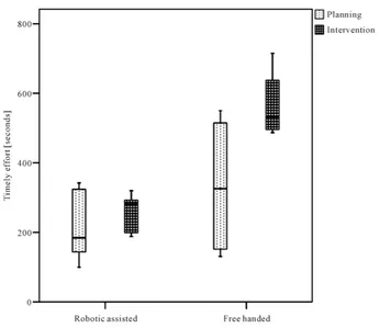
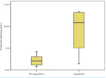
A total of 32 percutaneous punctures were conducted upon the four liver targets. The radiologist used the in plane technique 22 times, and the subcapsular lesion located in segment 8 was punctured in single oblique tech-nique all 8 times. The lesion in S5 was also targeted by two of the radiologists with an angulated trajectory. The average total procedure time for freehand technique was 443 seconds while robot-assisted needle interventions took 405 seconds (p = 0.64). Detailed timely effort of planning and performing the procedures are shown in Figure 3andFigure 4. The average needle tip deviation was 5.6 mm without using the robotic device (versus 4 mm, p = 0.11). When focusing solely on the angulated punctures of the clip in segment 8, the differences showed statistical significance for total time (490 sec vs. 900 sec, p < 0.01) and needle tip deviation (3.7 mm vs. 10.8 mm, p < 0.01). Since the robot-assisted procedures were performed without image guidance, the radiolo-gists were not exposed to scattered radiation. Using the freehand technique, the median measured radiation ex-posure during image guidance was 2.4 µSv Effective Dose with a median of 6 images taken per procedure. Again, the more complex angulated procedures caused an increase in scattered x-rays, leading to a median do-sage of 10.9 µSv with a median of 24 images that were taken in each procedure to control the needle position (Figure 5andFigure 6). Image guidance during procedures conducted using the freehand technique caused an additional radiation exposure for the corpse of 23.2 mGycm for in plane procedures and 123.8 mGycm for an-gulated procedures.
4. Discussion
In this cadaver study, we evaluated a newly available robotic guidance system that is suitable for planning and executing liver interventions based on a previously acquired CT dataset. Efficacy was assessed by comparing percutaneous robot-assisted needle placements to interventions performed using the standard freehand tech-nique.
Figure 3. The time consumed during the planning and performing of liver biopsies using the standard freehand technique and with the help of the robot guidance tool.
[image:5.595.141.487.405.704.2]B. Schulz et al.
Figure 5. Number of images acquired during the standard free hand procedure for needle
guidance. Angulated interventions required considerably more images.
Figure 6. The subsequently increased scattered radiation exposure for the interventionalist.
terms of difficulty. Therefore, we believe that oblique but safer trajectories will be chosen more often than is currently the case with the use of such robotic devices.
[image:6.595.144.486.363.614.2]proce-dures relied solely on the planned and aligned trajectory path. As stated before, the general use of aprons is ubi-quitous; however, without the constant use of lead glasses and gloves, a certain amount of scattered X-rays will hit the attending staff during the procedure.
Important limitations of the robotic-assisted interventions are primarily the inability of the device to compen-sate for patient movement after having performed the planning-scan. Especially for interventions in organs that are affected by diaphragm movements, precise breathing commands would be essential for the planning scan as well as during the procedure when manipulating the needle. Sudden movements of the patient would hinder or even preclude the robot-based intervention, since the planning relies on a previously acquired dataset. A second planning scan would be necessary to update the anatomical conditions, leading to a significant increase in radia-tion dose for the patient. Another restricradia-tion of the system is the cumbersome dimensions and setup. Wheeling the robot onto the docking plate and initializing and setting up the device takes about 10 minutes. However, we noticed that if the robot is not properly aligned to the docking plate, the booting up procedure has to be repeated. Finally, the range of the device is somewhat limited: due to a lack of space, the device is usually placed on the opposite side of the body, which may inhibit very lateral interventions or punctures in the posterior-anterior di-rection. Limitations of this study include the nature of its design: without any movement of the liver, the device successfully planned and guided liver biopsies in all procedures; however, clinical studies are mandatory to as-sess its efficacy for patients who receive biopsy in breath-hold technique.
Nowadays, CT-guided robot-assisted interventions have to compete against devices used in Magnet Reson-ance Imaging (MRI)- or Cone Beam CT (CBCT)-based intervention suites [7]-[12]. The most significant ad-vantages of MRI interventions are the avoidance of any radiation burden and excellent tissue discrimination. However, MRI-based interventions are elaborate due to the narrow access paths and the complex guidance strug-gling with artifacts to visualize the actual needle position. CBCT-based suites lack tissue differentiation capabil-ities compared to CT images. Furthermore, during the procedure, only projection radiography images are avail-able, otherwise repetitive 3D scans would have to be performed, causing an additional dose penalty for the pa-tient [13].
5. Conclusion
In conclusion, the proposed robotic assistance device successfully performed liver biopsies in this corpse study. Accuracy and timely effort were comparable with those of manual procedures and superior regarding angulated and thus more complex interventions. In combination with zero scattered radiation exposure for the attending staff, this device seems promising for CT-guided needle interventions.
References
[1] Siragusa, D.A. and Friedman, A.C. (2001) Imaging-Guided Percutaneous Biopsy of Hepatic Masses. Techniques in Vascular and Interventional Radiology, 4, 172-185. http://dx.doi.org/10.1016/S1089-2516(01)90023-X
[2] Chintapalli, K.N., Montgomery, R.S., Hatab, M., Katabathina, V.S. and Guiy, K. (2012) Radiation Dose Management: Part 1, Minimizing Radiation Dose in CT-Guided Procedures. American Journal of Roentgenology, 198, W347-W351.
http://dx.doi.org/10.2214/AJR.11.7958
[3] Cleary, K., Melzer, A., Watson, V., Kronreif, G. and Stoianovici, D. (2006) Interventional Robotic Systems: Applica-tions and Technology State-of-the-Art. Minimally Invasive Therapy & Allied Technologies, 15, 101-113.
http://dx.doi.org/10.1080/13645700600674179
[4] Rasmus, M., Huegli, R.W., Bilecen, D. and Jacob, A.L. (2007) Robotically Assisted CT-Based Procedures. Minimally Invasive Therapy & Allied Technologies, 16, 212-216. http://dx.doi.org/10.1080/13645700701520636
[5] Yanof, J., Haaga, J., Klahr, P., Bauer, C., Nakamoto, D., Chaturvedi, A., et al. (2001) CT-Integrated Robot for Inter-ventional Procedures: Preliminary Experiment and Computer-Human Interfaces. Computer Aided Surgery, 6, 352-359.
http://dx.doi.org/10.3109/10929080109146304
[6] Wood, B.J., Locklin, J.K., Viswanathan, A., Kruecker, J., Haemmerich, D., Cebral, J., et al. (2007) Technologies for Guidance of Radiofrequency Ablation in the Multimodality Interventional Suite of the Future. Journal of Vascular and Interventional Radiology, 18, 9-24. http://dx.doi.org/10.1016/j.jvir.2006.10.013
[7] Schulz, B., Eichler, K., Siebenhandl, P., Gruber-Rouh, T., Czerny, C,, Vogl, T.J., et al. (2013) Accuracy and Speed of Robotic Assisted Needle Interventions Using a Modern Cone Beam Computed Tomography Intervention Suite: A Phan-tom Study. European Radiology, 23, 198-204. http://dx.doi.org/10.1007/s00330-012-2585-0
B. Schulz et al.
High-Field MRI System—First Clinical Results (Roboterunterstutzte Punktion in einem Hochfeld-Kernspintomo- grafen—Erste klinische Ergebnisse). Fortschr Röntgenstr, 184, 42-47. http://dx.doi.org/10.1055/s-0031-1281774
[9] Zangos, S., Eichler, K., Thalhammer, A., Schoepf, J.U., Costello, P., Herzog, C., et al. (2007) MR-Guided Interven-tions of the Prostate Gland. Minimally Invasive Therapy & Allied Technologies, 16, 222-229.
http://dx.doi.org/10.1080/13645700701520669
[10] Penzkofer, T., Isfort, P., Bruners, P., Wiemann, C., Kyriakou, Y., Kalender, W.A., et al. (2010) Robot Arm Based Flat Panel CT-Guided Electromagnetic Tracked Spine Interventions: Phantom and Animal Model Experiments. European Radiology, 20, 2656-2662. http://dx.doi.org/10.1007/s00330-010-1837-0
[11] Pfleiderer, S.O., Marx, C., Vagner, J., Franke, R.P., Reichenbach, J.R. and Kaiser, W.A. (2005) Magnetic Reson-ance-Guided Large-Core Breast Biopsy inside a 1.5-T Magnetic Resonance Scanner Using an Automatic System: In Vitro Experiments and Preliminary Clinical Experience in Four Patients. Investigative Radiology, 40, 458-463.
http://dx.doi.org/10.1097/01.rli.0000167423.27180.54
[12] Tovar-Arriaga, S., Tita, R., Pedraza-Ortega, J.C., Gorrostieta, E. and Kalender, W.A. (2011) Development of a Robotic FD-CT-Guided Navigation System for Needle Placement—Preliminary Accuracy Tests. The International Journal of Medical Robotics and Computer Assisted Surgery, 7, 225-236. http://dx.doi.org/10.1002/rcs.393