805
Introduction
The actin skeleton is required for many cellular processes, including cytokinesis, endocytosis and exocytosis; cell shape and polarity; cell process formation; and motility (Jacinto and Baum, 2003). During the egg-to-blastula stage in Xenopus, each cell assembles a cortical actin network of filament bundles, which is required for maintenance of overall rigidity and shape of the whole embryo (Kofron et al., 2002). In previous work we have shown that the cadherin-binding protein plakoglobin is necessary and sufficient for maintaining the cortical actin skeleton, and acts downstream of the cytoplasmic signaling intermediate cdc42 (Kofron et al., 2002). Loss of either of these proteins causes loss of shape and rigidity of the embryo, which collapses under its own weight. Examination of the cytoskeleton of such embryos reveals the loss of cortical actin, but not the microtubule or intermediate filament skeletons, of the blastomeres. Conversely, overexpression of cdc42 or plakoglobin increases the density of the cortical actin skeleton, and the rigidity of the embryo (Kofron et al., 2002).
Of particular interest is the mechanism by which each cell of the embryo assembles a similar cortical actin network. The number of cells increases rapidly by repeated cell divisions in the early embryo, and yet each cell, as it forms, assembles an actin skeleton appropriate to its contribution to the overall shape and rigidity of the whole embryo. In general, two mechanisms for this can be envisaged. First, each cell could inherit actin assembly instructions from the egg. Second, intercellular signaling could maintain the appropriate density and pattern of cortical actin filaments. In general, little is known about how cells of supracellular arrays all maintain
actin skeletons appropriate for the shape, size and rigidity of the array. Xenopusembryos offer an attractive system in which to study this.
It has been known for many years that phospholipids can participate in intercellular signaling (Vogt, 1963), and their diverse roles have only recently been realized as more model systems have become available (Im et al., 2000; Yang et al., 2002). The phospholipid LPA can induce different cellular responses, depending upon cell type and context. These include smooth muscle contraction, cell proliferation, platelet aggregation, cell migration and neurite retraction (Goetzl, 2001; Xie et al., 2002). In particular, LPA signaling has been shown to influence both the actin cytoskeleton and cellular morphology. Increased LPA signaling in fibroblasts increases the formation of stress fibers. In different neural cell lines, it causes rapid process retraction, cell rounding or actin reorganization (Fukushima et al., 2002; Ridley and Hall, 1992; Yan et al., 2003). Overexpression of the Xenopus XLPA1
receptor in a rat neuroblastoma line that lacks endogenous LPA receptors, causes cell rounding, retracted neurites and an increase in stress fibers (Kimura et al., 2001).
LPA signals through G-protein-coupled receptors (GPCR) belonging to the rhodopsin-like class A receptors. These are seven transmembrane domain (TMD) proteins that bind specific G proteins to elicit responses (Anliker and Chun, 2004). The first LPA receptor was identified as a sheep orphan GPCR (Edg-2) and subsequently as the mouse ortholog of rec1.3 (Macrae et al., 1996; Masana et al., 1995). It was also identified in a screen for GPCRs associated with neuron production, as a transcript expressed in the ventricular zone of the developing mouse cortex, and demonstrated to be an LPA-specific receptor (Hecht et al., 1996). Overexpression of this
The mechanisms that control shape and rigidity of early embryos are not well understood, and yet are required for all embryonic processes to take place. In the Xenopus
blastula, the cortical actin network in each blastomere is required for the maintenance of overall embryonic shape and rigidity. However, the mechanism whereby each cell assembles the appropriate pattern and number of actin filament bundles is not known. The existence of a similar network in each blastomere suggests two possibilities: cell-autonomous inheritance of instructions from the egg; or
mutual intercellular signaling mediated by cell contact or diffusible signals. We show that intercellular signaling is required for the correct pattern of cortical actin assembly in Xenopusembryos, and that lysophosphatidic acid (LPA) and its receptors, corresponding to LPA1 and LPA2 in mammals, are both necessary and sufficient for this function.
Key words: Lysophosphatidic acid, Actin cytoskeleton, G-protein-coupled receptor, Xenopus
Summary
Lysophosphatidic acid signaling controls cortical actin assembly
and cytoarchitecture in Xenopus embryos
Brett Lloyd1,2, QingHua Tao1, Stephanie Lang1and Chris Wylie1,*
1Cincinnati Children’s Hospital Research Foundation, 3333 Burnett Avenue, Cincinnati, OH 45229, USA 2PSTP Program, University of Cincinnati College of Medicine, PO Box 670555, Cincinnati, OH 45267, USA *Author for correspondence (e-mail: christopher.wylie@cchmc.org)
Accepted 1 December 2004
Development 132, 805-816
Published by The Company of Biologists 2005 doi:10.1242/dev.01618
Research article
De
transcript in cell lines induced serum-dependent cell rounding, which was mimicked by addition of LPA. Verification that this was an LPA receptor was provided by studies in yeast and gain-of-function studies using the human ortholog (An et al., 1997; Erickson et al., 1998). Structural studies have suggested key residues to be important for phospholipid binding and LPA specificity (Wang et al., 2001). To date, three LPA receptors have been identified in mammals and renamed LPA1, LPA2and
LPA3 (Lynch, 2002). These share sequence homology with a
more divergent fourth receptor (Anliker and Chun, 2004). In Xenopus, a single LPA receptor and its pseudoallele have so far been identified. These are most closely related to mammalian LPA1 (and designated here as XLPA1A and
XLPA1B). Both genes are expressed maternally and throughout
embryogenesis (Kimura et al., 2001).
In this work, we show that LPA signaling is both necessary and sufficient for maintenance of the normal cortical actin skeleton in the early Xenopusembryo. First, we show that an additional LPA receptor, most closely related to LPA2
(designated here as XLPA2) is expressed after the onset of
zygotic transcription. No homolog of mammalian LPA3 was
identified. We show that either addition of LPA ligand, or overexpression of Xenopus LPA receptors, increases the density of the cortical actin network in the early embryo and increases the rate of wound healing. Conversely, depletion of XLPA1and XLPA2receptors in the blastula reduces the density
of the cortical actin network. Cell disaggregation mimics the effect of LPA receptor depletion, and adding soluble LPA to dissociated cells reverses the effect. These data suggest an intercellular signaling mechanism for global patterning of the cortical actin network in the early Xenopusembryo.
Materials and methods
Oocytes and embryos
Ovaries were removed from mature females and stage VI oocytes were defolliculated and injected with antisense or morpholino oligonucleotides. For double injections, oocytes were incubated at 18°C for 24 hours after injection with antisense oligo and then injected with morpholino oligo. Oocytes were matured using 1 µM progesterone and fertilized using the host transfer technique as reported previously (Holwill et al., 1987). Embryos were dejellied in a 2% cysteine/0.1 MMR solution (pH=7.8) and maintained in 0.1 MMR. Embryo stages cited are as described by Nieuwkoop and Faber (Nieuwkoop and Faber, 1967). For mRNA and morpholino injections after fertilization, embryos were transferred into a 2% ficoll/0.5 MMR solution and injected into the animal cytoplasm of each blastomere at the two-cell stage.
Analysis of the actin skeleton
Vitelline membranes were removed from stage 9 embryos in a 1 MMR solution on agarose dishes. Embryos were fixed for 30 minutes in FG fixative (3.7% formaldehyde/0.25% glutaraldehyde/0.2% Triton X-100 in PIPES buffer (Gard et al., 1997) before excision of animal caps to examine the undisturbed actin skeleton. Alternatively, animal caps were excised and cultured for 10 minutes before fixation in FG fix, to allow the analysis of the response of the actin skeleton to wounding. In each case, the cortical actin skeleton was analyzed exactly as described by Kofron et al. (Gard et al., 1997; Kofron et al., 2002). For lipid experiments, LPA, phosphatidic acid and phosphatidylethanolamine (Avanti Polar Lipids) were reconstituted in 0.4% lipid-free BSA (Sigma) in 1MMR and 0.4% lipid-free BSA was added to all solutions as a carrier. After caps were cut, they were
incubated for 10 minutes in a lipid or control solution before analysis of cortical actin.
Oligonucleotides
Twelve antisense oligonucleotides complementary to both XLPA1A and XLPA1B mRNA were tested for their ability to deplete the maternal messages by injecting into the marginal zones of oocytes, incubating for 24 hours at 18°C, and assaying for mRNA depletion using RTPCR. Antisense oligonucleotides that depleted both mRNAs to less than 20% of normal levels were phophorothioate-modified, purified by HPLC, and resuspended in sterile, filtered water. The sequence of the oligo selected for use was as follows (where asterisks represent phosphorothioate linkages): LPA1-10MP, 5′ T*C*A*TT-GTAGTAGCAC*T*G*G 3′.
Morpholino oligonucleotides were designed that targeted both XLPA1Aand XLPA1Bor XLPA2. These were resuspended in sterile, filtered water and injected at doses of 10-40 ng into either oocytes or embryos: XLPA1A and 1B MO, 5′ TTCACTTCAGATGTCAGTC-ATGCTG 3′; XLPA2 MO, 5′ ACCTCCAATGTTACAGCGCAG-CCTC 3′.
RNA constructs
Clones encoding both X. tropicalis XLPA1and XLPA2were identified by blasting the murine sequences for LPA1against X. tropicaliscDNA libraries at the Sanger Institute site (http://www.sanger.ac.uk/). The following clones for XLPA1 (TNeu092p02) and XLPA2 (TNeu013j17) were isolated, sequenced and DNA was linearized with Asp718. Dominant-negative forms of the human small Rho GTPases were excised from the pKH3 vector (a generous gift from Yi Zheng) using BamHI and EcoRI and inserted into the pCS2+ vector. DNA was linearized with ApaI. In vitro transcription was performed using the SP6 mMessage Machine (Ambion). Samples were treated for 15 minutes with DNase I, purified by phenol:chloroform extraction and resuspended in sterile filtered water.
RT-PCR
Total RNA was isolated from either two oocytes or embryos at specified stages in a proteinase K solution as described (Kofron et al., 2002) and subsequently treated with DNase I. cDNA was synthesized using oligo dT primers from 1 µg total RNA. The cDNA samples were analyzed on the MJ Research Opticon. Uninjected samples were used to generate a standard curve for each primer set and all data were normalized to either ornithine decarboxylase or plakoglobin as a control. Water and no reverse transcriptase controls were run each time and found to produce no product. PCR reactions were run on a 1.8% agarose gel to verify amplification of the correct size fragment and look for the formation of primer dimers. Primer pairs that were used are as follows.
XLPA1: F, 5′CTTGGAGTCCCGTGTGTTTT 3′; R, 5′ TGGCT-GCAGAAGTCTGTGAC 3′
XLPA2: F, 5′TTCTTCTGCAACAGGGGTTC 3′; R, 5′ GGGCCT-CACTCCAACTGTTA 3′
ODC: F, 5′GCC ATT GTG AAG ACT CTC TCC ATT C 3′; R, 5′ TTC GGG TGA TTC CTT GCC AC 3′
Plakoglobin: F, 5′GCT CGC TGT ACA ACC AGC ATT C 3′; R, 5′GTA GTT CCT CAT GAT CTG AAC C 3′
Cell dissociation assays
Vitelline membranes were removed from mid-blastulae (stage 8). Five animal caps were cut, and dissociated in 67 mM phosphate buffer for 3 minutes (Snape et al., 1987). Dissociated cells were transferred into 1Ca2+/Mg2+-free MMR on a 1% agarose dish. After 1 hour, cells were transferred into 0.1-1 µM LPA in Ca2+/Mg2+-free MMR in glass dishes for five minutes, or allowed to reassociate in 1MMR. Cells were removed from the LPA solutions and maintained in 1 Ca2+/Mg2+-free MMR for different time intervals before fixation. Cells were fixed for 4 minutes in FG fix, washed with 1PBS+0.1%
De
Tween-20, and stained with Alexa 488-phalloidin. To determine if Ca2+or Mg2+affected the actin cytoskeleton of dissociated cells, the cells were transferred back into 1 MMR 15 minutes after dissociation, incubated for 30 minutes, and fixed and stained as above.
Statistics
Using the Laser Scanning Microscope software (Zeiss), projections were made from z-stacks of single cells or animal caps. The mean intensity was recorded over a 5000 µm2area for at least 15 dissociated cells in each group. For animal caps, the mean intensity was recorded over a 0.62 mm2area for gain-of-function experiments and a 1000
µm2area with the low threshold set to 100. The mean intensities were averaged and are reported as mean±s.e.m. Student’s t-test was used to determine significance and P<0.05 was considered to be statistically significant.
Results
Actin-containing structures in cells of the Xenopus blastula
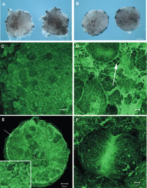
These were examined in fixed animal caps excised from Xenopusblastulae after fixation for 30 minutes in FG fixative. Alternatively, animal caps were excised and allowed to heal for 10 minutes before fixation. Figure 1 shows a dissecting microscope view of caps fixed before isolation (Fig. 1A) and after 10 minutes culture (Fig. 1B), by which time healing has started, the wound margins have become smooth and the outer surfaces of the caps are becoming visible as the cap rounds up. At the late blastula stage, each cell lining the roof of the blastocoel cavity had a dense cortical network of actin filament bundles (Fig. 1C,D) (see also Kofron et al., 2002). Cells extend occasional filopodia (arrowed in Fig. 1D). In caps that were allowed to heal for 10 minutes before fixation, actin-rich purse-strings formed around the margins of the caps (arrowed in Fig. 1E). In addition to forming a purse-string, cells in healing caps also extended many actin-rich processes, which obscured cell boundaries (Fig. 1E, see inset). Occasionally, cells were identified that had rounded up and were undergoing cytokinesis in the plane of the roof of the blastocoel (outlined in Fig. 1E). In these cells, actin rich contractile rings were seen (Fig. 1F). Outside the contractile rings, the cortical actin skeleton of a dividing cell was significantly less dense than that of controls, and was replaced by a coarser network of filament bundles (Fig. 1F).
Currently, the mechanism(s) by which each blastomere assembles these components, either in the intact embryo, or in response to wounding, is not known.
Intercellular signaling controls the density of the cortical actin network
Because each cell of the blastula has a similar pattern and density of cortical actin (Fig. 1C,D), we tested the possibility that intercellular signaling maintains or initiates this pattern. We removed animal caps from early blastulae and dissociated them into single cells by removing the divalent cations required for cell adhesion. The cells were kept apart, fixed after different times in culture and the cortical actin network stained using Alexa-488 phalloidin. The cortical actin network in dissociated cells changed over the course of 30-60 minutes from the dense cortical network seen in undissociated caps from sibling embryos (Fig. 2A,B), to a coarser network of thick filament bundles, similar to those of dividing cells in intact animal caps
(compare Fig. 1F with Fig. 2C). To avoid the potential artifact that the actin skeleton is reduced by the Ca2+/Mg2+-free saline,
we compared dissociated cells that had been cultured in Ca2+/Mg2+-free MMR before fixation with those that were
transferred into 1MMR at low density after disaggregation for 30 minutes before fixation. There was no significant difference in the intensity of phalloidin staining in the two groups of cells (data not shown). Subsequent reaggregation of single cells by transfer at high density to 1MMR resulted in reassembly of the high-density cortical actin network characteristic of intact caps (Fig. 2D). This suggests that intercellular signaling, either through soluble ligands or by cell contact, is required to maintain the density and pattern of cortical actin assembly in each cell of the intact embryo.
LPA ligand and receptor are both functional in the Xenopus blastula
[image:3.612.313.561.76.394.2]It is well established that LPA signaling influences the actin
Fig. 1. F-actin-containing structures in early Xenopusembryos. (A) Animal caps removed from late blastulae (stage 9) and photographed immediately after excision (0 minutes) or 10 minutes after healing (B). Scale bar: 125 µm. (C,D) Low- and
high-magnification views of F-actin staining of the actin network of a late blastula animal cap. Scale bars: 20 µm in C; 5 µm in D. (E) F-actin staining of an animal cap that had been removed from the embryo and allowed to heal for 10 minutes. Arrow indicates the purse-string. Inset shows a high-power view of the inner cells of the cap. Scale bar: 50 µm. (F) High-magnification image of area outlined in E showing the contractile ring and coarser actin filament network in a dividing cell. Scale bar: 5 µm.
De
cytoskeleton in many cell types in vitro. However, the functions of LPA signaling in vivo during embryogenesis are not well understood. To test whether it plays a role in the cortical actin network of early Xenopusembryos, we first carried out gain-of-function experiments using both the ligand and its receptors. Purified 18:1 oleyl-LPA, bound to lipid-stripped bovine serum albumin was added to animal caps isolated from late blastula and early gastrula stage embryos. Animal caps were excised, cultured for 10 minutes in the presence or absence of LPA, then fixed and stained for F-actin with Alexa 488-phalloidin (Fig. 3A).
In the presence of 1 µM LPA, there was a dramatic increase in F-actin in the cortical actin network throughout the animal cap and in the purse-strings (Fig. 3C). This resulted in faster healing in the LPA-treated animal caps (compare Fig. 3B with 3C). At high magnification, the actin network in LPA treated caps was thicker and less organized compared with controls, and cell boundaries were obscured by the abundance of actin in many regions (compare Fig. 3D with 3E). These effects were dose dependent in the range of 0.1-5 µM LPA. Two related phospholipids were used as controls for specificity. After treatment with 5 µM phosphatidic acid (PA), the caps either displayed no change or a slight decrease in cortical actin (Fig. 3F). PA-treated caps were flatter than controls, and there was no effect on the rate of wound healing. At higher magnifications, the cortical actin network was similar in density to control embryos and the cells contained similar patterns of F-actin (not shown). Phosphatidylethanolamine
(PE) had no effects, either on wound healing or on the cortical actin network (not shown). There was a significant increase in the intensity of phalloidin staining from 878±112 to 1154±160 in the 1 µM LPA treatment group, but no change with 5 µM PA (735±62) (Fig. 4A). All data represents four independent experiments with five caps per group in each experiment. These data show that LPA is sufficient to increase cortical actin at early blastula stages, and this suggests the receptors for LPA signaling are present and functional in the embryo.
Identification of a second LPA receptor in early Xenopus embryos
Two genes encoding LPA receptors have been described in Xenopus laevis: XLPA1A and XLPA1B(Kimura et al., 2001).
These both show 90% homology to the human LPA1receptor
[image:4.612.46.291.72.328.2]and are 98% identical in amino acid sequence to each other. As Xenopus laevis is allotetroploid, these are most likely pseudoalleles and represent the duplicated orthologs of the
Fig. 2.Dissociation of blastula stage animal caps causes a reduction in the density of cortical actin, which is rescued by reaggregation of the cells. (A,B) Low- (A) and high- (B) magnification images of the F-actin network in situ in caps before disaggregation. (C) The coarser F-actin network found in single cells 45 minutes after dissociation. (D) Reassociation of the animal cap cells caused the cortical actin network to return to normal levels. Scale bars: 20 µm in A; 5 µm in
B-D. Fig. 3. Addition of LPA to animal caps causes an increase in cortical
actin and more rapid wound-healing. (A) Experimental design. Animal caps were excised from late blastulae, incubated for 10 minutes in either control or LPA solutions, fixed and stained with Alexa-488 phalloidin to visualize F-actin. Caps incubated in 1 µM LPA (C) had increased cortical actin and more rapid wound-healing, compared with control caps (B). (D,E) Higher magnification images of control (B) and 1 µM LPA-treated (C) caps. The increased cortical actin and surface projections caused by LPA are shown.
(F) Incubation of animal caps in 5 µM phosphatidic acid for 10 minutes did not increase cortical actin or expedite wound-healing. Scale bars: 50 µm in B,C,F; 10 µm in D,E.
De
[image:4.612.313.560.74.399.2]mammalian LPA1 receptor. The
sequences for murine LPA2and LPA3
were used to screen the X. tropicalis cDNA databases at the Sanger Institute (http://www.sanger.ac.uk/). Two X. tropicalis clones were identified (TNeu013j17 and TGas026e21) with significant homology to mouse LPA2.
These were obtained and sequenced and found to encode the same mRNA. As only TNeu013j17 contained the full coding sequence, this was used for experiments described here. The full sequence of X. tropicalis XLPA2
mRNA was deposited into GenBank as Accession Number AY652941.
The predicted protein was found to be 62% identical and 16% similar to mouse LPA2 at the protein level and
thus was designated X. tropicalis XLPA2. It contains 344 amino acids,
has a predicted molecular mass of 39.5 kDa, and is predicted to have seven putative transmembrane domains (TMD) (Fig. 5A). XLPA2 is most
divergent from the mammalian orthologs in the fourth and fifth TMDs and at the C terminus. Based on structural models, LPA receptors have been shown to contain three residues that interface with LPA (Wang et al., 2001). XLPA2 contains the conserved
arginine and lysine in the third and seventh TMD, respectively, that are thought to interact with the head group of LPA; and a glutamine in the third domain that confers LPA specificity (highlighted in red in Fig. 5A). Like mammalian LPA2
receptors, it also lacks the longer extracellular N terminus of LPA1.
No orthologs of mouse LPA3 were found in egg, gastrula,
neurula or tadpole libraries.
Expression of LPA receptors during Xenopus laevis development
Total RNA was isolated from a series of developmental stages, and expression levels of XLPA1and XLPA2analyzed by
real-time RTPCR. As reported previously, XLPA1was found to be
most abundant in the oocyte (Kimura et al., 2001). After the mid-blastula transition (MBT) and the onset of zygotic transcription, levels of the XLPA1transcript fall, but low levels
of XLPA1 expression continued until at least stage 45.
Conversely, XLPA2mRNA was not detected in oocytes or early
embryos. Expression commenced at MBT, and remained constant until at least stage 45 (Fig. 5B). Results are representative of a single experiment. This experiment was repeated in three independent experiments and the same result was obtained each time.
Overexpression of X. tropicalis LPA receptors mimics addition of LPA ligand
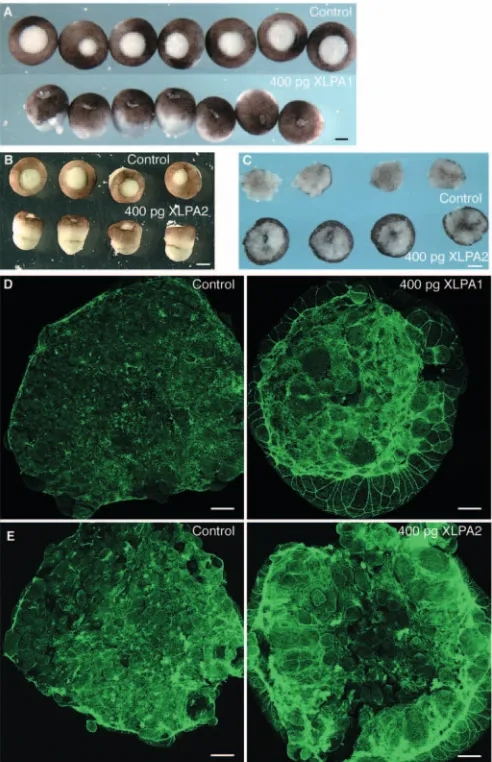
To assay for the presence of functional LPA ligand at the blastula stage, we injected 400 pg of either XLPA1or XLPA2
receptor mRNA at the two-cell stage (200 pg/ blastomere), and excised animal caps for analysis of the actin skeleton at the late blastula stage. After removal of the vitelline membrane, embryos injected with either XLPA1 or XLPA2 became
elongated along the animal-vegetal axis (Fig. 6A,B). They were also more compact than controls, and the animal caps healed faster than control caps (Fig. 6C). The effects on animal caps of LPA receptor overexpression were identical to those caused by addition of LPA to the animal caps; denser networks of cortical actin, thicker purse-strings, no change in contractile rings and faster wound-healing (Fig. 6D,E). Overexpression of LPA receptors caused a significant increase in phalloidin intensity over a 0.62 mm2area from 1133±177 to 1372±302 or 1610±348 for XLPA1 and XLPA2, respectively (Fig. 4B).
All data represent four independent experiments with five caps per group in each experiment. Therefore, overexpression of the LPA receptor is sufficient to increase cortical actin and the rate of wound healing in the early embryo, and demonstrates the presence of endogenous ligand.
LPA signaling is necessary, as well as sufficient, for cortical actin polymerization in the Xenopus blastula
The presence of a maternal store of LPA1mRNA in the oocyte
suggested that LPA signaling may be controlled, at least until the blastula stages, by maternally encoded genes. So, for loss of function experiments, we first depleted the stored maternal LPA1 mRNA using antisense oligodeoxynucleotides. Twelve
oligos, each complementary to both XLPA1A and XLPA1B,
[image:5.612.214.561.73.319.2]were synthesized and tested for their ability to deplete both
Fig. 4.Quantitation of changes in the intensity of phalloidin staining. There is a significant increase in the intensity of phalloidin staining if caps are treated with LPA (A) or if either LPA receptor is overexpressed in caps (B), but not with treatment of the related phospholipid phosphatidic acid (A). (C,D) There is a significant decrease in the intensity of phalloidin staining by targeting the mRNA with either a phosphorothioate oligo or morpholino oligonucleotides. Asterisks indicate significance at P<0.05. All data are representative of a single experiment and all experiments were repeated at least three times with five caps per group.
De
XLPA1 mRNAs after injection into the oocyte cytoplasm. One
was selected and modified by replacing the 5′ and 3′ phosphodiester linkages with phosphorothioate linkages. Doses of 5, 7.5 and 10 ng were injected into manually defolliculated full-grown oocytes, which were fertilized 48 hours later by the host transfer technique (Holwill, 1987). XLPA1 mRNA levels were reduced to 16% of control levels at
the two-cell stage (Fig. 7A). At stage 10, XLPA1 mRNA in the
controls had decreased significantly, resulting in a relative increase in the depleted embryos to 33% of control levels.
The cortical actin in late blastula embryos was assayed either by fixation before removal of the animal cap, or by fixation 10 minutes after excision of the animal cap (to assay the response to wounding). In both cases, levels of cortical actin, including the purse-string that formed in response to wounding, as well as cortical actin in each cell, were reduced, compared with control embryos. Animal caps from depleted embryos, and the bases from which they were excised, healed more slowly than controls (Fig. 7B,C). At higher magnification, actin filament bundles in the cell cortices were dramatically reduced in density, compared with the controls (Fig. 7D). The formation of actin-rich filopodia was unaffected in these embryos. Neither the overexpression nor depletion of LPA receptors affected the number or size of contractile rings seen in dividing cells. The caps contained the normal number of cells and there was no evidence of undividing cells in these embryos (data not shown). The effects of the thioate oligo were reversed (Fig. 7C) by depleting XLPA1, fertilizing the oocytes by host transfer and
injecting 400 pg of XLPA1 mRNA at the two-cell stage, by
which time the antisense oligo had degraded (Raats et al., 1997). The average mean intensity of phalloidin staining decreased from 1532±395 to 978±202 and 877±191 for the 7.5 ng and 10 ng dose of XLPA1-10MP, respectively (Fig. 4C).
Data are representative of three independent experiments with five animal caps per group. These experiments show that signaling through XLPA1 is
required to maintain the normal density of the cortical actin network in the early Xenopusembryo.
Despite the reduction of cortical actin at the blastula stage, embryos depleted only of the maternal XLPA1
were able to gastrulate and develop normally to tadpole stages (Fig. 7E). This could be due to re-establishment of receptor levels as the maternal store is replaced by zygotic transcription of XLPA1 and/or XLPA2. To test this
possibility, we synthesized antisense morpholino oligos, which block translation of their target mRNAs throughout early development (Heasman et al., 2000), complementary to each mRNA separately (XLPA1-MO and XLPA2-MO). These were
injected at either the two-cell stage of development into the animal hemisphere at doses from 10-40 ng, or into oocytes that were then fertilized using the host transfer technique.
At doses of 20-40 ng of the XLPA1-MO, there was a
generalized decrease in the amount of F-actin staining throughout all cells in the animal caps (Fig. 9A), similar to caps depleted of maternal XLPA1. Purse-strings were present after
animal cap excision, but at reduced levels compared with control caps (Fig. 9A). At high power, cells in LPA1-depleted
caps were found to have lost the dense cortical network of actin filament bundles, but retained a coarser network similar to that seen in dividing cells, and in dissociated cells. In addition, fewer cell processes were present (Fig. 9B). These data are representative of four independent experiments with five animal caps per group.
Depletion of XLPA2by the morpholino oligo had no effect
before the onset of zygotic transcription, consistent with the fact that onset of transcription starts at the mid-blastula stage (Fig. 5). However, at late blastula and early gastrula stages, it caused effects similar to depletion of XLPA1 (Fig. 9B).
[image:6.612.43.323.77.365.2]Injection of 15 ng of both morpholino oligos together caused effects similar to 40 ng of either morpholino alone (Fig. 9B). Injection of both morpholinos reduced the average mean intensity of phalloidin staining from 1122±87 to 1002±59 (Fig. 4D). These data suggest that after the mid-blastula stage, the combined levels of the two LPA receptors are necessary to maintain the pattern and density of cortical actin in the embryo. In contrast to removal of only the maternal LPA1 mRNA,
Fig. 5. Identification of a Xenopus tropicalis LPA receptor that is similar to mammalian LPA2. (A) Alignment of X. tropicalisXLPA2with mouse and human LPA2. Putative transmembrane domains (TMD) are indicated above the sequence. Residues important for LPA binding and specificity in other species are highlighted in red. X. tropicalis XLPA2is 62% identical and 16% similar to murine LPA2. (B) Expression using real-time RTPCR of XLPA1and XLPA2mRNA during early development. Each bar represents the amount of mRNA present, normalized to the loading control ornithine decarboxylase. As reported previously, XLPA1 is abundant maternally and continues to be expressed at a low level throughout development. Expression of XLPA2 begins at the mid-blastula stage and continues to at least stage 45. St.2, two-cell stage; St.9, late blastula; St.10, early gastrula; St.12, late gastrula; St.15, early neurula; St.20, late neurula; St.35, tail-bud; St.45, swimming tadpole.
De
embryos that were injected with XLPA1or XLPA2morpholino
oligos, which block translation of the zygotic mRNA as well, did show later developmental defects. These were first evident during gastrulation (Fig. 9C), which proceeded more slowly, with blastopores remaining open longer than those of control embryos. Defects became more severe during neurulation (Fig. 9D), with defects ranging from slower closure of the neural folds, to significantly reduced neural fold formation. By the tail-bud stage (Fig. 9E), LPA1 or LPA2-depleted embryos
showed reduction in body length and major defects in many organ rudiments. These pleiotropic effects are most likely due to an expanding number of LPA-mediated morphogenetic events during later stages.
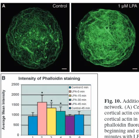
Addition of soluble LPA to isolated cells restores the cortical actin density to in vivo levels
Loss of LPA signaling reduces the density of the cortical skeleton, and mimics the effect of dissociating the cells, suggesting that LPA is an endogenous intercellular signal that controls the density of the cortical actin skeleton. To test this, cortical actin skeletons were compared between intact embryos, cells from embryos that had been dissociated at the mid-blastula stage and kept apart for 1 hour, and cells kept apart for 1 hour and then incubated for 5 minutes in 0.1 or 1
µM LPA. The cortical actin skeleton was significantly reduced in dissociated cells compared with intact embryos, and was rescued by subsequent addition of LPA to the dissociated cells (Fig. 10A). The mean fluorescence intensity for each cell was determined over a 5000 µm2area and averaged for each group. Addition of LPA to dissociated cells caused a statistically significant rise from 933±180 to 1626±349. Washing out the LPA, and keeping the cells dissociated caused a drop in cortical actin back to the level in dissociated cells after 45 minutes (Fig. 10B). The experiment was repeated three times with the same result. These data show that continuous signaling by LPA is required to maintain the normal pattern and level of cortical actin.
Dominant negative Rho and Rac, but not cdc42, block the overexpression effects of LPA receptors
The effects of LPA on the actin cytoskeleton in Swiss 3T3 fibroblasts are mediated through the small Rho GTPases, including activation of RhoA and Rac1. A dominant-negative form of RhoA (RhoA-N19) blocked the formation of stress fibers in response to LPA, while the formation of lamellipodia was blocked by expression of a dominant-negative Rac1 (Rac-N17) (Ridley and Hall, 1992; Ridley et al., 1992).
To determine whether LPA signaling in early Xenopus embryos acts through similar pathways, we expressed these same dominant-negative constructs, assayed their effects on the actin skeleton and asked if overexpression of LPA receptors could rescue these effects. We injected mRNA for either XLPA2 alone, a dominant-negative GTPase alone or for both
mRNAs at the two-cell stage and analyzed the actin skeleton at stage 9. Overexpression of RhoA-N19 alone resulted in a loss of purse-strings (arrow in Fig. 8B, upper middle panel), delayed wound healing and an increase in cellular processes (Fig. 8A,B, lower left panel). At higher doses, cell division was blocked and occasionally large cells were seen that had not divided (not shown). When XLPA2 and RhoA-N19 were
injected together, the Rho-N19 blocked the effect of XLPA2on
wound healing, but not the increase in overall cortical actin (Fig. 8A,B). This suggests that RhoA is downstream of LPA signaling in the formation of purse strings and wound healing, but not in the pathway leading to assembly of the cortical network of actin.
Overexpression of Rac-N17 alone also resulted in loss of purse-strings (Fig. 8A, upper right panel). In addition, there was a dramatic loss in the cortical actin network in each cell (Fig. 8B, lower middle panel). Co-injection of XLPA2 mRNA
[image:7.612.314.560.74.455.2]did not rescue this effect, showing that Rac is downstream of
Fig. 6. Overexpression of either X. tropicalis XLPA1or XLPA2 mRNA causes an increase in cortical actin and more rapid wound healing, mimicking LPA treatment. (A) The bases (after excision of animal caps) of embryos injected with 400 pg of XLPA1elongated along the animal-vegetal axis after the vitelline membrane was removed and were more rigid than control embryos. These bases healed faster than controls. Scale bar: 350 µm. (B) Bases from embryos injected with XLPA2mRNA showed a similar phenotype as bases from XLPA1-injected embryos. Scale bar: 450 µm. (C) Animal caps from XLPA2-injected embryos rounded up faster than control caps after 10 minutes of healing. Scale bar: 125 µm. Injection of either XLPA1(D) or XLPA2(E) mRNA increased the levels of cortical actin in the animal cap cells and in the purse-string compared with controls. Scale bar: 50 µm.
De
LPA signaling leading to assembly of the cortical actin network (Fig. 8A,B). When XLPA2 was co-injected with
dominant-negative forms of cdc42, there was no blockade of the overexpression effects of XLPA2(data not shown).
Discussion
The data presented show that intercellular signaling is required to maintain the normal cortical actin pattern and density in each blastomere during early Xenopusdevelopment, and that LPA signaling is both necessary and sufficient for this. LPA is a bioactive lipid, known to be involved in intercellular signaling. It is generated outside the cell by ectoenzymes, and acts upon specific G-protein-coupled receptors. Four LPA receptors have been identified in humans and mice. These have been known previously by a variety of names and re-classified more systematically recently as LPA1-4 (Chun et al., 2002). In
Xenopus, one receptor, with high homology to LPA1, has
already been identified (Kimura et al., 2001). We report here a second receptor with high homology to mammalian LPA2.
Interestingly, XLPA1 is stored as a maternal mRNA, while
XLPA2commences expression at the mid-blastula stage. As the
experiments described here suggest they play redundant roles in maintaining the actin skeleton, it is interesting that they are not coordinately regulated at these early stages.
LPA-mediated signaling has been implicated in a wide range of cell behavior, including proliferation, survival, motility, cell shape and differentiation (Anliker and Chun, 2004; Fukushima et al., 2002; Tigyi, 2001; Ye et al., 2002). Targeted mutation of LPA receptors in the mouse has shown that LPA signaling is required for normal development (Contos et al., 2000; Contos et al., 2002). Redundancies in receptor function and the pleiotropic effects of their removal have made it difficult to identify specific cellular events in specific organs that require LPA signaling. However, it is clear that in its absence, normal development does not occur. One specific event found to require LPA signaling in vivo was survival of Schwann cells in the sciatic nerve (Contos et al., 2000). In the present study, we have used the early Xenopusembryo as a relatively simple and tractable system to identify a specific role for LPA signaling in vivo. Upregulation of either the ligand or its receptor increased the density of cortical actin, indicating the presence of functional receptor and ligand in the embryo. Downregulation of the two LPA receptors had the converse effect, indicating that LPA signaling is both necessary and sufficient for maintenance of the correct density and pattern of cortical actin.
[image:8.612.46.340.65.486.2]It is of interest that either dissociation of the blastula cells or depletion of the LPA receptors, caused loss of the high-density cortical actin network, but left a coarser network of actin filaments remaining in the blastomeres. When LPA is added to dissociated cells, or they are allowed to aggregate again, a denser network, similar to that found in vivo, was assembled. This suggests that there are cell-autonomous
Fig. 7.Maternal XLPA1is required in vivo for modulating actin assembly in early Xenopus development. (A) Injection of 10 ng of an antisense phosphorothioate oligodeoxynucleotide (LPA1-10MP) into oocytes caused a reduction in XLPA1mRNA to 16% of control levels after fertilization. At the beginning of gastrulation (stage 10), this reduction was one-third of control levels. (B) Bases from embryos depleted of maternal XLPA1healed more slowly than controls. Scale bar: 280 µm. (C) Animal caps from depleted embryos (middle) showed decreased levels of F-actin in the animal cap and in the purse-string after wounding. These caps were larger than controls (left) due to slower wound-healing. The effects of the oligo were rescued by injecting X. tropicalis XLPA1mRNA back into fertilized eggs that had been depleted of XLPA1(right). This demonstrates that the effects are specific to depletion of XLPA1. Scale bars: 50 µm. (D) High-magnification image of the cortical actin network in control (left) and XLPA1-depleted caps (right) after 10 minutes of healing. The density of actin filaments is greatly reduced in the depleted caps compared with controls. Scale bars: 10
µm. (E) Depletion of the maternal stores of XLPA1does not lead to long-term developmental defects. Embryos were able to gastrulate (left) and develop to tail-bud stages (right). Control embryos are red and XLPA1 depleted are brown. Scale bars: 350 µm (left) and 750
µm (right).
De
mechanisms, either mediated by autocrine signaling or constitutively active signaling intermediates, that maintain a basal level of actin assembly, and LPA signaling between cells converts this to the dense network seen in cells that are connected to other cells in the embryo. In this context, it is interesting that cells rounding up to divide lose the denser network, suggesting that LPA signaling may be switched off to allow them to do this. At the moment, we have no direct evidence for this hypothesis, nor of its mechanism.
Intercellular signaling can be mediated through cell-cell contacts, secreted signals that function in an autocrine or paracrine fashion, or both. It has been shown that cell-cell contacts, in particular adherens junctions, modulate the cortical actin skeleton (Gumbiner, 1990; Gumbiner, 1996). In this work, we have not determined the roles of adherens junctions. However, the loss-of-function data presented here shows that LPA signaling is a necessary signal for regulating the density of the network. In dissociated cells, LPA is sufficient to increase the density of the actin cytoskeleton without cell contact. In addition, loss of LPA receptors in the whole embryo results in a coarser network, without affecting cell adhesion, suggesting that cell-cell contacts are still present. Despite this, it is likely that cell junctions will provide information to the cell, in addition to intercellular lipid signaling, to establish the correct pattern and density of actin filaments.
We find that there is redundancy in signaling through the XLPA1 and XLPA2 receptors with respect to the
changes in the actin cytoskeleton. Both receptors, when overexpressed, produced a similar increase in cortical actin and more rapid wound healing. In addition, a high dose of each morpholino individually caused a similar phenotype to a lower dose of both morpholinos together. This suggests that the quantity, rather than the nature, of LPA receptors is crucial for the actin cytoskeleton, and that one receptor may compensate for the other. No late developmental phenotype was apparent when the phosphorothioate oligo was used to deplete only the maternal store of XLPA1. This was
most likely due to the onset of XLPA1 and XLPA2
production after the MBT. Redundancy also exists between murine LPA receptors. The Edg4–/– mouse (mouse homologs of LPA1 and LPA2 are known as Edg2 and Edg4, respectively) showed no obvious gross or histological phenotype and the Edg2–/–/Edg4–/– mouse only showed an increase in frontal hematomas compared with the Edg2–/–mouse (Contos et al., 2002). In addition, when LPA was added to mouse embryonic fibroblasts isolated from the meninges, stress fibers formed throughout the cell. This response was only blocked in fibroblasts isolated from the Edg2–/–/Edg4–/– mouse and not from the individual knockouts (Contos et al., 2002).
It is likely that LPA signaling is required for more than the formation of the cortical actin skeleton in the blastula. It is an advantage of this model system that the function in cortical actin skeleton can be studied at an early stage, in the absence of a background of
pleiotropic roles of LPA. However, the extensive later developmental defects caused by blockade of LPA1and LPA2
[image:9.612.276.562.137.533.2]suggest that LPA signaling is required in different regions of the embryo as more cell types form, and multiple types of cell behavior develop. It will be of interest to identify these, and
Fig. 8.Dominant-negative forms of RhoA and Rac1 block the overexpression effects of XLPA2. (A) Low-power magnification. Scale bar: 20 µm. (Left panels) A control cap (upper) and a cap injected with 100 pg of XLPA2 mRNA (lower) into each cell at the two-cell stage. (Upper middle) Overexpression of RhoA-N19 blocks purse-string formation and delays wound healing with no change on cortical actin. (Lower middle) The overexpression effects of XLPA2on wound healing are blocked by RhoA-N19, but not the increase in cortical actin. (Right panels) Rac-N17 also prevents purse-string assembly and reduces the amount of cortical actin (upper) and blocks the effects of XLPA2(lower). (B) High-power
magnification. Scale bar: 5 µm. (Upper left) Cellular network in a cap injected with a low dose of XLPA2mRNA. (Lower left) Injection of RhoA-N19 results in an increase in cellular processes and (upper middle) prevents the formation of an actin purse-string (arrow). (Upper right) Cells in caps injected with both XLPA2and RhoA-N19 still have cell processes similar to RhoA-N19 alone and no purse-string. (Lower middle) Rac-N17 caused a decrease in the amount of cortical actin, decreased the number of cell processes, and caused the cells to become rounded. (Lower right) When co-injected with XLPA2, Rac-N17 blocks the increases in cortical actin and formation of rigid network, but cell processes are still evident.
De
the mechanisms whereby LPA signaling is spatially and temporally controlled during embryogenesis.
LPA receptors require the function of the small Rho GTPases XRho and XRac to elicit the overexpression effects of increased cortical actin, increased wound healing and thick animal caps. It has been well established that in many cell types LPA signaling functions through RhoA in a Gα12/13pathway
(Contos et al., 2002; Kimura et al., 2001; Ridley and Hall,
1992; Yan et al., 2003). Additional evidence demonstrates that LPA may also activate Rac through a Gαi/o-mediated pathway to exert its
effects (Van Leeuwen et al., 2003). Although we have not determined which G proteins are used in our model, it is possible that XRho and XRac are being activated in the embryo by similar mechanisms as in single cells.
Both addition of LPA to animal caps and overexpression of either LPA receptor increased the rate of wound healing. One mechanism that LPA may be affecting is assembly of a purse-string. Brock et al. first described the formation of an actinomyosin purse-string that is assembled rapidly to provide the driving force to close embryonic wounds (Brock et al., 1996). However, previous work in Xenopusembryos suggests that in superficial wounds, where the deep layer of cells is not breached, the purse-string does not provide the driving force for wound closure (Davidson et al., 2002). Instead, contraction and ingression of the deep cells may pull the wound closed. The results presented here do not discriminate between purse-string-mediated and non-purse-purse-string-mediated mechanisms of wound healing. They show only that LPA signaling is required for normal purse string assembly and for wound healing.
[image:10.612.44.358.73.382.2]It has been hypothesized previously that LPA signaling may play a role in wound healing. Regular application of LPA to a surface wound in a rat model accelerated wound closure and a thickening of the epithelial layer after wounding (Balazs et al., 2001). In our gain-of-function experiments, the thickness of the animal cap was increased in a similar manner and the caps rounded up faster than controls. In loss-of-function
[image:10.612.44.281.501.736.2]Fig. 10. Addition of LPA to dissociated cells increases the density of the cortical actin network. (A) Cells treated for 5 minutes with 1 µm LPA show an increase in the density of cortical actin compared with control cells. Scale bar: 5 µm. (B) Quantitation of the levels of cortical actin in individual dissociated cells measured by pixel intensity of Alexa 488-phalloidin fluorescence. Bars 1 and 6 represent dissociated cells, not treated with LPA, at the beginning and end of the experiment respectively. Bars 2-5 represent cells treated for 5 minutes with LPA, which was then washed out, and cells fixed 0, 15, 30 and 45 minutes later. Asterisks represent a significant difference (P<0.05) compared with control.
Fig. 9.Zygotic expression of LPA receptors is necessary to maintain the density of the actin cytoskeleton at the late blastula stage and for normal Xenopusdevelopment. (A) Depletion of XLPA1by injection of 40 ng of a morpholino oligonucleotide (MO) into oocytes or eggs caused a reduction in the amount of cortical actin at the late blastula stage. Scale bar: 50 µm. (B) Injection of 30 ng of LPA2MO into eggs reduced the density of actin by the same amount as did the LPA1 MO. Injection of 15 ng of both MO simultaneously caused a reduction in actin comparable to the higher doses of the individual MOs. Scale bar: 10 µm. (C) Embryos depleted of either LPA receptor or both exhibited delays in gastrulation and closure of the blastopore. Scale bar: 280 µm. (D) An array of defects was seen in depleted embryos during the neurula stages from reduction in the size of neural folds to failure of the neural folds to form. Scale bar: 350 µm. (E) At late tail-bud stages (St. 37-8), embryos displayed pleiotropic defects including a shortened anteroposterior axis, reduced heads, and an open neural tube. Scale bar: 450 µm.
De
experiments, wound healing was delayed, but the embryo could still heal. It is possible that there are redundant signaling systems that compensate for the loss of LPA signaling during wound healing, such as signaling by related phospholipids. In the Edg2–/–/Edg4–/– mouse, normal wound healing was observed compared with control mice, but this may also due to functional redundancy and complexity of the mouse model (Contos et al., 2002).
In Drosophila, substantial changes in cell shape by the leading edge cells are required to draw the wound closed, while in final stages filopodia between cells may bridge the wound and assist in closure (Wood et al., 2002). In Xenopusoocytes, wound closure is mediated by drawing the wound closed in a circular fashion via an actinomyosin purse string composed of F-actin and myosin II (Bement et al., 1999). The signals that control these responses have yet to be elucidated. The experiments documented here show that LPA signaling is required in vivo for cellular responses to wounding in the early Xenopus embryo.
In conclusion, these experiments show that intercellular signaling by LPA and its two receptors provides an essential mechanism for coordinating the pattern and density of actin assembly in individual cells of a supracellular array as it forms from a single cell, thus controlling its overall architecture and rigidity. This mechanism is likely to be used many times in development to generate specific architectural shapes from groups of individual cells.
The authors acknowledge financial support for this work from: the NIH (RO1HD044764, T32HD07463) and The Cincinnati Children’s Hospital Research Foundation.
References
An, S., Dickens, M. A., Bleu, T., Hallmark, O. G. and Goetzl, E. J. (1997). Molecular cloning of the human Edg2 protein and its identification as a functional cellular receptor for lysophosphatidic acid. Biochem. Biophys. Res. Commun.231, 619-622.
Anliker, B. and Chun, J. (2004). Lysophospholipid G protein-coupled receptors. J. Biol. Chem.279, 205-258.
Balazs, L., Okolicany, J., Ferrebee, M., Tolley, B. and Tigyi, G. (2001). Topical application of the phospholipid growth factor lysophosphatidic acid promotes wound healing in vivo. Am. J. Physiol. Regul. Integr. Comp. Physiol.280, R466-R472.
Bement, W. M., Mandato, C. A. and Kirsch, M. N. (1999). Wound-induced assembly and closure of an actomyosin purse string in Xenopus oocytes. Curr. Biol.9, 579-587.
Brock, J., Midwinter, K., Lewis, J. and Martin, P. (1996). Healing of incisional wounds in the embryonic chick wing bud: characterization of the actin purse-string and demonstration of a requirement for Rho activation. J. Cell Biol.135, 1097-1107.
Chun, J., Goetzl, E. J., Hla, T., Igarashi, Y., Lynch, K. R., Moolenaar, W., Pyne, S. and Tigyi, G. (2002). International Union of Pharmacology. XXXIV. Lysophospholipid receptor nomenclature. Pharmacol Rev.54, 265-269.
Contos, J. J., Fukushima, N., Weiner, J. A., Kaushal, D. and Chun, J.
(2000). Requirement for the lpA1 lysophosphatidic acid receptor gene in normal suckling behavior. Proc. Natl. Acad. Sci. USA 97, 13384-13389.
Contos, J. J., Ishii, I., Fukushima, N., Kingsbury, M. A., Ye, X., Kawamura, S., Brown, J. H. and Chun, J. (2002). Characterization of lpa(2) (Edg4) and lpa(1)/lpa(2) (Edg2/Edg4) lysophosphatidic acid receptor knockout mice: signaling deficits without obvious phenotypic abnormality attributable to lpa(2). Mol. Cell. Biol.22, 6921-6929.
Davidson, L. A., Ezin, A. M. and Keller, R. (2002). Embryonic wound healing by apical contraction and ingression in Xenopus laevis. Cell Motil. Cytoskel.53, 163-176.
Erickson, J. R., Wu, J. J., Goddard, J. G., Tigyi, G., Kawanishi, K., Tomei,
L. D. and Kiefer, M. C. (1998). Edg-2/Vzg-1 couples to the yeast pheromone response pathway selectively in response to lysophosphatidic acid. J. Biol. Chem.273, 1506-1510.
Fukushima, N., Ishii, I., Habara, Y., Allen, C. B. and Chun, J. (2002). Dual regulation of actin rearrangement through lysophosphatidic acid receptor in neuroblast cell lines: actin depolymerization by Ca(2+)-alpha-actinin and polymerization by rho. Mol. Biol. Cell13, 2692-2705.
Gard, D. L., Cha, B. J. and King, E. (1997). The organization and animal-vegetal asymmetry of cytokeratin filaments in stage VI Xenopus oocytes is dependent upon F-actin and microtubules. Dev. Biol.184, 95-114.
Goetzl, E. J. (2001). Pleiotypic mechanisms of cellular responses to biologically active lysophospholipids. Prostaglandins64, 11-20.
Gumbiner, B. (1990). Generation and maintenance of epithelial cell polarity. Curr. Opin. Cell Biol.2, 881-887.
Gumbiner, B. M. (1996). Cell adhesion: the molecular basis of tissue architecture and morphogenesis. Cell84, 345-357.
Heasman, J., Kofron, M. and Wylie, C. (2000). Beta-catenin signaling activity dissected in the early Xenopus embryo: a novel antisense approach. Dev. Biol.222, 124-134.
Hecht, J. H., Weiner, J. A., Post, S. R. and Chun, J. (1996). Ventricular zone gene-1 (vzg-1) encodes a lysophosphatidic acid receptor expressed in neurogenic regions of the developing cerebral cortex. J. Cell Biol. 135, 1071-1083.
Holwill, S., Heasman, J., Crawley, C. and Wylie, C. (1987). Axis and germ line deficiencies caused by u.v. irradiation of Xenopus oocytes cultured in vitro. Development100, 735-743.
Im, D. S., Ungar, A. R. and Lynch, K. R. (2000). Characterization of a zebrafish (Danio rerio) sphingosine 1-phosphate receptor expressed in the embryonic brain. Biochem. Biophys. Res. Commun.279, 139-143.
Jacinto, A. and Baum, B. (2003). Actin in development. Mech. Dev. 120, 1337-1349.
Kimura, Y., Schmitt, A., Fukushima, N., Ishii, I., Kimura, H., Nebreda, A. R. and Chun, J. (2001). Two novel Xenopus homologs of mammalian LP(A1)/EDG-2 function as lysophosphatidic acid receptors in Xenopus oocytes and mammalian cells. J. Biol. Chem.276, 15208-15215.
Kofron, M., Heasman, J., Lang, S. A. and Wylie, C. C. (2002). Plakoglobin is required for maintenance of the cortical actin skeleton in early Xenopus embryos and for cdc42-mediated wound healing. J. Cell Biol.158, 695-708.
Lynch, K. R. (2002). Lysophospholipid receptor nomenclature. Biochim. Biophys. Acta1582, 70-71.
Macrae, A. D., Premont, R. T., Jaber, M., Peterson, A. S. and Lefkowitz, R. J. (1996). Cloning, characterization, and chromosomal localization of rec1.3, a member of the G-protein-coupled receptor family highly expressed in brain. Mol. Brain Res.42, 245-254.
Masana, M. I., Brown, R. C., Pu, H., Gurney, M. E. and Dubocovich, M. L. (1995). Cloning and characterization of a new member of the G-protein coupled receptor EDG family. Receptors Channels3, 255-262.
Nieuwkoop, P. D. and Faber, J. (1967). Normal Table of Xenopus laevis (Daudin). Amsterdam, The Netherlands: North Holland Publishing.
Raats, J. M., Gell, D., Vickers, L., Heasman, J. and Wylie, C. (1997). Modified mRNA rescue of maternal CK1/8 mRNA depletion in Xenopus oocytes. Antisense Nucleic Acid Drug Dev.7, 263-277.
Ridley, A. J. and Hall, A. (1992). The small GTP-binding protein rho regulates the assembly of focal adhesions and actin stress fibers in response to growth factors. Cell70, 389-399.
Ridley, A. J., Paterson, H. F., Johnston, C. L., Diekmann, D. and Hall, A.
(1992). The small GTP-binding protein rac regulates growth factor-induced membrane ruffling. Cell70, 401-410.
Snape, A., Wylie, C. C., Smith, J. and Heasman, J. (1987). Changes in the states of commitment of single animal pole blastomeres of Xenopus laevis. Dev. Biol. 119, 503-510.
Tigyi, G. (2001). Physiological responses to lysophosphatidic acid and related glycero-phospholipids. Prostaglandins64, 47-62.
Van Leeuwen, F. N., Olivo, C., Grivell, S., Giepmans, B. N., Collard, J. G. and Moolenaar, W. H. (2003). Rac activation by lysophosphatidic acid LPA1 receptors through the guanine nucleotide exchange factor Tiam1. J. Biol. Chem.278, 400-406.
Vogt, W. (1963). Pharamacologically active acidic phospholipids and glycolipids. Biochem. Pharmacol.12, 415-420.
Wang, D. A., Lorincz, Z., Bautista, D. L., Liliom, K., Tigyi, G. and Parrill, A. L. (2001). A single amino acid determines lysophospholipid specificity of the S1P1 (EDG1) and LPA1 (EDG2) phospholipid growth factor receptors. J. Biol. Chem.276, 49213-49220.
Wood, W., Jacinto, A., Grose, R., Woolner, S., Gale, J., Wilson, C. and
De
Martin, P. (2002). Wound healing recapitulates morphogenesis in Drosophila embryos. Nat. Cell Biol.4, 907-912.
Xie, Y., Gibbs, T. C. and Meier, K. E. (2002). Lysophosphatidic acid as an autocrine and paracrine mediator. Biochim. Biophys. Acta1582, 270-281.
Yan, H., Lu, D. and Rivkees, S. A. (2003). Lysophosphatidic acid regulates the proliferation and migration of olfactory ensheathing cells in vitro. Glia
44, 26-36.
Yang, A. H., Ishii, I. and Chun, J. (2002). In vivo roles of lysophospholipid receptors revealed by gene targeting studies in mice. Biochim. Biophys. Acta
1582, 197-203.
Ye, X., Fukushima, N., Kingsbury, M. A. and Chun, J. (2002). Lysophosphatidic acid in neural signaling. NeuroReport13, 2169-2175.