Japanese Spotted Fever than in Those with Tsutsugamushi Disease
Katsunori Tai,aHiromichi Iwasaki,bSatoshi Ikegaya,aNobuhiro Takada,cYukiko Tamaki,dKenji Tabara,eTakanori Uedaa
First Department of Internal Medicine,aDepartment of Infection Control and Prevention,band Senior Fellow Laboratory,cFaculty of Medical Sciences, University of Fukui, Eiheiji, Fukui, Japan; Tamaki Hospital, Tanabe, Japand; Shimane Prefecture Institute of Public Health and Environmental Science, Matsue, Japane
Tetracyclines are administered to cure Japanese spotted fever (JSF) and tsutsugamushi disease (TD). It is generally said that the
clinical course of JSF is worse than that of TD despite antibiotic treatment. The precise mechanism underlying the more severe
clinical course of JSF is not fully understood. We therefore examined whether the differential cytokine profile between these two
infectious diseases contributes to the difference in clinical severity. The serum concentrations of various cytokines (tumor
ne-crosis factor alpha [TNF-␣], interleukin-6 [IL-6], and gamma interferon [IFN-␥]) and chemokines (IL-8, interferon-inducible
protein 10 [IP-10], monocyte chemoattractant protein 1 [MCP-1], macrophage inflammatory protein 1
␣
[MIP-1
␣
], MIP-1

, and
eotaxin) were measured in 32 TD and 21 JSF patients. The results showed that serum levels of TNF-
␣
in the acute phases of TD
and JSF were significantly increased, with a higher concentration of TNF-␣
in patients with JSF (mean, 39.9 pg/ml) than in those
with TD (mean, 13.8 pg/ml). Comparatively higher levels of other cytokines and chemokines (IL-6, IFN-␥, IL-8, IP-10, MCP-1,
MIP-1
␣
, and MIP-1

) were also observed in the acute phase of JSF. The clinical severity score (3.67
ⴞ
1.71) of JSF patients was
higher than that of TD patients (1.47
ⴞ
0.77). Our findings revealed that the cytokine and chemokine levels in the acute phase of
JSF were significantly higher than those in the acute phase of TD. The differential cytokine levels may be related to the difference
in clinical severity between JSF and TD.
S
crub typhus, also known as tsutsugamushi disease (TD), is a
mite-borne infection caused by
Orientia tsutsugamushi
that
occurs frequently in Southeast Asia. The vector of this disease is
the trombiculid mite, also known as tsutsugamushi, which resides
in mountainous areas and around rivers throughout Southeast
Asia. In Japan, the number of cases of TD is approaching 500 per
year (
1
,
2
). TD is characterized by fever, rash, and eschar (
3
). Both
minocycline and doxycycline are effective for treating TD and
eradicating
O. tsutsugamushi
, and marked defervescence is
ob-served in the majority of patients within 24 h after
commence-ment of tetracyclines (
4
,
5
).
Japanese spotted fever (JSF), caused by
Rickettsia japonica
, was
first reported in Japan in 1984 (
6–9
). Areas where JSF is endemic
are located along the southwestern and central coastal areas of
Japan, which have a warmer climate (
9
). The clinical
manifesta-tions (high fever, rash, and tick bite eschar) of JSF are similar to
those of TD (
3
,
10
). Tetracyclines are recommended for clinically
suspected cases of JSF (
5
), and complications like pneumonia,
meningitis, disseminated intravascular coagulation, and systemic
inflammatory response syndrome (SIRS) leading to multiple
or-gan failure may result if tetracyclines are not administered early
(
11
,
12
).
It is generally said that the clinical course of JSF is worse than
that of TD despite antibiotic treatment. The mortality rate of TD
was reported to be 7% in untreated classical cases, while the
mor-tality rate of Rocky Mountain spotted fever in the preantibiotic era
was 20% to 25%, and the recent mortality rate, due to delay in
diagnosis and therapy, was reported to be 5% (
13
). Compared
with these mortality rates, it is important to recognize that the
mortality rate of JSF may be similar to that in patients with Rocky
Mountain spotted fever, which is the most lethal rickettsiosis. The
precise mechanism underlying the more severe clinical course of
JSF is not fully understood, and in the future, a larger number of
patients should be investigated. It has been hypothesized that fatal
JSF is associated with hypercytokinemia (
14
); however, no studies
that elaborate cytokine levels in the acute phase of JSF exist.
In the present study, we measured the concentrations of several
cytokines and chemokines in patients with TD and JSF before and
after the administration of minocycline and compared the levels
in the acute phases of JSF and TD. We believe our study is of great
interest to physicians involved in the treatment of patients with
rickettsial infection.
MATERIALS AND METHODS
Patient characteristics and diagnosis.This prospective study was ap-proved by the Institutional Review Board of the Faculty of Medical Sci-ence of the University of Fukui. Serum cytokine and chemokine levels were examined in 53 Japanese patients (25 males and 28 females; age range, 13 to 86 years) with confirmed rickettsial disease, which included 32 patients with confirmedO. tsutsugamushiinfection diagnosed between 2003 and 2009 in Tanabe City, Wakayama Prefecture, Japan, and 21 pa-tients with confirmedR. japonicainfection diagnosed between 2007 and 2008 in the mountainous region of Misen, Shimane Prefecture, Japan. Detailed history taking and a physical examination for rash and eschar were performed in all patients (Table 1). The diagnosis of rickettsiosis was based on a rise in serum IgM antibody titer or a 4-fold rise in serum IgG antibody titer to strains ofO. tsutsugamushiorR. japonicausing an indi-rect immunoperoxidase antibody test performed on paired serum sam-ples collected during the acute (1 to 7 days after disease onset) and
con-Received21 November 2013Returned for modification9 December 2013 Accepted18 February 2014
Published ahead of print26 March 2014
Editor:A. M. Caliendo
Address correspondence to Katsunori Tai, ktai@u-fukui.ac.jp.
Copyright © 2014, American Society for Microbiology. All Rights Reserved.
doi:10.1128/JCM.03238-13
on May 16, 2020 by guest
http://jcm.asm.org/
valescent phases (14 to 21 days after onset). Its sensitivity was 99.1% and specificity was 98.9% for reactivity to IgM and IgG (15,16).
Modified clinical severity scoring of rickettsial infections.We used the clinical severity scoring system which was presented by Kern et al. for Mediterranean spotted fever, with minor modifications to adjust it to tsutsugamushi disease (17). Disease severity was evaluated using a modi-fied scoring system based on the clinical scoring system reported by Iwa-saki et al. (18). In brief, the score was calculated as the sum of different point values assigned to specific criteria: points were assigned to the clin-ical manifestations of fever (1 or 2 points), severe myalgia (1 point), lymphadenopathy (1 point), hepatosplenomegaly (1 point), liver dys-function (1 point), thrombocytopenia (1 or 2 points), disseminated intra-vascular coagulation (2 points), and central nervous system disorder (3 points) (Table 2). These scores, with total possible points ranging from 0 to 15, were determined on the basis of laboratory data and major symp-toms on admission.
Cytokines and chemokines in patients with TD and JSF.Sera taken from patients with TD and JSF were collected at the first clinic visit (acute phase) and 2 weeks after initiation of minocycline therapy (convalescent phase) and were frozen within 8 h of receipt and stored at⫺80°C until analysis. In our experience, the freezing of sera does not affect the results on retesting (19). All serum cytokine and chemokine levels were measured simultaneously using multiplex bead immunoassays. Multiplex bead im-munoassays (Bio-Plex suspension array system; Bio-Rad Laboratories, Inc., CA, USA) were used to quantify cytokines and chemokines simulta-neously by following the manufacturer’s instructions (20). This novel immunoassay uses color-coded beads and permits the simultaneous de-tection of up to 100 cytokines and chemokines in a single well of a 96-well
microplate in just 3 h (21–23). The serum levels of the following cytokines and chemokines were assessed: tumor necrosis factor alpha (TNF-␣), in-terleukin-6 (IL-6), gamma interferon (IFN-␥), IL-8, interferon-inducible protein 10 (IP-10), monocyte chemoattractant protein 1 (MCP-1), mac-rophage inflammatory protein 1␣(MIP-1␣), MIP-1, and eotaxin. The concentrations of each cytokine and chemokine in the acute and conva-lescent phases are reported.
Statistical analysis.All data are presented as the mean⫾standard deviation (SD). Student’sttest was performed to analyze differences in the data using statistical software (Microsoft Excel 2010; Microsoft
[image:2.585.333.509.72.570.2]Corpora-FIG 1Comparison of the peak temperatures (A) and clinical severity scores (B) of patients with tsutsugamushi disease (TD) and Japanese spotted fever (JSF).
TABLE 1Clinical characterization of TD and JSFa
Criterion
No. (%) of patients with: TD (n⫽32) JSF (n⫽21) Eschar 28 (87.5) 20 (95.2) Skin rash 30 (93.8) 21 (100) Fever (⬎38°C) 21 (65.6) 21 (100) Lymph node swelling 7 (21.9) 7 (33.3) Thrombocytopenia (PLT⬍50,000/mm3) 13 (40.6) 4 (19.0)
Liver dysfunction (ALT⬎40 IU/liter) 18 (56.3) 21 (100) Disseminated intravascular coagulation 2 (6.3) 4 (19.0) Central nervous system disorder 1 (3.1) 3 (14.3)
aThe revised clinical severity scores (mean⫾standard deviation) were 1.47⫾0.77 for
TD patients and 3.67⫾1.71 for JSF patients. ALT, alanine aminotransferase; PLT, platelet.
TABLE 2Revised clinical severity scoring system
Criteriona Score
Fever of:
⬎38°C 1
⬎39°C 2
Severe myalgia 1
Lymph node swelling 1
Hepatosplenomegaly 1
Liver dysfunction (ALT⬎40 IU/liter) 1 Thrombocytopenia with PLT counts of:
⬍100⫻109/liter 1
⬍50⫻109/liter 2
Disseminated intravascular coagulation 2 Central nervous system disorder 3
aALT, alanine aminotransferase; PLT, platelet.
Higher Cytokine Levels in Japanese Spotted Fever
on May 16, 2020 by guest
http://jcm.asm.org/
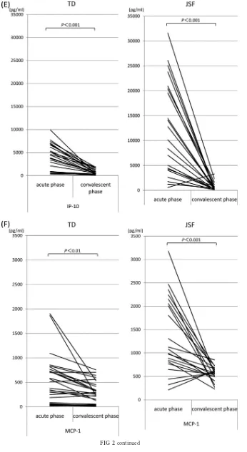
[image:2.585.40.286.79.190.2] [image:2.585.39.285.559.716.2]FIG 2Serum cytokine and chemokine levels during the acute and convalescent phases of TD and JSF. Serum cytokines TNF-␣(A), IL-6 (B), and IFN-␥(C) and chemokines IL-8 (D), IP-10 (E), MCP-1 (F), MIP-1␣(G), MIP-1(H), and eotaxin (I) were measured using multiplex bead immunoassays. The concentrations of cytokines and chemokines in the acute and convalescent phases are shown. N.S., not significant.
on May 16, 2020 by guest
http://jcm.asm.org/
[image:3.585.121.461.65.685.2]FIG 2continued
Higher Cytokine Levels in Japanese Spotted Fever
on May 16, 2020 by guest
http://jcm.asm.org/
[image:4.585.118.465.60.721.2]FIG 2continued
on May 16, 2020 by guest
http://jcm.asm.org/
[image:5.585.120.466.66.700.2]FIG 2continued
Higher Cytokine Levels in Japanese Spotted Fever
on May 16, 2020 by guest
http://jcm.asm.org/
[image:6.585.120.464.61.715.2]tion, Redmond, WA, USA). APvalue of⬍0.05 was considered statistically significant.
RESULTS
Clinical features of TD and JSF.
All clinically suspected cases of
rickettsial infection underwent indirect immunoperoxidase
antibody testing performed on paired serum samples collected
during the acute and convalescent phases. The IgM antibody
titers were more than 1:640 during the acute phase in each case,
and IgG antibody titers also increased more than 1:640 in the
convalescent phase (data not shown). The following strains of
O. tsutsugamushi
were identified: Gilliam (
n
⫽
0), Karp (
n
⫽
2),
Kato (
n
⫽
0), Irie/Kawasaki (
n
⫽
24), Hirano/Kuroki (
n
⫽
6), and
Shimokoshi (
n
⫽
0). Patient demographics and clinical and
labo-ratory data are summarized in
Table 1
.
O. tsutsugamushi
and
R.
japonica
differ in their antigenic determinants, and accordingly,
there was no overlap of the serologic assays for TD and JSF. The
peak temperatures of fever in patients with JSF were higher
than those in patients with TD (39.2
⫾
0.7°C versus 38.1
⫾
0.7°C)
(
Fig. 1A
). Moreover, patients with JSF had a higher clinical
sever-ity score (3.67
⫾
1.71) than patients with TD (1.47
⫾
0.77) (
Fig.
1B
). Patients were given 100 to 200 mg/day minocycline
intrave-nously or
per os
for 3 to 10 days, and all responded well to
treat-ment.
Serum cytokine and chemokine levels during minocycline
treatment of TD and JSF.
The serum levels of TNF-
␣
, IL-6,
IFN-
␥
, IL-8, IP-10, MCP-1, MIP-1
␣
, and MIP-1

were
ele-vated in almost all patients during the acute phase of rickettsial
disease (
Fig. 2A
to
I
). Compared with the acute phase, the
serum concentrations of these cytokines and chemokines were
significantly decreased in the convalescent phase. In contrast,
eotaxin levels were unchanged in the acute and convalescent
phases. We next examined whether serum levels of cytokines
and chemokines were significantly different between TD and
JSF in the acute phase. The mean concentrations of cytokines
and chemokines in patients infected with
O. tsutsugamushi
or
R.
japonica
are shown in
Table 3
. The serum concentrations of
TNF-
␣
in patients with JSF (35.0 pg/ml) were significantly higher
than those in patients with TD (13.8 pg/ml). Similarly, the serum
levels of cytokines IL-6 (65.8 pg/ml versus 11.6 pg/ml) and IFN-
␥
(220.3 pg/ml versus 73.4 pg/ml) and chemokines IL-8 (46.3 pg/ml
versus 13.2 pg/ml), IP-10 (12,298.3 pg/ml versus 3,215.9 pg/ml),
MCP-1 (1,365.8 pg/ml versus 467.5 pg/ml), MIP-1
␣
(53.9 pg/ml
versus 15.0 pg/ml), and MIP-1

(127.7 pg/ml versus 88.5 pg/ml)
in JSF patients were significantly higher than those in TD patients.
However, there was no significant difference in the concentration
of eotaxin between the acute phases of TD and JSF (67.1 pg/ml
versus 68.2 pg/ml). On the other hand, in the convalescent phase,
the serum levels of TNF-
␣
, IL-8, and MCP-1 in JSF patients were
significantly higher than those in TD patients, and there were no
significant differences in other cytokines and chemokines between
TD and JSF patients (
Table 3
).
Positive correlations exist between TNF-
␣
and IL-8 (
r
⫽
0.876)
(
Fig. 3A
) and between TNF-
␣
and IP-10 (
r
⫽
0.704) (
Fig. 3B
) in
the acute phase of JSF. In contrast, no significant correlation was
observed between TNF-
␣
and other cytokines and chemokines in
the acute phase of TD.
FIG 2continued
on May 16, 2020 by guest
http://jcm.asm.org/
[image:7.585.125.462.67.377.2]DISCUSSION
In the present study, the levels of cytokines and chemokines were
significantly higher in the acute phase of JSF than in the acute
phase of TD. Consistent with this, a positive correlation between
TNF-
␣
and IL-8 (
r
⫽
0.876), as well as between TNF-
␣
and IP-10
(
r
⫽
0.704), was observed in the acute phase of JSF. We speculate
that the severity of JSF is related to hypercytokinemia. Systemic
inflammatory reactions are regulated by a complicated cytokine
network, and the production of proinflammatory cytokines and
chemokines is essential in the immune response against
patho-gens. Regulation of cytokine and chemokine production is
neces-sary for an appropriate host response; however, tissue damage
may result from activation of immune cells due to the
overpro-duction of cytokines and chemokines. The unregulated and
exces-sive systemic release of numerous cytokines and chemokines in a
life-threatening infection can cause systemic disorders and lead to
fatal consequences like septic shock or SIRS (
24
,
25
). We estimate
that hypercytokinemia may contribute to the disease severity of
rickettsiosis, including JSF. For instance, fulminant TD has been
associated with hemophagocytic syndrome (
10
), and this
syn-drome may represent a hyperreaction of the immune system
me-diated by an overactivated cytokine network during the advanced
stages of rickettsial infection (
19
,
26–28
). We previously reported
that a downregulation of these cytokines and chemokines is
ob-served after the administration of tetracyclines, demonstrated a
correlation between the severity of TD and the serum
concentra-tion of TNF-
␣
in the acute phase of the disease, and showed that
TNF-
␣
levels are predictive of the severity of TD (
18
,
19
,
29
,
30
). In
relation to other spotted fever group infections, it is reported that
Mediterranean spotted fever is characterized by a Th1 cytokine
profile, and the patient’s immune system responds with
proin-flammatory and immunoregulatory cytokine production (IL-1

,
TNF-
␣
, IL-6, IFN-
␥
, IL-8, IL-10, and IL-12) that accompanies the
rickettsial vasculitis and contributes to the healing process (
31
).
The cytokine profiles for JSF and MSF were not much different
from each other. No cytokines were characteristic of JSF, in
con-trast with what was observed for other rickettsioses. It is generally
said that the clinical course of JSF is worse than that of TD despite
antibiotic treatment, and in cases of severe JSF with complications
where monotherapy with either minocycline or doxycycline was
not effective, such patients can be successfully treated using
tetra-cyclines combined with quinolones (ciprofloxacin, levofloxacin,
etc.) (
32
). However, in this study, although we examined
correla-tions between the severity of JSF and some cytokine levels, we
could not show any significant difference. It is unclear whether the
severity of JSF is related to the serum concentrations of cytokines
and chemokines in the acute phase of the disease. Although the
precise mechanism underlying the disease severity of JSF is not
well understood, our study provides insight into the possible
con-tribution of cytokines and chemokines in the pathogenesis of this
disease.
[image:8.585.39.287.88.333.2]In conclusion, the present study showed that the serum
cyto-kine and chemocyto-kine levels were significantly higher in JSF than in
TD. We suspect that the differences in cytokine and chemokine
TABLE 3Differences in cytokine and chemokine levels between TD andJSF patients Disease phase and cytokine or chemokinea
Avg level⫾SD (pg/ml) in patients with:
P
valueb
TD (n⫽32) JSF (n⫽21) Acute
TNF-␣ 13.8⫾13.5 35.0⫾27.5 ⬍0.001 IL-6 11.6⫾17.2 65.8⫾73.4 ⬍0.01 IFN-␥ 73.4⫾78.1 220.3⫾228.0 ⬍0.001 IL-8 13.2⫾10.5 46.3⫾35.3 ⬍0.001 IP-10 3,215.9⫾2,786.1 12,298.3⫾9,539.4 ⬍0.001 MCP-1 467.5⫾492.0 1,365.8⫾816.7 ⬍0.001 MIP-1␣ 15.0⫾25.3 53.9⫾41.0 ⬍0.001 MIP-1 88.5⫾63.5 127.7⫾54.0 ⬍0.05 Eotaxin 67.1⫾31.7 68.2⫾40.1 NS Convalescent
TNF-␣ 4.1⫾4.4 6.8⫾6.9 ⬍0.05 IL-6 0⫾0 2.2⫾7.0 NS IFN-␥ 3.5⫾3.7 16.7⫾7.5 NS IL-8 6.1⫾7.5 18.5⫾23.3 ⬍0.01 IP-10 550.8⫾534.2 730.9⫾805.2 NS MCP-1 253.9⫾229.1 553.3⫾153.3 ⬍0.001 MIP-1␣ 7.3⫾13.9 9.5⫾16.9 NS MIP-1 63.7⫾46.2 65.7⫾29.6 NS Eotaxin 77.7⫾31.8 65.3⫾40.4 NS
aNormal levels: TNF-␣,⬍2.3 pg/ml; IL-6,⬍3.0 pg/ml; IFN-␥,⬍7.3 pg/ml; IL-8,⬍3.0
pg/ml; IP-10, MCP-1, MIP-1␣and MIP-1, normal levels not determined. bPvalues were determined byttest. NS, not significant.
FIG 3Relationships between TNF-␣and IL-8 (A) (r⫽0.876) and between TNF-␣and IP-10 (r⫽0.704) (B) in the acute phase of JSF.
Higher Cytokine Levels in Japanese Spotted Fever
on May 16, 2020 by guest
http://jcm.asm.org/
[image:8.585.136.447.550.711.2]levels contribute to the difference in disease severity between JSF
and TD.
ACKNOWLEDGMENTS
This work was supported by grant H24-Shinkou-Ippan-008 to H.I. for research on emerging and reemerging infectious diseases from the Min-istry of Health, Labor and Welfare of Japan and a Grant-in-Aid for Scien-tific Research (C) KAKENHI 24591478 from the Japan Society for the Promotion of Science.
We thank Seitaro Nasu of Nasu Clinic for provision of serum of rick-ettsia patients and the colleagues in our laboratory for excellent technical assistance and helpful discussions.
The authors declare no conflict of interest.
REFERENCES
1.Iwasaki H, Yano T, Kaneko S, Egi M, Takada N, Ueda T. 2001. Epidemiological analysis on many cases of tsutsugamushi disease found in Hiroshima Prefecture, Japan. Kansenshogaku Zasshi75:365–370. 2.Tamura A, Ohashi N, Urakami H, Miyamura S.1995. Classification of
rickettsia-tsutsugamushi in a new genus, Orientia gen. nov., as Orientia tsutsugamushi comb. nov. Int. J. Syst. Bacteriol.45:589 –591.http://dx.doi .org/10.1099/00207713-45-3-589.
3.Iwasaki H, Ueda T, Uchida M, Nakamura T, Takada N, Mahara F.1991. Atypical lymphocytes with a multilobated nucleus from a patient with tsutsugamushi disease (scrub typhus) in Japan. Am. J. Hematol.36:150 – 151.http://dx.doi.org/10.1002/ajh.2830360216.
4.Miyamura S, Ohta T, Tamura A.1989. Comparison of in vitro suscep-tibilities of Rickettsia prowazekii, R. rickettsii, R. sibirica and R. tsutsuga-mushi to antimicrobial agents. Nihon Saikingaku Zasshi44:717–721.http: //dx.doi.org/10.3412/jsb.44.717.
5.Suto T, Hatakeyama H, Ito R, Nakamura Y, Mahara F.1989. In vitro susceptibility of a strain of Rickettsia recently isolated from a case of Jap-anese spotted fever to chemotherapeutic agents. Kansenshogaku Zasshi
63:35–38.
6.Mahara F, Koga K, Sawada S, Taniguchi T, Shigemi F, Suto T, Tsuboi Y, Ooya A, Koyama H, Uchiyama T. 1985. The first report of the rickettsial infections of spotted fever group in Japan: three clinical cases. Kansenshogaku Zasshi59:1165–1171.
7.Uchida T, Mahara F, Tsuboi Y, Oya A. 1985. Spotted fever group rickettsiosis in Japan. Jpn. J. Med. Sci. Biol.38:151–153.http://dx.doi.org /10.7883/yoken1952.38.151.
8.Mahara F.1997. Japanese spotted fever: report of 31 cases and review of the literature. Emerg. Infect. Dis.3:105–111.http://dx.doi.org/10.3201 /eid0302.970203.
9.Mahara F.2006. Rickettsioses in Japan and the Far East. Ann. N. Y. Acad. Sci.1078:60 –73.http://dx.doi.org/10.1196/annals.1374.007.
10. Iwasaki H, Hashimoto K, Takada N, Nakayama T, Ueda T, Nakamura T.1994. Fulminant Rickettsia tsutsugamushi infection associated with hemophagocytic syndrome. Lancet343:1236.http://dx.doi.org/10.1016 /S0140-6736(94)92456-2.
11. Bone RC, Sibbald WJ, Sprung CL.1992. The ACCP-SCCM consensus conference on sepsis and organ failure. Chest101:1481–1483.http://dx .doi.org/10.1378/chest.101.6.1481.
12. Cavaillon JM, Adib-Conquy M, Cloez-Tayarani I, Fitting C. 2001. Immunodepression in sepsis and SIRS assessed by ex vivo cytokine pro-duction is not a generalized phenomenon: a review. J. Endotox. Res.7:85– 93.http://dx.doi.org/10.1177/09680519010070020201.
13. Kuroda T, Suzuki S, Konno M, Amano T, Mikame M.1991. A case of scrub typhus with disseminated intravascular coagulation, meningitis and pulmonary fibrosis. Nihon Naika Gakkai Zasshi80:1816 –1817.http://dx .doi.org/10.2169/naika.80.1816.
14. Iwasaki H, Mahara F, Takada N, Fujita H, Ueda T.2001. Fulminant Japanese spotted fever associated with hypercytokinemia. J. Clin. Micro-biol.39:2341–2343.http://dx.doi.org/10.1128/JCM.39.6.2341-2343.2001. 15. Kim YJ, Yeo SJ, Park SJ, Woo YJ, Kim MW, Kim SH, Chang IA, Jeon SH, Park BJ, Song GJ, Lee MG, Kim IS, Kim YW.2013. Improvement of the diagnostic sensitivity of scrub typhus using a mixture of recombi-nant antigens derived from orientia tsutsugamushi serotypes. J. Korean Med. Sci.28:672– 679.http://dx.doi.org/10.3346/jkms.2013.28.5.672. 16. Watthanaworawit W, Turner P, Turner C, Tanganuchitcharnchai A,
Richards AL, Bourzac KM, Blacksell SD, Nosten F.2013. Short report: a prospective evaluation of real-time PCR assays for the detection of Ori-entia tsutsugamushi and Rickettsia spp. for early diagnosis of rickettsial infections during the acute phase of undifferentiated febrile illness. Am. J. Trop. Med. Hyg.89:308 –310.http://dx.doi.org/10.4269/ajtmh.12-0600. 17. Kern WV, Oristrell J, SeguraPorta F, Kern P.1996. Release of soluble
tumor necrosis factor receptors in Mediterranean spotted fever rickettsi-osis. Clin. Diagn. Lab. Immunol.3:233–235.
18. Iwasaki H, Mizoguchi J, Takada N, Tai K, Ikegaya S, Ueda T.2010. Correlation between the concentrations of tumor necrosis factor-alpha and the severity of disease in patients infected with Orientia tsutsuga-mushi. Int. J. Infect. Dis.14:E328 –E333.http://dx.doi.org/10.1016/j.ijid .2009.06.002.
19. Iwasaki H, Takada N, Nakamura T, Ueda T.1997. Increased levels of macrophage colony-stimulating factor, gamma interferon, and tumor ne-crosis factor alpha in sera of patients with Orientia tsutsugamushi infec-tion. J. Clin. Microbiol.35:3320 –3322.
20. Salvatore CM, Techasaensiri C, Tagliabue C, Katz K, Leos N, Gomez AM, McCracken GH, Hardy RD.2009. Tigecycline therapy significantly reduces the concentrations of inflammatory pulmonary cytokines and chemokines in a murine model of mycoplasma pneumoniae pneumonia. antimicrob. Agents Chemother.53:1546 –1551.http://dx.doi.org/10.1128 /AAC.00979-08.
21. Kim HO, Kim HS, Youn JC, Shin EC, Park S.2011. Serum cytokine profiles in healthy young and elderly population assessed using multi-plexed bead-based immunoassays. J. Transl. Med.9:113.http://dx.doi.org /10.1186/1479-5876-9-113.
22. Wagner B, Freer H.2009. Development of a bead-based multiplex assay for simultaneous quantification of cytokines in horses. Vet. Immunol. Immunopathol. 127:242–248. http://dx.doi.org/10.1016/j.vetimm.2008 .10.313.
23. Reis S, Sampaio ALF, Henriques MDM, Gandini M, Azeredo EL, Kubelka CF.2007. An in vitro model for dengue virus infection that exhibits human monocyte infection, multiple cytokine production and dexamethasone immunomodulation. Mem. Inst. Oswaldo Cruz102:983– 990.http://dx.doi.org/10.1590/S0074-02762007000800014.
24. Cohen J, Abraham E.1999. Microbiologic findings and correlations with serum tumor necrosis factor-alpha in patients with severe sepsis and septic shock. J. Infect. Dis.180:116 –121.http://dx.doi.org/10.1086/314839. 25. Bone RC, Balk RA, Cerra FB, Dellinger RP, Fein AM, Knaus WA,
Schein RM, Sibbald WJ, ACCP/SCCM Consensus Conference Commit-tee.1992. Definitions for sepsis and organ failure and guidelines for the use of innovative therapies in sepsis. The ACCP/SCCM Consensus Con-ference Committee. American College of Chest Physicians/Society of Crit-ical Care Medicine. Chest101:1644 –1655.
26. Chung DR, Lee YS, Lee SS.2008. Kinetics of inflammatory cytokines in patients with scrub typhus receiving doxycycline treatment. J. Infect.56:
44 –50.http://dx.doi.org/10.1016/j.jinf.2007.09.009.
27. Kramme S, Van An L, Khoa ND, Van Trin L, Tannich E, Rybniker J, Fleischer B, Drosten C, Panning M.2009. Orientia tsutsugamushi bac-teremia and cytokine levels in Vietnamese scrub typhus patients. J. Clin. Microbiol.47:586 –589.http://dx.doi.org/10.1128/JCM.00997-08. 28. Kodama K, Senba T, Yamauchi H, Nomura T, Chikahira Y. 2003.
Clinical study of Japanese spotted fever and its aggravating factors. J. In-fect. Chemother.9:83– 87.http://dx.doi.org/10.1007/s10156-002-0223-5. 29. Tai K, Iwasaki H, Ikegaya S, Ueda T. 2013. Minocycline modulates cytokine and chemokine production in lipopolysaccharide-stimulated THP-1 monocytic cells by inhibiting I kappa B kinase alpha/beta phos-phorylation. Transl. Res. 161:99 –109. http://dx.doi.org/10.1016/j.trsl .2012.10.001.
30. Iwasaki H, Inoue H, Takada N, Mahara F, Ueda T. 2000. Cytokine modulation induced by minocycline in Tsutsugamushi disease. Kansen-shogaku Zasshi74:598 – 600.
31. Popivanova NI, Murdjeva MA, Baltadzhiev IG, Haydushka IA.2011. Dynamics in serum cytokine responses during acute and convalescent stages of Mediterranean spotted fever. Folia Med. (Plovdiv)53:36 – 43.
http://dx.doi.org/10.2478/v10153-010-0035-9.
32. Seki M, Ikari N, Yamamoto S, Yamagata Y, Kosai K, Yanagihara K, Kakugawa T, Kurihara S, Izumikawa K, Miyazaki Y, Higashiyama Y, Hirakata Y, Tashiro T, Kohno S.2006. Severe Japanese spotted fever successfully treated with fluoroquinolone. Intern. Med.45:1323–1326.
http://dx.doi.org/10.2169/internalmedicine.45.1831.