Staphylococcus aureus
(MRSA) Strains Suitable in Regions of High
MRSA Endemicity
Jeong-Uk Kim,aChoong-Hwan Cha,aHae-Kyong An,aHo-Jun Lee,aMi-Na Kimb
Department of Laboratory Medicine, Gangneung Asan Hospital, University of Ulsan College of Medicine, Gangneung, South Koreaa
; Department of Laboratory Medicine, Asan Medical Center, University of Ulsan College of Medicine, Seoul, South Koreab
A multiplex real-time PCR assay that simultaneously detects themecA, staphylococcal cassette chromosome (SCCmec)-open reading frame X (orfX) junction, and staphylococcal 16S rRNA genes was developed and evaluated using 444 staphylococcal strains. We demonstrated that this assay resulted in fewer false-positive results than a single-locus real-time PCR assay that am-plified the SCCmec-orfXjunction. This assay would be useful in a clinical laboratory in a region of high endemicity for methicil-lin-resistantStaphylococcus aureus(MRSA) infections.
T
he spread of methicillin-resistant Staphylococcus aureus(MRSA) among hospital and community settings poses a threat to public health worldwide. Rapid, accurate detection and
appropriate intervention reduce the prevalence of MRSA (1–3).
Recently, rapid methods for molecular detection of MRSA have been developed. A single-locus real-time PCR assay that amplifies
the staphylococcal cassette chromosome (SCCmec)-open reading
frame X (orfX) junction was first proposed by Huletsky et al. (4),
and now, there are commercially available assays that identify
MRSA based on the detection of the SCCmec-orfXjunction (5–7).
These assays have an advantage over double-locus assays, based on
the simultaneous detection of themecA gene and aS. aureus
-specific gene, for the direct detection of MRSA from screening specimens. Double-locus assays have been associated with false-positive MRSA detections in clinical samples, including nasal
swabs that contain both methicillin-susceptible S. aureus
(MSSA) and methicillin-resistant coagulase-negative
staphylo-cocci (MRCoNS) (8). However, false-positive MRSA results
have been also reported in single-locus assays (6,7,9–12), for
example, due to MSSA isolates containing SCCmecremnants
that were misidentified as MRSA (11–16).
South Korea has been a region of MRSA infection endemicity
for many years. The rates of methicillin resistance amongS. aureus
isolates recovered from clinical specimens ranged from 67.8% to
74.1% during the 2000s (17). SCCmecis a mobile element that can
be inserted into and excised from the chromosome. It was
re-ported that partial excision of SCCmecfrom epidemic MRSA
strains results in MSSA isolates (13,15). Thus, in regions of high
endemicity, the single-locus assay for direct detection of MRSA may have high false-positive results because of the presence of
MRSA-derived MSSA strains that carry remnants of SCCmec
ele-ments. To address this, we developed a multiplex real-time PCR
assay that simultaneously detects themecA, SCCmec-orfX
junc-tion, and staphylococcal 16S rRNA genes. The assay was based on
the hypothesis that a pure MRSA strain has constantmecA,
SCC-mec-orfXjunction, and 16S rRNA copy numbers, represented by
the threshold cycle (CT) value, and that the exact relationship
betweenCTvalues of each target may be established.mecAand
SCCmec-orfX were targeted to reduce the false-positive MRSA
results caused by the presence of SCCmecremnants among MSSA
isolates that do not carrymecA. The staphylococcal 16S rRNA
gene was targeted to indicate coexisting staphylococcal strains in clinical samples.
This assay was evaluated using 444 strains, which included both reference strains from various international collections and clinical isolates from laboratory collections. The reference strains were 8 strains of MRSA (CCARM 3792, CCARM 3795, CCARM 3798, CCARM 3803, CCARM 3805, CCARM 3877, CCARM 3897, and CCARM 3911), 4 strains of MSSA (KCTC 1621, KCTC 1916, KCTC 1928, and ATCC 29213), and 11 strains of methicil-lin-susceptible coagulase-negative staphylococci (MSCoNS) (Staphylococcus epidermidis, KCCM 35494;Staphylococcus
simu-lans, KCCM 41686;Staphylococcus capitis, KCCM 41466;
Staphy-lococcus warneri, KCTC 3340;Staphylococcus haemolyticus, KCTC
3341;Staphylococcus xylosus, KCTC 3342;Staphylococcus
interme-dius, KCTC 3344; Staphylococcus saprophyticus, KCTC 3345;
Staphylococcus cohnii, KCTC 3574;Staphylococcus caprae, KCTC
3583; andStaphylococcus auricularis, KCTC 3584). Twenty-nine
MSSA isolates carrying SCCmecremnants, which had been
con-firmed by SCCmectyping (18,19), were tested as control strains.
The clinical isolates consisted of 209 MRSA, 109 MSSA, and 74 MRCoNS strains and were recovered mostly from wound, spu-tum, blood, and urine samples. Identification and susceptibility testing of these staphylococcal isolates were performed using the MicroScan WalkAway 96 (Siemens Healthcare Diagnostics Inc., West Sacramento, CA) and the Vitek 2 (bioMérieux Inc., Dur-ham, NC) automated identification and susceptibility testing sys-tems.
The reference strains and the clinical isolates were grown on blood agar plates (Asan Pharmaceutical, Seoul, South Korea) at 37°C for 24 h. Two or three bacterial colonies of the reference
Received21 September 2012Returned for modification11 November 2012
Accepted17 December 2012
Published ahead of print26 December 2012
Address correspondence to Jeong-Uk Kim, [email protected].
Copyright © 2013, American Society for Microbiology. All Rights Reserved.
doi:10.1128/JCM.02495-12
on May 16, 2020 by guest
http://jcm.asm.org/
strains and isolates were harvested with a 1-l loop and suspended in 0.5 ml of distilled water. The suspension was heated in a boiling
water bath for 10 min and centrifuged at 13,000⫻gfor 5 min. The
supernatant was used for the real-time PCR.
Base sequences of the SCCmec-orfXjunction,mecA, and
staph-ylococcal 16S rRNA genes were obtained from NCBI GenBank and aligned with Sequencher 5.0 software (Gene Codes Co., Ann Arbor, MI). Based on sequence alignment, we identified regions of interest and designed primers and probes manually or with the
Primer 3 program (http://frodo.wi.mit.edu/primer3/). The
real-time PCR primers and probes designed and used in this study are
shown inTable 1.
The time PCRs were conducted with a Rotor-Gene Q real-time PCR instrument (Qiagen Inc., Germantown, MD). The PCR
mixture contained 0.5l of primer-probe mix, 5l of 2⫻
Rotor-Gene Multiplex PCR master mix (Qiagen Inc., Germantown,
MD), and 1.0l of template DNA in a total volume of 10l. The
PCR parameters were 95°C for 5 min followed by 40 cycles of 95°C
for 15 s and 60°C for 15 s, and green, yellow, and crimson
fluores-cence were measured. After completion of PCR,CTvalues of the
mecA, SCCmec-orfX, and 16S rRNA genes were recorded from the
Rotor-Gene Q software. Statistical tests, including determinations
of thercorrelation coefficient and descriptive statistics, were
per-formed using SPSS 13.0 software (SPSS Inc., Chicago, IL).Pvalues
below the 5% level were considered statistically significant. The analytical sensitivity of the real-time PCR was determined by 10-fold serial dilutions of a subculture of MRSA strain CCARM 3792. The strain was grown overnight on a blood agar plate, sus-pended in saline to a density equivalent to a 0.5 McFarland
tur-bidity number, and serially diluted 10-fold from 102to 107. DNA
was extracted from 200l of the bacterial dilutions using the
QIAcube with a QIAamp DNA minikit (Qiagen Inc.,
German-town, MD) and eluted in 50l. In parallel, 100l of the dilutions
was plated on blood agar and incubated at 37°C for 24 h. There-after, CFU were counted.
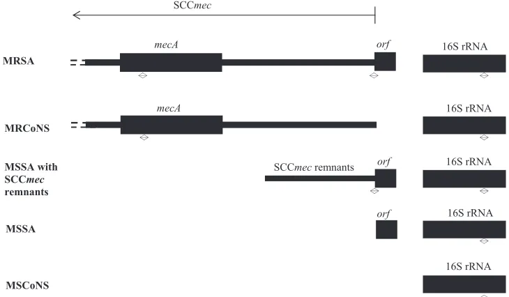
The real-time PCR assay was initially evaluated with 8 MRSA, 4
FIG 1Schematic diagram showing the relevant genetic elements detected by the multiplex real-time PCR assay in MRSA, MRCoNS, MSSA with SCCmec
[image:2.585.41.545.76.225.2]remnants, MSSA, and MSCoNS. The two-way arrows indicate the amplified regions.
TABLE 1The real-time PCR primers and probes for the detection of MRSA
Oligonucleotide Sequence (5=¡3=)a Concn (M) Target(s)b
FSCC_A GCGGAGGCTAACTATGTCAA 0.5 I, II, IVa, IVb, IVc, IVg, VI, VIII
FSCC_B ATATGTAATTCCTCCACATCTCATT 0.5 III, V, VII
FSCC_C GGCTGAAGTAACCGCATCA 0.5 IVe
FSCC_D TTCATAATATGTGCTACGCAATC 0.5 X
FSCC_E CGGCAATTCTCATAAACCTC 0.5 IX, XI
BorfX GCAAAATGACATTCCCACA 0.5 orfX
PorfX HEX-TCAATTAACACAACCCGCATCAT-BHQ1 0.2 orfX
FmecA GAATGCAGAAAGACCAAAGC 0.5 mecA
BmecA TTCTTTGGAACGATGCCTAT 0.5 mecA
PmecA FAM-TTGGCCAATACAGGAACAGCA-BHQ1 0.2 mecA
F16SrRNA CTTACCAAATCTTGACATCCTTT 0.5 16S rRNA
B16SrRNA CTCGTTGCGGGACTTAAC 0.5 16S rRNA
P16SrRNA Cy5.5-CGTCAGCTCGTGTCGTGAGAT-BHQ2 0.2 16S rRNA
aHEX, hexachloro-6-carboxyfluorescein; FAM, 6-carboxyfluorescein; BHQ1, black hole quencher 1; BHQ2, black hole quencher 2. b
The Roman numerals indicate the SCCmectypes amplified by the primer.
on May 16, 2020 by guest
http://jcm.asm.org/
[image:2.585.110.475.486.699.2]MSSA, and 11 MSCoNS reference strains and 29 MSSA control
strains carrying SCCmecremnants. Only the expected PCR products
were amplified from each reference strain. However, SCCmec-orfX
was not detected in 6 of 29 control strains. The results of the evaluation of 392 clinical isolates were as follows. Three targets were simultaneously detected in all 209 (100%) MRSA isolates and in 4 (5.4%) MRCoNS isolates. Of the 109 MSSA isolates, the
mecAand 16S rRNA genes were detected at the same time in 2
(1.8%) isolates, and both the SCCmec-orfXand 16S rRNA genes
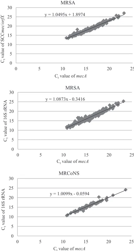
were detected in 11 (10.1%) isolates. TheCTvalues ofmecAwere
compared to theCTvalues of the SCCmec-orfXand 16S rRNA
genes. The correlation coefficient determined betweenmecA CT
values and SCCmec-orfX CTvalues was high for MRSA isolates
(r⫽0.959;P⬍0.0001), and correlation coefficients determined
betweenmecA CTvalues and 16S rRNACTvalues were high for
MRSA isolates (r⫽0.970;P⬍0.0001) and MRCoNS isolates (r⫽
0.963;P⬍0.0001). The results are shown inFig. 1and2andTable
2. Thus, theCTdifferences between the SCCmec-orfXandmecA
genes (CTSCC) and between the 16S rRNA andmecAgenes (CT16S)
FIG 2Correlation betweenCTvalues ofmecA, SCCmec-orfX, and 16S rRNA
genes in 209 MRSA and 74 MRCoNS isolates.
TABLE 2 MRSA detection results for clinical isolates by multiplex real -time PCR a Species No. of isolates mecA SCC mec-orfX 16S rRNA CT difference between SCC mec-orfX and mecA ( CT SCC ) CT difference between 16S rRNA and mecA ( CT 16S ) No. of isolates showing positive result Threshold cycle ( CT ) value No. of isolates showing positive result Threshold cycle ( CT ) value No. of isolates showing positive result Threshold cycle ( CT ) value MRSA 209 209 16.71 ⫾ 2.44 209 19.02 ⫾ 2.59 209 17.35 ⫾ 2.73 2.70 ⫾ 0.50 (1.58 ⬃ 4.99) 1.08 ⫾ 0.70 ( ⫺ 0.86 ⬃ 3.45) MSSA 109 2 35.40 ⫾ 1.36 11 29.32 ⫾ 2.45 109 17.95 ⫾ 2.87 ⫺ 18.58 ⫾ 1.28 ( ⫺ 19.48 ⬃⫺ 17.67) MRCoNS 74 74 15.48 ⫾ 2.36 4 34.98 ⫾ 2.35 74 15.61 ⫾ 2.41 17.78 ⫾ 1.84 (15.37 ⬃ 19.69) 0.10 ⫾ 0.51 ( ⫺ 1.41 ⬃ 2.46) aThe mecA ,SCC mec-orfX ,and 16S rRNA threshold cycle values represent the means ⫾ standard deviations. The CT SCC and CT 16S threshold cycle values represent ranges of means ⫾ standard deviations.
on May 16, 2020 by guest
http://jcm.asm.org/
[image:3.585.54.270.67.470.2]were used to assess the presence of MRSA. ACTSCCⱖ4.7 (mean⫹
4 standard deviations [SD]) indicated that MRSA and staphylo-cocci other than MRSA were present simultaneously, whereas a
CT16Sⱕ⫺1.72 (mean⫺4 SD) indicated that MRSA and
staphy-lococci lacking themecAgene coexisted.
The detection limit of the assay was determined using genomic
DNA purified from a 1:104dilution of a stock solution of MRSA
strain CCARM 3792 and was found to be 20 CFU per PCR. Real-time PCR assays, including the BD GeneOhm MRSA
as-say and Cepheid’s Xpert MRSA asas-say that target theorfXofS.
aureusand the right-extremity junction of SCCmec, are currently
used for infection control (20–23). Previous studies reported
false-negative and -positive results in these single-locus assays ranging from 0.0% to 7.3% and from 0.0% to 5.4%, respectively
(4,6,24–26). False-negative and -positive results can affect the
whole infection control program, bringing about the spread of MRSA in the hospital and unnecessary isolation and
decoloniza-tion procedures. If a MRSA with an unknown SCCmectype is
present in the samples, there could be false-negative results.
Cur-rently, 11 different types of SCCmechave been recognized inS.
aureus(http://www.sccmec.org/); we designed 5 forward primers,
1 reverse primer, and 1 probe to detect all known types of SCCmec.
SCCmecis a 21- to 67-kb genetic fragment that integrates into the
chromosome of MRSA at the integration site sequence for SCC
(ISS), which is located at the 3=end oforfX, and carries the central
determinant for broad-spectrum-lactam resistance encoded by
themecAgene (27–29). It is unstable and able to be excised.
Exci-sion of the SCCmeccan be complete or partial, with some
ele-ments left behind at the ISS. Since MRSA strains are resistant to
multiple drugs, the excision of SCCmecfrom such isolates results
in MSSA isolates retaining resistance to antibiotics other than
ß-lactams (12–16,30). In the study on determining the proportion
and diversity of multidrug-resistant MSSA (MR-MSSA) strains derived from MRSA strains, Donnio et al. investigated 247 MR-MSSA isolates from 60 French hospitals using the IDI-MRSA real-time PCR assay, the forerunner of the BD GeneOhm MRSA assay,
and found that 68% of isolates were positive (15). According to
Shore’s study, 7 MR-MSSA isolates harboring SCCmecremnants
identified by SCCmec typing PCR were tested with the BD
GeneOhm MRSA and Xpert MRSA assays, and 3 isolates yielded
positive results in both assays (14). In the present study, 29 MSSA
isolates tested as control strains were MR-MSSA and carried
SCCmec remnants that had been confirmed by the multiplex
PCR-based SCCmectyping. In 23 of 29 MR-MSSA control strains,
the SCCmec-orfXjunction was detected. Six control strains with
negative results might contain SCCmecremnants that lacked the
target-specific region of 5 forward primers. The possibility of the
presence of SCCmecremnants in MR-MSSA should always be
considered when a real-time PCR assay targeting the SCCmec
-orfXjunction is used for the rapid detection of MRSA from clinical
specimens, since this might give a high number of false-positive results.
To our knowledge, the incidence of false positives has not been reported in South Korea for single-locus real-time PCR assays. We expected the false-positive results to be higher than in countries of low MRSA infection endemicity; as predicted, they were as high as 10.1% in the clinical isolates of staphylococci that had been con-secutively collected in our laboratory over 3 months. If such a high rate of false-positive results occurs, the diagnostic value of single-locus real-time PCR assays seems unsatisfactory for a laboratory in
a region of high MRSA infection endemicity. Therefore, we
consid-ered simultaneous amplification of themecAgene and SCCmec
-orfXjunction to rule out MSSA isolates that carry SCCmec
rem-nants and lack themecAgene. However, this could also lead to a
false-positive result when MRCoNS and MSSA carrying SCCmec
remnants coexist in the clinical samples. Thus, the staphylococcal 16S rRNA gene was added to the targets to lessen false-positive results. In the case of a pure MRSA strain, relative quantifications of the three target genes would be constant, whereas in cases of
mixed populations of MRCoNS and MSSA carrying SCCmec
rem-nants, they would be mostly variable.
Three primer-probe pairs targeting the 16S rRNA gene were
designed. Of those, the pair having aCTvalue very close to theCT
ofmecAwas chosen. In most of the MRSA isolates, theCTof
SCCmecwas the largest, followed by those of the 16S rRNA and
mecAgenes. ThemecAgene was chosen as a reference gene for
relative quantifications. Consequently, theCTdifferences between
the SCCmec-orfX andmecAgenes (CTscc) and between the 16S
rRNA andmecAgenes (CT16S) were constant. We tested whether
mixed populations of MRCoNS and MSSA carrying SCCmec
rem-nants can be distinguished by relative quantifications. Mixed cocktails of staphylococcal genomic DNA samples were made to amplify the three targets, including mixtures of genomic DNA from MRSA and staphylococci other than MRSA and mixtures of
MRCoNS and MSSA carrying SCCmecremnants. Then, the
sim-ulated samples were analyzed. The results of analysis for mixtures
of genomic DNA from MRCoNS and MSSA carrying SCCmec
remnants are shown inTable 3. The data showed that MRCoNS
and MSSA carrying SCCmecremnants were simultaneously
pres-ent only withCTSCCⱖ4.7 andCT16Sⱕ⫺1.72, and mixed
popu-lations of MRSA and MRCoNS could not be differentiated from
those of MRCoNS and MSSA carrying SCCmecremnants with
CTSCCⱖ4.7 andCT16Sⱖ⫺1.71. The data from other mixed DNA
samples were in accord with proposedCTcalculations; aCTSCCⱖ
4.7 indicated that MRSA and staphylococci other than MRSA
were present simultaneously, whereas aCT16Sⱕ⫺1.72 indicated
that MRSA and staphylococci lacking themecAgene coexisted.
In this study, unexpected amplimers were obtained in 4 MRCoNS isolates and 2 MSSA isolates. A total of 4 MRCoNS isolates yielded simultaneous amplification of the three targets,
and theCTSCCvalues were 19.69, 17.50, 18.55, and 15.37;CT16S
values were 0.61,⫺0.97, ⫺0.31, and⫺0.09, respectively. In 2
MSSA isolates, themecAwas amplified and theCT16Svalues were
⫺17.67 and⫺19.48, respectively. In order to know whether or not
these 6 isolates were unusual genotypic strains, stored isolates were regrown on blood agar plates for 24 h. Template DNA was extracted from a single colony. Only expected products were
am-plified from the reprepared samples; both themecAand 16S rRNA
genes were detected in 4 MRCoNS isolates and the 16S rRNA gene in 2 MSSA isolates. The results showed that the unexpected am-plimers were due to the mixed staphylococci. Consequently, 4 MRCoNS isolates showing amplification of all of the three targets would be mixtures of MRSA and MRCoNS or of MRCoNS and
MSSA carrying SCCmecremnants. In South Korea, it is assumed
that MSSA strains carrying SCCmecremnants comprise
approxi-mate 3% of theS. aureusisolates recovered from clinical
speci-mens because about 30% ofS. aureusisolates are MSSA and 10%
of MSSA carry SCCmecremnants. Becker’s study reported that
nasal cocolonization by MSSA and MRCoNS was observed in
3.4% of patients (8). Furthermore, it was known from analyzing
on May 16, 2020 by guest
http://jcm.asm.org/
simulated samples that MRCoNS must comprise more than 90%
of a mixed population to have aCTSCCvalue over 15. Accordingly,
it is reasonable to assume that there is very little chance of ampli-fying all three of the targets from mixed populations of MRCoNS
and MSSA carrying SCCmecremnants. In the case of 2 MSSA
isolates showing amplification of themecA, it would seem that
they contained mixed populations of MRCoNS and MSSA. In summary, as a preliminary study for the introduction of a direct MRSA molecular detection system to our laboratory in a region with high MRSA infection endemicity, a multiplex
real-time PCR assay that simultaneously detects themecA, SCCmec
-orfXjunction, and staphylococcal 16S rRNA genes was developed
and evaluated using 444 staphylococcal strains. The key issue was whether this assay can reduce false positives caused by MSSA
car-rying SCCmecremnants. The evaluation data showed that this
assay resulted in fewer false-positive results than a single-locus
real-time PCR assay that amplified the SCCmec-orfX junction.
This assay would be useful in a clinical laboratory in a region with high MRSA infection endemicity, although further evaluation with clinical specimens is necessary before it can be applied in the laboratory.
REFERENCES
1.Bootsma MC, Diekmann O, Bonten MJ.2006. Controlling
methicillin-resistantStaphylococcus aureus: quantifying the effects of interventions and rapid diagnostic testing. Proc. Natl. Acad. Sci. U. S. A.103:5620 –5625.
2.Harbarth S, Masuet-Aumatell C, Schrenzel J, Francois P, Akakpo C,
Renzi G, Pugin J, Ricou B, Pittet D.2006. Evaluation of rapid screening
and pre-emptive contact isolation for detecting and controlling methicil-lin-resistantStaphylococcus aureusin critical care: an interventional co-hort study. Crit. Care10:R25. doi:10.1186/cc3982.
3.Chowers MY, Paitan Y, Gottesman BS, Gerber B, Ben-Nissan Y, Shitrit
P.2009. Hospital-wide methicillin-resistant Staphylococcus aureus con-trol program: a 5-year follow-up. Infect. Concon-trol Hosp. Epidemiol.30: 778 –781.
4.Huletsky A, Giroux R, Rossbach V, Gagnon M, Vaillancourt M, Bernier
M, Gagnon F, Truchon K, Bastien M, Picard FJ, van Belkum A,
Ouellette M, Roy PH, Bergeron MG.2004. New real-time PCR assay for
rapid detection of methicillin-resistantStaphylococcus aureusdirectly from specimens containing a mixture of staphylococci. J. Clin. Microbiol.
42:1875–1884.
5.Peterson LR, Liesenfeld O, Woods CW, Allen SD, Pombo D, Patel PA,
Mehta MS, Nicholson B, Fuller D, Onderdonk A. 2010. Multicenter
evaluation of the LightCycler methicillin-resistantStaphylococcus aureus
(MRSA) advanced test as a rapid method for detection of MRSA in nasal surveillance swabs. J. Clin. Microbiol.48:1661–1666.
6.Rossney AS, Herra CM, Brennan GI, Morgan PM, O’Connell B.2008.
Evaluation of the Xpert methicillin-resistantStaphylococcus aureus
(MRSA) assay using the GeneXpert real-time PCR platform for rapid de-tection of MRSA from screening specimens. J. Clin. Microbiol.46:3285– 3290.
7.Rossney AS, Herra CM, Fitzgibbon MM, Morgan PM, Lawrence MJ,
O’Connell B.2007. Evaluation of the IDI-MRSA assay on the SmartCycler
real-time PCR platform for rapid detection of MRSA from screening spec-imens. Eur. J. Clin. Microbiol. Infect. Dis.26:459 – 466.
8.Becker K, Pagnier I, Schuhen B, Wenzelburger F, Friedrich AW, Kipp
F, Peters G, von Eiff C.2006. Does nasal cocolonization by
methicillin-resistant coagulase-negative staphylococci and methicillin-susceptible
Staphylococcus aureusstrains occur frequently enough to represent a risk of false-positive methicillin-resistantS. aureusdeterminations by molec-ular methods? J. Clin. Microbiol.44:229 –231.
9.Laurent C, Bogaerts P, Schoevaerdts D, Denis O, Deplano A, Swine C,
Struelens MJ, Glupczynski Y.2010. Evaluation of the Xpert MRSA assay
for rapid detection of methicillin-resistantStaphylococcus aureusfrom na-res swabs of geriatric hospitalized patients and failure to detect a specific SCCmectype IV variant. Eur. J. Clin. Microbiol. Infect. Dis.29:995–1002.
10. Oberdorfer K, Pohl S, Frey M, Heeg K, Wendt C.2006. Evaluation of a
single-locus real-time polymerase chain reaction as a screening test for specific detection of methicillin-resistantStaphylococcus aureusin ICU patients. Eur. J. Clin. Microbiol. Infect. Dis.25:657– 663.
11. Stamper PD, Louie L, Wong H, Simor AE, Farley JE, Carroll KC.2011.
Genotypic and phenotypic characterization of methicillin-susceptible
Staphylococcus aureusisolates misidentified as methicillin-resistant Staph-ylococcus aureusby the BD GeneOhm MRSA assay. J. Clin. Microbiol.
49:1240 –1244.
12. Wong H, Louie L, Lo RY, Simor AE.2010. Characterization of
Staphy-lococcus aureusisolates with a partial or complete absence of staphylococ-cal cassette chromosome elements. J. Clin. Microbiol.48:3525–3531.
13. Donnio PY, Oliveira DC, Faria NA, Wilhelm N, Le Coustumier A, de
Lencastre H.2005. Partial excision of the chromosomal cassette
contain-ing the methicillin resistance determinant results in methicillin-susceptibleStaphylococcus aureus. J. Clin. Microbiol.43:4191– 4193.
14. Shore AC, Rossney AS, O’Connell B, Herra CM, Sullivan DJ,
Hum-phreys H, Coleman DC.2008. Detection of staphylococcal cassette
chro-mosome mec-associated DNA segments in multiresistant methicillin-susceptible Staphylococcus aureus(MSSA) and identification of
Staphylococcus epidermidisccrAB4 in both methicillin-resistantS. aureus
and MSSA. Antimicrob. Agents Chemother.52:4407– 4419.
15. Donnio PY, Fevrier F, Bifani P, Dehem M, Kervegant C, Wilhelm N,
Gautier-Lerestif AL, Lafforgue N, Cormier M, Le Coustumier A.2007.
Molecular and epidemiological evidence for spread of multiresistant me-thicillin-susceptibleStaphylococcus aureusstrains in hospitals. Antimi-crob. Agents Chemother.51:4342– 4350.
16. Lindqvist M, Isaksson B, Grub C, Jonassen TO, Hallgren A. 2012.
[image:5.585.40.546.78.223.2]Detection and characterisation of SCCmecremnants in multiresistant me-thicillin-susceptibleStaphylococcus aureuscausing a clonal outbreak in a Swedish county. Eur. J. Clin. Microbiol. Infect. Dis.31:141–147.
TABLE 3Results of the multiplex real-time PCR assay for mixtures of genomic DNA from MRCoNS and MSSA carrying SCCmecremnants
DNA samplea
Proportion
CTdifference between
SCCmec-orfXandmecA(CTSCC)b
CTdifference between 16S rRNA
andmecA(CT16S)b
MRCoNS (%) MSSA (%)
A 95 5 15.99⫾1.48 (13.03⬃16.94) 0.87⫾0.50 (0.20⬃1.34)
B 90 10 14.60⫾1.52 (11.75⬃16.15) 0.74⫾0.17 (0.51⬃0.92)
C 80 20 13.15⫾1.74 (10.33⬃15.34) 0.90⫾0.44 (0.36⬃1.38)
D 70 30 12.32⫾1.96 (9.36⬃15.00) 0.59⫾0.23 (0.19⬃0.85)
E 60 40 11.54⫾1.79 (8.79⬃14.29) 0.45⫾0.51 (⫺0.13⬃1.10)
F 50 50 12.25⫾4.42 (8.34⬃20.75) 0.46⫾0.62 (⫺0.65⬃1.01)
G 40 60 10.41⫾2.41 (7.38⬃14.59) 0.27⫾0.22 (⫺0.01⬃0.57)
H 30 70 9.25⫾2.23 (6.46⬃12.43) ⫺0.19⫾0.40 (⫺0.72⬃0.22)
I 20 80 7.81⫾2.00 (4.94⬃9.71) ⫺0.56⫾0.91 (⫺1.85⬃0.56)
J 10 90 6.91⫾1.64 (4.85⬃8.59) ⫺1.55⫾1.00 (⫺2.81⬃ ⫺0.47)
K 5 95 5.66⫾1.34 (3.84⬃7.01) ⫺2.06⫾0.69 (⫺2.99⬃ ⫺0.95)
aSix samples each of 11 types of DNA were used.
b
The threshold cycle values represent ranges of means⫾standard deviations.
on May 16, 2020 by guest
http://jcm.asm.org/
17. Korea Centers for Disease Control and Prevention.2010. 2010 labora-tory surveillance for vancomycin resistant staphylococcus aureus. VRSA Newsl.10:3.
18. Chen L, Mediavilla JR, Oliveira DC, Willey BM, de Lencastre H,
Kreiswirth BN.2009. Multiplex real-time PCR for rapid Staphylococcal
cassette chromosome mec typing. J. Clin. Microbiol.47:3692–3706.
19. Milheiriço C, Oliveira DC, de Lencastre H.2007. Update to the
multi-plex PCR strategy for assignment of mec element types inStaphylococcus aureus. Antimicrob. Agents Chemother.51:3374 –3377.
20. Ho TH, Huang YC, Lin TY.2011. Evaluation of the BD GeneOhm
StaphSR assay for detection ofStaphylococcus aureusin patients in inten-sive care units. J. Microbiol. Immunol. Infect.44:310 –315.
21. Keshtgar MR, Khalili A, Coen PG, Carder C, Macrae B, Jeanes A, Folan
P, Baker D, Wren M, Wilson AP. 2008. Impact of rapid molecular
screening for meticillin-resistantStaphylococcus aureusin surgical wards. Br. J. Surg.95:381–386.
22. Kluytmans J.2007. Control of meticillin-resistantStaphylococcus aureus
(MRSA) and the value of rapid tests. J. Hosp. Infect.65(Suppl 2):100 –104.
23. Schulz M, Nonnenmacher C, Mutters R. 2009. Cost-effectiveness of
rapid MRSA screening in surgical patients. Eur. J. Clin. Microbiol. Infect. Dis.28:1291–1296.
24. Kolman S, Arielly H, Paitan Y.2010. Evaluation of single and double-locus
real-time PCR assays for methicillin-resistantStaphylococcus aureus(MRSA) surveillance. BMC Res. Notes3:110. doi:10.1186/1756-0500-3-110.
25. Ornskov D, Kolmos B, Bendix Horn P, Nederby Nielsen J, Brandslund
I, Schouenborg P.2008. Screening for methicillin-resistant
Staphylococ-cus aureusin clinical swabs using a high-throughput real-time PCR-based method. Clin. Microbiol. Infect.14:22–28.
26. Söderquist B, Neander M, Dienus O, Zimmermann J, Berglund C,
Matussek A, Molling P.2012. Real-time multiplex PCR for direct
detec-tion of methicillin-resistantStaphylococcus aureus(MRSA) in clinical samples enriched by broth culture. APMIS120:427– 432.
27. Hiramatsu K, Cui L, Kuroda M, Ito T.2001. The emergence and
evo-lution of methicillin-resistantStaphylococcus aureus. Trends Microbiol.
9:486 – 493.
28. Hiramatsu K, Katayama Y, Yuzawa H, Ito T.2002. Molecular genetics of
methicillin-resistantStaphylococcus aureus. Int. J. Med. Microbiol.292: 67–74.
29. International Working Group on the Classification of Staphylococcal
Cassette Chromosome Elements (IWG-SCC). 2009. Classification of
staphylococcal cassette chromosome mec (SCCmec): guidelines for re-porting novel SCCmec elements. Antimicrob. Agents Chemother.53: 4961– 4967.
30. Donnio PY, Louvet L, Preney L, Nicolas D, Avril JL, Desbordes L.2002.
Nine-year surveillance of methicillin-resistantStaphylococcus aureusin a hospital suggests instability ofmecADNA region in an epidemic strain. J. Clin. Microbiol.40:1048 –1052.